DOI: 10.5935/2359-4802.20170025
363
International Journal of Cardiovascular Sciences. 2017;30(4):363-366
CASE REPORT
Mailing Adress: Levent Cerit
Near East University. Postal Code 07100, Nicosia – Chipre E-mail: drcerit@hotmail.com, drcerit@yahoo.com
A Transcatheter Aortic Valve Implantation Without Aortography Guidance Due To
Vascular Access Constraints Or Limitations
Oktay Ergene,1 Hamza Duygu,2 Volkan Emren,3 Ugur Kocabas,4 Levent Cerit2
Dokuz Eylül University,1 Izmir, Turquia; Near East University,2 Nicosia, Chipre; Afyon Kocatepe University School of Medicine,3 Afyonkarahisar, Turquia;
Ataturk Training and Research Hospital Department of Cardiology,4 Izmir – Turquia
Manuscript received November 27, 2016, revised manuscript January 24, 2017, accepted January 24, 2017.
Transcatheter Aortic Valve Replacement; Dissection; Aortic Valve Stenosis.
Keywords
Introduction
Aortic valve replacement (AVR) is an established treatment modality in patients with severe symptomatic aortic stenosis (AS).1,2 Given the increased mortality and morbidity of AVR for the high-risk patients and the poor long-term results of balloon aortic valvuloplasty, transcatheter aortic valve implantation (TAVI) is increasingly accepted treatment option who are not candidates for AVR.3 We describe here a case of succesfully implanted Edwards SAPIEN XT transcatheter heart valve without aortography guidance due to multiple arterial access problems and complicated by iatrogenic De Bakey type III aortic dissection and severe paravalvular leak.
Case report
A 79-year-old woman was admitted with dyspnea (NYHA III) over a period of 3 months. She had a history of hypertension, diabetes mellitus, hyperlipidemia and chronic renal failure. A grade 3/6 systolic murmur was heard over the aortic area. Rhythm was sinus and electrocardiography fulfilled the criteria of left ventricular hypertrophy. Transthoracic echocardiography showed a severely calcified aortic stenosis with valve area of
0.6 cm2, maximum gradient of 92 mmHg, mean gradient
of 55 mmHg, left ventricular ejection fraction of 60%, aortic annular diameter of 20 mm, and ascending aorta of 35 mm.
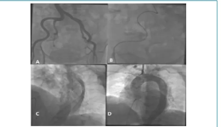
Patient was evaluated by a heart team and deemed to be high risk for AVR (STS:12, euroscore:21) and accordingly decided to proceed to TAVI. Coronary angiography demonstrated the stenosis of LAD by 40%, first diagonal 40%, circumflex 85%, and proximal right coronary artery 50%. A mild to moderate aortic regurgitation was detected by aortography. The distance between left main coronary ostium (LMC) and aortic annulus was 11 mm. Right iliac artery was severly tortuous and no stenotic lesion was detected (Figure 1A, 1B).
We aimed to revascularize critical circumflex lesion before the procedure and percutaneous coronary intervention was performed. One month later, TAVI procedure scheduled. Under general anesthesia a left femoral cutdown was performed because of the severly tortuous right iliac artery. A pacemaker was placed from
the right femoral vein.However, left Amplatz catheter
over a Terumo and Exchange stiff wire was not got through to arcus aorta and an attemt to cross the aortic
valve failed.Accordingly we noticed that the catheter
364
Figure 1 – Angiographic images showing severe tortuous iliac artery (A. B), retrograd aortic dissection (C), and aortography via right femoral artery (D).
Ergene et al. Transcatheter aortic valve implantation Int J Cardiovasc Sci. 2017;30(4):363-366
Case Report
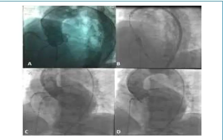
accompanied by rapid ventricular pacing (Figure 2A). Immediately after, X-ray tube’s angle and the patient left fixed positon, and prosthetic valve was started to implant without aortography. A 23 mm Edwards SAPIEN XT prosthetic aortic valve was deployed with rapid pacing at 180 bpm in accordance with this index drawings without aortography guidance (Figure 2B). But, aortogram following implantation showed severe paravalvular aortic regurgitation (Figure 2C).In order to solve this problem, prosthetic valve was crossed with AL-1 catheter and a straight and stiff wire, and successfully dilated with a 23 mm aortic balloon. Severe aortic regurgitation decreased to mild degree (Figure 2D). Transthorasic echocardiography obtained one day after the procedure revealed a dramatic decrease in aortic valve gradient (20/10 mmHg) and no aortic regurgitation. The patient was discharged from hospital on 6th postoperative day without any complication.
Discussion
TAVI is increasingly becoming an accepted option in the treatment of patients with severe symtomatic
AS who are not eligible for surgery due to prohibitive
risks and frailtiy.4 Although TAVI is less invasive
than conventional open heart surgery, it has several complications including the occlusion of coronary ostia, annular rupture, ventricular perforation, paravalvular regürgitation, heart block, device malposition or embolisation, stroke, major bleeding, vascular injury, and acute kidney injury.4
365
Figure 2 – Deployment of prosthetic aortic valve based on index drawings (A, B), severe paravalvular aortic regurgitation following implantation (C), and aortogram showing mild regurgitation just after aortic balloon dilatation (D).
Ergene et al.
Transcatheter aortic valve implantation
Int J Cardiovasc Sci. 2017;30(4):363-366
Case Report
marked the LMC ostium and aortic anulus on screen by a colourful pen during aortography accompanying with rapid pacing, then patient and X-ray tube fixed, and immediately after, the prosthesis valve implanted in accordance with this index drawing without aortography guidance. Another possible option could further define the annulus (landing zone) is to perform balloon aortic valvuloplasty (pre-dilatation) and to mark in the screen the waist and use it as a marker.
Mild or moderate aortic regurgitation can be seen after implantation. However hemodynamically severe post-procedural aortic regurgitation is rare.4 These are generally caused by inadequate inflation of the balloon or by calcium deposits that prevent the valve unit to properly seat and seal within the annulus.3,7,8 Severe paravalvular leak assuming incomplet expansion of prosthetic valve was declined dramatically after balloon dilatation in our patient.
Also in case of thoracoabdominal dissection a different second approach (transapical or transaortic) could be suggested. These approaches give the possibility to do the procedure with pig tail on the right femoral artery and
more important avoid any contact with the new dissected fragile thoracoabdominal aorta/aortic arch and Novaflex system. Additionally, transesophageal echocardiography could have been used to position the valve.
In conclusion, this case demonstrates that maybe safe to implant percutaneous aortic valve with a single fixed contrast fluoro image after an acute major vascular complication occurred, and transradial approach is an important option during TAVI procedures and can also reduce the bleeding risks as a secondary vascular access site.
Author contributions
366
1. Bonow RO, Carabello BA, Chatterjee K, de Leon AC Jr, Faxon DP, Freed MD, et al; American College of Cardiology/American Heart Association Task Force on Practice Guidelines. 2008 focused update incorporated into the ACC/AHA 2006 guidelines for the management of patients with valvular heart disease: a report of the American College of Cardiology/American Heart Association Task Force on Practice Guidelines (Writing Committee to Revisethe 1998 Guidelines for the Management of Patients With Valvular Heart Disease). J Am Coll Cardiol. 2008; 52(13):e1-142.
2. Vahanian A, Baumgartner H, Bax J, Butchart E, Dion R, Filippatos G, et al; Task Force on the Management of Valvular Hearth Disease of the European Society of Cardiology.; ESC Committee for Practice Guidelines. Guidelines on the management of valvular heart disease: the Task Force on the Management of Valvular Heart Disease of the European Society of Cardiology. Eur Heart J. 2007;28(2):230-68.
3. Webb JG, Pasupati S, Humphries K, Thompson C, Altwegg L, Moss R, et al. Percutaneous transarterial aortic valve replacement in selected high-risk patients with aortic stenosis. Circulation. 2007;116(7):755-63.
4. Holmes DR Jr, Mack MJ, Kaul S, Agnihotri A, Alexander KP, Bailey SR, et al. 2012 ACCF/AATS/SCAI/STS expert consensus document on transcatheter aortic valve replacement. J Am Coll Cardiol. 2012;59(13):1200-54.
5. Masson JB, Kovac J, Schuler G, Ye J, Cheung A, Kapadia S, et al. Transcatheter Aortic valve implantation: review of the nature, management, and avoidance of procedural complications. JACC Cardiovasc Interv. 2009;2(9):811-20.
6. Holmes DR Jr, Nishimura RA, Reeder GS. In-hospital mortality after balloon aortic valvuloplasty: frequency and associated factors. J Am Coll Cardiol. 1991;17(1):189-92.
7. Chiam PT, Ruiz CE. Percutaneous transcatheter aortic valve implantation: assessing results, judging outcomes, and planning trials: the interventionalist perspective. JACC Cardiovasc Interv. 2008;1(4):341-50.
8. Ong SH, Mueller R, Gerckens U. Iatrogenic dissection of the ascending aorta during TAVI sealed with the CoreValve revalving prosthesis. Catheter Cardiovasc Interv. 2011;77(6):910-4.
References
Ergene et al. Transcatheter aortic valve implantation Int J Cardiovasc Sci. 2017;30(4):363-366
Case Report
Potential Conflict of Interest
No potential conflict of interest relevant to this article was reported.
Sources of Funding
There were no external funding sources for this study.
Study Association