ISSN/$–see front matter © 2013 Sociedade Brasileira de Ortopedia e Traumatologia. Published by Elsevier Editora Ltda. All rights reserved. www.rbo.org.br/
doi: 10.1016/j.rboe.2012.03.002
*Corresponding author at: Rua Teresina, 30/1202, Edificio Spazio Gran Ville, Goiania, GO. CEP: 74.815-715. E-mail: [email protected]
Original Article
Clinical evaluation of patients with osteomyelitis after open
fractures treated at the Hospital de Urgências de Goiânia,
Goiás
Pablo Erick Alves Villa,
1Thiago Roberto Nunes,
1Fernando Prudente Gonçalves,
1Jefferson Soares Martins,
2Guilhermo Sócrates Pinheiro de Lemos,
3Frederico Barra de Moraes
4*1Resident Physician in Orthopedics and Traumatology, Emergency Hospital of Goiânia, State Health Department, Goiânia, GO, Brazil. 2Orthopedist and Head of Residence in Orthopedics and Traumatology, Emergency Hospital of Goiânia, State Health Department, Goiânia, GO, Brazil. 3Infectologist and Head of the Hospital Infection Control Committee, Emergency Hospital of Goiânia, State Health Department, Goiânia, GO, Brazil. 4Orthopedist; MSc in Health Sciences from UFG/UnB; Doctoral Student and Preceptor of Residence in Orthopedics and Traumatology,
Emergency Hospital of Goiânia, State Health Department, Goiânia, GO, Brazil.
Work performed in the Emergency Hospital of Goiânia, State Health Department, Goiânia, GO, Brazil.
A RT I C L E I N F O Article history:
Received December 26 2011 Approved March 8 2012
Keywords: Osteomyelitis Fractures, Open Orthopedics
a b s t r a c t
Objective: To evaluate clinically patients with chronic osteomyelitis after open fractures, treated in the Hospital of urgencies in Goiania. Methods: A cross-sectional study, with data collection through questionnaire, from a review of medical records. We collected data on the type of trauma and the clinical characteristics of the patient. The hour of attendance and the injuries on the patients were collected, and then classified according to Gustilo and Anderson (1976). Samples of the lesion during the surgical procedure were collected for culture of pathogenic microorganisms. The analyzes were performed using STATA/SE version 8.0. Descriptive analysis was performed (absolute and relative frequencies) and to verify existence of association between variables was performed using thur-square or Fisher’s Exact Test. This study was approved by the Research Ethics Committee of the Hospital and Emergency in Goiania. Results: There was predominance of male adult, presenting open fractures with increased involvement of the leg bones or in two or more bones (polytrauma). The majority of patients presented with a lesion type III (high-energy trauma). There was loss of excessive time since the time of the accident until the initial surgical care. We detected the presence of gram-positive cultures of material obtained after the diagnosis of osteomyelitis. Conclusions: The control of factors such as antibiotics, exposure time, bacterial resistance to the antimicrobial used, extensive tissue damage and location of the fracture are extremely important to the predictive effect of infection in open fractures.
Introduction
Increasing popularization of means of transport, with increasing numbers of vehicles of ever greater power, has led to higher incidence of exposed fractures. These are severe injuries with a high socioeconomic impact.1 These fractures involve
high energy in their mechanism of occurrence, which causes greater devitalization of the bone tissue and its protective sheath. This favors infection by germs, as well as making their consolidation more difficult. In this type of fracture, there is communication between the bone or hematoma and the external environment, which is generally contami-nated by bacteria.2
Evolution to complete consolidation, without infection of the exposed fracture depends on various factors, such as the degree of contamination of the fracture, the individual’s nutritional state, the time that elapsed between the traumatic event and correct attendance, and especially, the soft-tissue sheath around the fracture. These soft tissues are the conduit through which an adequate blood supply arrives at the bone, containing repair and defense cells.3
Adequate classification and documentation at the time of the trauma are essential steps in establishing approaches that will be decisive in achieving good evolution of the condition. Another important aspect of this classification is that it enables comparison between different samples.2
The treatment begins with correct pre-hospital attendance, followed by use of antibiotics as early as possible (which should be started at the hospital’s emergency reception), surgical debridement, exhaustive surgical-mechanical cleaning, abundant irrigation, initial stabilization, definitive stabilization and skin coverage.4
Thus, although exposed fractures are very common within orthopedic settings, understanding them goes beyond the bone-trauma binomial to involve intrinsic and extrinsic characteristics that have a high relationship with the time of the traumatic event and the initial attendance after the event. Therefore, this study had the aim of clinically evaluating patients with chronic osteomyelitis after exposed fractures who were treated at the Emergency Hospital of Goiania, Goiás.
Methods
This was a cross-sectional study conducted among 95 patients with chronic osteomyelitis after exposed fractures who were hospitalized for surgical treatment at the Emergency Hospital of Goiania. These patients were attended between January 2006 and January 2010, among a total of 9,500 patients. In the present study, the need for hospitalization for surgical treatment of exposed fractures that evolved with osteomyelitis was evaluated as the inclusion criterion. Other cases that were treated in this hospital’s emergency clinic by means of regional anesthetic blockade, mainly those with wounds to the extremities of the hands and feet, were excluded.
The sample was formed by convenience, and the data were gathered in 2011, through filling out a questionnaire, from reviewing the medical records held by the Hospital Infection Control Committee (HICC) of this hospital.
Personal data on the patients were gathered, along with data relating to the type of trauma and the patients’ clinical characteristics (preoperative bath, time elapsed between the trauma and the surgery, data on the operation, presence or absence of microorganisms and antibiotic therapy used). The time of attendance and lesions encountered in the patient were described, and the case was then classified in accordance with Gustilo and Anderson,5 namely:
Type I: wound of 1 cm (or less) on the skin, with minimal detachment of periosteum or soft tissues. Minimal comminution. Inside out.
Type II: wound of between 1 cm and 10 cm on the skin with moderate detachment of periosteum or soft tissues and comminution of the fracture.
Type III: wound larger than 10 cm, with high energy associated with it. Extensive injury to soft tissues and periosteal detachment and/or crushing. IIIA: significant contamination. Adequate coverage of soft tissues, despite presence of lacerations and flaps. IIIB: significant contamination. Extensive injuries with major periosteal detachment, but with external loss of soft tissues that does not allow skin coverage, generally requiring reconstruction procedures later on. IIIC: fracture with arterial injuries requiring repair.
In relation to the diagnostic criteria for infection, samples were collected from the lesion during the surgical procedure, for culturing microorganisms in the patients with diagnoses of osteomyelitis. These were obtained by means swab smears, along with collection of a small part of the affected bone, which was deposited in a test tube with 1 mL of 0.9% physiological serum. The sample was sent to the laboratory for analysis.
To isolate the microorganisms, BHI (brain-heart infusion) broth and plates containing blood agar and MacConkey agar were used. The material collected using swabs and the sediment from the aspirated material were sown in these culturing media, which were centrifuged at 3,500 rpm for 15 minutes. The plates and the tubes with sown material were incubated at 36 degrees Celsius for up to 48 hours, and the growth in the BHI broth was subcultured on blood agar and MacConkey agar plates. The microorganisms isolated were identified using conventional methods.6
A database was constructed using the Excel software. The data analyses were processed using the STATA/SE software, version 8.0. Descriptive analysis was performed (absolute and relative frequencies) in order to identify the behavior of the variables studied. To investigate whether there were any associations between the variables, Pearson’s chi-square or Fisher’s exact test was used. In all the tests, a significance level of 5% was used (alpha = 0.05), and tests were taken to be statistically significant when p < 0.05.
Results
Ninety-five patients with exposed fractures who evolved with chronic osteomyelitis were studied. Their mean age was 33 years (range: 15 to 76).
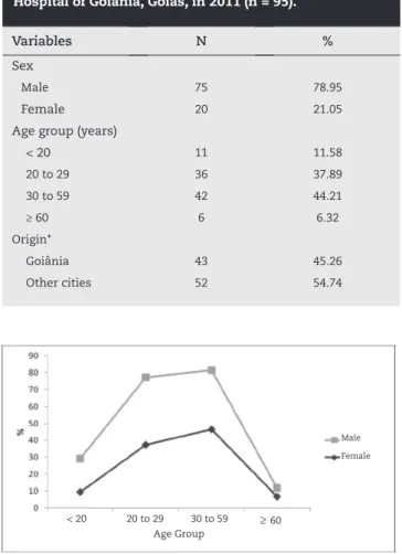
The majority of the patients were male: 75 (78.95%). The age group most affected was between 30 and 59 years (44.21%), followed by the age group from 20 to 29 years (37.89%). Regarding origin, 52 patients (54.74%) were from cities in the state of Goiás other than the state capital (Table 1). After stratification by sex, the age group from 30 to 59 was also the one most affected, both in females and in males (Fig. 1).
Among the men, the body segments that most frequently presented osteomyelitis after exposed fractures were the lower leg bones (32%) and the femur (26.7%). Among the women, there was a higher percentage (40%) of osteomyelitis after exposed fractures in two or more bones (femur and/or tibia), while in 30% of the patients, only the femur was affected (Figure 2).
From analysis on some of the patients’ characteristics according to the type of exposed fracture among the patients with a diagnosis of osteomyelitis, it was observed that individuals in the age group from 30 to 59 years presented greater frequency of type III lesions (51.9%). It was also seen that in patients with type III exposed fractures, the part of the body most affected was the lower leg (36.5%), followed by fractures in two or more bones (28.5%) and then the femur (26.9%). In patients with type II exposed fractures, the femur was more affected (33.3%), while in patients with type I exposed fractures, there were more injuries to the lower leg (50%). There was a statistically significant difference between the exposed fractures of the femur and the other injuries (p = 0.036) (Table 2).
Regarding preoperative baths, it was observed that 90% of the patients with osteomyelitis after type III exposed fractures did not have one. Regarding the time spent waiting for surgery, it was seen that 30.4% of the patients with type III lesions waited for the procedure for 12 to 24 hours (Table 3).
In the 43 cases in which a sample was collected for culturing during the surgical procedure, growth of Staphylococcus aureus
and two or more other etiological agents (multiple microbial agents) was observed in fractures of types I, II and III. However, attention was drawn to the presence of multiresistant
Staphylococcus aureus in type III exposed fractures. Regarding the length of antibiotic use from the time of diagnosis to the clinical cure from osteomyelitis, it was observed that the patients with type III lesions presented greater lengths of antibiotic use (more than six months) (Table 4).
Among the antibiotics used at the time of the patient’s admission, it was observed that, independent of the type of exposed fracture, the majority of the patients used first-generation cephalosporin, followed by a double regimen (first-generation cephalosporin + gentamicin). Among the patients with type III exposed fractures, 50% used first-generation cephalosporin alone. Regarding the definitive antibiotic therapy, it was seen that antibiotics were most used on the fractures alone. However, it was observed that for 10.4% of the patients with type III fractures, it was necessary to use antibiotic regimens, consisting of vancomycin and ciprofloxacin in association (Table 4).
Variables N %
Sex
Male 75 78.95
Female 20 21.05
Age group (years)
< 20 11 11.58
20 to 29 36 37.89
30 to 59 42 44.21
≥ 60 6 6.32
Origin*
Goiânia 43 45.26
Other cities 52 54.74
Table 1 - Characterization according to sex, age and origin among patients with chronic osteomyelitis after exposed fractures who were treated at the Emergency Hospital of Goiânia, Goiás, in 2011 (n = 95).
Fig. 1 - Age distribution according to the sex of the patients with chronic osteomyelitis after exposed fractures who were treated at the Emergency Hospital of Goiânia, Goiás, in 2011 (n = 95).
Fig. 2 - Part of the body affected according to the sex of the patients with chronic osteomyelitis after exposed fractures who were treated at the Emergency Hospital of Goiânia, Goiás, in 2011 (n = 95).
40.0
26.7
2.7 2.7 25.0 32.0 30.0 26.7
4.0 5.0 5.3
Male Female
Humerus Firearm Fem ur
Two or mor e bones
Clavicle/pelvis Low
er le g
Hand/foot
Part of body affected
Male
Female
20 to 29
< 20 30 to 59 ≥ 60
Variables
Type of Fracture
p value* I II III
N(%) N(%) N(%)
Age group (years)
<20 2 (33,3) 1 (11,1) 5 (9,6)
0,304
20 a 29 2 (33,3) 5 (55,6) 17 (32,7)
30 a 59 2 (33,3) 2 (22,2) 27 (51,9)
≥ 60 0 (0,0) 1 (11,1) 3 (5,8)
Part of body affected
Humerus 0 (0,0) 2 (22,2) 2 (3,8)
Forearm 1 (16,7) 1 (11,1) 1 (1,9)
Femur 1 (16,7) 3 (33,3) 14 (26,9)
Lower leg 3 (50,0) 0 (0,0) 19 (36,5) 0,036
Hand/foot 0 (0,0) 0 (0,0) 1 (1,9)
Clavicle/pelvis 0 (0,0) 1 (11,1) 0 (0,0)
Two or more bones 1 (16,7) 2 (22,2) 15 (28,5)
* Fisher’s exact test..
Table 2 - Age group and part of body affected among patients with chronic osteomyelitis, according to type of exposed fracture treated at the Emergency Hospital of Goiânia, Goiás, in 2011 (n = 67).
Variables
Type of fracture
p value* I II III
N(%) N(%) N(%)
Preoperative batha
Yes 1 (16,7) 2 (25,0) 4 (10,0)
0,494
No 5 (83,3) 6 (75,0) 36 (90,0)
Waiting time for surgeryb
Up to 6 hours 1 (20,0) 2 (25,0) 11 (23,9)
0,252
6 to 12 hours 1 (20,0) 3 (37,5) 19 (41,3)
12 to 24 hours 1 (20,0) 2 (25,0) 14 (30,4)
> 1 day 2 (40,0) 1 (12,5) 2 (4,3)
a Data missing in relation to 13 participants; b Data missing in relation to 8 participants; * Pearson’s chi-square test.
Discussion
The majority of the patients were male (78.95%), and this result was similar to what was reported by Muller et al.,7 who
found that 86.3% of their sample were male. Regarding age, the mean age in the present study was 33 years (range: 15 to 76), while Moore et al.8 found a mean age of 31 years. In a study
conducted by Arruda et al.,9 the majority of the patients were
also male, in the proportions of 6.6 men for each woman, and most of them were in an economically active age group (from 21 to 30).
The type of trauma causing the fracture is often related to the severity of the injury caused, and often to the severity of the patient’s condition. In the present study, 26.7% of the men and 40% of the women presented injuries to twp or more bones (multiple trauma). Moore et al.,8 found that 67.3% presented Variables
Type of fracture
p value I II III
N(%) N(%) N(%)
Etiological agenta
Staphylococcus aureus 2 (50,0) 1 (20,0) 3 (8,8)
Staphylococcus aureus (multiresistant) 0 (0,0) 1 (20,0) 21 (61,8)
Enterobacter sp. 0 (0,0) 1 (20,0) 1 (2,9)
Klebsiella sp 1 (25,0) 0 (0,0) 2 (5,9)
0,042
Acinectobacter sp. 0 (0,0) 0 (0,0) 1 (2,9)
Pseudomonas sp. 0 (0,0) 0 (0,0) 2 (5,9)
Multiple microbial agents 1 (25,0) 2 (40,0) 4 (11,8)
Initial antibiotic useb
Ciprofloxacin + clindamycin 1 (33,3) 0 (0,0) 1 (4,2)
First-generation cephalosporin 1 (33,3) 5 (71,4) 12 (50,0)
0,474
Clindamycin + gentamicin 0 (0,0) 1 (14,3) 3 (12,5)
First-generation cephalosporin +
gentamicin 1 (33,3) 1 (14,3) 3 (12,5)
Others (antibiotic regimens) 0 (0,0) 0 (0,0) 5 (20,8)
Definitive antibiotic usec
Vancomycin 1 (0,0) 2 (28,6) 19 (39,6)
Ciprofloxacin 0 (0,0) 2 (28,6) 8 (16,7)
Teicoplanin 1 (25,0) 0 (0,0) 5 (10,4)
Vancomycin + teicoplanin 0 (0,0) 0 (0,0) 6 (12,5) 0,053
Ciprofloxacin + rifampicin 1 (25,0) 1 (14,3) 0 (0,0)
Amikacin 0 (0,0) 0 (0,0) 2 (4,2)
Imipenem 1 (25,0) 1 (14,3) 1 (2,1)
Ciprofloxacin + clindamycin 0 (0,0) 0 (0,0) 3 (4,2)
Others (antibiotic regimens) 1 (25,0) 1 (14,3) 5 (10,4)
Length of antibiotic use (months)d
< 1 2 (50,0) 0 (0,0) 11 (34,4)
0,192
1 a 2,9 2 (50,0) 1 (33,3) 4 (12,5)
3 a 5,9 0 (0,0) 1 (33,3) 6 (18,7)
≥ 6 0 (0,0) 1 (33,3) 11 (34,4)
a Data missing in relation to 24 participants; b Data missing in relation to 33 participants; c Data missing in relation to 8 participants; d Data missing in relation to 28 participants; * Fisher’s exact test.
multiple trauma, while Gustilo10 found this in 30%, probably
because they studied samples at hospitals located close to very busy highways with greater incidence of high-speed accidents. In recent studies, high-speed accidents have been correlated as the biggest causes of severe exposed fractures. These more recent citations denote changes in the types of accident that generate fractures, in line with changes to modern life and advances in technology. This often limits the comparisons that might be made between older and more recent studies, taking into consideration the progressive changes in aggressiveness of the trauma and consequently in the severity of the fractures and the patients’ conditions.11,12
In relation to the classification of the exposed fractures, 51.9% of the patients aged 30 to 59 years in the present study had type III lesions, and this was similar to what was observed by Moore et al.,8 who found that 50.9% had type III lesions,
with a likely influence on the incidence of infection. On the other hand, in a study conducted by Gustilo,10 only 25.5% of the
patients presented type III exposed fractures. This discrepancy occurred through the effect of variables such as the location of the healthcare clinics, degree of urbanization, risk factors and time of conducting the studies.
In the present study, the most severe fractures (type III) occurred mainly in the lower-leg bones, probably through the commonest trauma mechanism, i.e. falling from a motorbike, thus corroborating the findings of Arruda et al.9 It is known
and also formed part of our findings that fractures of the lower leg and femur that occur as type III injuries, are in themselves a major set of predictive factors for infection over the course of the treatment. These fractures require specific care from the time that the patient arrives at the hospital onwards, so that all approaches are designed such that complications, including infections, are prevented.
The exposure duration, i.e. the length of time between the occurrence of the accident and the start of surgical treatment, may be a factor predictive of infection. In the present study, for 30.4% of the patients with type III lesions, this exposure duration was between 12 and 24 hours. In a study by Muller et al.,7 the mean exposure duration was 5 hours and 39
minutes, and an average of 3 hours and 11 minute of precious times were probably consumed by bureaucracy and other measures, such as waiting for X-rays and preparation of the surgical theater, among other matters, which could have been dealt with more speedily and have led to possible diminution of the incidence of infections. Gustillo13 found that the mean
time was 4 hours and 24 minutes between the occurrence of the trauma and the start of the surgical treatment, and that 21.15% were attended after waiting for more than six hours. In our sample, 41.38% of the patients with type III exposed fractures began their treatment between 6 and 12 hours after the accident.
The reasons for greater occurrence of osteomyelitis in exposed fractures, particularly in those of type III, are likely to be connected with the time wasted between the time of the accident and the start of the initial operation (as mentioned above), or to the other particular characteristics of the wounds (high degree of contamination, accident in a rural area, first attendance, etc.) and the removal and transportation conditions.7
In the present study, growth by some pathogenic microorganisms in the different types of exposed fracture was observed. There was growth by Staphylococcus aureus and two or more etiological agents (multiple microbial agents) in exposed fractures of types I, II and III, and predominance of multiresistant Staphylococcus aureus (commonly present in the skin) in type III exposed fractures. These results did not differ much from those presented by Gustilo,10 who found
that Staphylococcus aureus (Gram-positive) predominated, with 60% Gram-positive agents overall. Clifford14 stated that 60% to
70% of the entry cultures were positive, with predominance of Staphylococcus aureus and Enterococcus sp. (Gram-negative). Moore et al.8 reported having found that 71% of the germs were
Gram-positive.
In the present study, it was seen that, independent of the type of exposed fracture, the majority of the patients used first-generation cephalosporin, followed by a double regimen (first-generation cephalosporin + gentamicin), as the initial antibiotic therapy. A study conducted by Muller et al.7 showed
that cefalotin was preferred in 59.8% of the cases, and was well indicated for coverage of Gram-positive germs; the combination of crystalline penicillin + amikacin, which was used on 16.2% of the occasions, may cover positive and Gram-negative germs, but with the disadvantage that crystalline penicillin is generally not a good choice for Staphylococcus. Another, more expensive option would be to use an association of clindamycin + amikacin, with the advantage of coverage against Gram-positive and anaerobic bacteria s (clindamycin) and Gram-negative bacteria (amikacin). Another option would be to choose a single antibiotic in cases of contaminations in urban areas, with preference for cefalotin or cefazolin, while saving the cited associations for cases of contamination in rural areas, in which Gram-negative and anaerobic germs occur more frequently.
In the present study, regarding the definitive antibiotic therapy, it was observed that the antibiotics most used in cases of type II fractures were vancomycin alone, followed by ciprofloxacin alone. However, it was seen that 10.4% of the patients with type III fractures needed to use antibiotic regimens consisting of vancomycin and ciprofloxacin in association.
Conclusions
According to the results from this study, the patients were predominantly male adults presenting exposed fractures. The leg bones were more affected, or two or more bones (multiple trauma). The majority of the patients presented type III injuries (high-energy trauma). It was noted that excessive time elapsed between the accident and the start of emergency surgical treatment, and that Gram-positive germs predominated in the cultures from the material obtained.
antibiotic therapy, exposure duration, bacterial resistance to the antimicrobial agent used, major tissue damage and facture location are extremely important for annulling the predicted effect of infections in exposed fractures.
Acknowledgements
To the Emergency Hospital of Goiania for making it viable to conduct the study; and to the patients who were the study subjects.
Conflicts of interest
The authors declare that there was no conflict of interests in conducting this study.
R E F E R E N C E S
1. Imran Y, Vishvanathan T. Does the right leg require extra protection? Five-year review of type 3 open fractures of the tibia. Singapore Med J. 2004;45(6):280-2.
2. Paccola CAJ. Fraturas expostas. Rev Bras Ortop. 2001;36(8):283-91.
3. Kinik H, Karaduman M. Cierny-Mader Type III chronic osteomyelitis: the results of patients treated with debridement, irrigation, vancomycin beads and systemic antibiotics. Int Orthop. 2008;32(4):551-8.
4. Enninghorst N, McDougall D, Hunt JJ, Balogh ZJ. Open tibia fractures: timely debridement leaves injury severity as the only determinant of poor outcome. J Trauma. 2011;70(2):352-7. 5. Gustilo RB, Anderson JT. Prevention of infection in the
treatment of one thousand and twenty-five open fractures of long bones: retrospective and prospective analyses. J. Bone Joint Surg Am. 1976;58(4):453-8.
6. Koneman EW, Allen SD, Janda WM, Shreckenberg PC, Winn Jr. WC. Diagnostico microbiologico. Rio de Janeiro: MEDSI; 2001. 7. Muller SS, Sadenberg T, Pereira GJC, Sadatsune T, Kimura
EE, Novelli Filho JLV. Estudo epidemiologico, clinico e microbiologico prospectivo de pacientes portadores de fraturas expostas atendidos em hospital universitario. Acta Ortop Bras. 2003;11(3):158-69.
8 Moore TJ, Mauney C, Barron J. The use of quantitative bacterial counts in opens fractures. Clin Orthop. 1989;(248):227-30.
9. Arruda LRP, Silva MAC, Malerba FG, Fernandes MC, Turibio FM, Matsumoto MH. Fraturas expostas: estudo epidemiologico e prospectivo. Acta Ortop Bras. 2009;17(6):326-30.
10. Gustilo RB. Management of open fractures in orthopaedic infection: diagnosis and treatment. Philadelphia: Saunders; 1989. p. 87-117.