EFFECT OF SHORT-TERM ZINC SUPPLEMENTATION
ON ZINC AND SELENIUM TISSUE DISTRIBUTION
AND SERUM ANTIOXIDANT ENZYMES
Andrey A. Skalny
1,2, Alexey A. Tinkov
2,3,4, Yulia S. Medvedeva
5, Irina B. Alchinova
5,
Mikhail Y. Karganov
5, Anatoly V. Skalny
2,3,5, Alexandr A. Nikonorov
41Federal State Scientifi c Institution Institute of Toxicology, Federal Medico-Biological Agency
Bekhtereva 1, St. Petersburg 192019, Russia
2Russian Society of Trace Elements in Medicine, ANO Centre for Biotic Medicine
Zemlyanoy Val 46, Moscow 105064, Russia
3Laboratory of Biotechnology and Applied Bioelementology, Yaroslavl State University
Sovetskaya 14, Yaroslavl 150000, Russia
4Department of Biochemistry, Orenburg State Medical University
Sovetskaya 6, Orenburg 460000, Russia
5Laboratory of Physicochemical and Ecological Pathophysiology, Institute of General Pathology and Pathophysiology
Baltiyskaya 8, Moscow 125315, Russia
6Institute of Bioelementology (Russian Satellite Centre of Trace Element – Institute for UNESCO), Orenburg State University
Pobedy Av. 13, Orenburg 460352, Russia
ABSTRACT
Background. A signifi cant association between Zn and Se homeostasis exists. At the same time, data on the
infl uence of zinc supplementation on selenium distribution in organs and tissues seem to be absent. There-fore, the primary objective of the current study is to investigate the infl uence of zinc asparaginate supplemen-tation on zinc and selenium distribution and serum superoxide dismutase (SOŹ) and glutathione peroxidase (żPx) activity in Wistar rats.
Material and methods. γ6 rats were used in the experiment. The duration of the experiment was 7 and 14
days in the fi rst and second series, respectively. The rats in żroup I were used as the control ones. Animals in żroups II and III daily obtained zinc asparaginate (ZnA) in the doses of 5 and 15 mg/kg weight, respectively. Zinc and selenium content in liver, kidneys, heart, muscle, serum and hair was assessed using inductively coupled plasma mass spectrometry. Serum SOŹ and żPx activity was analysed spectrophotometrically using Randox kits.
Results. Intragastric administration of zinc asparaginate signifi cantly increased liver, kidney, and serum zinc
content without affecting skeletal and cardiac muscle levels. Zinc supplementation also stimulated selenium retention in the rats’ organs. Moreover, a signifi cant positive correlation between zinc and selenium content was observed. Żinally, zinc asparaginate treatment has been shown to modulate serum żPx but not SOŹ activity.
Conclusion. The obtained data indicate that zinc-induced increase in żPx activity may be mediated through
modulation of selenium status. However, future studies are required to estimate the exact mechanisms of zinc and selenium interplay.
INTRODUCTION
Zinc and selenium are essential trace elements due to their cofactor function in a number of enzymatic sys-tems (Kaim et al., β01γ). In particular, these metals are cofactors for Cu, Zn-superoxide dismutase (SOŹ) and glutathione peroxidase (żPx) that play a signifi cant role in maintenance of redox homeostasis (Bettger, 199γ). Zinc and selenium defi ciency is characterised by various clinical signs due to the participation of trace elements in numerous metabolic pathways (Lee, β01β). The inci-dence of poor zinc and selenium status is high in certain territories (Źiplock, 199γ; Prasad, β00γ). It is notable that combined zinc and selenium defi ciency is frequent-ly observed in a number of pathologic states (Barretto et al., β008; Çavdar et al., β009; Khalili et al., β008) as well as in relatively healthy population (Źe Jong et al., β001). Such a fact indicates a signifi cant associa-tion between Zn and Se homeostasis. Moreover, a num-ber of fundamental studies have investigated a chemical basis of this association (Blessing et al., β004; Żeroci et al., β005). źarlier studies have indicated the possibility of mutual infl uence of zinc and selenium in the case of supplementation. In particular, it has been shown that zinc supplementation in dialysis patients increases se-rum selenium concentration (żuo et al., β01γ). A simul-taneous administration of selenium and zinc resulted in enhanced trace elements retention in organs and tissues (Chmielnicka et al., 1988). Moreover, selenium supple-mentation has also been shown to increase tissue seleni-um in animals fed both zinc-adequate and zinc-defi cient diet (Żatmi et al., β01γ). At the same time, data on the infl uence of zinc supplementation on selenium distribu-tion in organs and tissues seem to be absent. Therefore, the primary objective of the current study is to investi-gate the infl uence of zinc asparaginate supplementation on zinc and selenium distribution and serum SOŹ and żPx activity in Wistar rats.
MATERIAL AND METHODS
γ6 male Wistar rats were used in the experiment. The research was approved by the Local źthics Committee. The animals have been acclimatized to the laboratory conditions for two weeks prior to the experiment. The animals were maintained in a laboratory on a regular 1β:1β h light-dark cycle (lights on at 8.00 a.m.) and fed
a standard diet ad libitum. żranulated chow PK-1β0
(“Laboratorkorm” Ltd., Moscow, Russia) containing γ07 kcal/100 g (β0% protein, 70% carbohydrate, 10% fat) was used as a standard diet. Zinc and selenium content in the rat chow was 78.6 ±5.1 and 0.17 ±0.04 μg/g, respectively. The animals received pure drinking water with general mineralization < β50 mg/l.
Two series of experiments were performed. The duration of the experiment was 7 and 14 days in the fi rst and second series, respectively. The rats in żroup I were used as the control ones. Animals in żroups II and III daily obtained zinc asparaginate (ZnA)
Zn(C4NO4H6)β ∙ Zn(OH)β in the doses of 5 (ZnA5) and
15 mg/kg weight (ZnA15), respectively. Zinc aspar-aginate in starch was given by intragastric gavage (at 10.00 a.m.) using silicone fl exible catheters.
Blood was collected via venesection of the jugular vein with subsequent separation of serum. Rats’ liver, kidneys, heart, and muscles (m. gastrocnemius) were collected at the end of the experiment. The organs and tissues were separated from connective tissue and rinsed with ice-cold physiological saline. Hair was col-lected from the cranial part of the spine. The obtained samples were used for subsequent chemical analysis.
The system was prepared in accordance with the manufacturer’s recommendations. Calibration was performed using 0.5, 5, 10, and 50 μg/l solutions of zinc and selenium prepared from Universal Źata Ac-quisition Standards Kit (Perkinźlmer Inc., Shelton, CT 06484, USA) by addition of distilled deionized water acidifi ed with 1% nitric acid. Internal standardization
was performed using yttrium (89Y) isotope Yttrium
(Y) Pure Single-źlement Standard (Perkinźlmer Inc., Shelton, CT 06484, USA) on a matrix containing 8% 1-butanol (Merck KżaA, 64β71 Źarmstadt, żerma-ny), 0.8% Triton X-100 (Sigma-Aldrich, Co., St. Lou-is, MO 6γ10γ USA), 0.0β% tetramethylammonium hydroxide (Alfa-Aesar, Ward Hill, MA 018γ5 USA), and 0.0β% ethylenediaminetetraacetic acid (Sigma-Aldrich, Co., St. Louis, MO 6γ10γ USA). Reference materials were additionally used for quality control. Standard żBW09101 hair sample (Shanghai Institute of Nuclear Research, Shanghai, China) was used as a reference material during hair trace element analysis. Laboratory control of serum analysis was performed using ClinCheck Plasma Control, lot 1β9, levels 1 and β (RźCIPź Chemicals + Instruments żmbH, żermany).
Blood serum was also used for estimation of SOŹ and żPx activity using Randox kits (Randox Labora-tories Ltd., Crumlin, United Kingdom) on an automat-ed biochemical analyser Tokyo Boeki (Tokyo Boeki Machinery Ltd., Tokyo, Japan).
The obtained data are expressed as mean values and the respective standard deviations (mean ±SŹ). One-way ANOVA was used for data processing in or-der to reveal signifi cant infl uence of zinc
supplemen-tation on the studied parameters (p trend of the overall
tendency). Żisher’s Least Signifi cant Źifference test was used for group mean comparisons. Correlation analysis was performed using Spearman’s correlation coeffi cient. The signifi cance level for all statistical
analyses was set as p < 0.05. Statistical treatment of
the data obtained was performed using Statistica 10 (StatSoft Inc., Tulsa, Oklahoma, USA).
RESULTS
The infl uence of zinc asparaginate supplementation on zinc content in organs and tissues
The obtained data demonstrated an increase in zinc content as a result of intragastric gavage of zinc aspar-aginate (Table 1). In particular, administration of 5 and
15 mg/kg ZnA for 7 days resulted in 11 and βγ% el-evation of liver zinc content in comparison to the con-trol values. Similar effect was observed after 14-days treatment. Liver zinc levels in rats obtaining 15 mg/kg ZnA signifi cantly exceeded the respective values ob-tained for the fi rst and second groups by 19 and 15%.
Kidney zinc content was also affected by zinc sup-plementation. 7-days administration of 15 mg/kg ZnA resulted in a signifi cant elevation of kidney Zn levels by 6 and 1β% as compared to the I and II żroup values, re-spectively. Treatment with 15 mg/kg zinc asparaginate for 14 days was associated with a signifi cant 19 and 9% increase in kidney zinc content in comparison to the re-spective values in the control and ZnA5 groups.
M. gastrocnemius zinc content was not signifi -cantly affected by administration of zinc asparaginate in the experimental groups. Moreover, the overall ten-dency was not signifi cant.
Zinc treatment for 7 days was accompanied by a relative decrease in heart zinc content. Oppositely, administration of ZnA for 14 days led to an increase in heart zinc values. However, both tendencies were not signifi cant.
Intragastric gavage of zinc signifi cantly affected serum metal concentration. In particular, treatment with 5 and 15 mg/kg zinc asparaginate for 7 and 14 days resulted in a respective signifi cant γ5 and 45%, and 55 and 107% increase in serum Zn when com-pared to the control values.
Zinc asparaginate supplementation for 7 days did not result in any signifi cant changes in rats’ hair zinc con-tent. At the same time, treatment with 5 and 15 mg/kg ZnA for 14 days increased hair zinc content by 10 and 1β% in comparison to the control level.
It is notable that no signifi cant differences in or-gans zinc content between the respective groups of an-imals treated for 7 and 14 days were observed. At the same time, the overall tendency to treatment-induced increase in zinc content in liver, kidneys and serum was more expressed after 14 days of treatment in ac-cordance with one-way ANOVA results.
The infl uence of zinc asparaginate supplementation on selenium content in organs and tissues
In particular, supplementation with 15 mg/kg ZnA for 7 and 14 days resulted in a signifi cant β8 and 17% increase in liver selenium content in comparison to the control group values. However, zinc-induced se-lenium deposition in liver was less expressed after 14 days of treatment.
It is notable that treatment with 5 mg/kg zinc as-paraginate decreased selenium content in the studied organs. In particular, rats from żroup II were char-acterised by a 16, 70, and β9% decrease in kidney, muscle, and heart content as compared to the control values, respectively. At the same time, intragastric Table 1. Infl uence of zinc supplementation on Zn and Se distribution in organism
Parameter Control (I) ZnA5 (II) ZnA15 (III) p trend
7 days
Zn liver β8.75 ±γ.74 γ1.8β ±β.15 γ5.44 ±4.9γa 0.0β6*
Zn kidney 18.88 ±1.15 18.0β ±0.7β β0.11 ±1.γ7a,b 0.017*
Zn muscle 8.8β ±β,0β 8.70 ±1.9β 10.46 ±γ,16 0.414
Zn heart 17.1γ ±0.77 16.77 ±1.55 16.1γ ±0.61 0.β8β
Zn serum 1.15 ±0.β7 1.55 ±0.ββa 1.67 ±0.β8a 0.0β1*
Zn hair 150.50 ±8.80 151.17 ±6.59 144.8γ ±9.70 0.γ86
Se liver 0.6γ ±0.09 0.58 ±0.05 0.80 ±0.07a,b <0.001*
Se kidney 1.β5 ±0.1β 1.04 ±0.β0a 1.β7 ±0.17b 0.057
Se muscle 0.1β ±0.01 0.04 ±0.01a 0.14 ±0.0βa,b <0.001*
Se heart 0.γ1 ±0.0γ 0.ββ ±0.0γa 0.γγ ±0.0γb <0.001*
Se serum 0.45 ±0.04 0.4γ ±0.06c 0.50 ±0.0γb 0.054
Se hair 0.βγ ±0.01 0.βγ ±0.0γ 0.β6 ±0.0βb 0.087
14 days
Zn liver β8.7β ±β.β4 β9.77 ±1.66 γ4.γ0 ±γ.14a,b 0.00β*
Zn kidney 17.γβ ±1.58 18.86 ±0.97 β0.54 ±1.81a,b 0.007*
Zn muscle 10.67 ±1,γ9 11.94 ±γ,6β 1β.70 ±6,81 0.849
Zn heart 15.99 ±0.69 16.46 ±0.59 16.9β ±1.94a 0.446
Zn serum 0.91 ±0.14 1.41 ±0.γ0a 1.88 ±0.γ0a,b <0.001*
Zn hair 1β4.78 ±11.88 1γ6.04 ±5.49 1γ9.89 ±1γ.8γa 0.077
Se liver 0.60 ±0.05 0.64 ±0.08 0.71 ±0.04a,c 0.0γ5*
Se kidney 1.16 ±0.08 1.14 ±0.11 1.07 ±0.1γc 0.γγ0
Se muscle 0.β5 ±0.05 0.γβ ±0.04a 0.16 ±0.05a,b <0.001*
Se heart 0.β9 ±0.04 0.γβ ±0.0βc 0.γ0 ±0.04 0.β84
Se serum 0.4γ ±0.0γ 0.41 ±0.05 0.4γ ±0.04 0.469
Se hair 0.β8 ±0.0γ 0.γ0 ±0.0γ 0.γγ ±0.0γa,b 0.0β8*
Values are expressed as mean ±SŹ.
aSignifi cant difference in comparison to Control (I) animals (p < 0.05). bSignifi cant difference in comparison to ZnA5 (II) animals (p < 0.05).
administration of 15 mg/kg ZnA resulted in a β8 and β0% elevation of liver and muscle selenium content in comparison to the respective control values. Moreover, the obtained values of selenium content in organs and tissues of rats exposed to 15 mg/kg ZnA signifi cantly exceeded the respective parameters in the żroup II (ZnA5). It has been noted that the overall tendency to zinc-induced changes in selenium content in liver, heart, and muscle was signifi cant in accordance with one-way ANOVA. Such tendency in the case of kid-ney and serum selenium levels was fairly signifi cant.
The reverse character of changes in serum and mus-cle selenium levels was observed after zinc treatment for 14 days. In particular, supplementation of 5 mg/kg zinc asparaginate resulted in increased selenium deposition in organs, whereas administration of 15 mg/kg ZnA was associated with a decrease in these parameters. Thus, the rats from żroup II were characterized by a γ0% elevation of muscle selenium levels as compared to the respec-tive control values. At the same time, treatment with 15 mg/kg weight ZnA decreased muscle selenium content by γ5 and 55% as compared to the żroup I and II val-ues, respectively. The overall tendency to zinc-induced decrease in muscle selenium levels was signifi cant. The overall tendency to zinc-associated increase in hair selenium content was not signifi cant after 7 days of treatment. At the same time, administration of 15 mg/kg weight zinc asparaginate signifi cantly increased hair selenium content by 1γ% as compared to the żroup II values. After β-week treatment the tendency
was signifi cant. In particular, hair Se content in rats obtaining 15 mg/kg zinc asparaginate exceeded the re-spective values in żroup I and II by 18 and 10%.
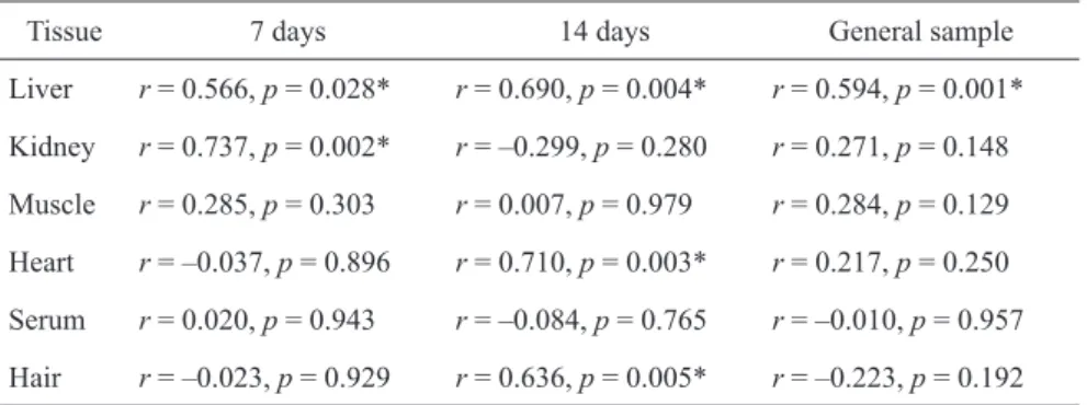
Correlation between zinc and selenium content in organs and tissues
In order to specify the association between zinc and selenium in organs and tissues we have performed cor-relation analysis (Table β). A signifi cant direct associa-tion between Se and Zn in liver was observed in ani-mals in both series of experiments. Positive correlation between these metals in kidneys was also observed in rats involved in an experiment for 7 days. Źirect asso-ciation between zinc and selenium content in heart and hair was observed after 14 days of treatment.
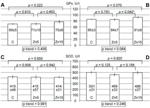
The infl uence of zinc supplementation on serum SOD and GPx activity
It has been estimated that zinc supplementation signifi -cantly affects serum antioxidant enzymes activity (Żig. 1). Źespite a 9% increase in żPx activity in rats obtain-ing 15 mg/kg zinc asparaginate for 7 days, the overall tendency was not signifi cant (Żig. 1A). At the same time, żroup III values of serum żPx activity were 7 and 8% higher than the ones obtained for żroups I and II, respectively. The tendency to zinc-induced increase in żPx activity was nearly signifi cant (Żig. 1B). At the same time, intragastric administration of zinc aspar-aginate did not signifi cantly affect serum SOŹ activity after 7 (Żig. 1C) and 14 days of experiment (Żig. 1Ź).
Table 2. Correlation between zinc and selenium content in the studied tissues and
or-gans in rats treated for 7 and 14 days
Tissue 7 days 14 days żeneral sample
Liver r = 0.566, p = 0.0β8* r = 0.690, p = 0.004* r = 0.594, p = 0.001* Kidney r = 0.7γ7, p = 0.00β* r = –0.β99, p = 0.β80 r = 0.β71, p = 0.148 Muscle r = 0.β85, p = 0.γ0γ r = 0.007, p = 0.979 r = 0.β84, p = 0.1β9 Heart r = –0.0γ7, p = 0.896 r = 0.710, p = 0.00γ* r = 0.β17, p = 0.β50 Serum r = 0.0β0, p = 0.94γ r = –0.084, p = 0.765 r = –0.010, p = 0.957 Hair r = –0.0βγ, p = 0.9β9 r = 0.6γ6, p = 0.005* r = –0.ββγ, p = 0.19β
Correlation analysis revealed a signifi cant as-sociation between serum selenium and żPx activity in rats being treated with zinc both for 7 (r = 0.651, p = 0.006) and 14 days (r = 0.554, p = 0.017). At the same time, zinc and selenium concentration in other tissues and organs did not signifi cantly correlate with żPx and SOŹ activity.
DISCUSSION
The obtained data indicate a signifi cant infl uence of in-tragastric administration of zinc asparaginate on zinc content in various organs and tissues of rats. Total zinc content in the studied organs decreases in the following order: liver > kidney > heart > muscle > serum. These fi ndings are in agreement with previous studies. In par-ticular, it has been shown that administration of zinc oxide nanoparticles results in primary accumulation of zinc in liver as compared to other parenchymatous or-gans (Baek et al., β01β). Moreover, the obtained data
conform to the role of liver as a key organ in zinc ho-meostasis (Żaa et al., β008).
A dose-dependent increase in serum zinc content also confi rms earlier data indicating the effect of oral zinc sulfate supplementation on serum metal concen-tration in volunteers (Samman and Roberts, 1987).
The observed treatment-induced increase in kid-ney zinc content is in agreement with previous studies (Chen et al., 1977). It is proposed that such an effect may occur due to a role of kidneys in zinc excretion (Hambidge et al., 1998).
Źespite the presence of data indicating the role of muscles as regulatory sites of zinc homeostasis (Krebs et al., 1995), short term zinc administration did not re-sult in signifi cant changes of metal levels in muscles (m. gastrocnemius). Taking into account a short period of treatment, the obtained data indicate that muscle tis-sue (both myocardium and skeletal muscles) do not play a signifi cant role in homeostatic regulation of zinc balance in acute period of zinc treatment.
Fig. 1. źffect of zinc supplementation on serum żPx and SOŹ activity. żraph represents
The results of the current study have demonstrated the infl uence of zinc administration on selenium status in rats. Moreover, the obtained data indicate a posi-tive association between zinc and selenium levels in the organism. This observation is in agreement with the previous study indicating the increase of liver and serum selenium levels in response to zinc treatment (żala yn-Sidorczuk et al., β01β). At the same time, the exact mechanisms of zinc-induced increase in se-lenium levels are unknown.
Hypothetically, the infl uence of zinc on selenium status may occur through modulation of one of the stages of selenium homeostasis: absorption, retention, or excretion. źarlier study has indicated that selenium absorption decreased as the zinc content in the diet increased (House and Welch, 1989). However, zinc administration decreased selenium retention only at higher zinc:selenium ratio (10:1) whereas no antago-nism was observed at a β:1 ratio (źybl et al., 1986). At the same time, Chmielnicka and the coauthors have demonstrated lower urinary selenium excretion after zinc treatment (Chmielnicka et al., 1988).
The obtained data indicate a positive infl uence of zinc treatment on serum żPx activity. This observa-tion is in agreement with the earlier study demon-strating an increase in serum zinc and żPx activity in volunteers obtaining zinc supplementation (Kara et al., β010). The observed direct correlation between serum selenium level and żPx activity confi rm ear-lier data (Luoma et al., 1984; Kim et al., β011). Such an association is based on the structural role of sele-nium in żPx molecule (Tappel, β014). Taking into account a signifi cant association between zinc treat-ment, serum selenium, and żPx activity, it may be proposed that zinc-induced increase in żPx activ-ity may be mediated through modulation of sele-nium status. The absence of signifi cant association between zinc levels and serum SOŹ activity is in agreement with the earlier statement (Bettger, 199γ). At the same time, it should be mentioned that only total serum superoxide dismutase activity was ana-lysed in the current investigation, while isolated Cu, Zn-SOŹ characteristics were not studied. Taking into account the obtained results it proposed that antioxi-dant action of zinc may be associated with increased żPx but not SOŹ activity.
żenerally, the results of the current study indicate: 1. Intragastric administration of zinc asparaginate
signifi cantly increases liver, kidney, and serum zinc content without affecting skeletal and cardiac muscle levels.
β. Zinc supplementation signifi cantly increases sele-nium retention in the rats’ organs. Moreover, a sig-nifi cant positive correlation between zinc and sele-nium content exists.
γ. Zinc asparaginate treatment modulates serum żPx but not SOŹ activity.
REFERENCES
Baek, M., Chung, H. ź., Yu, J., Lee, J. A., Kim, T. H., Oh, J. M., Lee, W. J., Paek, S. M., Lee, J. K., Jeong, J., Choy, J. H., Choi, S. J. (β01β). Pharmacokinetics, tissue distri-bution, and excretion of zinc oxide nanoparticles. Int. J. Nanomed., 7, γ081–γ097.
Barretto, J. R., Silva, L. R., Leite, M. ź., Boa-Sorte, N., Pimentel, H., Purifi cação, A. C., Carvalho, ż., Żontes, M. I., Amorim, T. (β008). Poor zinc and selenium status in phenylketonuric children and adolescents in Brazil. Nutr. Res., β8, γ, β08–β11.
Bettger, W. J. (199γ). Zinc and selenium, site-specifi c versus general antioxidation. Can. J. Physiol. Pharmacol., 71, 9, 7β1–7β4.
Blessing, H., Kraus, S., Heindl, P., Bal, W., Hartwig, A. (β004). Interaction of selenium compounds with zinc fi nger proteins involved in ŹNA repair. źur. J. Bio-chem., β71, 15, γ190–γ199.
Çavdar, A. O., żözdaşoğlu, S., Babacan, ź., Mengübaş, K., Ünal, ź., Yavuz, ż., Taçyildiz, N. (β009). Zinc and se-lenium status in pediatric malignant lymphomas. Nutr. Cancer., 61, 6, 888–890.
Chen, R. W., Vasey, ź. J., Whanger, P. Ź. (1977). Accumula-tion and depleAccumula-tion of zinc in rat liver and kidney metal-lothionens. J. Nutr., 107, 5, 805–81γ.
Chmielnicka, J., Zareba, ż., Witasik, M., Brze nicka, ź. (1988). Zinc-selenium interaction in the rat. Biol. Trace źlem. Res., 15, β67–β76.
Źe Jong, N., żibson, R. S., Thomson, C. Ź., Żerguson, ź. L., McKenzie, J. ź., żreen, T. J., Horwath, C. C. (β001). Selenium and zinc status are suboptimal in a sample of older New Zealand women in a community – based study. J. Nutr., 1γ1, 10, β677–β684.
źybl, V., Sýkora, J., Mertl, Ż. (1986). In vivo interaction of selenium with zinc. Acta Pharm. Toxicol., 59, 7, 547–548.
Żaa, ż., Nurchi, V. M., Ravarino, A., Żanni, Ź., Nemolato, S., żerosa, C., Van źyken, P., żeboes, K. (β008). Zinc in gastrointestinal and liver disease. Coord. Chem. Rev., β5β, 10, 1β57–1β69.
Żatmi, W., Kechrid, Z., Nazıroğlu, M., Żlores-Arce, M. (β01γ). Selenium supplementation modulates zinc lev-els and antioxidant values in blood and tissues of dia-betic rats fed zinc-defi cient diet. Biol. Trace źlem. Res., 15β, β, β4γ–β50.
Żeroci, ż., Badiello, R., Żini, A. (β005). Interactions be-tween different selenium compounds and zinc, cadmium and mercury. J. Trace źlem. Med. Biol., 18, γ, ββ7–βγ4. żala yn-Sidorczuk, M., Brzóska, M. M., Rogalska, J., Ro-szczenko, A., Jurczuk, M. (β01β). źffect of zinc supple-mentation on glutathione peroxidase activity and sele-nium concentration in the serum, liver and kidney of rats chronically exposed to cadmium. J. Trace źlem. Med. Biol., β6, 1, 46–5β.
żuo, C. H., Chen, P. C., Hsu, ż. S., Wang, C. L. (β01γ). Zinc supplementation alters plasma aluminum and sele-nium status of patients undergoing dialysis: a pilot study. Nutrients, 5, 4, 1456–1470.
Hambidge, K. M., Krebs, N. Ż., Miller, L. (1998). źvalua-tion of zinc metabolism with use of stable-isotope tech-niques: implications for the assessment of zinc status. Am. J. Clin. Nutr., 68, β, 410–41γ.
House, W. A., Welch, R. M. (1989). Bioavailability of and interactions between zinc and selenium in rats fed wheat grain intrinsically labeled with 65Zn and 75Se. J. Nutr., 119, 6, 916–9β1.
Kaim, W., Schwederski, B., Klein, A. (β01γ). Bioinorganic chemistry – inorganic elements in the chemistry of life: An introduction and guide. John Wiley: Chichester. Kara, ź., żunay, M., Cicioglu, İ., Ozal, M., Kilic, M.,
Mogulkoc, R., Baltaci, A. K. (β010). źffect of zinc
supplementation on antioxidant activity in young wres-tlers. Biol. Trace źlem. Res., 1γ4, 1, 55–6γ.
Khalili, H., Soudbakhsh, A., Hajiabdolbaghi, M., Źashti--Khavidaki, S., Poorzare, A., Saeedi, A. A., Sharifi far, R. (β008). Nutritional status and serum zinc and selenium levels in Iranian HIV infected individuals. BMC Infect. Źis., 8, 165.
Kim, H. H., Yang, H. R., Kim, H. Y. P. (β011). Selenium status and glutathione peroxidase activity in Korean in-fants. Kor. J. Nutr., 44, β, 11β–118.
Krebs, N. Ż., Miller, L. V., Naake, V. L., Lei, S., Westcott, J. ź., Żennessey, P. V., Hambidge, K. M. (1995). The use of stable isotope techniques to assess zinc metabolism. J. Nutr. Biochem., 6, 6, β9β–γ01.
Lee, J. H. (β01β). Micronutrient defi ciency syndrome: zinc, copper and selenium. Pediatr. żastroenterol. Hepatol. Nutr., 15(γ), 145–150.
Luoma, P. V., Sotaniemi, ź. A., Korpela, H., Kumpulainen, J. (1984). Serum selenium, glutathione peroxidase ac-tivity and high-density lipoprotein cholesterol – effect of selenium supplementation. Res. Commun. Chem. Pathol. Pharmacol., 46, γ, 469–47β.
Prasad, A. S. (β00γ). Zinc defi ciency. BMJ, γβ6, 7γ86, 409–410.
Samman, S., Roberts, Ź. C. (1987). The effect of zinc sup-plements on plasma zinc and copper levels and the re-ported symptoms in healthy volunteers. Med. J. Aust., 146, 5, β46–β49.
Tappel, A. (β014). Selenium-glutathione peroxidase: prop-erties and synthesis. In M. ŹeLuca, H. Lardy, R. L. Cross (źds), Current Topics in Cellular Regulation. Vol. β4. źnzyme Catalysis and Control (pp. 87–96). Orlando, Żlorida: Academic Press.
Zhao, L. J., Ren, T., Zhong, R. ż. (β01β). Źetermination of lead in human hair by high resolution continuum source graphite furnace atomic absorption spectrometry with microwave digestion and solid sampling. Analyt. Lett., 45, β467–β481.
Received – Przyjęto: 22.05.2015 Accepted for print – Zaakceptowano do druku: 26.06.2015
For citation – Do cytowania