The Temporal Requirements for Insulin
Signaling During Development in
Drosophila
Alexander W. Shingleton1*, Jayatri Das1, Lucio Vinicius2, David L. Stern1
1Ecology and Evolutionary Biology, Princeton University, Princeton, New Jersey, United States of America,2Leverhulme Centre for Human Evolutionary Studies, University of Cambridge, Cambridge, United Kingdom
Recent studies have indicated that the insulin-signaling pathway controls body and organ size inDrosophila, and most metazoans, by signaling nutritional conditions to the growing organs. The temporal requirements for insulin signaling during development are, however, unknown. Using a temperature-sensitive insulin receptor (Inr) mutation in
Drosophila,we show that the developmental requirements for Inr activity are organ specific and vary in time. Early in development, before larvae reach the ‘‘critical size’’ (the size at which they commit to metamorphosis and can complete development without further feeding), Inr activity influences total development time but not final body and organ size. After critical size, Inr activity no longer affects total development time but does influence final body and organ size. Final body size is affected by Inr activity from critical size until pupariation, whereas final organ size is sensitive to Inr activity from critical size until early pupal development. In addition, different organs show different sensitivities to changes in Inr activity for different periods of development, implicating the insulin pathway in the control of organ allometry. The reduction in Inr activity is accompanied by a two-fold increase in free-sugar levels, similar to the effect of reduced insulin signaling in mammals. Finally, we find that varying the magnitude of Inr activity has different effects on cell size and cell number in the fly wing, providing a potential linkage between the mode of action of insulin signaling and the distinct downstream controls of cell size and number. We present a model that incorporates the effects of the insulin-signaling pathway into theDrosophilalife cycle. We hypothesize that the insulin-signaling pathway controls such diverse effects as total developmental time, total body size and organ size through its effects on the rate of cell growth, and proliferation in different organs.
Citation: Shingleton AW, Das J, Vinicius L, Stern DL, (2005) The temporal requirements for insulin signaling during development inDrosophila.PLoS Biol 3(9): e289.
Introduction
Development in multicellular animals is a process that involves both tight control and flexibility in the regulation of cell size and cell number. Tight control is necessary to produce an animal in which each organ is of an appropriate size relative to the whole body. Flexibility is necessary to produce an animal in which the whole body is of an appropriate size relative to environmental conditions. One key environmental factor shown to influence size is nutrition. In animals as diverse as humans and flies, malnutrition delays development and reduces adult body and organ size. Recent studies indicate that the insulin-signaling pathway (Gene Ontology ID GO:0008286) coordinates growth with nutri-tional condition in most metazoans, and is remarkably conserved. InDrosophila, expression of insulin-like peptides is nutritionally regulated [1], as is expression of IGF1 (insulin-like growth factor 1) in developing rats [2]. Flies carrying mutations of the insulin receptor (Inr) and mice carrying mutations of the IGF1 receptor show delayed development and growth deficiency with a reduction in body and organ size [3–6]. These and similar‘‘knock out’’experiments have demonstrated the gross effects of the insulin-signaling path-way on adult phenotype. Nevertheless, little is known of how the pathway acts during development to affect changes in the adult. In mice, such elucidation is hampered by the inaccessibility of the developing fetus enclosed in the uterus. In flies, however, developing larvae can be easily studied and manipulated.
Here we explore the temporal requirement for insulin signaling in developingDrosophila melanogaster. InDrosophila,
development proceeds through three larval instars to pupariation, pupation, and finally adult eclosion. Feeding is restricted to the first (L1), second (L2), and most of the third (L3) instar. Early in the third instar, the larvae reach a ‘‘critical size’’ at which point they have acquired sufficient nutrients to complete development in the absence of food [7– 10]. After critical size is attained, larvae stop feeding, leave the food, and search for a pupariation site. There is, however, a delay between the time critical size is reached and the time larvae stop feeding and reach their maximum body size. Both critical size and the time between reaching critical size and the cessation of feeding are unaffected by nutrition [8,10]. Consequently, nutrition ostensibly affects final adult size through the amount of feeding in the fixed period between critical size and pupariation [10].
Because feeding affects final size only in the period between critical size and pupariation [8,10], one may predict that insulin signaling affects size only during the same period. However, organ growth continues well after pupariation [11,12] and is autonomously modulated by the
insulin-Received October 1, 2004; Accepted June 17, 2005; Published August 16, 2005 DOI: 10.1371/journal.pbio.0030289
Copyright:Ó2005 Shingleton et al. This is an open-access article distributed under
the terms of the Creative Commons Attribution License, which permits unrestricted use, distribution, and reproduction in any medium, provided the original work is properly cited.
Abbreviations: DD, degree day; JH, juvenile hormone; PI3K, phosophoinositide 3-kinase; TSP, temperature sensitive period
Academic Editor: Laura Johnston, Columbia University, United States of America
signaling pathway [5,13]. Insulin signaling may therefore continue to influence size after pupariation. Details of the temporal requirements for insulin signaling are unknown in
Drosophila, but a recent study on the Drosophila forkhead
transcription factor (dFOXO) suggests that they vary during development [14]. Unphosphorylated dFOXO negatively regulates growth, but activation of the insulin receptor signaling pathway induces dFOXO phosphorylation, exclud-ing it from the nucleus and inhibitexclud-ing its activity [15,16]. Constitutive activation of dFOXO in the first and second instars causes developmental arrest, whereas dFOXO activa-tion in the third instar causes a reducactiva-tion in adult size [14]. Work onCaenorhabditis elegansalso indicates that the timing requirements for insulin signaling might vary during the lifecycle. InC. elegans, the pathway acts during early develop-ment to regulate diapause, and during adulthood to influence ageing [17]. Resolving the links between larval size, nutrition, and developmental progression on the one hand, and the time-dependence of insulin-pathway gene effects on the other, requires a more precise understanding of the temporal requirements for insulin signaling during development.
We have used a temperature-sensitive mutation of the insulin receptor to investigate the role of the insulin-signaling pathway during different stages of development in
Drosophila. By reducing insulin receptor activity at different
points in development, we identify the periods of develop-ment during which insulin signaling affects adult phenotype. We show that total developmental time is affected by changes in insulin signaling only if those changes occur early in development, before a larva has reached critical size. In contrast, final body and organ size are affected by changes in insulin signaling only if those changes occur late in develop-ment, once a larva has passed critical size. Insulin signaling continues to influence organ size, but not body size, after larvae have stopped feeding and throughout much of pupal development. Not all organs respond equally to changes in insulin signaling, however. We find that the genitals show a limited response to suppression of insulin signaling inInrE19/
InrGC25 flies, implicating insulin signaling in the control of allometries.
Results
Temperature-Sensitive Suppression of the Insulin Receptor
During a study of the interaction of environmental factors and the insulin-signaling pathway, we discovered that flies trans-heteroallelic for insulin receptor mutationsInrE19 and InrGC25 show a temperature-sensitive suppression of the insulin pathway. We initially reared InrE19/InrGC25 flies, wild-type Oregon-R flies, and flies homozygous forchico1, at 188C, 258C, and 298C, and measured the wing areas of the adults. Chico is the fly ortholog of the insulin receptor substrate.chico1is a null mutation, and homozygotes show a partial loss of insulin receptor function with a more than 50% reduction in body size relative to wild-type and a much reduced viability [5]. Both Oregon-R andchico1homozygote flies showed a smaller wing area with higher rearing temper-ature (Figure 1). The response to tempertemper-ature is the same in both genotypes (the lines relating wing size to temperature are parallel in Figure 1), indicating that temperature influences size independent of the insulin-signaling pathway.
However, InrE19/InrGC25 flies have a wing size similar to Oregon-R flies when reared at 188C but a wing size similar to
chico1mutants when reared at 258C (Figure 1). This suggests
thatInrE19/InrGC25flies show temperature-sensitive suppres-sion of Inr activity. Changing the rearing temperature does not, however, simply switch Inr‘‘on’’or‘‘off’’inInrE19/InrGC25 flies. At 18 8C, InrE19/InrGC25 flies have smaller wings than wild-type flies, suggesting that the insulin receptor is still partially suppressed at this temperature. Additionally, very fewInrE19/InrGC25flies survived to adulthood when raised at 25 8C. and none survived at 298C. The difference between InrE19/InrGC25 flies reared at 18 8C and 25 8C appears, therefore, to be a consequence of the degree to which Inr is suppressed.
To confirm that these temperature-dependent effects were due to changes in Inr activity, we assessed the activity of the insulin-signaling pathway in InrE19/InrGC25 flies reared at different temperatures. First, we looked at the cellular localization of dFOXO in InrE19/InrGC25 late third instar larvae reared at 178C and at 248C. Down-regulation of the insulin-signaling pathway dephosphorylates dFOXO and causes it to move from the cytoplasm to the nucleus. Antibody stains with anti-dFOXO show an increase in nuclear versus cytoplasmic localization of dFOXO in InrE19/InrGC25 larvae when reared at 248C relative to 178C (Figure 2A and 2B). We did not observe change in nuclear versus cytoplasmic staining forInrE19orInrGC25/TM6B-Tbcontrols reared at 17
8C versus 25 8C (Figure 2C and 2D). Second, we assessed Inr activity inInrE19/InrGC25second instar larvae using thetGPH reporter gene [18].tGPHis under the control of theDrosophila -tubulin promotor and produces a GFP (green fluorescent protein) fused to the pleckstrin homology domain of the receptor for phosphoinositides-1. Under the action of phosophoinositide 3-kinase (PI3K), this fusion protein, GPH, becomes localized to the cell membrane. PI3K is itself activated by the insulin receptor, and so increased local-ization of GPH to the cell membrane indicates increased Inr activity. In InrE19/InrGC25 larvae reared at 15 8C, GPH is localized to the membrane, but this localization is lost
Figure 1.Temperature Sensitivity inInrE19/InrGC25Flies
Increasing the rearing temperature ofInrE19/InrGC25females from 188C to 258C causes a reduction in wing area from approximately wild-type (Oregon-R [Ore-R]) to that of an insulin pathway mutant(chico). Wing size is expressed as percentage area of Oregon-R female wing at 258C. NoInrE19/InrGC25flies survived rearing at 298C. The standard errors are smaller than the markers.
when the larvae are moved to 258C for 12 h (Figure 2E and 2F). Localization does not appear to be lost at 258C inInrE19
or InrGC25/TM6B-Tb controls (Figure 2G and 2H). The
phenotypic effects of temperature onInrE19/InrGC25flies do, therefore, appear to be a consequence of temperature-sensitive suppression of the insulin-signaling pathway. How-ever, we do not know the molecular basis for the temperature sensitivity of the InrE19/InrGC25 transheterozygotes. It may arise from temperature-sensitive expression of one or both of
the mutant alleles or from decreased function of the mutant receptor at higher temperatures.
Due to the low viability ofInrE19/InrGC25flies reared at 25
8C, all subsequent experiments involved comparisons of flies reared at 178C with flies reared at 248C. Further, we used the
InrE19/ TM3 siblings reared under identical conditions as
controls.InrE19/ TM3flies show only a moderate reduction in
Inr activity and have a slight reduction in body size relative to wild-type [4]. Because temperature affects overall body-size in
Figure 2.Increasing the Temperature ofInrE19/InrGC25
Flies Suppresses the Insulin-Signaling Pathway
The dFOXO panel shows localization of dFOXO protein in the fat body, the propidium iodide panel shows the position of the nuclei, and the merge panel clarifies the degree of dFOXO localization to the nuclei.
(A) Endogenous dFOXO in the fat body ofInrE19/InrvGC25third instar larvae has weak nuclear localization at 178C.
(B) Increase in rearing temperature causes a decrease in cytoplasmic distribution and an increase in nuclear localization of dFOXO, consistent with a decrease in the level of insulin signaling
(C and D) Temperature has no detectable effect on dFOXO localization inInrE19/TM3control flies.
(E) GPH membrane localization reveals high levels of insulin signaling in the fat body ofInrE19/InrGC25second instar larvae reared at 158C. GPH is in green, DNA is stained blue. (F) This localization is lost when the larvae are moved to 258C for 12 h, consistent with a decrease in the level of insulin signaling.
wild-type flies [19], it is necessary to distinguish between changes inInrE19/InrGC25phenotype that are a consequence of changes in the level ofInrexpression from those resulting from changes in rearing temperature. To do this we report the phenotype ofInrE19/InrGC25flies as a percentage of the phenotype ofInrE19/ TM3 control flies that have undergone temperature shifts at the same developmental time.
The Changing Role of Insulin Signaling during Development
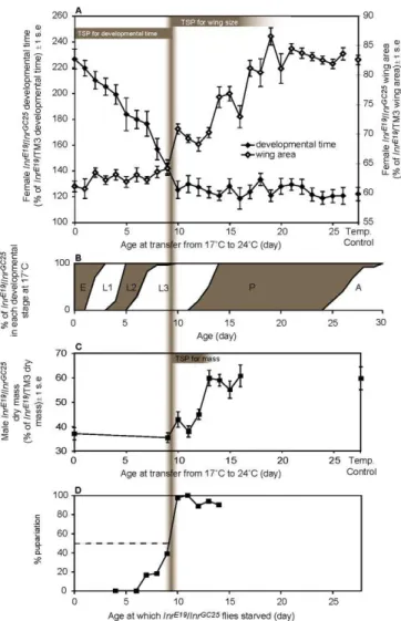
We used the temperature-sensitive Inr mutants to inves-tigate the role of the insulin pathway during Drosophila development. We reduced Inr activity during different periods of development by transferring InrE19/InrGC25 flies from a permissive 17 8C to a restrictive 24 8C at different points in development. After the switch, the flies were left to complete development at 24 8C. We were able to identify temperature-sensitive periods (TSPs) during which increas-ingly earlier switches from 17 8C to 24 8C resulted in increasingly abnormal phenotypes.
Total developmental time is sensitive to Inr activity only before the middle of the third larval instar (Figure 3A). SwitchingInrE19/InrGC25flies from 178C to 248C changes the time to adult eclosion during the first 9 d of development; the earlier the switch, the greater the delay in eclosion. After the ninth day of development at 17 8C, when the flies are approximately 40% through the third instar (Figure 3B), a shift to the restrictive temperature does not delay adult eclosion. At 178C,InrE19/InrGC25flies show a slight delay in eclosion relative to InrE19/ TM3 flies, suggesting that Inr
activity is still a little impaired at this temperature. The delay inInrE19/InrGC25flies reared at both 178C and 248C relative
to the InrE19/ TM3 controls occurs predominantly through extension of the third instar (Figure 4).
In contrast, adult wing area is sensitive to Inr activity only during late third instar and early pupation (see Figure 3A). Switching InrE19/InrGC25 flies from 17 8C to 24 8C changes adult wing size between day 9 and 20; the earlier the shift, the smaller the wings The precise TSP of femaleInrE19/InrGC25 flies for wing area may be smaller than implied by Figure 3A because the data are based on population rather than individual measures. The wings may be insensitive to changes in Inr activity as early as day 17. Before day 9, however, shifting the flies to the restrictive temperature earlier in development has no additional effect on adult wing size. At 178C, the wings ofInrE19/InrGC25flies are slightly smaller than
inInrE19/ TM3control flies, again suggesting that Inr activity
is marginally reduced at this temperature.
We tested whether total body mass was sensitive to Inr activity over the same period as wing size. We compared the dry mass of adultInrE19/InrGC25 males reared under several thermal conditions: 24 8C, 17 8C, and a series of samples switched from 17 8C to 24 8C on days 9 through 16 (pupariation is at approximately day 13 at 178C). Adult body mass is sensitive to reduction in Inr activity between day 9 and day 13 at 178C (see Figure 3C). Shifting InrE19/InrGC25
flies to the restrictive temperature after pupariation, and therefore after larvae have stopped feeding, has no influence on adult mass. Shifting the flies earlier than day 9 has no additional effect on adult body mass.
Inr activity therefore influences total development time, adult wing size, and adult mass for different periods of
Figure 3.Suppression of Inr Expression in InrE19/InrGC25
Flies Affects Developmental Time and Adult Size
(A) Developmental time and adult wing size of InrE19/InrGC25 females switched from 178C to 248C increasingly late in development, expressed as percentage of developmental time and adult wing size ofInrE19/ TM3 females maintained under identical thermal conditions. Temperature-control flies were maintained at 178C throughout development. TSPs of femaleInrE19/InrGC25
for wing area and delayed eclosion can be seen as regions of the chart where switching from 178C to 248C increasingly early in development results in increasingly abnormal phenotypes (that is, where the gradient of the relationship between switch day and phenotype is non-zero). For delayed adult eclosion, the TSP of female
InrE19/InrGC25 is before the ninth day of development at 17 8C. For reduced wing size, the TSP of femaleInrE19/InrGC25is between the ninth and approximately the 20th day of development at 178C.
(B) The stages of development ofInrE19/InrGC25flies at 178C (A, adult; E, embryo; L1, first instar; L2, second instar; L3, third instar; P, pupae). The point at which suppression of the insulin pathway changes from delaying adult development to reducing adult wing size occurs approximately 40% into the third instar (vertical gray bar)
(C) Dry mass ofInrE19/InrGC25
males switched from 178C to 24 8C at different points in development, expressed as percentage of dry mass of
InrInrE19/ TM3males maintained under identical thermal conditions. The TSP of maleInrE19/InrGC25
for reduced adult mass is after the ninth day of development but before pupariation.
(D) Proportion of 178CInrE19/InrGC25larvae pupariating when completely starved at different points in development. The point at which 50% of larvae pupariate in the absence of food marks the critical size. The critical size is reached approximately 40% through the third instar and coincides with the end of the TSP for delayed eclosion and the beginning of the TSP for reduced wing size and adult dry mass (vertical gray bar). All pupariating larvae successfully completed metamorphosis and eclosed as adults.
development. We tested whether the time at which down-regulation of the insulin pathway switches from delaying development to reducing the size of the resulting flies coincides with attainment of critical size. In practice, the critical size is determined as the size at which 50% of larvae proceed to pupariation in the absence of food [8]. We measured the timing of critical size in InrE19/InrGC25 flies reared at 178C under our experimental conditions and found that it is attained at approximately day 9 (see Figure 3D), coinciding with the time at which Inr activity switches from affecting developmental time to body size. Therefore, larval critical size coincides with the shift in Inr function.
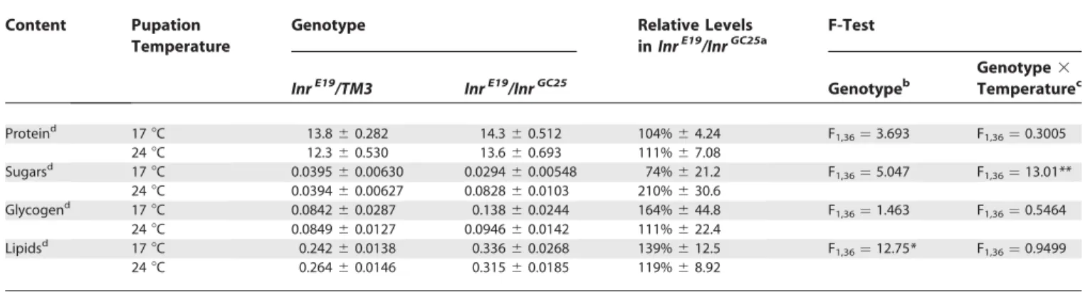
A Reduction in Inr Activity Changes Body Chemistry
To better understand why reduction of Inr activity after pupariation influenced adult wing size but not overall body mass, we measured the protein, lipid, glycogen, and sugar content of InrE19/InrGC25 flies reared at 17 8C up to pupariation, then either switched to 248C or maintained at 178C.
Reducing Inr activity after pupariation results in an approximate doubling of free-sugar concentration inInrE19/ InrGC25flies (Table 1). There is no similar response in protein, glycogen, or lipid levels. Lipid levels are, however, elevated in both InrE19/InrGC25 flies maintained at 17 8C and those
switched to 248C at pupariation relative toInrE19/ TM3flies (Table 1).InrE19/InrGC25flies grown at a restrictive 24 8C for their entire development also have elevated lipid levels (138%
6 13.3 of InrE19/ TM3 flies). This suggests that the slight deficiency in insulin signaling at 178C may account for the entire effect on lipid levels.
A Reduction in Inr Activity Affects Different Organs Differently
Different organs typically grow at different rates in developing animals, a phenomenon called ‘‘allometry.’’ Patterns of allometry in populations can also result from
organs growing at the same rate but starting and stopping growth at different times in development [20]. We inves-tigated whether the insulin-signaling pathway might be involved in the regulation of allometry by examining whether different organs respond differently to reductions in Inr activity induced by a temperature shift ofInrE19/InrGC25flies from 17 8C to 24 8C. We examined organs located at the anterior, median, and posterior of males: the maxillary palp, wing, and genital arch. We chose the maxillary palp rather than another anterior organ because it is similar in size to the genital arch, allowing us to control for absolute size in this comparison.
Figure 5 shows the relative areas of wings, genital arch, and maxillary palps of male InrE19/InrGC25and male InrE19/ TM3 flies reared at either 178C or 248C. At 178C, all three organs are smaller inInrE19/InrGC25males thanInrE19/ TM3males. At 24 8C, the wing and maxillary palps are further reduced in InrE19/InrGC25 males, consistent with a further reduction in the level of Inr activity. In contrast, there is no difference in the size of the genital arches, relative toInrE19/ TM3males, in flies reared at 178C and 248C.
This suggests that the size of the genital arches may not be regulated by Inr activity. However, the arches are smaller in InrE19/InrGC25males than inInrE19/ TM3males at both rearing temperatures. It is possible that this size difference is a consequence of genetic background, unrelated to differences in Inr activity in the two genotypes. Alternatively, the genital arches may show a limited response to changes in Inr activity. They may be sensitive to a mild reduction in Inr activity experienced by InrE19/InrGC25 flies reared at 17 8C, but be insensitive to a further reduction experienced by InrE19/ InrGC25flies reared at 248C.
To distinguish between these two hypotheses, we generated large Minute clones homozygous forchico1. Homozygouschico1
clones produce phenotypes identical to mutantInrclones [13] and autonomously cause a dramatic size reduction in fly wings and eyes, whereas the heterozygouschico1cells behave
as wild-type [5]. We identified males that were homozygous
for chico1 throughout one side of the genitals and
hetero-zygous forchico1on the other. If genital size is independent of the insulin-signaling pathway, then the genitals from either compartment should be identical in size. Each comparison was made within a single male, automatically controlling for total body size. We found that genital arches consisting of mutant chico1 clones were 16% smaller than paired genital arches on the same male (genital arch area:chico1mutant¼ 2,840660lm2,chico1wild-type¼3,2306110lm2,
paired-samplettest:n¼6,t¼1.67,p¼0.0214). This reduction of 16% is consistent with the 16% reduction observed in InrE19/ InrGC25 males relative to controls (Figure 5). In contrast, maxillary palps consisting of mutantchico1clones were 45% smaller than paired palps on the same male (maxillary palp area:chico1mutant¼4,8206100lm2,chicowild-type¼8,640
6170lm2,n¼7). The genital arches do, therefore, show a
limited response to changes in insulin signaling. They are sensitive to a mild reduction in Inr activity, such as observed inInrE19/InrGC25flies reared at 178C. Further reduction in Inr
activity, such as observed inInrE19/InrGC25flies reared at 24 8C, has no additional effect on genital arch size but does have an effect on maxillary palp and wing size.
Figure 4. Developmental Delay in 24 8C InrE19/InrGC25 Flies Occurs primarily through Elongation of the Third Larval Instar
Area shows percentage of individuals (n¼20) in each developmental stage at different times in development. Time is shown in DDs to control for the effect of temperature on developmental rate.
A Reduction in Inr Activity Affects Cell Size and Cell Number Independently
To investigate the cellular basis for the effect of reduction of Inr activity on size, we compared the size and number of epidermal wing cells inInrE19/InrGC25 flies withInrE19/ TM3 flies, reared at 17 8C and 24 8C. The wing-size difference betweenInrE19/InrGC25andInrE19/ TM3flies reared at 178C is due to smaller cells but not fewer cells in the wings ofInrE19/
InrGC25males (compare relative sizes at 178C, Figure 6, Table S2). InInrE19/InrGC25females reared at 178C, cell size is also smaller than inInrE19/ TM3females, but there are also slightly
fewer cells. In both males and females, the additional difference in wing-size observed inInrE19/InrGC25relative to
InrE19/ TM3 flies reared at 24 8C is due to fewer, but not
smaller, cells: Cell size in InrE19/InrGC25 wings relative to
InrE19/ TM3wings is the same at both 178C and 248C (Figure
Table 1.Protein, Sugar, Glycogen, and Lipid Content ofInrE19/InrGC25andInrE19/ TM3Flies Reared at 17
8C until Pupariation and then Switched to 248C or Maintained at 178C
Content Pupation
Temperature
Genotype Relative Levels
inInrE19/InrGC25a
F-Test
InrE19/TM3 InrE19/InrGC25 Genotypeb
Genotype3
Temperaturec
Proteind 178C 13.860.282 14.360.512 104%64.24 F
1,36¼3.693 F1,36¼0.3005
248C 12.360.530 13.660.693 111%67.08
Sugarsd 178C 0.039560.00630 0.029460.00548 74%621.2 F
1,36¼5.047 F1,36¼13.01**
248C 0.039460.00627 0.082860.0103 210%630.6
Glycogend 178C 0.084260.0287 0.13860.0244 164%644.8 F
1,36¼1.463 F1,36¼0.5464
248C 0.084960.0127 0.094660.0142 111%622.4
Lipidsd 178C 0.24260.0138 0.33660.0268 139%612.5 F
1,36¼12.75* F1,36¼0.9499
248C 0.26460.0146 0.31560.0185 119%68.92
aExpressed as percent ofInrE19/ TM3control.
bTests whether protein, sugar, glycogen, or lipid levels differ betweenInrE19/InrGC25andInrE19/ TM3independent of temperature.
cTest whether the effect of temperature on body chemistry differs betweenInrE19/InrGC25andInrE19/ TM3flies, that is, whether a reduction in Inr activity in temperature-sensitiveInrE19/InrGC25flies influences body chemistry.
dAll concentrations are milligrams per milligram of total dry mass.
*p,0.01, **p,0.001.
DOI: 10.1371/journal.pbio.0030289.t001
Figure 5.Different Organs Respond Differently to Suppression ofInrActivity Bars show organ area inInrE19/InrGC25
males as a percentage of area inInrE19/TM3
males, to control for temperature effects. Bars with different letters indicate organs that differ: A, B, and C are significantly different ata¼0.05 (Tukey-Kramer pairwise comparison). Mean areas of all organs given in Table S1. s.e., standard error.
6, Table S2). Reduction in wing area through suppression of the insulin pathway in InrE19/InrGC25 flies is therefore predominantly a consequence of smaller cells at 178C but fewer cells at 248C.
Discussion
Suppressing the insulin-signaling pathway extends devel-opmental time and reduces final adult size inDrosophila. By varying the activity of Inr we have demonstrated that these effects depend on when in development the suppression occurs; Inr suppression affects total developmental time early in development, and body and organ size late in development. The transition from affecting developmental time to affect-ing body and organ size occurs when the fly reaches the critical size, the point at which development can be completed in the absence of food. The effect of reduced Inr activity on size varies from organ to organ, implicating the insulin pathway in the control of allometry inDrosophila. In
addition, varying the magnitude of Inr activity has different effects on cell size and cell number.
Insulin Signaling, Critical Size, and Developmental Delay
We found that a reduction in Inr activity after critical size does not delay development (see Figure 3A). Therefore reducing Inr activity affects total developmental time by delaying the time at which larvae reach critical size. Critical size has been identified as the key stage in insect maturation that determines the point at which, in holometabolous insects, a larva becomes committed to metamorphosis [21]. In Drosophila it is also the minimal viable weight necessary to survive pupation. Consequently, because larvae can complete development without any additional feeding after reaching its critical size, the critical size sets the lower limit of final adult size.
How insects measure critical size is largely unknown. Our results indicate that a reduction in Inr activity delays the point at which Drosophila larvae reach critical size. We hypothesize that critical size measurement involves a specific organ or organs, and that it is the slow growth of this organ or organs that delays development in Inr mutants. Possible candidates include the imaginal discs and the fat body. Regeneration of damaged imaginal discs delays pupariation
in Drosophila [22,23], indicating that some or all of the
imaginal discs need to grow to a particular size before a larva can pupariate. Complete removal of the discs does not, however, delay pupariation [23]. Another organ must there-fore measure critical size and initiate pupariation, with the immature imaginal discs inhibiting pupariation. Slow growth of either the imaginal discs or a ‘‘critical-size organ’’could delay development inInrmutants. Recent studies suggest this ‘‘critical-size organ’’could be the fat body, which functions as a nutrient sensor and is involved in the coordination of organismal growth [24]. Suppressing Inr/PI3K signaling in the fat body alone is sufficient to inhibit larval growth and mimics the effects of starvation [18].
Insulin Signaling and Final Body and Organ Size
A reduction in Inr activity after critical size reduces final adult size and organ size (see Figure 3A and 3C). Inr activity influences adult body size and wing size for different periods of development. As expected, adult body size is influenced by Inr activity only between critical size and pupariation, after which the larva does not feed and becomes a‘‘closed system,’’ neither gaining nor losing mass. However, insulin signaling continues to influence the final size of adult organs well into the pupal stage.InrE19/InrGC25flies switched from 178C to 24
8C at pupariation have the same mass as InrE19/InrGC25flies maintained at 17 8C, but have reduced wings. At the same time, the temperature-shifted flies have a much higher free-sugar concentration as adults. These two findings appear to be linked. Considerable cell proliferation in the imaginal discs occurs after the larva has stopped feeding [11,12,25], and this proliferation relies entirely on stored nutrients as an energy source. Nutrient storage occurs predominantly in the fat body cells, which accumulate reserves of proteins, lipids, and glycogen (the major carbohydrate storage compound) during the third larval instar [18]. Both starvation and suppression of the insulin pathway cause these nutrients to be mobilized for use by growing cells [18]. The finding that InrE19/InrGC25 mutants reared at 24 8C have elevated free-Figure 6.A Reduction in Inr Activity Affects Cell Size and Cell Number
Independently
(A) At 178C, the difference in wing area betweenInrE19/InrGC25 and
InrE19/TM3flies is due to a difference in cell size, whereas at 248C the difference is due to an additional difference in cell number. Bars show wing area, cell area, and cell number in InrE19/InrGC25 flies as a percentage of area or number inInrE19/ TM3flies.
(B) At 178C the reducedInractivity inInrE19/InrGC25
flies reduces cell area to approximately 85% the area inInrE19/ TM3
flies, whereas at 248C there is no further reduction in cell area, but there is a reduction in cell number to approximately 75% of the number inInrE19/ TM3flies. Mean wing and cell area, and cell number are given in Table S2.
sugar levels, but do not have elevated glycogen levels, indicates that they are able to mobilize their carbohydrate reserves, but that the free-sugars are not taken up by growing organs, and remain in the haemolymph.
Insulin Signaling and Cell Size and Cell Number
Any mechanism that influences organ size does so by changing cell size, cell number, or both. Although mutations ofInr,PI3K, andchicoreduce both cell size and cell number [4,5,13,26], downstream components of the insulin-signaling pathway can affect cell size and cell number independently. The RPS6-p70-protein kinase (S6K) branch of the pathway appears to influence cell size but not cell number [27], whereas the dFOXO branch appears to influence cell number but not cell size [15,16]. Because these signaling branches diverge downstream of the insulin receptor, it has not been clear how insulin signaling could affect cell size and number differentially. These changes are ultimately a consequence of changes in the relative rates of cell growth and division [28]. For example, the reduction in cell size, but not cell number, inS6Kmutants implies a reduction in the rate of cell growth but not of cell division. Conversely, the reduction in cell number, but not cell size, when dFOXO is over-expressed implies that the rates of cell growth and division are reduced equally. Finally, a reduction in both cell size and cell number will result if both the rates of cell growth and division are reduced, if the former is reduced to a greater extent than the latter.
Our results support the hypothesis that insulin signaling differentially affects cell size and cell number via different levels of insulin receptor activity. Slightly reduced levels of activity, as inInrE19/InrGC25flies raised at 178C, reduce cell size, possibly through a reduction in the rate of cell growth but not of cell division. Further reductions in the levels of activity, as inInrE19/InrGC25flies raised at 248C, reduces cell number only, possibly through a subsequent balanced reduction in both the rate of cell growth and cell division. These data are consistent with a result from Bohni et al. [5]. They showed that the wings ofchico2homozygotes are smaller due to a reduction of both cell size (17%) and cell number (27%). However, a further reduction in insulin signaling through the removal of a single copy of Inr enhances the small-size phenotype exclusively through a reduction in cell number but not cell size. Although we cannot exclude the possibility that the differences in cell size between InrE19/
InrGC25 and InrE19/ TM3 flies are a consequence of genetic
background unrelated to differences in Inr activity, the reduction in cell number alone is clearly due to reduction in insulin signaling (see Figure 5).
Insulin Signaling and Allometry
Variation in insulin signaling appears to affect the allometric relationship between organs. For example, at 17
8C, maleInrE19/InrGC25flies have body size, wings, maxillary palps, and genital arches approximately 85% of wild-type. Increasing the rearing temperature of InrE19/InrGC25 flies from 178C to 24 8C, however, causes a further reduction in wing, maxillary palp, and overall body size but does not affect the size of the genital arches (Figure 5). Consequently, flies reared at 248C have larger genitals relative to their body and wings compared to flies reared at 17 8C. The apparently restricted response of the genitals to changes in insulin
signaling is not a consequence of the particular alleles used in this study:chico-mutant clones also have much less of an effect on size when they are in the genital arches than when they are in the maxillary palps.
The mechanism by which different organs respond differ-ently to changes in insulin signaling is unclear. Organs may vary in their expression of the insulin receptor gene or may limit the activity of certain downstream components of the insulin signaling pathway. In 17 8C InrE19/InrGC25males, the genital arches, wings, and maxillary palps are all reduced by approximately the same amount relative to 178CInrE19/ TM3 males. In the wing, this reduction in area is a consequence of a reduction in cell size. A further decrease in Inr activity (through an increase in rearing temperature to 248C) reduces wing area through a reduction in cell number alone. If the genital arches are like the wing, then their response to insulin signaling may be restricted to changes in cell size and not cell number. The cells of the genital arches may therefore be deficient in components of the insulin-signaling pathway that regulate cell number but not components that regulate cell size.
A Model of the Insulin-Signaling Regulation of Growth and Development
The insulin-signaling pathway appears to play a different role after critical size than before. Similarly, a temperature-sensitive lethal mutation,l(1)ts-1126,which reduces the rate of cell proliferation in Drosophila, delays pupariation when larvae are moved to a restrictive temperature before the third instar, but reduces adult size when larvae are moved to a restrictive temperature late in the third instar [29]. The two effects of reduced Inr activity may therefore result from the same process: a reduction in the rate of cell growth and proliferation. We have developed a model of the insulin-signaling regulation of growth and development inDrosophila (Figure 7). (A similar model has recently been developed by Davidowitz and Nijhout [30] to explain variation in body size in response to temperature in the tobacco hornworm,
Manduca sexta.) A reduction in Inr activity prior to critical
size slows cell growth and proliferation and delays the time at which the larvae reaches critical size. Critical size is not substantially influenced by nutritional conditions [8] or insulin signaling in Drosophila,although this does not seem to be the case for all insects, for example,M. sexta[21]. Once critical size is reached, the time to pupariation and adult eclosion is fixed, as are the remaining periods of growth prior to adult differentiation of individual imaginal discs. The duration of these intervals are uninfluenced by nutritional conditions or insulin signaling. A reduction in Inr activity during these periods also slows cell growth and proliferation, but now reduces the amount of growth attained before differentiation, resulting in smaller organs and a smaller fly. Because different structures grow for different periods, they are sensitive to Inr activity at different times. For example, wing size begins to be insensitive to changes in Inr activity at approximately the same time as cell proliferation ceases, around 25% into the pupal stage. Adult body mass becomes insensitive to changes in Inr activity just before pupariation, when the larvae stops feeding and final body size is fixed.
signaling. Our data show that the length of the period between critical size and pupariation, the remaining period of growth of the body as a whole, is not substantially affected by a reduction in Inr activity. InDrosophilaand other insects, this interval is controlled by endocrine events. InDrosophila,a small peak in the ecdysteroid titer coincides with attainment of critical size [31], followed by a second peak 12 h later, just before the larvae leave the food [32]. InM. sextathis second peak acts directly on the nervous system to initiate wandering behavior [33], and the same is likely true forDrosophila[34]. The period in which Inr activity can influence final body size is therefore terminated by hormones. Importantly, hormones other than insulin also control the period of cell proliferation in the imaginal discs. For example, inM. sexta, ecdysteroids govern the phases of eye development during metamorphosis [35,36]. When the ecdysteroid titer rises above a minimum threshold just before pupariation, it stimulates a wave of cell proliferation to pass across the eye primordium. This proliferation is sustained until the ecdysteroid titer rises above a maximum level in the middle of pupal development, whereupon cell proliferation stops and maturation of the ommatidia begin. These and similar data in other insects [37– 39], including Drosophila [40–43], suggest that cell prolifer-ation in imaginal discs may be temporally regulated by thresholds of sensitivities to fluctuating levels of ecdysteroids and juvenile hormone (JH) [44]. Different organs have different thresholds of sensitivity and hence grow for differ-ent periods of time. Insulin signaling may therefore control body and organ size by regulating the amount of growth attained during these periods of cell proliferation.
This model requires that changes in ecdysteroid and JH levels are unaffected by the insulin-signaling pathway. It is known that adultInrandchicomutant flies have reduced levels of JH and impaired ovarian ecdysone synthesis [45,46]. However, the same hormone fluctuations that putatively control the period of cell proliferation also initiate
pupar-iation [34] and, because the timing of puparpupar-iation is unaffected by Inr activity, it seems likely that the temporal dynamics of the hormonal cascade in the larvae are also unaffected by Inr activity. We predict, therefore, that the insulin-signaling pathway regulates cell proliferation in imaginal discs but that the duration of proliferative phases are controlled by other endocrine cues, such as JH and ecdysteroids, that are themselves unaffected by the insulin-signaling pathway.
This model demonstrates how the pleiotropic effects of insulin signaling on developmental time and final body and organ size can be separated, and may be available for independent evolutionary modification. For example, changes in the relative size of an organ may occur by organ-specific modifications in its growth response to insulin signaling, through organ-specific changes in the expression of Inr, adjustments in Inr activity, or adjustments in the expression and activity of downstream components of the insulin-signaling pathway. Alternatively, changes in the period of an organ’s growth, through alterations in its sensitivity to other endocrine cues, may have a similar effect [44]. In metazoans in general, each organ has a unique timetable for cellular events in tissue development. Our model, and the data upon which it is based, indicate that in order to understand the effects of insulin signaling on adult phenotype it is necessary to understand how temporal changes in insulin signaling interact with this timetable.
Materials and Methods
Mutant stocks.InrGC25is a chromosomal inversion with a
break-point upstream of the Inr. InrE19is an uncharacterized mutation
induced by ethyl methanosulfonate. Both were described in Chen et
al. [4] and obtained from the Bloomington Stock Center. Thechico1
allele is a P-element insertion allele whose phenotype is similar to the nullchico2allele [5] and was kindly provided by Ernst Hafen. The tub-GFP-PHflies were kindly supplied by Bruce Edgar. The flies were maintained as chico1/CyO [5], InrE19
, and InrGC25 [4] balanced over
Figure 7.A Model of the Insulin-Signaling Regulation of Growth and Development
(A) Under normal conditions, imaginal discs grow to a critical size, which initiates an increase in the ecdysteroid titer. When ecdysteroid levels rise above a maximum threshold, the discs cease cell proliferation and undergo differentiation, fixing their final size. A, adult; E, embryo; L1–L3, first to third larval instar; P, pupa.
(B) InInrmutants, growth of imaginal discs to critical size is slowed, retarding development. When critical size is reached, the ecdysteroid titer again increases, rising above the maximum threshold for cell proliferation in the imaginal discs. Temporal changes in the ecdysteroid titer are unaffected by insulin signaling. Because the rate of cell proliferation is slowed, the imaginal discs are smaller when they begin to differentiate, reducing final organ size. Different discs have different thresholds of sensitivity to ecdysteroid and so cease cell proliferation at different times. Hormones other than ecdysteroids may also be involved.
TM6B-Tb, TM3, TM3-pAct-GFP, and tub-GFP-PH;InrE19/ TM3 [18] at 178C on standard yeast cornmeal agar medium.
Immunolocalization.We crossedInrE19/TM6-TbwithInrGC25
/TM6-Tbflies and reared them at either 178C or 248C. When the larvae
reached third instar, we genotyped them as either insulin receptor mutants (InrE19/InrGC25) or wild-type (InrE19or
InrGC25/TM6B-Tb). The fat bodies were dissected out in PBS, fixed in 4% paraformaldehyde
for 10 min, and stored in absolute methanol at208C. They were
permeabilized with PBT (0.3% Triton X in PBS) for 30 min, washed in BBT (0.3% bovine serum albumin in PBT) for 30 min, then blocked in NGS/BBT (3% normal goat serum in BBT) for 30 min. They were stained with anti-dFOXO 2095 [16] (kindly provided by O. Puig) (1:1,000 in PBT) and fluorescein anti-rabbit (1:500 in PBT) (Vector Labs, Burlingame, California, United States). DNA was stained with
propidium iodide (1:1,000 in PBS with 1lg of RNase A). We mounted
the fat bodies in Vectashield (Vector Labs) for observation under a confocal microscope (Perkin Elmer UltraVIEW RS3; PerkinElmer Life and Analytical Sciences, Boston, Massachusetts, United States).
GPH localization. We crossed tub-GFP-PH;InrE19/TM6B-Tb with InrGC25/TM6B-Tb flies and reared them at 158C. When the larvae reached second instar, they were removed from their food, washed,
and genotyped as insulin receptor mutants (tub-GFP-PH;InrE19/
InrGC25) or wild-type (tub-GFP-PH;InrE19or
InrGC25/TM6B-Tb). They
were then transferred to fresh food and maintained at 158C or 258C for 24 h. We then dissected the larvae in 100% methanol kept at 158C
or 258C depending on the temperature treatment of the larvae, and
stored their fat bodies without additional fixing in 100% methanol at
208C. We quickly washed the fat bodies in 50% methanol in PBS, and then 100% PBS before mounting them in Vectashield with Hoechst 33342 (2:1,000) to stain DNA. We observed the fat bodies under a confocal microscope (Zeiss LSM 510; Zeiss, Gena, Germany).
Temperature shift.We reared flies at a number of temperature regimes. They were either maintained at 178C, 188C, 248C, or 258C
or switched from 178C to 248C on day 1 to day 27 of development.
We set up four bottles for each temperature regime, each containing standard yeast cornmeal agar medium. Two were the product of male InrE19/TM6B-Tband femaleInrGC25/ TM3and two were the product of femaleInrE19/TM6B-Tband maleInrGC25/ TM3. Each bottle contained
between 100 and 200 eggs laid over a 4-h period. Temperature shifts were performed according to the median age of flies in each bottle. Larvae were reared on standard yeast cornmeal agar medium. Total
developmental time was not significantly different betweenInrE19/
InrGC25 flies from the two different crosses, so the data from all
bottles maintained under the same temperature regime were pooled (data not shown).
Measurements and data handling.Bottles were inspected daily at 4:00 PM and any flies that had eclosed in the previous 24 h were collected and preserved in 80% ethanol. Flies were then genotyped and the eclosion times to the nearest day of eachInrE19/InrGC25and
InrE19/ TM3control fly was recorded. We usedInrE19/ TM3rather than InrGC25/TM6B-Tb flies as controls because TM6-Tb has Tubby as a
marker, which affects developmental timing and adult shape [47]. Ten to 15InrE19/InrGC25andInrE19/ TM3female wings from each
temper-ature regime were dissected and mounted in lactic acid:ethanol (4:5). TenInrE19/InrGC25andInrE19/TM3males reared at 248C and 15InrE19/
InrGC25andInrE19/ TM3males reared at 178C were also dissected and their wings, maxillary palps, and genital arches mounted in either lactic acid:ethanol (wings) or Hoyer’s Solution (maxillary palps and genital arches). Digital images of the wings, maxillary palps, and genital arches were captured and measured using IPLab 3.9 (Scanylitics, Fairfax, Virginia, United States). Wing cell size was
estimated using the number of trichomes in a 0.01 mm2 square
between the veins IV and V of the dorsal wing blade. An index of the total number of wing cells was estimated by multiplying the number of trichomes in the area by the total wing size and dividing by 0.01. Dry masses of individual male flies were measured with an analytical balance (Mettler Toledo AX26 DeltaRange; Mettler-Toledo, Colum-bus, Ohio, United States).
The total developmental time for each fly was converted into degree-days (DDs) to control for the effects of temperature on growth rates. DD is a measure of the heat accumulation above a minimal temperature (To), and is calculated as:
Xk
r¼j
ðTrT0Þnr ð1Þ
whereTris the experimental temperature, andnris the number of
days maintained atTr, summed across all experimental temperatures jtok. In this case, we used only two experimental temperatures, 178C and 248C. We calculated the value ofT0such that the developmental
time of control InrE19/TM3 flies was constant, irrespective of
temperature regime. To do this we regressed total developmental time in DDs ofInrE19/TM3flies against the time of their temperature shift from 178C to 248C, using different values ofT0. The value ofT0
that minimized the slope of this regression line was found to be 9.978 8C. WhenT0is 9.9788C, the average developmental time of InrE19/
TM3 flies is 156.04 6 0.338 DD. We then converted the total
developmental time for eachInrE19/InrGC25fly into DDs usingT0¼
9.978, and expressed it as the average percentage of developmental time ofInrE19/ TM3flies (156.04 DD), along with the standard error of
the percentages.
Rearing temperature also affects body, organ, and cell size and cell number [19]. Consequently, all these measurements inInrE19/InrGC25 flies were expressed as a percentage of the value inInrE19/ TM3flies maintained under the same temperature regime. Wing area ofInrE19/ InrGC25females and dry mass ofInrE19/InrGC25males switched from 17
8C to 248C increasingly late in development, was expressed as the
percentage of wing area and dry mass ofInrE19/ TM3flies switched
from 178C to 248C at the same percent of total development at 178C. See Figure S1 for details.
Clonal analysis. We crossed males carrying the alleles chico1/CyO with females carrying the allelesf36a;M(2)Z fþ37C/CyO. Larvae were subjected to X-rays (1,000 rad) at 24–72 h after egg-laying. Genitals and maxillary palps of male offspring without balancer chromosomes were studied forf36abristles.
Developmental staging ofInrE19/InrGC25andInrInrE19/ TM3at 178C
and 248C.We crossedInrE19/ TM3-pAct-GFPflies withInrGC25/
TM3-pAct-GFPflies and collected the eggs over a period of 4 h. After 24 h, we selectedInrE19/InrGC25andInrE19orInrGC25/ TM3-pAct-GFPeggs
using a fluorescence microscope to detect GFP activity. The eggs were
transferred to 12 mm ø Petri dishes containing standard yeast
cornmeal agar medium. Each dish contained 20 eggs of only one
genotype and was maintained at either 178C or 248C. We prepared
three dishes: InrE19/InrGC25 maintained at 17 8C; InrE19/InrGC25
maintained at 248C; andInrE19orInrGC25/ TM3-pAct-GFPmaintained
at 178C. From day 2, ten individuals were randomly selected from
each Petri dish and their developmental stage was recorded using mouthpart development, before being returned to the Petri dish.
Body chemistry assays. White prepupae of InrE19/InrGC25 and
InrE19/ TM3 flies raised at 17 8C were transferred to moistened
Kimwipes (Kimberly-Clark, Neenah, Wisconsin, United States) in Petri dishes. Prepupae of each genotype were split between further
development at 178C and 248C. We collected adults within 8 h of
eclosion, after their cuticle had hardened, and immediately froze them in liquid nitrogen for later analysis. Wet and dry masses of individual flies were measured with an analytical balance (Mettler Toledo AX26 DeltaRange). All metabolic assays were performed on
individual dried males (n¼10 for each assay). Glycogen and sugar
content were measured using a protocol of van Handel [48]. Lipid content was quantified using another protocol of van Handel [49]. To
determine protein concentration, flies were homogenized in 100ll
0.1 M Na2HPO4. Tenll of each sample was combined with the
Bio-Rad protein assay dye reagent (Bio-Bio-Rad Laboratories, Hercules, California, United States) following the manufacturer’s instructions and spectrophotometrically assayed at 595 nm.
Supporting Information
Figure S1.Fitted Values for Wing Aarea ofInrE19/TM3Control Flies
Wing area ofInrE19/InrGC25flies in Figure 2 is expressed as percentage
of wing area ofInrE19/ TM3flies kept under the same temperature
regime, using the fitted values shown on the plot. Temperature affects wing area differently before and after critical size. Consequently, fitted values were calculated by regressing wing area ofInrE19/ TM3
flies against transfer age before critical size (35% development) and after critical size. A similar method was used to determine the dry mass ofInrE19/ TM3males, except a single regression analysis was used to calculate the fitted values.
Found at DOI: 10.1371/journal.pbio.0030289.sg001 (59 KB PDF).
Table S1. Wing, Genital Arch, and Maxillary Palp Area of InrE19/
InrGC25andInrE19/ TM3Males Reared at 178C and 248C
Found at DOI: 10.1371/journal.pbio.0030289.st001 (37 KB DOC).
Accession Numbers
The FlyBase (http://flybase.bio.indiana.edu/search/) accession numbers for the genes and gene products discussed in this paper are Chico (Fbgn0024248), chico1 (FBal0031303), dFOXO (FBgn0038197), Inr (FBgn0013984), InrE19 (FBal0094021), InrGC25 (FBal0010755), PI3K (Fbgn0015279), and S6K (FBgn0015806). The National Center for Biotechnology Information (NCBI) (http://www.ncbi.nlm.nih.gov/) accession number for IGF1 receptor is NM_010513.
Acknowledgments
We thank Fred Nijhout and three anonymous referees for critical and helpful comments on early versions of this manuscript. We thank
Miguel Gaspar for his assistance with imaging. AWS was supported by Princeton University Council on Science and Technology, JD was supported by a Howard Hughes Medical Institute Predoctoral Fellowship. DLS acknowledges the National Institutes of Health, the David and Lucile Packard Foundation, and Princeton University for financial support. LV thanks Michael Akam and his lab for guidance, and St John’s College (Cambridge), the Cambridge Overseas Trust, and the Overseas Award Scheme for funding.
Competing interests.The authors have declared that no competing interests exist.
Author contributions.JD, LV, DLS, and AWS conceived, designed, and performed the experiments. AWS analyzed the data and wrote
the paper. &
References
1. Ikeya T, Galic M, Belawat P, Nairz K, Hafen E (2002) Nutrient-dependent expression of insulin-like peptides from neuroendocrine cells in the CNS contributes to growth regulation inDrosophila. Curr Biol 12: 1293–1300. 2. Shambaugh GE 3rd, Radosevich JA, Glick RP, Gu DS, Metzger BE, et al.
(1993) Insulin-like growth factors and binding proteins in the fetal rat: Alterations during maternal starvation and effects in fetal brain cell culture. Neurochem Res 18: 695–703.
3. Baker J, Liu JP, Robertson EJ, Efstratiadis A (1993) Role of insulin-like growth factors in embryonic and postnatal growth. Cell 75: 73–82. 4. Chen C, Jack J, Garofalo RS (1996) TheDrosophila insulin receptor is
required for normal growth. Endocrinology 137: 846–856.
5. Bohni R, Riesgo-Escovar J, Oldham S, Brogiolo W, Stocker H, et al. (1999) Autonomous control of cell and organ size by CHICO, aDrosophilahomolog of vertebrate IRS1–4. Cell 97: 865–875.
6. Liu JP, Baker J, Perkins AS, Robertson EJ, Efstratiadis A (1993) Mice carrying null mutations of the genes encoding insulin-like growth factor I (Igf-1) and type 1 IGF receptor (Igf1r). Cell 75: 59–72.
7. Beadle G, Tatum E, Clancy C (1938) Food level in relation to rate of development and eye pigmentation inDrosophila melanogaster. Biol Bull 75: 447–462.
8. De Moed GH, Kruitwagen C, De Jong G, Scharloo W (1999) Critical weight for the induction of pupariation inDrosophila melanogaster: Genetic and environmental variation. J Evol Biol 12: 852–858.
9. Bakker K (1961) An analysis of factors which determine success in competition for food among larvae ofDrosophila melanogaster. Arch Neerl Zool 14: 200–281.
10. Robertson FW (1963) Ecological genetics of growth inDrosophila.6. Genetic correlation between duration of larval period and body size in relation to larval diet. Genet Res 4: 74–96.
11. Garcia-Bellido A, Merriam JR (1971) Parameters of the wing imaginal disc development ofDrosophila melanogaster. Dev Biol 24: 61–87.
12. Postlethwait JH, Schneiderman HA (1971) A clonal analysis of development inDrosophila melanogaster: morphogenesis, determination, and growth in the wild-type antenna. Dev Biol 24: 477–519.
13. Brogiolo W, Stocker H, Ikeya T, Rintelen F, Fernandez R, et al. (2001) An evolutionarily conserved function of theDrosophila insulin receptor and insulin-like peptides in growth control. Curr Biol 11: 213–221.
14. Kramer JM, Davidge JT, Lockyer JM, Staveley BE (2003) Expression of DrosophilaFOXO regulates growth and can phenocopy starvation. BMC Dev Biol 3: 5.
15. Junger MA, Rintelen F, Stocker H, Wasserman JD, Vegh M, et al. (2003) The DrosophilaForkhead transcription factor FOXO mediates the reduction in cell number associated with reduced insulin signaling. J Biol 2: 20. 16. Puig O, Marr MT, Ruhf ML, Tjian R (2003) Control of cell number by
Drosophila FOXO: Downstream and feedback regulation of the insulin receptor pathway. Genes Dev 17: 2006–2020.
17. Dillin A, Crawford DK, Kenyon C (2002) Timing requirements for insulin/ IGF-1 signaling inC. elegans. Science 298: 830–834.
18. Britton JS, Lockwood WK, Li L, Cohen SM, Edgar BA (2002)Drosophila’s insulin/PI3-kinase pathway coordinates cellular metabolism with nutri-tional conditions. Dev Cell 2: 239–249.
19. French V, Feast M, Partridge L (1998) Body size and cell size inDrosophila: The developmental response to temperature. J Insect Physiol 44: 1081– 1089.
20. Stern DL, Emlen DJ (1999) The developmental basis for allometry in insects. Development 126: 1091–1101.
21. Davidowitz G, D’Amico LJ, Nijhout HF (2003) Critical weight in the development of insect body size. Evol Dev 5: 188–197.
22. Ursprung H, Hadorn E (1962) [Further research on model growth in combination with partly dissociated wing imaginal disks of Drosophila melanogaster.Dev Biol 4: 40–66.
23. Simpson P, Berreur P, Berreur-Bonnenfant J (1980) The initiation of pupariation inDrosophila: dependence on growth of the imaginal discs. J Embryol Exp Morph 57: 155–165.
24. Colombani J, Raisin S, Pantalacci S, Radimerski T, Montagne J, et al. (2003) A nutrient sensor mechanism controlsDrosophilagrowth. Cell 114: 739–749. 25. Garcia-Bellido A, Merriam JR (1971) Clonal parameters of tergite
develop-ment in Drosophila. Dev Biol 26: 264–276.
26. Weinkove D, Neufeld TP, Twardzik T, Waterfield MD, Leevers SJ (1999) Regulation of imaginal disc cell size, cell number and organ size by Drosophilaclass I(A) phosphoinositide 3-kinase and its adaptor. Curr Biol 9: 1019–1029.
27. Montagne J, Stewart MJ, Stocker H, Hafen E, Kozma SC, et al. (1999) DrosophilaS6 kinase: A regulator of cell size. Science 285: 2126–2129. 28. Neufeld TP (2003) Shrinkage control: Regulation of insulin-mediated
growth by FOXO transcription factors. J Biol 2: 18.
29. Simpson P, Schneiderman HA (1976) A temperature sensitive mutation that reduces mitotic rate inDrosophila melanogaster. Rouxs Arch Dev Biol 179: 215–236.
30. Davidowitz G, Nijhout HF (2004) The physiological basis of reaction norms: The interaction among growth rate, the duration of growth and body size. Integr Comp Bio 44: 443–449.
31. Berreur P, Porcheron P, Berreur-Bonnenfant J, Simpson P (1979) Ecdysteroid levels and pupariation inDrosophila melanogaster. J Exp Zool 210: 347–352.
32. Schwartz MB, Imberski RB, Kelly TJ (1984) Analysis of metamorphosis in Drosophila melanogaster: Characterization of giant, an ecdysteroid-deficient mutant. Dev Biol 103: 85–95.
33. Dominick OS, Truman JW (1985) The physiology of wandering behaviour inManduca sexta.II. The endocrine control of wandering behaviour. J Exp Biol 117: 45–68.
34. Riddiford LM (1993) Hormones andDrosophiladevelopment. In: Bate M, Martinez Arias A, editors. The development ofDrosophila melangogaster.New York: Cold Spring Harbor Laboratory Press. pp 899–939.
35. Champlin DT, Truman JW (1998) Ecdysteroids govern two phases of eye development during metamorphosis of the moth,Manduca sexta. Develop-ment 125: 2009–2018.
36. Champlin DT, Truman JW (1998) Ecdysteroid control of cell proliferation during optic lobe neurogenesis in the mothManduca sexta. Development 125: 269–277.
37. Berreur P, Bougues R (1975) Effects of ecdysone on in vivo growth of wing disks ofCalliphora erythrocephala. J Insect Physiol 21: 915–919.
38. Oberlander H, Leach CE, Shaaya E (2000) Juvenile hormone and juvenile hormone mimics inhibit proliferation in a lepidopteran imaginal disc cell line. J Insect Physiol 46: 259–265.
39. Kawasaki H (1995) . Kawasaki H (1995) Ecdysteroid concentration inducing cell proliferation brings about the imaginal differentiation in the wing disc of Bombyx mori in vitro Dev Growth Differ 37: 575–580.
40. Peel DJ, Milner MJ (1992) The response ofDrosophilaimaginal disk cell-lines to ecdysteroids. Rouxs Arch Dev Biol 202: 23–35.
41. Currie DA, Milner MJ, Evans CW (1988) The growth and differentiation in vitro of leg and wing imaginal disc cells from Drosophila melanogaster. Development 102: 805–814.
42. Chihara CJ, Fristrom JW, Petri WH, King DS (1972) The assay of ecdysones and juvenile hormones onDrosophilaimaginal disks in vitro. J Insect Physio 18: 1115–1123.
43. Cullen CF, Milner MJ (1991) Parameters of growth in primary cultures and cell lines established fromDrosophilaimaginal discs. Tissue Cell 23: 29–39. 44. Emlen DJ, Allen CE (2003) Genotype to phenotype: Physiological control of
trait size and scaling in insects. Integr Comp Biol 43: 617–634.
45. Tu MP, Yin CM, Tatar M (2002) Impaired ovarian ecdysone synthesis of Drosophila melanogasterinsulin receptor mutants. Aging Cell 1: 158–160. 46. Tatar M, Kopelman A, Epstein D, Tu MP, Yin CM, et al. (2001) A mutant
Drosophila insulin receptor homolog that extends life-span and impairs neuroendocrine function. Science 292: 107–110.
47. Craymer L (1980) [New mutants report]. Drosoph Inf Serv 55: 197–200. 48. Van Handel E (1985) Rapid determination of glycogen and sugars in
mosquitoes. J Am Mosq Control Assoc 1: 299–301.