Jebmh.com
Original Article
J. Evid. Based Med. Healthc., pISSN- 2349-2562, eISSN- 2349-2570/ Vol. 3/Issue 45/June 06, 2016 Page 2261
A CROSS-SECTIONAL STUDY OF RETROPHARYNGEAL ABSCESS
Mitta Sreenivasulu1, V. Krishna Chaitanya2
1Associate Professor, Department of ENT, Narayana Medical College & General Hospital, Nellore, Andhra Pradesh. 2Associate Professor, Department of ENT, Narayana Medical College & General Hospital, Nellore, Andhra Pradesh.
ABSTRACT
For many ENT-clinicians, it is very difficult to diagnose the stage of infection and options available for treating retropharyngeal space abscesses (RPSAs) with very limited literature available to focus on the treatment options in limited resource setup. The main cutting edge of the study aims to correlate post-surgical complications of RPSAs and also to know the age related incidence.
MATERIALS AND METHODS
A prospective and retrospective study was undertaken in the Department of ENT, Narayana Medical College & General Hospital, Nellore, Andhra Pradesh during the period of 2011-15. The incumbent laboratory parameters like throat swab culture was done for all the suspected patients along with Complete blood counts (CBC) & X-ray neck.
RESULTS
A total sixty suspected paediatric patients were prospectively and retrospectively studied for the period of four years, out of which males 35 and females were 25 respectively. The mean age of the patient was 8.96±1.25 years (IQR 4-14 years) median age was 10 years. Radiological examination and Computed tomography (CT) scan was done for greater accuracy. Blocked airway is most common postsurgical intervention and it was found to be statistically significant (p<0.00) with respect to lower age group of the population.
CONCLUSION
The present study concludes that proper positioning and avoidance of unnecessary manipulation is essential for preventing the postoperative complications of RPSAs. The spread of infection to the spine can lead to replicate the osteomyelitis and vertebral erosion, which in turn results in subluxation and subsequent spinal cord injury, rupture of the abscess with inhalation of contents can lead to aspiration pneumonia (or rarely asphyxiation) and spread of infection to mediastinum can lead to mediastinitis. More research could be intervened to prevent the infections at poor resource setup.
KEYWORDS
Retropharyngeal Abscess, Demographic Profile, Surgical Intervention, Prospective Design.
HOW TO CITE THIS ARTICLE: Sreenivasulu M, Chaitanya VK. A cross-sectional study of retropharyngeal abscess. J. Evid. Based Med. Healthc. 2016; 3(45), 2261-2264. DOI: 10.18410/jebmh/2016/501
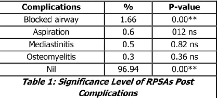
INTRODUCTION: Retropharyngeal abscess is a serious and occasionally life treating infection due to the anatomic location and the potential for obstruction of the upper airway. Deep infections of the head and the neck have been recognised since the time of the Greek physician Galen.1 According to Holmes (1907), Galen referred to a case of retropharyngeal abscess.1 Knowledge of the retropharyngeal space and its relationship to the other compartments is important in understanding the presentation, treatment and complications of deep neck infections. The retropharyngeal space of Gillette or the posterior space of Grodzinski, or Holyoke's space lies behind the pharynx between the buccopharyngeal fascia, which covers the constrictor muscle, and the prevertebral fascia.
It extends from the base of the skull to the tracheal bifurcation.2 The space is divided into two lateral compartments (The space of Gillette) by a fibrous raphe. Lymph glands lie in the space in 4 groups, 2 on each side of the midline. It is claimed that the lateral group of the two is the most important, constantly present in children and give rise to abscess formation. Acute retropharyngeal abscess occurs mainly in infancy and in children usually under 4-5 years of age.3 Waugh regards the tonsil as the source of infection, as the condition is never found in children whose tonsils have been removed.
Definite enlargement of the tonsils is present in about 80% of patients who suffer from retropharyngeal abscess.3 It usually needs to be drained and treated with antibiotics. Because RPSAs typically occur in deep tissue, they are difficult to diagnose by physical examination alone. The signs and symptoms of retropharyngeal abscess are difficulty in breathing, difficulty in swallowing, drooling, fever, severe throat pain, stridor (a high-pitched wheezing during inhalation), muscle spasms around the ribs when breathing.
Financial or Other, Competing Interest: None. Submission 29-02-2016, Peer Review 15-03-2016, Acceptance 25-05-2016, Published 06-06-2016. Corresponding Author:
Dr. Mitta Sreenivasulu,
Associate Professor, Department of ENT,
Narayana Medical College & General Hospital, Quarters-98, Nellore, Andhra Pradesh.
Jebmh.com
Original Article
J. Evid. Based Med. Healthc., pISSN- 2349-2562, eISSN- 2349-2570/ Vol. 3/Issue 45/June 06, 2016 Page 2262 For many ENT-clinicians, it is very difficult to diagnose
the stage of infection and also for treating retropharyngeal space abscesses (RPSAs) with very limited literature available to focus the treatment options in limited resource setup. The main cutting edge of the study aims to correlate post-surgical complications of RPSAs and also to know the age related incidence.
MATERIALS AND METHODS: A prospective and
retrospective study was undertaken in the Department of ENT, Narayana Medical College, Nellore, Andhra Pradesh during the period of 2011-15. The incumbent laboratory parameters like throat swab culture was done for all the suspected patients, Complete blood counts (CBC), X-ray radiological impressions and Computed tomography (CT) scan.4,5,6 was done with greater accuracy. The demographic profile and other defined parameters were collected from the pretested questionnaires and also exposed risk factors were collected and correlated with infection distribution. The collected data was analysed by using SAS-16.50 version7. Univariate analysis was performed to test the hypothesis.
RESULTS:
Fig. 1: Gender Distribution
Fig. 2: Family Economic Status
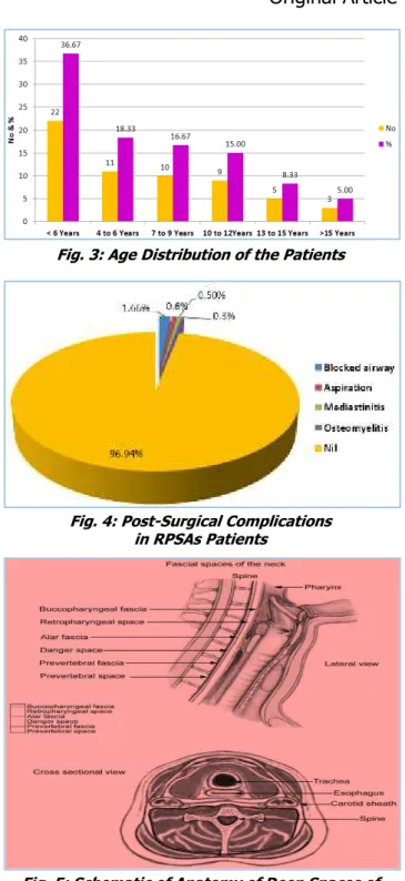
Fig. 3: Age Distribution of the Patients
Fig. 4: Post-Surgical Complications in RPSAs Patients
Fig. 5: Schematic of Anatomy of Deep Spaces of Neck, As Illustrated in Lateral and Cross-Sectional
Views Fascial planes Surround Potential Spaces
Jebmh.com
Original Article
J. Evid. Based Med. Healthc., pISSN- 2349-2562, eISSN- 2349-2570/ Vol. 3/Issue 45/June 06, 2016 Page 2263
Complications % P-value
Blocked airway 1.66 0.00**
Aspiration 0.6 012 ns
Mediastinitis 0.5 0.82 ns
Osteomyelitis 0.3 0.36 ns
Nil 96.94 0.00**
Table 1: Significance Level of RPSAs Post Complications
** Significant at 1% level, ns-Non significant.
The results revealed that a total sixty suspected paediatric patients were prospectively and retrospectively studied for the period of four years, out of which males were 35 and females were 25 respectively. The mean age of the patient was 8.96±1.25 years (IQR 4-14 years), median age was 10 years. As per the demographic profile, the RPSAs are categorised into three groups viz., low income 76.0%, medium 14.0% and high income was 10% presented Fig (1) & (2). The maximum number of children were affected in the age group between 3-6 years (36.67%) and it was found to be statistically significant (p<0.01) followed by 6 to 8 years and >10 years respectively. The post-surgical complications were correlated by univariate analysis, the results revealed that blocked airway accounted 1.66%, aspiration was 0.60%, mediastinitis was 0.50% and osteomyelitis was 0.30% respectively. Blocked airway is most common after post-surgical intervention and it was found to be statistically significant (p<0.00) with respect to lower age group of the population.
DISCUSSION: RPA is the collection of pus in retropharyngeal space.8,9 It can progress suddenly from an indolent, contained disorder to a rapidly progressive and life threatening infection. Several studies have reported that over 95% of RPA cases occur in children under the age of 6 years. In children, RPA is mostly associated with viral upper respiratory tract infection, pharyngitis and otitis media. All these infections cause adenopathy of retropharyngeal lymph nodes and suppuration giving rise to RPA formation.8,9 The most frequently reported organisms previously in RPA
culture included group A β-haemolytic streptococci, S.
aureus, Haemophilus influenza and Klebsiella species.10,11 Grisaru-Soen et al6 along with others reported that children with retropharyngeal abscess most commonly present with
restricted neck movements, fever and cervical
lymphadenopathy and rarely with respiratory tract symptoms such as stridor or airway obstruction. Lateral cervical radiograph was diagnostic in 80% and a cervical CT scan in 95%.12,13 ,14
The present study has documented to focus on uncomplicated cases of retropharyngeal abscess who have been relatively healthy paediatric patients; the prognosis was good with complete recovery without sequelae. Complicated cases are associated with significant changes noticed after the surgical intervention. A total 4.0% of the patients had acquired with defined complications and positively associated with risk factors.
Many authors suggest that the assessment of a patient with a potential deep neck space infection was more difficult and found to be statistically significant (p<0.01). Airway compromises should be immediately identified and addressed. Al Sabah et al5 proposed that all patients should be given a trial of medical treatment with intravenous clindamycin. Surgery should be reserved for those who do not respond. Sometimes hoarseness, stridor and respiratory obstruction may also develop either due to anterior displacement of posterior pharyngeal wall by the abscess or secondary laryngeal oedema. Large abscesses were noticed during the study period. The abscess was ruptured and causing asphyxiation or aspiration and pneumonia like infections. The proper positioning and avoidance of unnecessary manipulation is essential for preventing the complications of RPSAs. Close monitoring of patients with airway compromise is very important. In our study, the postoperative complications we came across were blocked airway, aspiration, mediastinitis and osteomyelitis.
CONCLUSION: The present study concludes that proper positioning and avoidance of unnecessary manipulation is essential for preventing the postoperative complications of RPSAs. The spread of infection to the spine can lead to replicate osteomyelitis and vertebral erosion, which in turn results in subluxation and subsequent spinal cord injury, rupture of the abscess with inhalation of contents can lead to aspiration pneumonia (or rarely asphyxiation) and spread of infection to mediastinum can lead to mediastinitis. More research could be intervened to prevent the infections at poor resource setup.
REFERENCES
1. Manuel Grodinsky. Retropharyngeal and lateral
pharyngeal abscesses: an anatomic and clinical study. Ann Surg 1939;110(2):177-199.
2. Taiwo GA Ijaduola. Patterns of retropharyngeal
abscess in Nigerian child. J Nat Med Assoc 1986;78(1):72-73.
3. Guthrie D. Acute retropharyngeal abscess in
childhood. Br Med J 1926;2(3441):1174-1175. 4. Lalakea MI, Messner AH. Retropharyngeal abscess
management in children: current practices.
Otolaryngol Head Neck Surg 1999;121(4):398-405.
5. Al-Sabah B, Bin Salleen H, Hagr A, et al.
Retropharyngeal abscess in children: 10-year study. J Otolaryngol 2005;33(6):352-355.
6. Grisaru-Soen G, Komisar O, Aizenstein O, et al. Retropharyngeal and parapharyngeal abscess in
children-epidemiology, clinical features and
treatment. Int J Pediatr Otorhinolaryngol
2010;74(9):1016-1020
7. Ko-Villa E, Munyarugero E. Anaesthetic management
Jebmh.com
Original Article
J. Evid. Based Med. Healthc., pISSN- 2349-2562, eISSN- 2349-2570/ Vol. 3/Issue 45/June 06, 2016 Page 2264 8. Seid AB, Dunbar JS, Cotton RT. Retropharyngeal
abscesses in children revisited. Laryngoscope 1979;89(11):1717-1724.
9. Gates GA. Deep neck infection. Am J Otolaryngol 1983;4(6):420-421.
10. Asmar BI. Bacteriology of retropharyngeal abscess in children. Pediatr Infect Dis J 1990;9(8):595-597. 11. Brook I. Microbiology of retropharyngeal abscesses in
children. Am J Dis Child 1987;141(2):202-204.
12. Craig FW, Schunk JE. Retropharyngeal abscess in children: clinical presentation, utility of imaging, and current management. Pediatrics 2003;111(6 pt 1):1394-1398.
13. John Grosso, Charles M Meyer.
http://archpedi.ama-assn.org/cgi/reprint/144/12/1349.
14. McLeod C, Stanley KA. Images in emergency