Original Article
Artigo Original
Speech-language pathology assessment for
tracheal decannulation in patients suffering
from traumatic brain injury
Avaliação fonoaudiológica para decanulação
traqueal em pacientes acometidos por
traumatismo cranioencefálico
Isabel de Lima Zanata1
Rosane Sampaio Santos2
Jair Mendes Marques2
Gisela Carmona Hirata3
Daiane Aparecida dos Santos4
Keywords
Traumatic brain injury Tracheostomy Weaning Dysphagia Speech-language Therapy
Descritores
Traumatismo Cranioencefálico Traqueostomia Desmame Disfagia Fonoaudiologia
Correspondence address: Isabel de Lima Zanata
Rua Lothario Boutin, 90, Curitiba, (PR), Brazil, CEP: 81750-450. E-mail: [email protected]
Received: August 26, 2014
Accepted: September 01, 2014 Study carried out at Hospital do Trabalhador – HT – Curitiba (PR), Brazil.
1 Hospital do Idoso Zilda Arns - Curitiba (PR), Brazil. 2 Universidade Tuiuti do Paraná – UTP - Curitiba (PR), Brazil. 3 Hospital Infantil Waldemar Monastier - Curitiba (PR), Brazil. 4 Hospital do Trabalhador do Paraná – HT - Curitiba (PR), Brazil.
Financial support: nothing to declare.
Conlict of interests: nothing to declare.
ABSTRACT
Purpose: To describe the effect of Speech-Language Pathology (SLP) management on the tracheal decannulation process in patients with traumatic brain injury (TBI). Methods: Prospective controlled clinical study. Two groups
of patients with TBI conirmed by computed axial tomography were included in the study group (G1) and control group (G2) composed of 30 individuals each, with 25 (83.3%) male and 5 (16.7%) female individuals in both
groups. Patients’ age ranged from 18 to 53 years old – mean age was 32 years. A SPL assessment tool was developed for tracheostomized patients with TBI, composed of investigation of awareness level, cognition and
swallowing (annex 1) and conduct. G1 underwent the assessment proposed by the study, and G2 was assessed
by retrospective analysis of medical records without SLP evaluation. In this population, the variables time with tracheostomy and total days of hospitalization were the measurement markers for the effect of SLP conduct with this instrument. Results: It was veriied that G1 presented mean reduction of 4.2 days with tracheostomy
and of 4.4 days in length of hospital stay when compared to G2. However, these igures are not statistically signiicant (p = 0.2031). Conclusion: The group that was evaluated and received the SLP conduct proposed in the instrument presented a reduction in the time of permanence with tracheostomy, as well as in hospital stay.
RESUMO
Objetivo: Descrever o efeito da conduta fonoaudiológica no processo de decanulação traqueal em indivíduos com traumatismo cranioencefálico (TCE). Método: Estudo clínico transversal prospectivo controlado. Participaram
deste estudo dois grupos de indivíduos com TCE conirmado por tomograia axial computadorizada, sendo o grupo de estudo (G1) e o grupo controle (G2) compostos por 30 indivíduos cada, com 25 (83,3%) indivíduos do gênero masculino e 5 (16,7%) do gênero feminino em cada grupo. A faixa etária variou de 18 a 53 anos,
com média de 32 anos. Foi elaborado um instrumento de avaliação fonoaudiológica para indivíduos com TCE
traqueostomizados composto por investigação do nível de consciência, cognição e deglutição (anexo 1) e conduta. O G1 recebeu a avaliação proposta pelo estudo e o G2, análise retrospectiva de prontuário sem avaliação
fonoaudiológica. As variáveis tempo de permanência com a traqueostomia e total de dias de internamento foram os marcadores de mensuração do efeito da conduta fonoaudiológica com esse instrumento nessa população. Resultados: Veriicou-se que o G1 obteve uma redução média de 4,2 dias de permanência com a traqueostomia
INTRODUCTION
According to the National Head Injury Foundation, traumatic
brain injury (TBI) is an aggression to the brain caused by external
physical force that may lead to a reduced state of consciousness and results in the impairment of cognitive skills or physical
ability, and may be classiied, according to the Glasgow score
coma scale, as mild (13-15), moderate (9-12) and severe (3-8)(1). The main causes of TBI are car accidents, but it can be
caused by falls, aggressions, white weapon or gunire bullets.
The patient’s prognosis depends on anatomoclinical and evolutionary aspects of trauma, among which we highlight the
extent of the injury, the initial score on the scale of Glasgow, the
response to treatment, the presence of global or focal lesions, associated injuries, age, comorbidities and time of clinical and surgical interventions(1,2).
People affected by TBI may present temporary or permanent disabilities. The main consequence is brain damage due to edema or bleeding due to trauma, which results in increased intracranial pressure, causing diverse sequelae which severity depends on the affected area(3).
Dysphagia is among the observed sequelae in trauma patients. Besides the concern with nutrition, the importance of dysphagia focuses on the prevention of tracheal aspiration. It has
been pointed that between 40% and 60% of cases with TBI has
dysphagia. Intubation and tracheostomy can also be associated, which are procedures needed and common in severe TBI(4).
In addition to the concern of nutritional support adequate to the person with TBI, it is essential that the interdisciplinary team carefully evaluate the most appropriate nutrition in each case. When the individual presents with dysphagia as one of the TBI sequelae, the early oral food intake permission may lead to clinical complications that can compromise the nutritional and pulmonar clinical setting. The dysphagia diagnosis may be
deined by clinical and objective assessment, such as swallowing
videofluoroscopy and functional nasolaringofibroscopy. The rehabilitation of people with dysphagia aims to allow
eficient and safe oral intake, contributing to the nutritional state
stabilization and to eliminate the risks of clinical complications due to laryngotracheal aspiration(5).
The results depend on variables as consciousness level, patient age, possibility of clinical complications, use of tracheostomy and mechanical ventilation(6,7).
Tracheostomy, in turn, is indicated when there is obstruction of the upper airways when the patient needs prolonged mechanical
ventilation and/or have dificulty weaning, for excessive
tracheobronchial secretions and the patient needs continuous airway protection against tracheal aspiration(8).
Dysphagia has close relationship with the tracheostomy, not only because this procedure is indicated for patients with swallowing problems and tracheal aspiration, but also because the very tracheostomy may cause aspiration because it interferes directly in the pharyngeal phase of swallowing(9).
Approximately 10% of critically ill patients are submitted
to tracheostomy and ventilatory support and facilitating the passage of air in the airways, allowing a better quality of life for patients and minimizing possible injuries from prolonged
mechanical ventilation. However, the frequency of tracheostomy in patients affected by TBI contrasts with the lack of criteria for tracheal decannulation(10).
It is observed that it is common to follow speciic rules
tracheostomy indication, but there is still no decisive rules for decannulation process. In most hospitals, an interdisciplinary team is responsible for this process and the interaction between health professionals can speed up the removal of the tracheal tube, making it safer for the patient, with less risk of failure and complications.
The tracheostomy decannulation is considered as the
moment when the cuff begins to delate, through the plastic
cannula to the metal, until the removal of the tracheostomy cannula and the stoma occlusive curative. The decision of when to start decannulation tracheostomy should result from teamwork, emphasizing that predictors of failure need to be absent, such as sedation, mechanical ventilation, respiratory failure, presence of airway obstruction by edema, tumor or other causes, previous surgery of the head and neck, vocal fold paralysis and glottis stenosis or subglottic that must be remedied and the upper airway, restored to the proper passage
of air low. The management of tracheal decannulation varies
in each institution, some consider it feasible if the patient keeps the cannula tracheostomy occluded for 48 hours or more, while others consider it as a line valve is tolerated(11).
Thus, an early integrated approach is necessary, with the participation of the SLP in order to optimize the patient’s general condition, favoring rehabilitation and reduction in hospitalization time(12). Several studies in the last decade(8,10,11,13-18) point out to the need of objective protocols for tracheal decannulation.
A study performed with tracheostomy patients with TBI
showed six criteria for tracheal decannulation. Among the
language pathology criteria are: level of consciousness with
a score on the Glasgow coma scale greater than 8, present
condition to maintain the cuff delated and cannula occluded,
keeping a respiratory pattern, absence of orotracheal discharge
or in minimum amount, with luid aspect, phonation with no
clinical signs of “wet” voice, swallowing with no clinical signs
of tracheal aspiration and presence of eficient voluntary cough. The authors correlated the six criteria with decannulation and,
following the data analysis, observed that all criteria were
signiicant when deciding for the decannulation. This led to
the creation of the Speech-Language Pathology Protocol for Tracheal Decannulation (SPTD) of patients suffering from TBI(19).
This study aimed to describe the effect of the SLP assessment in the tracheal decannulation process in two groups of people with tracheostomy and traumatic brain injury (TBI).
METHODS
It is a cross-sectional and comparative study. The sample
was composed of the study group (G1) with 30 patients admitted
from January to December 2012, affected by traumatic brain
injury (TBI), conirmed by computed tomography (CT), being 25 (83.3%) male and 5 (16.7%) were female. The age of the
and standard deviation of 10,8 years. The degree of TBI was evaluated by the medical team and entered in the patient record.
Medical records of 30 patients who constituted the control
group (G2), hospitalized in the period from January to December 2011, affected by TBI, conirmed by CT, were also analyzed, being 25 (83.3%) male and 5 (16.7%) female. The age of the
patients varied from 19 to 52 years, average of 32.1 years and standard deviation of 11,0 years. The degree of TBI was in the medical record.
The study was conducted in Hospital do Trabalhador do Paraná in the city of Curitiba, in the department of Neurology, including patients admitted to hospital or units of intensive or semi-intensive therapy, and evaluated upon request and/or released by the treating physician. The analysis of the medical records was performed in the hospital archives.
The study was conducted in two stages: the irst stage
consisted of clinical assessment, according to the data of the
SLP Tool for Tracheal Decannulation (STTD) (Annex 1).
The SLP assessment was performed when requested and/or released by the physician responsible. The average time between the tracheostomy and the SLP assessment for tracheal decannulation was 15.6 days, with two days the minimum time
and 38.0 days, the maximum time.
The assessment consisted in the application of STTD from
the analysis of the patient’s identiication data, considering the
variables: age, gender, diagnosis, anatomical site of the lesion
and the degree of the TBI, and observing the following six
tracheal decannulation criteria:
1 - Level of consciousness: Assessed with the Glasgow coma
scale(20). The measurement was made by the medical team and, at the time of language pathology assessment, the most current score was observed in the patient’s medical records.
The level of consciousness was considered insuficient for
airway protection and therefore for the decannulation when the score was equal or less than 8 points(10).
2 - Breathing: The cannula material was observed - plastic or metal. Regarding the cuff, if inlated or delated and, from
this, if the patient was able to keep it delated. In case of
negative, the decannulation was not indicated, regardless
the other criteria. In afirmative cases, it was observed
if the patient was able to keep a respiratory pattern in the cannula occlusion, what was essential to initiate the decannulation process. The maintenance of the breathing pattern was considered when the patient was able to maintain
oxygen saturation levels in the blood (SpO2) above 90% (21).
The oxygen saturation in the blood was measured by pulse oximetry and monitored throughout the assessment by the
speech therapist.
3 - Orotracheal discharge: In the presence of orotracheal discharge, the volume, appearance and color were observed. There are no objectives methods of measurement and
discharge classiication in the literature and, therefore, it was classiied subjectively by the speech therapist in little or a lot and qualiied as thick or luid and clear or yellowish. It is
considered that for a secure decannulation, the discharge must
have acceptable bulk and appearance - small amount and
luid discharge. The color (clear or yellowish) may indicate
absence or improvement of infection(22). Patients with high volume discharge are more likely to have penetration and tracheal aspiration and decannulation failure can be attributed
to excessive discharge(13).
4 - Phonation: We irst observed if the patient was responsive
or not. If yes, we evaluated the presence of “wet” voice quality using the emission of the sustained vowel /e/. If “wet” voice was observed, we observed if the patient was able to spontaneously clear the throat with no risk of
tracheal aspiration, and evaluated the other criteria to deine
the possibility of decannulation, as this feature is frequently associated to increased aspiration risk(23).
5 - Swallowing: During the clinical swallowing assessment, the patient remained in the sitting position and occluded tracheotomy, staying at your disposal tools such as cup, tablespoon, plastic syringe and straw. Consistencies used for the clinical swallowing assessment followed the pattern of the American Dietetic Association and we used instant food thickener of the brand Thick & Easy (Hormel Health Labs, Swiss): liquid: water; pudding: 200 ml of water thickened with 15 g of thickener Thick&Easy(24).
The inorganic dye blue aniline was added to both consistencies to contrast with the pink color of the mucosa. Three sequences of swallowing were included in the assessment, 5 ml, 10 ml and free sip of each food consistency. A minimum of three swallowing samples for each food consistency was collected. An interval of 3 minutes at a consistency and subsequent was obeyed.
The assessment was discontinued if the patient experiences
nausea, vomiting or clinical instability. After the assessment
were noted clinical signs of aspiration - cough relex, dyspnea
or “wet” voice(25). In the presence of any of these clinical signs of tracheal aspiration if hearkened-for other criteria to suggest decannulation.
6 - Cough: the presence of voluntary cough was observed
following the speech therapist request and veriied, whether
it was effective or ineffective. The same was found to be
effective regarding the ability of the patient to expel air
from the material during the food supply if necessary. The decannulation was indicated if the assessment associated to other criteria was effective. Voluntary cough refers to cough produced under control and is not related to tracheal aspiration. Its presence alone is not synonymous of airway
clearing, but can identify expectoration ability(6).
The criteria level of consciousness and breathing were considered decisive for the beginning of decannulation process. It was necessary that the patient has to score on the scale
Glasgow coma greater than 8, present condition of keeping
the cuff delated and, in case of occlusion of the cannula, keep
After application of STTD it was suggested to medical staff if the patient had a condition or not to begin the decannulation process. From that moment, the speech therapist responsible for the service performed the follow-up. If the doctor responsible
agreed with the conduct, the process of delating the cuff was
initiated, observing the clinical stability of the patient and indicating the change of plastic to metal cannula. The occlusion training was performed with the metal cannula for 48 h, if the patient kept the respiratory pattern. After this period, considering the general state of the patient, the doctor opted to withdrawal the tracheostomy cannula and perform the stoma occluding curative.
With the tracheal stoma occluded, the speech therapist checked the time spent with the tracheostomy and when discharged from the hospital, the total time of hospitalization of the patient.
The second stage is the analysis of medical records of patients who did not receive language pathology assessment. This stage of the study was carried out through the analysis of medical records of patients who underwent tracheotomy and affected by TBI admitted in 2011 and who did not have language pathology assessment during that period, due to no such service at the time.
With access to medical records, the patient identiication
data were analyzed, considering the variables: age, gender, diagnosis, anatomical site of the lesion and the degree of the
TBI, it was also veriied the time spent with the tracheostomy
and the total time of hospitalization of the patient.
For the statistical analysis we used descriptive statistics
and inferential methods (Student’s t test, Fisher’s exact test and Pearson correlation). The signiicance level for all tests was 0.05 (5%).
This study was approved by the Research Ethics Committee (REC) of Hospital do Trabalhador do Paraná (HT) and approved under protocol n. 213.216. The study group individuals or their legal guardians signed the Informed Consent Form. There was
IC signature waiver for individuals in the G2 because as it was
a medical records procedure.
RESULTS
All subjects in this study were diagnosed with severe degree of TBI, and characterized by anatomical region and hemisphere lesion. Regarding the anatomical area of the injury, it is observed higher prevalence in the frontotemporoparietal region for the
G2 and frontal and temporal region in the G1.
In this study, both the G1 and the G2 had a mean age of approximately 32.1 years, also pointing to a higher prevalence (83.3%) in males. The study also indicated that increasing age
increases the length of hospitalization and with tracheostomy. There was no statistical correlation regarding the gender, length of time with the tracheostomy and length of hospitalization.
Patients in the G1 underwent STTD and all had a successful
decannulation. Thus, compared to the time spent with the tracheostomy and the total time of hospitalization between the
G2 and G1.
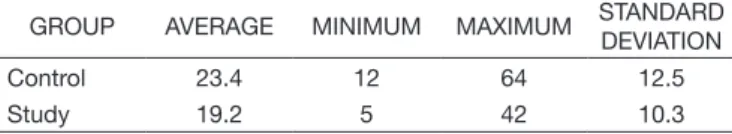
Regarding the total number of days spent with the tracheotomy,
the G2 had a mean of 23.4 days while the G1, only 19.2 days.
These results show that there was a comparative reduction of
4.2 days in the average length of time with tracheostomy in
the G1 patients who had SLP assessment compared to the G2
(Table 1).
Regarding the total days of hospitalization, the G2, that
had no SLP assessment in tracheal decannulation process,
averaged 33.1 days, while in the G1 the total was 28.7 days,
i.e., a reduction of 4.4 days in the total average, statistically not
signiicant, p = 0.2031 (Table 2).
DISCUSSION
The indings regarding the anatomical region of the lesion
agree with other study that emphasizes higher prevalence of brain injury in frontal and temporal regions(26). Regarding the cerebral hemisphere lesion we found no correlation in other studies.
With reference to age this research G1 average age resulted
in 32.1 years, consistent with other study(3). Regarding the
gender, we concluded predominance of TBI in men (83.3%),
which agrees with other studies(3,26).
Even on age, the study indicates that increasing age increases the length of stay and stay with the tracheostomy, which
corroborates indings of another study(27).
However, there was no statistical correlation regarding the gender, length of time with the tracheostomy and length of
hospitalization, which also corroborates indings of another
study(15).
Regarding the comparative reduction in average length of stay with tracheostomy, this study goes against a study conducted in a hospital in Switzerland, which evaluated patients with and without dysphagia tracheostomy multidisciplinary approach to tracheal decannulation. Patients were divided into two groups, one received this approach and the other was assessed retrospectively by the data in the chart, responding to the clinical protocol. The group of patients who received the
multidisciplinary approach has signiicantly reduced the average
length of stay with tracheostomy for 28 days, compared to the group without the approach, with an average of 33 days(14).
And regarding the reduction of days of hospitalization,
although it was not statistically signiicant, it is cost wise
important implications, especially for the growing number of patients with TBI.
Table 2. Descriptive statistics of total days of hospitalization in G1 (n = 30) and G2 (n = 30)
GROUP AVERAGE MINIMUM MAXIMUM STANDARD
DEVIATION
Control 33.1 17 85 15.3
Study 28.7 11 51 10.8
Source: author
Table 1. Descriptive statistics of total days with tracheostomy in G1 (n = 30) and G2 (n = 30)
GROUP AVERAGE MINIMUM MAXIMUM STANDARD
DEVIATION
Control 23.4 12 64 12.5
Study 19.2 5 42 10.3
Although accidents and violence reached almost epidemic proportions in Brazil, from the point of view of mortality and morbidity, they are still incipient efforts to try to estimate the economic impact of traumatic brain injury in the country. From an economic point of view, the costs produced by any
health problems can be classiied into direct and indirect
costs. Direct costs relate to hospital costs and indirect refer to lost productivity brought by health problems, such as loss of working days or even lower productivity generated by physical limitations. Trauma is responsible for the production loss of more years of life than heart disease and cancer combined(28).
From the perception and analysis of the economic impact on the budget of public health, it is necessary a change of attitude in the treatment of TBI individuals tracheostomy. It is crucial that a specialized multidisciplinary team is responsible for the tracheostomy decannulation process.
A Canadian study reported that with the establishment of a team specialized in tracheostomy caring for patients suffering from head trauma, there was a reduction in time and also in the incidence of complications, which corroborates other studies, as a survey conducted in the St. Mary’s Hospital, in London, in which after the implementation of a multidisciplinary service there was a reduction in the time spent with the tracheostomy from 34 to 24 days(29,30).
With the same purpose, researchers have studied the tracheostomy patients from St. Vincent’s Hospital, Australia, for three years and observed that the average length of hospitalization was
reduced from 42 to 34.5 days and also a signiicant reduction
in decannulation time during these years(17).
Still in Australia, in another hospital, a research found that the average hospital stay fell from 60 to 41.5 days and the average length of stay with the tracheostomy was reduced from 22.5 to 16.5 days(27).
The literature review shows that in the past decade more
studies began to emerge on this subject, from the dificulties
encountered in each institution, aiming at an improvement in
the service and that deined decannulation protocols are being sought to minimize risks and optimize beneits to the patient.
However, in Brazil they are still incipient studies covering
and deine criteria for tracheal decannulation to be followed.
This research concludes that the SLP clinical assessment is crucial for tracheal decannulation, which will reduce the time spent with the tracheostomy, accelerating decannulation and thus reducing time and cost of hospital stays for patients suffering from TBI. Thus, further researches are necessary with other populations, in order to validate the tool and its applicability.
CONCLUSION
The group that received the SLP assessment proposed in the tool showed reduced the permanence time with tracheostomy as well as the hospitalization length.
REFERENCES
1. Cambier J, Masson M, Dehen H. Neurologia. 11. ed. Rio de Janeiro:
Guanabara Koogan; 2005. Tradução Mundim FD.
2. Jones HR Jr. Neurologia de Netter. Porto Alegre: Artmed; 2006. 3. Barbosa IL, Andrade LM, Caetano JA, Lima JA, Vieira LJES, Lira SVG.
Fatores desencadeantes ao trauma crânio encefálico em um hospital de
emergência municipal. Rev Baiana de Saúde Pública. 2010;34(2):240-53. 4. Campos BBNS, Machado FS. Terapia nutricional no traumatismo
cranioencefálico grave. Rev Bras Ter Intensiva. 2012; 24(1):97-105. http://
dx.doi.org/10.1590/S0103-507X2012000100015.
5. Foley N, Teasell R, Salter K, Kruger E, Martino R. Dysphagia treatment post stroke: a systematic review of randomised controlled trials. Age Ageing. 2008;37(3):258-64. PMid:18456790. http://dx.doi.org/10.1093/ ageing/afn064.
6. Gomes GF. Identificação de fatores preditivos de pneumonia aspirativa em pacientes hospitalares com doença cerebrovascular complicada por disfagia orofaríngea [dissertação]. Curitiba (PR): Universidade Federal do Paraná; 2001. 86 p.
7. Abel R, Ruf S, Spahn B. Cervical spinal cord injury and deglutition disorders. Dysphagia. 2004;19(2):87-94. PMid:15382796. http://dx.doi. org/10.1007/s00455-003-0511-y.
8. De Leyn P, Bedert L, Delcroix M, Depuydt P, Lauwers G, Sokolov Y, et al. Tracheotomy: clinical review and guidelines. Eur J Cardiothorac Surg. 2007;32(3):412-21. PMid:17588767. http://dx.doi.org/10.1016/j. ejcts.2007.05.018.
9. Forte AP, Forte V. Impacto da traqueostomia na deglutição. In: Ferreira LP, BefiLopes DM, Limongi SCO, editores. Tratado de fonoaudiologia. São Paulo: Roca; 2005. p. 405-9.
10. O’Connor HH, White AC. Tracheostomy decannulation. Respir Care. 2010;55(8):1076-81. PMid:20667155.
11. Mendes TAB, Cavalheiro LV, Arevalo RT, Sonegth R. Estudo preliminar
sobre a proposta de um fluxograma de decanulação em traqueostomia com
atuação interdisciplinar. Einstein (Sao Paulo). 2008;6(1):1-6.
12. Oliveira E, Lavrador JP, Santos MM, Lobo-Antunes J. Traumatismo crânio encefálico: abordagem integrada. Acta Med Port. 2012;25(3):179-92. PMid:23069239.
13. Christopher KL. Tracheostomy decannulation. Respir Care. 2005;50(4):538-41. PMid:15807918.
14. Frank U, Mäder M, Sticher H. Dysphagic patients with tracheotomies: a multidisciplinary approach to treatment and decannulation management. Dysphagia. 2007;22(1):20-9. PMid:17024547. http://dx.doi.org/10.1007/ s00455-006-9036-5.
15. Mackiewicz-Nartowicz H, Mackiewicz-Milewska M, Lach S, Szymańska-Skrzypek A, Owczarek A, Sinkiewicz A. Decannulation factors in patients after serious brain injuries. Adv Pall Med. 2008;7:69-72.
16. Stelfox HT, Crimi C, Berra L, Noto A, Schmidt U, Bigatello LM, et al. Determinants of tracheostomy decannulation: an international survey. Crit Care. 2008;12(1):R26. PMid:18302759. http://dx.doi.org/10.1186/cc6802. 17. Tobin AE, Santamaria JD. An intensivist-led trachesotomy review team is associated with shorter decannulation time and length of stay: a prospective cohort study. Crit Care. 2008;12(2):R48. PMid:18402705. http://dx.doi. org/10.1186/cc6864.
18. Garrubba M, Turner T, Grieveson C. Multidisciplianry care for tracheostomy patients: a systematic review. Crit Care. 2009;13(6):R177. PMid:19895690.
http://dx.doi.org/10.1186/cc8159.
19. Zanata IL, Santos RS, Hirata GC. Tracheal decannulation protocol in patients affected by traumatic brain injury. Int Arch Otorhinolaryngol. 2014;18(2):108-14. http://dx.doi.org/10.1055/s-0033-1363467. 20. Teasdale G, Jennet B. Assesment of coma and impaired consciousness, a
pratical scale. Lancet. 1974;2(7872):81-4. PMid:4136544. http://dx.doi. org/10.1016/S0140-6736(74)91639-0.
21. Helayel PE, Oliveira GR Fo, Marcon L, Pedemeiras FH, Nicolodi MA, Pedemeiras SG. Gradiente SpO2 – SaO2 Durante Ventilação Mecânica em Anestesia e Terapia Intensiva. Rev Bras Anestesiol. 2001;51(4):305-10.
http://dx.doi.org/10.1590/S0034-70942001000400005.
evaluation of the swallow. Ann Otol Rhinol Laryngol. 2003;112(5):469-75. PMid:12784989. http://dx.doi.org/10.1177/000348940311200515. 23. Smith Hammond CA, Goldstein LB. Cough and aspiration of food and
liquids due to oral-pharyngeal dysphagia: ACCP evidence-based clinical practice guidelines. Chest. 2006;129(1 Suppl):154S-68S. PMid:16428705.
http://dx.doi.org/10.1378/chest.129.1_suppl.154S.
24. Ayoob K-T, Duyff RL, Quagliani D. Position of the American Dietetic Association: food and nutrition misinformation. J Am Diet Assoc. 2002;102(2):260-6. PMid:11846124. http://dx.doi.org/10.1016/S0002-8223(02)90062-3.
25. Marik P. Aspiration pneumonitis and aspiration pneumonia. N Engl J Med. 2001;344(9):665-71. PMid:11228282. http://dx.doi.org/10.1056/ NEJM200103013440908.
26. Choi JH, Jakob M, Stapf C, Marshall RS, Hartmann A, Mast H. Multimodal early rehabilitation and predictors of outcome in survivors of severe traumatic brain injury. J Trauma. 2008;65(5):1028-35. PMid:19001970.
http://dx.doi.org/10.1097/TA.0b013e31815eba9b.
27. Cameron TS, McKinstry A, Burt SK, Howard ME, Bellomo R, Brown DJ, et al. Outcomes of patients with spinal cord injury before and after introduction of an interdisciplinary tracheostomy team. Crit Care Resusc. 2009;11(1):14-9. PMid:19281439.
28. IPEA – Instituto de Pesquisa Econômica Aplicada. Impactos sociais e econômicos
dos acidentes de trânsito nas aglomerações urbanas – síntese da pesquisa
[Internet]. Brasília: IPEA; 2003 [citado em 2013 Jul 7]. Disponível em: http:// www.pedestre.org.br/downloads/IpeaSinteseAcidentesTransitoMaio2003. pdf
29. De Mestral C, Iqbal S, Fong N, LeBlanc J, Fata P, Razek T, et al. Impact of a specialized multidisciplinary tracheostomy team on tracheostomy care in critically ill patients. Can J Surg. 2011;54(3):167-72. PMid:21443833.
http://dx.doi.org/10.1503/cjs.043209.
30. Arora A, Hettige R, Ifeacho S, Narula A. Driving standards in tracheostomy care: A preliminary communication of the St Mary’s ENT-led multidisciplinary team approach. Clin Otolaryngol. 2008;33(6):596-9. PMid:19126136.
http://dx.doi.org/10.1111/j.1749-4486.2008.01814.x.
Author contributions
Annex 1. SPEECH THERAPY TOOL FOR TRACHEAL DECANNULATION IN TBI.
PATIENT IDENTIFICATION:
Name: ______________________________________________________________ Age: ______ BD: ___________ Record: _____________Gender: ( ) M ( ) F Diagnosis: _________________________________________________________ Injured anatomical region: _____________________________________________ Degree of traumatic brain injury: ____________________________________
1. CONSCIOUSNESS LEVEL
Glasgow: 3 4 5 6 7 8 9 10 11 12 13 14 NA
2. BREATH
Cannula type: ( ) plastic ( ) metal ( ) no cuff ( ) with cuff
( ) inlated ( ) delated
Maintenance condition cuff delated: ( ) Yes ( ) No
Tracheostomy closure, with cuff delated:
( ) Keep respiratory pattern ( ) Doesn’t keep respiratory pattern
3. ENDOTRACHEAL DISCHARGE
Endotracheal region with discharge: ( ) present ( ) absent Volume: ( ) small ( ) large
Aspect: ( ) thick ( ) luid
Color: ( ) light ( ) sallow
4. PHONATION
Patient orally responsive: ( ) No ( ) Yes Wet voice: ( ) Yes ( ) No
5. SWALLOWING
CONSISTENCY AND SWALLOWING CLINICAL FINDINGS:
CONSISTENCY/FINDINGS LIQUID PUDDING
Volume 5 ml 10 ml FS 5 ml 10 ml FS
Signs of aspiration
PRESENT?
Cough reflex Dyspnea “Wet” voice Caption: FS: free sip
6. COUGH
Voluntary cough: ( ) No () Yes ( ) eficient ( ) ineficient