Division of Oncology, Radiotherapy (InRad)1, Hospital das Clínicas, Faculty
of Medicine, University of São Paulo – São Paulo/SP, Brazil.
Bronchoscopy Unit, Pulmonary Division2, Hospital das Clínicas, Faculty
of Medicine, University of São Paulo – São Paulo/SP, Brazil. E-mail: [email protected]
Received for publication on February 24, 2005. Accepted for publication on May 05, 2005.
ORIGINAL RESEARCH
HIGH DOSE-RATE BRACHYTHERAPY AS A
TREATMENT OPTION IN PRIMARY TRACHEAL
TUMORS
Heloisa de Andrade Carvalho1, Viviane Figueiredo2, Wilson Leite Pedreira Jr.2,
and Salim Aisen1
Carvalho H de A, Figueiredo V, Pedreira Jr. WL, Aisen S. High dose-rate brachytherapy as a treatment option in primary tracheal tumors. Clinics. 2005;60(4):299-304.
PURPOSE: To present experience with high dose-rate endobronchial brachytherapy in the treatment of primary tracheal
tumors.
PATIENTS AND METHODS: Four patients with nonresected primary tracheal tumors are presented: 2 cases of squamous
cell carcinoma of the trachea, 1 of recurrent adenoid cystic carcinoma, and 1 with recurrent plasmacytoma. All received brachytherapy, alone or as a boost for primary irradiation, in 3 or 4 fractions of 7.5 Gy, calculated at a depth of 1 cm. Follow-up was considered to start from the end of brachytherapy.
RESULTS: Local control was achieved in all cases at the time of first bronchoscopic evaluation. Two patients with squamous cell carcinoma died at 6th and 33rd months after brachytherapy, respectively. The first had no evidence of disease, and the
latter had local recurrence. The other 2 patients were alive after 64 and 110 months of follow-up, respectively, both with no evidence of disease. Tracheal stenosis developed in these 2 cases, 22 and 69 months after brachytherapy. Tracheal stent placement was needed only for the patient with an adenoid cystic carcinoma.
CONCLUSIONS: Endobronchial high dose-rate brachytherapy may be used for tracheal tumors, even as a boost for external
beam irradiation, or in recurrences. Local control in 3 out of 4 patients indicates that individual cases may benefit from the treatment. Long-term survival may also be expected, mainly for tumors with adenoid cystic histology.
KEYWORDS: Radiotherapy. Brachytherapy. Tracheal tumors. High dose-rate. Complications.
Brachytherapy is the radiation treatment in which the radioactive material is located in contact with or inside the target lesion. Special applicators, needles, and catheters are used as guides for positioning the radioactive material in the patient. Endobronchial brachytherapy is performed through bronchoscopy, with the placement of one or more catheters in the lower airways. Brachytherapy has been widely used, especially after the development of high dose-rate (HDR) remote afterloading machines. In 1991, our group first introduced this method in Latin America.1 The
advan-tages obtained with the use of HDR brachytherapy include good patient tolerance, short treatment time, and a higher potential to achieve local control.
The highly conformal nature of brachytherapy enables the radiation oncologist to accomplish safe escalation of radiation doses to the tumor while minimizing doses to nor-mal surrounding structures. Most of the reported experience with endobronchial brachytherapy is related to treatment of primary or metastatic endobronchial lesions, either with curative or palliative intent. Very few articles report the use of brachytherapy for primary tracheal tumors.2-7 Due to the
rarity of the disease (0.2% - 0.5% of all upper respiratory tract malignancies),8,9 the role of endoluminal brachytherapy
in the treatment regimen of tracheal neoplasms has not been clearly defined yet.
mid-dle, or lower third), resectability, lymph node involvement, and surgical margins.10,11
Adenoid cystic carcinoma and squamous cell carcinoma are the most common tracheal tumors. Resection and post-operative radiation therapy are the treatment of choice.12,13
Complete resection is the desired goal, and benign tumors, for which cure can be expected, are best treated by resec-tion alone.
We report the experience of a single institution with high dose-rate brachytherapy in the management of nonresected primary tracheal tumors.
MATERIALS AND METHODS
From 1991 to 1997, four patients with primary tracheal tumors were treated with endobronchial/endotracheal HDR brachytherapy: two men with squamous cell carcinoma, and two women, one with adenoid cystic carcinoma, one with primary tracheal plasmacytoma (previously published as a case report).14 Brachytherapy was indicated as a boost
after external beam irradiation in 1 case of inoperable sq-uamous carcinoma of the lower third of the trachea, which was asymptomatic at that time. The other patient with sq-uamous cell carcinoma was previously irradiated for a head and neck tumor and received brachytherapy alone. He had an inoperable tumor located at the upper portion of the trachea, causing dyspnea, cough, and mild bleed-ing right below the supraclavicular radiation field limits. The 2 patients with adenoid cystic carcinoma and plas-macytoma, respectively, underwent brachytherapy as a sal-vage treatment for local recurrences after external beam irradiation years before (9 and 11 years, respectively). Di-agnosis of recurrence was made after complaints of dyspnea and cough with mild bleeding in the first case and cough in the second one. Only the patient with plasmacytoma received laser therapy for debulking before irradiation.14
Table 1 presents patient characteristics, and Table 2 presents previous radiation treatments.
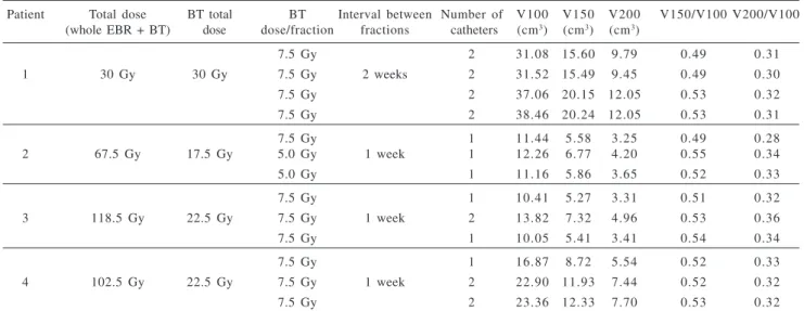
Endotracheal HDR brachytherapy was delivered in 3 or 4 fractions of 7.5 Gy each, at weekly intervals. Dose calcu-lation was made at 1 cm from the source axis, with optimi-zation on dose points. No centered catheters were available, and 6Fr catheters were used in all procedures. Nasal place-ment and fixation of the catheters with tape was used, ex-cept for the patient with previous larynx carcinoma, where the procedure was performed through the tracheotomy. One or two catheters were placed in the tracheal lumen and po-sitioned in the right and/or left main bronchus, respectively. Proximal and distal margins of 1 to 2 cm from the lesion were included in the treatment volume, to account for pos-sible variations due to respiratory cycles. Since there may be areas of very high radiation doses where the catheter sur-face is in contact with the airway mucosa, the volumes re-ceiving the total dose (100%), and those rere-ceiving 150% and 200% of the total dose were also evaluated (Table 3).
Follow-up was considered to start from the end of brachytherapy. The local response was evaluated by fiberbronchoscopy performed 30 to 40 days after the end of brachytherapy. Subsequent bronchoscopies were per-formed at 3 and 6 months after brachytherapy, and after that whenever indicated. Additionally, thoracic CT scans were performed every 6 months over the first 3 years, then yearly until at least 5 years of follow-up.
Table 1 - Description of patient characteristics
Patient Gender Age Histology DFS Tracheal Ext. Obstr. LN
(years) location Score*
1 M 4 3 SCC - Upper 4 cm 6 N0
2 M 7 8 SCC - Lower 3 cm 2 N0
3 F 5 3 ACC 9 Middle 2.5 cm 1 0 N0
4 F 6 1 Plasm 1 1 Middle 4 cm 2 N0
*Obstruction Score (Speiser, 199327): Tracheal obstruction > 50% = 10; < 50% = 6, and < 10% = 2.
DFS = disease free survival before first recurrence; Ext. = lesion extension; LN = lymph node involvement; M = male; F = female; SCC = squamous cell carcinoma; ACC = adenoid cystic carcinoma; Plasm = plasmacytoma
Table 2 - Previous external beam radiation treatment (patient # 1 did not receive external beam irradiation)
Patient Total dose Dose/fraction Tracheal location
2 50 Gy 2 Gy Lower
3 *60 Gy 2 Gy Middle
36 Gy 1.8 Gy Upper
4 **40 Gy 2 Gy Middle/lower
40 Gy Upper
RESULTS
All patients presented complete local response at the time of first bronchoscopic evaluation, with improvement of related symptoms in 2 of the 3 symptomatic cases. No biopsy was performed in order to avoid the risk of fistu-lae or bleeding of the irradiated areas. The 2 patients with squamous cell carcinoma died at 6th and 33rd months after
brachytherapy, respectively. The first, with no evidence of local disease and persistent cough, died of respiratory in-fection. No direct relationship with treatment was detected, but in the last bronchoscopic evaluation, 3 months after treatment, there was still moderate necrosis in the irradi-ated areas, perhaps facilitating an infection. The second patient, who received brachytherapy as a boost, died with local recurrence 2 years after treatment. Two patients were alive, with no evidence of disease at 64 and 110 months of follow-up. Tracheal stenosis was observed in these 2 patients—and was considered a severe complication—22 and 69 months after brachytherapy, respectively. Both had been extensively irradiated previously, with areas that re-ceived 60 to 90 Gy (Table 4). A tracheal stent was suc-cessfully placed for palliation in the patient with adenoid cystic carcinoma, but it was not necessary for the plasma-cytoma patient. During this period, this patient presented other isolated lesions in the upper respiratory tract (lar-ynx and orophar(lar-ynx), and was successfully treated with external beam irradiation. Nine years after brachytherapy, she developed chronic lymphocytic leukemia.
No relationship of the brachytherapy irradiated volumes and complications was observed.
DISCUSSION
Treatment of primary tracheal tumors has been optimized over the years. At first, most patients were treated with endo-scopic or surgical local excision followed by radiotherapy with orthovoltage or radon seed implantation. One-year sur-vival was less than 25%.15-17 Advances in surgical techniques
have made it possible to resect tracheal tumors that might have previously been considered unresectable.18,19 At present,
surgery has become the primary treatment for tracheal tumors.13,20 Advances in radiation therapy have shown
mod-est gains in the treatment of tracheal malignancies, either as definitive or as adjuvant treatment.11,13,20-24
Schneider13 reported in his series of 14 resected cases, a
5-year survival of 35.7% for 6 patients who needed adjuvant irradiation.
Kanematsu20 also reported, in 16 patients with adenoid
cystic carcinoma, a 5-and 10-year survival of 91% and 76%, respectively, for patients with resected tumors versus a 5-year
Table 4 - Results and survival after the end of brachytherapy
Patient Survival DFS Cause of Local Sequelae (months) (months) death recurrence
1 6 6 Respiratory No No
infection
2 3 3 2 4 Tumor Yes No
3 6 4 6 4 - No Stenosis
(22 months)
4 1 1 0 1 1 0 - No Stenosis
(69 months) DFS = disease free survival
Table 3 - Brachytherapy characteristics
Patient Total dose BT total BT Interval between Number of V100 V150 V200 V150/V100 V200/V100 (whole EBR + BT) dose dose/fraction fractions catheters (cm3) (cm3) (cm3)
7.5 Gy 2 31.08 15.60 9.79 0.49 0.31
1 30 Gy 30 Gy 7.5 Gy 2 weeks 2 31.52 15.49 9.45 0.49 0.30
7.5 Gy 2 37.06 20.15 12.05 0.53 0.32
7.5 Gy 2 38.46 20.24 12.05 0.53 0.31
7.5 Gy 1 11.44 5.58 3.25 0.49 0.28
2 67.5 Gy 17.5 Gy 5.0 Gy 1 week 1 12.26 6.77 4.20 0.55 0.34
5.0 Gy 1 11.16 5.86 3.65 0.52 0.33
7.5 Gy 1 10.41 5.27 3.31 0.51 0.32
3 118.5 Gy 22.5 Gy 7.5 Gy 1 week 2 13.82 7.32 4.96 0.53 0.36
7.5 Gy 1 10.05 5.41 3.41 0.54 0.34
7.5 Gy 1 16.87 8.72 5.54 0.52 0.33
4 102.5 Gy 22.5 Gy 7.5 Gy 1 week 2 22.90 11.93 7.44 0.52 0.32
7.5 Gy 2 23.36 12.33 7.70 0.53 0.32
survival of 40% when combined with radiation therapy. Two Russian series25,26 of 48 patients with squamous cell
carcinoma of the trachea and 16 cases of adenoid cystic car-cinoma had 5-year survival rates of 40.9% and 55.9%, re-spectively, for patients treated with combined surgery and radiotherapy.
Schraube et al.7 reported 5 patients treated with
endo-bronchial HDR brachytherapy: 4 combined with external beam irradiation, and 1 as a salvage treatment after surgery. External beam doses ranged from 46 to 60 Gy. Endobron-chial HDR brachytherapy was delivered in 5 fractions of 3 to 4 Gy each, twice a week, calculated at 1 cm from the source axis. They reported 4 of 5 complete responses, 2 of 3 deaths for recurrent or progressive disease, and 2 patients alive with no evidence of disease at 37th and 68th months
of follow-up.
The same group published results some years later, with 7 cases treated with brachytherapy as a boost for residual tumor after external irradiation. Local control was obtained in 5 of 7 patients, and late toxicity occurred in 3 patients (2 stenosis and 1 hemorrhage). Three-year actuarial survival was 32%.3
In this series, all 4 patients achieved immediate complete response, with palliation of symptoms and local control for at least 6 months. Palliation was achieved mainly due to improve-ment of obstruction. This might also have been evaluated by pulmonary function tests, with determination of the maximum flows, and thereby detecting impairment of high airways. But, in the present study, these tests were not performed.
The patients with adenoid cystic carcinoma and plas-macytoma had a better outcome, as expected. These patients also presented with tracheal stenosis, and one of them was
treated with local stent placement. They persisted with only mild limitations due to dyspnea. We considered the high doses of either external beam irradiation, with a partial dose overlap between fields, and endoluminal brachytherapy, to have been the main cause of stenosis. It is conceivable that more fractions with lower brachytherapy doses might have avoided or diminished this risk.
Nevertheless, Schraube7 also reported 2 of 5 tracheitis
with stenosis in his series, and Harms3 reported 1 case of
late hemorrhage using 4 or 5 fractions of brachytherapy with lower doses/fraction.
We observed that dose distribution using 2 catheters seemed to be better than using a single one. The 100% isod-ose curve may be widened to more than 2 cm diameter, al-lowing better coverage of tracheal walls, which was associ-ated with a lower surface dose (Table 3). This strategy may be used to diminish the risk of complications, especially when centralized catheters are not available.
CONCLUSIONS
Endobronchial HDR brachytherapy may be used for tracheal tumors as a boost for external beam irradiation or in recurrences. Total dose and dose/fraction have not yet been well defined; however, 3 or 4 fractions of 5 to 7.5 Gy (at 1 cm depth) are currently used. Although sur-vival impact was not demonstrated in this series, local control of 3 out of 4 patients indicates that individual cases may benefit from the treatment. Long-term survival with palliation may also be expected mainly in cases of adenoid cystic histology.
RESUMO
Carvalho H de A, Figueiredo V, Pedreira Jr. WL, Aisen S. Braquiterapia de alta taxa de dose como opção terapêutica nos tumores primários da traquéia. Clinics. 2005;60(4):299-304.
OBJETIVO: Apresentar a experiência do tratamento de 4
pacientes com tumores primários de traquéia, não operados, submetidos à braquiterapia endobrônquica de alta taxa de dose.
PACIENTES E MÉTODOS: Dois casos de carcinoma
espinocelular, uma recidiva de carcinoma adenóide cístico e uma recidiva de plasmocitoma primário da traquéia. Todos receberam braquiterapia endobrônquica, exclusiva ou como reforço de dose da radioterapia externa. Foram administradas 3 ou 4 frações de 7,5 Gy cada, calculados a 1 cm de profundidade. O seguimento foi considerado a partir do término da braquiterapia.
REFERENCES
1 . Aisen S, Carvalho HA, Chavantes MC, Esteves SCB, Haddad CMK, Pernomian AC, et al. Braquiterapia de alta taxa de dose. Rev Hosp Clín Fac Med S Paulo. 1992;47:121-4.
2 . Aggarwal A, Tewari S, Mehta AC. Successful management of adenoid cystic carcinoma of the trachea by laser and irradiation. Chest. 1999;116:269-70.
3 . Harms W, Latz D, Becker H, Herth F, Schraube P, Krempien R, Wannenmacher M. HDR-brachytherapy boost for residual tumour after external beam radiotherapy in patients with tracheal malignancies. Radiother Oncol. 1999;52:251-5.
4 . Kohek PH, Pakisch B, Glanzer H. Intraluminal irradiation in the treatment of malignant airway obstruction. Eur J Surg Oncol. 1994;20:674-80.
5 . Makarewicz R, Mross M. Radiation therapy alone in the treatment of tumors of the trachea. Lung Cancer. 1998;20:169-74. 6 . Mornex F, Coquard R, Danhier S, Maingon P, Husseini GE, Houtte
PV. Role of radiation therapy in the treatment of primary tracheal carcinoma. Int J Radiat Oncol Biol Phys. 1998;41:299-305. 7 . Schraube P, Latz D, Wannenmacher M. Treatment of primary
squamous cell carcinoma of the trachea: the role of radiation therapy. Radiother Oncol. 1994;33:254-8.
8 . Ampil FL. Primary malignant tracheal neoplasms: case reports and literature radiotherapy review. J Surg Oncol. 1986;33:20-3. 9 . Houston HE, Payne WS, Harrison EG Jr, Olsen AM. Primary cancers
of the trachea. Arch Surg. 1969;99:132-40.
10. Regnard JF, Fourquier P, Levasseur P. Results and prognostic factors in resections of primary tracheal tumors: a multicentric retrospective study. J Thorac Cardiovasc Surg. 1996;111:808-14.
11. Chao MWT, Smith JG, Laidlaw C, Joon DL, Ball D. Results of treating primary tumors of the trachea with radiotherapy. Int J Radiat Oncol Biol Phys. 1998;41:779-85.
12. Grillo HC, Mathisen DJ. Primary tracheal tumors: treatment and results. Ann Thorac Surg. 1990;49:69-77.
13. Schneider P, Schirren J, Muley T, Vogt-Moykopf I. Primary tracheal tumors: experience with 14 resected patients. Eur J Cardiothorac Surg. 2001;20:12-8.
14. Kairalla RA, Carvalho CRR, Parada AA, Alves VA, Saldiva PH. Solitary plasmacytoma of the trachea treated by loop resection and laser therapy. Thorax. 1988,43:1011-2.
15. Chow DC, Komaki R, Libshitz H, Mountain CF, Ellerbroek N. Treatment of primary neoplasms of the trachea: The role of radiation therapy. Cancer. 1993;71:2946-52.
16. Hajdú SI, Huvos AG, Goodner JT, Foote FW, Beattie EJ. Carcinoma of the trachea: clinicopathologic study of 41 cases. Cancer. 1970;25:1448-56.
17. McCafferty GJ, Parker LS, Suggit SC. Primary malignant disease of the trachea. J Laryngol. 1964;78:441-79.
18. Grillo HC, Dignan EF, Miura T. Extensive resection and reconstruction of mediastinal trachea without prosthesis or graft: an anatomical study in man. J Thorac Cardiovasc Surg. 1964;48:741.
19. Grillo HC. Tracheal surgery. J Thorac Cardiovasc Surg.1983;17:67-77.
20. Kanematsu T, Uehara T, Ushijima C, Asoh H, Yoshino I, Ichinose Y. Treatment outcome of resected and non-resected primary adenoid cystic carcinoma of the lung. Ann Thorac Cardiovasc Surg. 2002;8:74-7.
21. Baraka ME. Malignant tumors of the trachea. Ann R Coll Surg Engl. 1984;66:27-9.
22. Fields JN, Rigaud G, Emami BN. Primary tumors of the trachea: results of radiation therapy. Cancer. 1989;63:2429-33.
Dois pacientes com carcinoma espinocelular evoluíram a óbito em 6 e 33 meses após a braquiterapia, o primeiro, sem evidência de doença e o outro por recidiva local, respectivamente. As outras 2 pacientes encontravam-se vivas após 64 e 110 meses de seguimento, sem evidência de doença. Esses 2 casos apresentaram estenose traqueal em 22 e 69 meses após a braquiterapia, sendo necessária a colocação de prótese traqueal apenas na paciente com carcinoma adenóide cístico.
CONCLUSÕES: A braquiterapia endobrônquica de alta
taxa de dose pode ser utilizada tanto como reforço de dose da irradiação externa quanto em recidivas. O controle lo-cal obtido em 3 de 4 pacientes indica que casos individuais podem se beneficiar desse procedimento. Sobrevida a longo prazo pode ser observada, principalmente nos casos de histologia adenóide cística.
23. Pearson FG, Todd TR Jr, Cooper JD. Experience with primary neoplasms of the trachea and carina. J Thorac Cardiovasc Surg. 1984;88:511-8.
24. Rostom AY, Morgan RL. Results of treating primary tumors of the trachea by irradiation. Thorax. 1978;33:387-93.
25. Kharchenko VP, Galil-Ogly GA, Gvarishvili AA, II’in VI, Al’bekov RZ. Lechenie plockokletochnogo raka trachei. [Treatment of squamous cell carcinoma of the trachea]. Vopr Onkol. 1999;45:66-9.
26. Kharchenko VP, Galil-Ogly GA, Gvarishvili AA, II’in VI, Al’bekov RZ. Luchevaia terapila adenokistoznogo raka trakhei. [Radiotherapy for cystadenoma of the trachea]. Vopr Onkol. 1999;45:69-72.