AGT*M235T Polymorphism in Acute Ischemic Cardiac Dysfunction -
the Gisca Project
Claudia Guerra Murad Saud, Amália Faria dos Reis, Arlisa Monteiro de Castro Dias, Rosemery Nunes Cardoso,
Ana Cristina Klem Vargas Carneiro, Leandro Pereira de Souza, Ana Beatriz Monteiro Fonseca, Georgina Severo
Ribeiro, Carlos Augusto Cardozo de Faria
Universidade Federal Fluminense, Rio de Janeiro, RJ - Brazil
Mailing address: Claudia Guerra Murad Saud •
Estrada Caetano Monteiro, 1833 - Casa 40 - Badu - 24320-570 - Niterói, RJ - Brazil
E-mail: [email protected], [email protected]
Manuscript received March 24, 2009; revised manuscript received August 29, 2009; accepted December 18, 2009.
Abstract
Background: AGT*M235T polymorphism has been associated with high serum angiotensinogen (AGT) levels, systemic hypertension and cardiac dysfunction (CD).
Objective: To test the hypothesis of AGT*M235T polymorphism being associated with the risk of developing cardiac dysfunction (heart failure or asymptomatic left ventricular systolic dysfunction) after acute coronary syndrome (ACS) during hospitalization.
Methods: A total of 363 patients (mean age of 62 ± 12 years), of whom 233 (64%) were men and 130 (36%) were women, all from the same cohort and hospitalized for ACS, were studied. Clinical and genetic data from the 117 (32.2%) patients who developed cardiac dysfunction (case group) were compared to those of the 246 (67.8%) who did not develop this condition (control group). The AGT*M235T polymorphism was determined by sequence analysis and was in Hardy-Weinberg equilibrium.
Results: There was a significant difference in the distribution of genotypes among women, with a predominance of the *235MM genotype in the control group (p = 0.001) and of the *235T allele in the case group. In the logistic regression models, the diagnosis of anterior wall myocardial infarction at admission was related to an increased risk of CD in both genders, whereas unstable angina at admission.; absence of the *235T allele; blood glucose < 100 mg/dl; use of betablocker; serum creatinine level < 1.5 mg/dl; heart rate range ≥ 60 and < 90 bpm; and current cigarette smoking were related to a lower risk of CD.
Conclusion: This study suggests that the absence of the AGT *235T allele contributes to a reduced risk of cardiac dysfunction after acute coronary syndrome. (Arq Bras Cardiol 2010; 95(2): 144-152)
Key words: Angiotensinogen; polymorphism, genetic; heart failure; hypertension; ventricular dysfunction, left; acute coronary syndrome.
patients who develop cardiac dysfunction are at a high risk of death, which, according to Valiant’s study3, may be reduced by 20% by inhibiting the renin-angiotensin-aldosterone system (RAAS).
The RAAS plays a key role in the pathophysiology of the cardiovascular system, having angiotensinogen (AGT) as its substrate; angiotensin II is produced in the heart and is upregulated by mechanical load, causing changes in both the myocyte structure and the extracellular matrix4. Increased levels of angiotensin II have been associated with a worse outcome in patients with heart failure4,5. Considering these factors, variations in the genes of this system are obvious candidates for research on heart failure6-9. AGT*M235T polymorphism has three possible genotypes: MM, MT and TT. Individuals with the *235TT genotype show higher plasma levels of angiotensinogen and angiotensin II10-12. To date, few published studies have analyzed the association between AGT*M235T polymorphism and cardiac dysfunction (heart failure or ventricular systolic dysfunction), and results are controversial6,7,13.
Introduction
Diseases of the circulatory system - ischemic cerebrovascular and heart diseases - are the major cause of death in Brazil, accounting for 28% of deaths in 20041. For 2025, it is estimated that Brazil will have the sixth largest population of elderly individuals in the world, corresponding to 15% of the total population of the country, and that heart failure (HF) will be the major cause of death due to cardiovascular disease worldwide2.
In view of the lack of Brazilian studies analyzing AGT*M235T polymorphism and its association with ischemic cardiac dysfunction, the primary objective of the present study was to evaluate this association in hospitalized patients with ACS in the city of Niterói, Rio de Janeiro. The secondary objective was to analyze the interaction between gender and AGT*M235T polymorphism in this context.
Methodology
Study design
Observational case-control study nested in an initial prospective cohort14 obtained by the Fatores de Risco
Cardiovascular e Marcadores Genéticos nas Síndromes Coronarianas Agudas na População de Niterói, Rio de Janeiro
project15 (Cardiovascular Risk Factors and Genetic Markers in Acute Coronary Syndromes in the Population of Niterói, Rio de Janeiro).
• Criteria for inclusion in the cohort - men and women with age ≥ 20 years consecutively admitted to the emergency department or hospitalized within up to 72 hours of the onset of symptoms, and diagnosed with ACS.
• Criteria for exclusion from the cohort - patients with end-stage neoplastic disease, multiple trauma and dementia. The patients were followed-up during hospitalization. Time for enrollment in the study was pre-set at 12 consecutive months (from July 2004 to June 2005); this period was set as the determinant of the sample size.
Public hospitals offering 24-hour emergency services and with beds available for admission of cardiology patients were selected. Private hospitals offering 24-hour emergency service, a cardiology team, and coronary unit or ICU beds were selected. Hospitals with less than 50 beds were excluded, unless they specialized in cardiology. Only four private and three public hospitals met these criteria.
Deinition of variables
• Unstable angina (UA) - presence of electrocardiographic changes consistent with subendocardial injury or myocardial ischemia in two or more contiguous leads16 associated with typical rest angina pectoris17 or new-onset or crescendo angina17. Patients with typical angina pectoris associated with a confirmed previous history of myocardial infarction, angioplasty and/or coronary artery bypass grafting (CABG); or those at a high risk of coronary artery disease18 whose in-hospital tests showed induced myocardial ischemia or lesion ≥ 50% in coronary angioplasty were also considered.
• Non-ST elevation MI (NSTEMI) - presence of increased CK-MB and total CK levels (> twofold the local reference value), followed by a gradual decrease within 48 hours of the onset of symptoms; or > threefold the reference value within 48 hours of angioplasty18 associated with typical angina pectoris17 and/ or electrocardiographic abnormalities consistent with injury.
• ST elevation MI (STEMI) - abnormal levels of markers of myocardial necrosis, as described elsewhere19, associated with acute subepicardial injury or acute complete LBBB on electrocardiogram (ECG)15,20.
• Cardiac dysfunction (CD) - clinical manifestations of heart failure (Killip classes II to IV) and/or laboratory tests showing the presence of left ventricular systolic dysfunction (LVSD) during hospitalization21,22: echocardiography within the first 72 hours of hospital admission showing moderate or severe global systolic dysfunction associated with ejection fraction ≤ 40% using the Teichholz method23, or ejection fraction ≤ 40% using the Simpson method, or invasive contrast ventriculography during coronary angiography showing moderate or severe systolic dysfunction24.
• Heart failure with reduced ejection fraction (HFREF) - Killip classes II to IV in the presence of LVSD criteria described above. Heart failure with normal ejection fraction (HFNEF): Killip classes II to IV associated with ejection fraction > 40%, and impaired diastolic function on Doppler. Killip classification was adapted for the diagnosis of heart failure in patients with unstable angina.
Genetic analysis
Blood samples were collected in EDTA tubes at admission or in subsequent days, for genomic DNA isolation. The team in charge of the genotype evaluations was blind to the patients’ clinical information.
After genomic DNA extraction, integrity of the sample was analyzed using submarine electrophoresis (BIO-RAD electrophoresis system) in 0.8% agarose gel, and DNA strands were visualized in a transilluminator under ultraviolet light. The samples were then stored at 4o C for further sequencing. AGT*M235T polymorphism was tested using polymerase chain reaction (PCR) and sequence analysis of an amplified 157-base pair fragment of the AGT gene with the following oligonucleotides:
• sense: 5’- ACTGGATGTTGCTGCTGAGAAGA-3’ • antisense: 5’-GCCAGAGCCAGCAGAGAGGTTTG-3’25 The amplified fragments underwent sequence analysis in the MegaBace Analyzer (Amersham Biosciences), using Dynamic ET Dye Terminator cycle sequencing reagent; at the end of the run, the chromatogram was analyzed in the Sequence Analyzer (Figure 1).
Statistical analysis
Figure 1 -Chromatogram of the sequencing of the AGT*M235T polymorphism.
Homozygote CC
T235T
Heterozygote CT
M235T
Homozygote TT
M235M
Bioethical aspects
All patients included gave a written informed consent. The study was approved by the Research Ethics Committee of Faculdade de Medicina da Universidade Federal Fluminense
(UFF) (CEP CMM/HUAP no 59/03, July 18, 2003) and by the National Research Ethics Commission (Conep), Brasília (DF), in October 3, 2003.
The researchers did not interfere with the type of treatment chosen for each patient, which was determined by the medical staff of each institution.
Results
A total of 436 patients from five hospitals (two private and three public hospitals) who were followed up for a 12-month period were included. Angiotensin polymorphism could be identified in 410 patients. Of these, some reported a previous history of heart failure and were then excluded. Therefore, 363 patients were analyzed in this study.
Clinical and genetic data of the 117 patients (32.2%) with cardiac dysfunction (heart failure or LVSD) (case group) were compared to those of the 246 patients (67.8%) who did
not develop cardiac dysfunction (control group). Cases and controls were chosen from the same cohort.
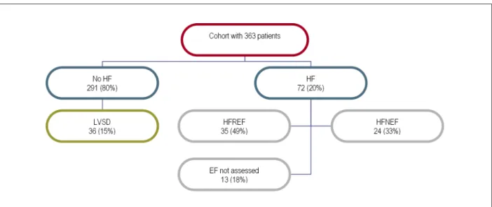
Of the 363 patients assessed (Figure 2), 291 (80%) had no signs or symptoms of heart failure, whereas 72 (20%) did. Of these 72 patients with HF, 35 (49%) developed HFREF and 24 (33%), HFNEF; ejection fraction of the other 13 (18%) was not measured during hospitalization. Of the 363 patients, 225 (62%) did not have LVSD, 80 (22%) had LVSD; the left ventricular systolic function was not assessed in 58 (16%) (Figure 2).
The overall characteristics of the sample are described in Table 1; clinical characteristics and risk factors of cases and controls are shown in Tables 2 and 3.
Age > 65 years predominated among cases (p = 0.008), thus showing that older patients had a higher probability of developing cardiac dysfunction (OR: 1.298 - 95% CI: 1.061-1.587). The diagnosis of unstable angina was more frequent among control patients (p < 0.001), whereas NSTEMI and anterior wall myocardial infarction were more frequent among cases (p = 0.002).
bpm (p = 0.024). The number of patients with systolic blood pressure < 90 mmHg was significantly higher in the case group (p = 0.048), due to inclusion of patients in cardiogenic shock. BMI (p = 0.028) and use of betablockers (p = 0.002) was lower in the case group.
The risk of death was 1.24 times greater among case patients in relation to controls (p < 0.001).
The genotypic frequencies of AGT*M235T polymorphism were in Hardy-Weinberg equilibrium and were not statistically different between the case and control groups.
Considering the genotype distribution according to gender (Table 4), a reduced frequency of the *235MM genotype (p = 0.013) and predominance of the *235T allele were observed among case women (p = 0.016). In male patients, the frequencies of genotypes and alleles were similar in cases and controls.
A significant difference was observed in the distribution of genotypes in relation to age only among women (Table 5), both in cases (p = 0.001) and in controls (p = 0.029), and there was a predominance of the *235TT genotype in patients ≤ 65 years. The other characteristics analyzed in relation to genotypes were not different between genders.
In logistic regressions in which the input variables were age range; heart rate; systolic blood pressure; cigarette smoking; type of ACS; genotypes and alleles; gender; previous CABG; previous myocardial infarction; history of systemic hypertension; intervention; creatinine; and blood glucose, the model with the best predictive power (R2 = 0.312) for the occurrence of cardiac dysfunction selected the following variables: unstable angina (OR: 0.416, p = 0.004), absence of the T allele (OR: 0.448, p = 0.044) and creatinine < 1.5 mg/dl (OR: 0.597, p = 0.012), which represented protective factors in relation to the endpoint. When the models were separated by gender, the intervention variable (thrombolytic agent, percutaneous angioplasty, or CABG during hospitalization) increased the risk of cardiac dysfunction only among women (OR: 2.239, p = 0.027).
Figure 2 -Organization chart of the distribution of heart failure in the study. HF - heart failure; HFREF - heart failure with reduced ejection fraction; HFNEF - heart failure with normal ejection fraction; LVED - left ventricular systolic dysfunction; EF - ejection fraction.
When the variables anterior wall myocardial infarction and use of betablocker and ACEI/ARB II during hospitalization were also included, the model with the best explanatory power for cardiac dysfunction (R2 0.350) selected anterior wall myocardial infarction as a risk factor (OR: 3.244; p = 0.001); the use of betablocker during hospitalization (OR: 0.490; p = 0.030), heart rate ≥ 60 and < 90 bpm (OR: 0.475; p = 0.002), and current cigarette smoking (OR: 0.449; p = 0.012) were selected as protective factors.
When a logistic regression was carried out by gender, the model with the best explanatory power in the male gender (R2 0.397) selected anterior wall myocardial infarction as a factor increasing the risk of cardiac dysfunction by 5.579 times (p < 0.001) and the use of betablocker, current cigarette smoking and blood glucose < 100 mg/dl as variables associated with a better prognosis. In the female gender, the model with the best predictive power (R2 0.346) selected the diagnosis of unstable angina and the *235MM genotype as factors reducing the risk of cardiac dysfunction (Table 6). With the inclusion of these new variables, intervention was no longer selected in the final model in women.
Discussion
The development of heart failure and/or left ventricular systolic dysfunction in the context of ACS, whether at admission or at any moment during hospitalization, results in a significant increase in the risk of in-hospital, 30-day, and long-term mortality21. Thus, we used this composite endpoint - cardiac dysfunction - for all analyses. The percentage of patients with HF (with normal or decreased ejection fraction) and asymptomatic left ventricular systolic dysfunction was reported in the results with the only purpose of better describing the study sample, without the intention of carrying out separate analyses in each of these subgroups.
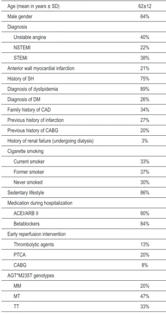
Table 1 - Clinical and genetic characteristics of the study sample (n = 363)
Age (mean in years ± SD) 62±12
Male gender 64%
Diagnosis
Unstable angina 40%
NSTEMI 22%
STEMI 38%
Anterior wall myocardial infarction 21%
History of SH 75%
Diagnosis of dyslipidemia 89%
Diagnosis of DM 26%
Family history of CAD 34%
Previous history of infarction 27%
Previous history of CABG 20%
History of renal failure (undergoing dialysis) 3%
Cigarette smoking
Current smoker 33%
Former smoker 37%
Never smoked 30%
Sedentary lifestyle 86%
Medication during hospitalization
ACEI/ARB II 90%
Betablockers 84%
Early reperfusion intervention
Thrombolytic agents 13%
PTCA 20%
CABG 8%
AGT*M235T genotypes
MM 20%
MT 47%
TT 33%
NSTEMI - non-ST elevation myocardial infarction; STEMI - ST elevation myocardial infarction; SH - systemic hypertension; DM - diabetes mellitus; CAD - coronary artery disease; ACEI - angiotensin converting enzyme inhibitor; ARB II - angiotensin II receptor blocker; PTCA - percutaneous transluminal coronary angioplasty: CABG - coronary artery bypass grafting; MM - 235 methionine-methionine genotype; MT - 235 methionine-methionine-threonine genotype; TT - 235 threonine-threonine genotype.
Table 2 - Clinical characteristics of the patients according to the outcome with or without cardiac dysfunction
Variables Controls (n = 246) Cases (n = 117) p
Gender
Male 162 (65.9%) 71 (60.7%)
0.351 Female 84 (34.1%) 46 (39.3%)
Age range
≤ 65 years 161 (65.4%) 59 (50.4%)
0.008 > 65 years 85 (34.6%) 58 (49.6%)
Diagnosis
Unstable angina 115 (46.7%) 30 (25.6%)
<0.001 NSTEMI 56(22.8%) 24(20.5%)
STEMI 75 (30.5%) 63 (53.8%) Anterior wall
myocardial infarction
33 (44%) 44 (71%) 0.002
BMI kg/m2 (± DP) 26.5 (± 4.7) 25.2 (± 3.9) 0.028
HR (bpm)
< 60 15 (6.7%) 6 (5.9%)
0.024 ≥ 60 < 90 160 (71.7%) 56 (54.9%)
≥ 90 < 120 37 (16.6%) 30 (29.4%) ≥ 120 <150 10 (4.5%) 9 (8.8%) ≥ 150 1 (0.4%) 1 (1.0%) SBP (mmHg)
< 90 4 (1.7%) 6 (5.4%)
0.048 ≥ 90 < 110 8 (3.3%) 10 (8.9%)
≥110 < 130 35 (14.5%) 16 (14.3%) ≥ 130 < 150 55 (22.7%) 20 (17.9%) ≥ 150 140 (57.9%) 60 (53.6%) Blood glucose (mg/dl)
<100 82 (34.9%) 26 (23.2%)
0.128 ≥100 and < 110 32 (13.6%) 14 (12.5%)
≥110 and <126 45 (19.1%) 28 (25%) ≥ 126 76 (32.3%) 44 (39.3%) Creatinine (mg/dl)
< 1.5 216 (89.3%) 91 (80.5%)
0.182 ≥ 1.5 < 2.5 15 (6.2%) 15 (13.3%)
≥ 2.5 < 3.5 3 (1.2%) 1 (0.9%) ≥ 3.5 < 4.5 2 (0.8%) 2 (1.8%) ≥ 4.5 6 (2.5%) 4 (3.5%) Genotype
0.165 MM 55 (22.4%) 17 (14.5%)
MT 110 (44.7%) 62 (53%) TT 81 (32.9%) 38 (32.5%)
Intervention 90 (36.6%) 52 (44.4%) 0.168 ACEI/ARB II 221 (89.8%) 107 (91.5%) 0.706 Betablocker 217 (88.2%) 88 (75.2%) 0.002 Deaths 6 (2.4%) 25 (21.4%) <0.001 Statistical calculations were made using only valid data for each variable. CD - cardiac dysfunction; NSTEMI - non-ST elevation myocardial infarction; STEMI - ST elevation myocardial infarction; BMI - body mass index; HR - heart rate; SBP - systolic blood pressure; ACEI - angiotensin converting enzyme inhibitor; ARB II - angiotensin II receptor blocker
angiography was made in only 41%. These rates are similar to those of Grace’s study26, in which 69% of the patients admitted in Killip I and 71.3% of the patients presenting with signs of heart failure at hospital admission were assessed for ejection fraction. According to Weir et al21, epidemiological studies usually do not provide a systematic measurement of ventricular function in all patients; however, they do not disregard the importance of the analysis of data from these studies.
Table 3 - Patients’ previous history and habits according to the outcome with or without cardiac dysfunction
Variables Controls (n = 246) Cases (n = 117) P
Previous history of
infarction 67 (28.2%) 28 (25%) 0.607 Previous history of
revascularization 57 (23.3%) 16 (13.8%) 0.036
Diagnosis of
dyslipidemia 210 (89%) 98 (88.3%) 0.857
History of DM 64 (26%) 32 (27.4%) 0.800
History of CRF 8 (3.3%) 3 (2.6%) 1.000
Family history of
CAD 75 (34.6%) 32 (33.7%) 0.898
History of SH 176 (75.5%) 84 (75%) 1.000
Sedentary lifestyle 211 (85.8%) 101 (86%) 1.000
Cigarette smoking
Current smoker 89 (36.2%) 30 (26%)
0.084 Former smoker 82 (3.3%) 51 (44%)
Never smoked 75 (30.5%) 36 (30%)
Statistical calculations were performed using only valid data for each variable. DM - diabetes mellitus; CRF - chronic renal failure undergoing dialysis; CAD - coronary artery disease; SH - systemic hypertension.
Table 4 - Genotypes and alleles of cases and controls according to gender
Men Women
AGT*M235T Cases (n = 71) Controls (n = 162) p Cases (n = 46) Controls (n = 84) P
MM 14 (19.7%) 35 (21.6%)
0.885
3 (6.5%) 20 (23.8%)
0.013
MT 32 (45.1%) 75 (46.3%) 30 (65.2%) 35 (41.7%)
TT 25 (35.2%) 52 (32.1%) 13 (28.3%) 29 (34.5%)
M allele 46 (64.8%) 110 (67.9%) 0.652 33 (71.7%) 55 (65.5%) 0.558
T allele 57 (80.3%) 127 (78.4%) 0.862 43 (93.5%) 64 (76.2%) 0.016
AGT*M235T - angiotensiongen M235T gene polymorphism; MM - methionine-methionine genotype; MT - methionine-threonine genotype; TT - threonine-threonine genotype.
Table 5 - Age range by genotypes of cases and controls according to gender
Women (n = 130)
Cases (n = 46) Controls (n = 84)
MM MT TT p MM MT TT P
Age range (years)
≤ 65 years - 33.3% 69.2%
0.029 35% 45.7% 82.8% 0.001
> 65 years 100% 67.7% 30.8% 65% 54.3% 17.2%
Men (n = 233)
Cases (n = 71) Controls (n = 162)
MM MT TT p MM MT TT P
Age range (years)
≤ 65 years 35.7% 62.5% 60%
0.218 68.6% 66.7% 76.9% 0.445
> 65 years 64.3% 37.5% 40% 31.4% 33.3% 23.1%
patients with STEMI underwent pharmacological thrombolysis. These percentages of interventions are slightly higher than those found by Escosteguy et al27 in Rio de Janeiro (2.1% angioplasties, 1% CABG, and 19.5% pharmacological thrombolysis).
In the bivariate analyses, no significant association was observed between performance of myocardial reperfusion procedures during hospitalization and cardiac dysfunction in the overall sample. The intervention variable showed increased risk of CD in the female gender only in the logistic regressions separated by gender. This finding may have resulted from the prolonged time elapsed for the performance of primary angioplasty in these hospitals (75% after more than 90 minutes)15, which reduces the beneficial effect expected of this intervention. This result may also be related to the fact that women in this sample were older, for this has a negative influence on the prognosis of ACS.
Table 6 - Logistic regression model by gender using genotypes in the variables
Variables - male
gender p OR 95% CI
Anterior wall myocardial infarction
< 0.001 5.579 2.553 to 12.192
Betablocker < 0.001 0.395 0.240 to 0.651
Current cigarette
smoking 0.010 0.364 0.168 to 0.787
Blood glucose <
100 mg/dl 0.023 0.386 0.170 to 0.877
Variables - female
gender p OR 95% CI
Unstable angina < 0.001 0.180 0.680 to 0.476
*235MM 0.055 0.222 0.048 to 1.033
contributed to a better prognosis after ACS, same as observed by Luciano et al29.
The frequencies of AGT*M235T polymorphism genotypes and alleles vary in the different studies published6,8,30, according to thepopulations analyzed. Natural selection of the genotypes more favorable to adaptation to local environmental characteristics and the different degrees of miscegenation in the different geographic regions are some of the possible explanations for these differences.
In the present study, the frequencies of the *235TT genotype were similar to those found in other Brazilian studies that analyzed SH31 and cardiovascular risk factors32, as well as to Batalla et al’s study33, in Spain, which analyzed the association between AGT*M235T and myocardial infarction in young individuals. Others, however, found different prevalences, as in the case-control study conducted in Minas Gerais30, which assessed 201 patients with coronary artery disease (with and without MI); in Pilbrow et al’s study8 on mortality in heart failure in New Zealand; and in Bauters et al’s6 study, in France, regarding ventricular remodeling three and 12 months after anterior wall myocardial infarction.
Unlike Bauters et al6, who studied 266 patients, and Ohminichi et al13, who analyzed 103 patients after myocardial infarction (within the first 7 days of the event and 3 months later), and did not find an association between AGT*M235T polymorphism and ventricular remodeling in the post-infarction follow-up, the present study showed that the absence of the *235T allele was associated with a reduced risk of cardiac dysfunction in an analysis of 363 patients (OR: 0.205; p = 0.044).
In the meta-analysis published in 2007 on polymorphisms and ischemic heart failure9, only Goldbergova et al7 mentioned AGT*M235T polymorphism in this context. These authors studied AGT G(-6)A andM235T polymorphisms in 158 patients with chronic heart failure (26.6% with angina pectoris and 53.2% after myocardial infarction) and 200 controls and observed that patients with AGT*GGMT had a 2.63 greater risk of developing HF in relation to controls
(pcorr = 0.01). In women, this association was 15 times higher (OR= 15.5, pcorr= 0.008).
In the bivariate analyses of the present study, *235MM had a significantly lower prevalence (p = 0.013) only among women, thus suggesting a protective effect. In logistic regressions separated by gender, the presence of the *235MM genotype was again associated with a reduced risk of cardiac dysfunction only among women (OR: 0.222), however with a borderline value (p = 0.055). These findings suggest a gender-genotype interaction, probably resulting from the effect of steroid hormones that affect the angiotensinogen gene expression in several tissues7,34.
The results of this study do not permit the conclusion that the absence of the *235T allele decreases the risk of ventricular dysfunction after an acute coronary event, because coronary artery disease is polygenic and multifactorial, and the environmental factors may modulate the expression of several genes35. However, this study points to the fact that this polymorphism may be one of the many factors which, associated with other genetic and environmental factors, may contribute to a worse prognosis.
In this study, only AGT*M235T was analyzed, and this is a poor representation in terms of characterization of a genetic profile. However, due to the lack of Brazilian publications, these results may contribute to broaden the knowledge on the relevance of the AGT*M235T polymorphism in the prognosis of ischemic heart diseases in the Brazilian population.
Study limitations
Measurement of the left ventricular systolic function could not be standardized in all hospitals participating in the study, due to differences in the resources available in these hospitals. In order to improve the sensitivity and specificity of the study of systolic function on echocardiography, qualitative analysis was combined with the ejection fraction measurement, based on Choy et al’s study23.
Since troponin assays, which are the ideal method for the diagnosis of MI, were not available in all hospitals, we used CK-MB determination, which is considered an alternative in these cases36. Because the enzyme determination kits varied among local laboratories, the maximum values reached were not used to estimate the size of infarction.
Conclusion
This study suggests that the absence of the *235T allele contributes to reduce the risk of acute ischemic cardiac dysfunction during hospitalization.
There was a gender-genotype interaction in the bivariate analyses, with a significant protective effect of the *235MM genotype only in women. However, in the logistic regressions carried out by gender, this represented a borderline result.
Potential Conflict of Interest
Sources of Funding
This study was partially funded by Pós-gradução em Ciências Cardiovasculares da UFF, Pró-reitoria em Pesquisa e Pós-graduação da UFF e Instituto Biossocial de Volta Redonda.
Study Association
This article is part of the thesis of master submitted by Claudia Guerra Murad Saud, from Universidade Federal Fluminense.
References
1. Ministério da Saúde. Secretaria Executiva. Datasus. Sistema de informações de mortalidade .[Acesso em 2007 ago 27]. Disponível em: http://www. datasus.gov.br.
2. Tavares LR, Victer H, Linhares JM, Barros CM, Oliveira MV, Pacheco LC, et al. Epidemiologia da insuficiência cardíaca descompensada em Niterói – Projeto EPICA- Niterói. Arq Bras Cardiol. 2004; 82 (2): 121-4.
3. Pfeffer MA, McMurray J, Leizorovicz A, Maggioni AP, Rouleau JL, Werf FV, et al. for the VALIANT Investigators*. Valsartan in Acute Myocardial Infarction Trial (VALIANT): rationale and design. Am Heart J. 2000; 140 (5): 727-34.
4. Feldman AM. The emerging role of pharmacogenomics in the treatment of patients with heart failure. Ann Thorac Surg. 2003; 76 (6): S2246-53.
5. Biolo A, Rhode LE. O impacto dos polimorfismos genéticos e da farmacogenética na avaliação e manejo da insuficiência cardíaca. Rev Soc CardioL RS. 2004; 3: 1-5.
6. Bauters C, Lamblin N, Ennezat PV, Mycinski C, Tricot O, Nugue O, et al. On behalf of the REVE Investigators Lille, Béthune, Dunkerque, Boulogne, St Omer, Arras, and Valenciennes, France. A prospective evaluation of left ventricular remodeling after inaugural anterior myocardial infarction as a function of gene polymorphisms in the renin-angiotensin-aldosterone, adrenergic, and metalloproteinase systems. Am Heart J. 2007; 153 (4): 641-8.
7. Goldbergova M, Spinarova L, Spinar J, Toman J, Vasku A, Vacha J. Association of two angiotensinogen gene polymorphisms, M235T and G(-6)A, with chronic heart failure. Int J Cardiol. 2003; 89 (2-3): 267-72.
8. Pilbrow AP, Palmer BR, Frampton CM, Yandle TG, Troughton RW, Campbell E, et al. Angiotensinogen M235T and T174M gene polymorphisms in combination doubles the risk of mortality in heart failure. Hypertension. 2007; 49 (2): 322-7.
9. Kitsios G, Zintzaras E. Genetic variation associated with ischemic heart failure: a huge review and meta-analysis. Am J Epidemiol. 2007; 166 (6): 619-33.
10. WinKelmann BR, Russ AP, Nauck M, Klein B, Böhm BO, Maier V, et al. Angiotensinogen M235T polymorphism is associated with plasma angiotensinogen and cardiovascular disease. Am Heart J. 1999; 137 (4 Pt 1): 698-705.
11. Jeunemaitre X, Soubrier F, Kotelevtsev YV, Lifton RP, Williams CS, Charru A, et al. Molecular basis of human hypertension: role of angiotensinogen. Cell. 1992; 71 (1): 169-80.
12. Serneri GG, Boddi M, Cecioni I, Vanni S, Coppo M, Papa ML, et al.. Cardiac angiotensin II formation in the clinical course of heart failure and its relationship with left ventricular function. Circ Res. 2001; 88 (9): 961-8.
13. Ohminichi N, Iwai N, Maeda K, Shimoike H, Nakamura Y, Izumi M, et al. Genetic basis of left ventricular remodeling after myocardial infarction. Int J Cardiol. 1996; 53 (3): 265-72.
14. Hulley SB, Cummings SR, Browner WS, Grady D, Hearst N, Newman TB. Delineando a pesquisa clínica: uma abordagem epidemiológica. 2ª. ed. Porto Alegre: Artmed; 2003. p. 113-23.
15. Reis AF, Salis LHA, Macrini LR, Dias AMC, Chilinque MGL, Saud CGM, e cols. Síndrome coronariana aguda: morbimortalidade e prática clínica em pacientes do município de Niterói (RJ). Rev SOCERJ. 2007; 20 ( 5): 360-71.
16. Braunwald E, Antman EM, Beasley JW, Califf RM, Cheitein MD, Hochman JS, et al. ACC/AHA 2002 Guideline update for the management of patients with unstable angina and non-ST-segment elevation myocardial infarction: A Report of the American College of Cardiology / American Heart Association Task Force on Practice Guidelines (Committee on the Management of Patients with Unstable Angina). J Am Coll Cardiol. 2002; 40 (7): 1366-74.
17. Cannon CP, Braunwald E. Unstable angina. In: Braunwald E, Zipes DP, Libby P (eds). Heart disease: a textbook of cardiovascular medicine. 6th ed. Philadelphia: W.B. Saunders; 2001. p. 1232-71.
18. Waters DD. Diagnosis and management of patients with unstable angina. In: Fuster V, Alexander RW, O’Rourke RA (eds). Hurst’s the heart. 10th ed. New York: McGraw Hill; 2001. p. 1237-74.
19. Alpert JS, Thygesen K, Antman E, Bassand JP. Myocardial infarction redefined – a consensus document of the Joint European Society of Cardiology / American College of Cardiology Committee for the redefinition of myocardial infarction. J Am Coll Cardiol. 2000; 36 (3): 959-69.
20. van de Werf F, Ardissino D, Betriu A, Cokkinos DV, Falk E, Fox A, et al.. Management of acute myocardial infarction in patients presenting with ST-segment elevation: The Task Force on the Management of Acute Myocardial Infarction of the European Society of Cardiology. Eur Heart J. 2003; 24 (1): 28-66.
21. Weir RAP, McMurrau JJV, Velazquez EJ. Epidemiology of heart failure and left ventricular systolic dysfunction after acute myocardial infarction: prevalence, clinical characteristics, and prognostic importance. Am J Cardiol. 2006; 97 (Suppl): 13F-25F.
22. Ramadan MM, Okura Y, Ohno Y, Suzuki K, Taneda K, Hoyano M, et al. Comparative analysis of systolic and isolated diastolic dysfunction: Sado heart failure study. Inter Heart J. 2008; 49 (4): 459-69.
23. Choy AM, Darbar D, Lang CC, Pringle TH, McNeill GP, Kennedy NSJ, et al. Detection of left ventricular dysfunction after acute myocardial infarction: comparison of clinical, echocardiographic, and neurohormonal methods. Br Heart J. 1994; 72 (1): 16-22.
24. Pfeffer MA, McMurray JJ, Velazquez EJ, Rouleau JL, Kleber L, Maggionni AP, et al. Valsartan, captopril, or both in myocardial infarction complicated by heart failure, left ventricular dysfunction, or both. N Engl J Med. 2003; 349 (20): 1893-906.
25. Bettinaglio P, Galbusera A, Capriolo J, Orisio S, Perna A, Arnoldi F, et al. Single strand conformation polymorphism (SSCP) as aquick and reliable method to genotype M235T polymorphism of angiotensinogen gene. Clin Biochem. 2002; 35 (5): 363-8.
26. Steg PG, Dabbous OH, Feldman LJ, Cohen-Solal A, Aumont MC, López-Sendón J, et al for the Global Registry of Acute Coronary Events (GRACE) Investigators. Determinants and prognostic impact of heart failure complicating acute coronary syndromes: observations from the global registry of acute coronary events. Circulation. 2004; 109 (4): 494-9.
27. Escosteguy CC, Portela MC, Medronho RA, Vasconcellos MTL. Infarto agudo do miocárdio: perfil clínico-epidemiológico e fatores associados ao obito hospitalar no município do Rio de Janeiro. Arq Bras Cardiol. 2003; 80 (6): 593-9.
28. Velazquez EJ, Francis GS, Armstrong PW, Aylward PE, Diaz R, O´Connor CM, et al, for the VALIANT registry. An international perspective on heart failure and left ventricular systolic dysfunction complicating myocardial infarction: the VALIANT registry. Eur Heart J. 2004; 25 (21): 1911-9.
29. Luciano KS, Pereira MR, Cosentino MB, Erdmann TR. Paradoxo dos fumantes com infarto agudo do miocárdio. Arquivos Catarinenses de Medicina. 2007; 36 (2): 34-40.
30. Araújo MA, Menezes BS, Lourenço C, Cordeiro ET, Gatti RR, Goulart LR. O gene do angiotensinogênio (M235T) e o infarto agudo do miocárdio. Rev Assoc Med Bras. 2005; 51: 164-9.
renina-angiotensina- aldosterona e fatores clínicos/antropométricos, [Tese]. Rio de Janeiro: Instituto Oswaldo Cruz, Fundação Oswaldo Cruz; 2004.
32. Pereira AC, Mota GFA, Cunha RS, Herbenhoff FL, Mill JG , Krieger JE. Angiotensinogen 235 allele “dosage” is associated with blood pressure phenotypes. Hypertension. 2003; 41 (1): 25-30.
33. Batalla A, Alvares R, Reguero JR, Hevia S, Iglesias-Cubero G, Alvares V, et al. Synergistic effect between apolipoprotein E and angiotensinogen gene polymorphisms in the risk for early myocardial infarction. Clinical Chemistry. 2000; 46 (12): 1910-5.
34. Pratt JH, Ambrosius WT, Tewksbury DA, Wagner MA, Zhou L, Hanna MP. Serum angiotensinogen concentration in relation to gonadal hormone, body size, and genotype in growing young people. Hypertension.1998; 32 (5): 875-9.
35. Talmud PJ. Gene-environment interaction and its impact on coronary heart disease risk. Nutr Metab Cardiovasc Dis. 2007; 17 (2): 148-52.