Sumário
A medicina transfusional em cães tornou-se uma prática importante nos últimos anos devido às suas demonstradas potencialidades no tratamento de pacientes críticos. Os médicos veterinários estão progressivamente a tornar-se familiarizados com os seus benefícios e com os riscos associados, mudando as suas práticas no sentido de uma utilização mais frequente e consciente de hemocomponentes. Esta evolução tem sido suportada por diversos estudos publicados sobre o uso e a eficácia dos produtos sanguíneos. Contudo os efeitos fisiológicos de dádivas frequentes e as consequências do processamento e armazenamento das unidades recolhidas carecem de investigação.
Esta tese teve como objetivo investigar a influência da depleção de sangue nas variáveis hemodinâmicas dos dadores de sangue, os efeitos de programas de dádivas frequentes nos parâmetros relacionados com a biodisponibilidade do ferro e nas variações do hemograma, os efeitos da recolha e do processamento das unidades de sangue na qualidade dos concentrados de eritrócitos, a influência do armazenamento destas unidades na percentagem de hemólise, os riscos das transfusões de sangue, cuja compatibilidade é desconhecida (através da identificação da prevalência do antigénio eritrocitário canino DEA 1.1), e os potenciais riscos e benefícios das transfusões de concentrado de eritrócitos.
Um total de 57 cães dadores de sangue foi avaliado durante 1 a 2 anos e 327 unidades de sangue inteiro foram recolhidas e processadas, permitindo o estudo da qualidade das unidades de concentrado de eritrócitos obtidas. Finalmente, os registos clínicos de 34 cães transfundidos com concentrado de eritrócitos foram revistos de forma a avaliar a sua eficácia.
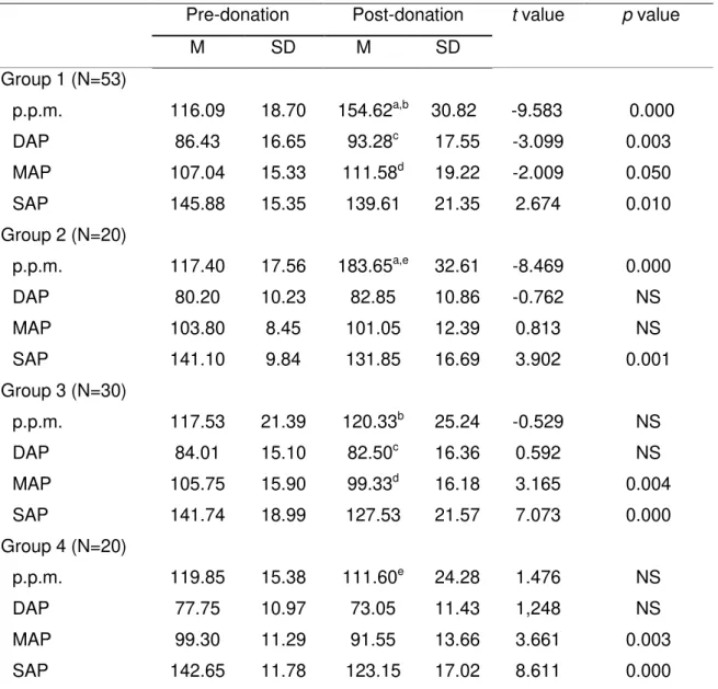
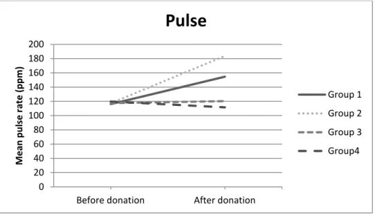
Os cães não-sedados que doaram 13% ou 15% do volume total de sangue (VTS) não evidenciaram variações potencialmente deletérias nas frequências do pulso nem nas pressões arteriais. No entanto, após a recolha de 15% do VTS em dadores sedados, foram registados potenciais efeitos nocivos, devido a aumentos significativos da frequência do pulso.
As dádivas de sangue trimestrais de 13% e 15% do VTS durante 2 anos foram consideradas seguras em cães, já que esta frequência permitiu uma resposta regenerativa da medula óssea capaz de restaurar a depleção de células sanguíneas em 10 dias após as dádivas, mantendo os parâmetros relacionados com a biodisponibilidade do ferro dentro do intervalo de referência calculado. A colheita de 13% do VTS cada 2 meses, durante 1 ano, induziu uma diminuição significativa das reservas de ferro.
sedação, da recolha de sangue, do tempo até ao processamento ou dos protocolos de centrifugação. Contudo, nas unidades de menor volume, com hematócrito mais elevado, com maior tempo até ao processamento e maior velocidade e tempo de centrifugação, tenderam a apresentar maior hemólise. Foram detetados valores de hematócrito significativamente mais elevados em unidades centrifugadas num programa mais rápido e longo (3500 G, 15 minutos).
A hemólise de unidades de concentrado de eritrócitos aumentou durante o armazenamento, especialmente da quinta para a sexta semana. De acordo com as normas de medicina humana, (é permitido um máximo de 1% de hemólise numa unidade a ser transfundida), os resultados apontam para a necessidade de avaliação da percentagem de hemólise em todas as unidades caninas de concentrado de eritrócitos com 36-42 dias de vida antes da sua administração, uma vez que quase 51% dessas unidades ultrapassou esse limite. Nenhum outro fator relacionado com o processo de colheita ou com as caraterísticas da unidade se relacionou com a hemólise. Da mesma forma, a hemólise detetada antes do armazenamento demonstrou não ter valor preditivo na hemólise registada após o período de armazenamento.
Os resultados obtidos sugerem que a elevada frequência do antigénio DEA 1.1 em cães portugueses (56,9%) é responsável por um elevado risco de sensibilização após uma primeira transfusão de sangue incompatível, e por uma probabilidade relevante de ocorrência de reação hemolítica aguda após uma segunda transfusão incompatível.
Os benefícios da transfusão de concentrado de eritrócitos foram evidenciados pelo aumento médio do packed cell volume (PCV) dos pacientes 3 horas após a transfusão. Além disso, todas as variáveis clínicas melhoraram dentro do mesmo período, embora só a pulsação, a cor das mucosas e a atividade mental foram significativamente diferentes dos resultados pré-transfusionais. Foram registadas apenas 3 reações transfusionais febris não hemolíticas, correspondendo a 6% do total das transfusões. A taxa de sobrevivência dos pacientes transfundidos foi de 71%, não estando relacionada com o volume ou idade das unidades de concentrado de eritrócitos, com a duração da transfusão, nem com o tempo de internamento ou o PCV dos pacientes.
Abstract
Transfusion medicine in dogs has become an important practice in the latest years, due to its life-saving demonstrated capacities. Veterinary practitioners are progressively becoming aware of its benefits, as well as its associated risks, and their routines are progressively changing towards a common and conscientious use of blood components. Such evolution has been based on several published studies about the use and efficacy of blood products, although the physiological effects of repeated donations and the consequences of laboratory processing and storage on the final product have been scarcely investigated.
This thesis aimed to investigate and clarify the influence of blood depletion in the hemodynamic variables of the donors, the effect of repeated blood donations in their iron status and hematologic parameters, the effects of collection and processing on the quality of packed red blood cells (pRBC) units, the influence of storage in pRBC hemolysis, the risks of untyped blood transfusions by identifying the prevalence of the dog erythrocyte antigen (DEA) 1.1, and the clinical benefits and potential risks of pRBC transfusions.
A total of 57 blood donors were evaluated during 1 to 2 years. In total, 327 whole blood units were processed and the obtained pRBC units were studied. Finally, clinical records from 34 dogs transfused with pRBC were reviewed to assess transfusion efficacy.
Non-sedated dogs donating 13% or 15% of total blood volume (TBV) did not evidence immediate harmful variations in pulse rates or arterial blood pressures. However, in sedated donors, potential deleterious hemodynamic effects were registered upon collection of 15% of TBV, with significant increases of pulse rates.
Quarterly blood donations of 13% and 15% of TBV during 2 years were considered safe for canine donors, as these regimens induced a bone marrow regenerative response, able to restore depleted blood cells within 10 days while maintaining iron status within the calculated reference range. Collection of 13% of TBV every 2 months during 1 year induced a significant decrease of iron stores.
The characteristics of pRBC units after processing were not significantly different when factors related to sedation, blood collection, time to processing or centrifugation protocols were studied, although units with lower volume, higher hematocrit, longer time to processing and higher centrifugation speed and time tended to present higher haemolysis. Significantly higher hematocrit values were detected in units centrifuged in a faster and longer program (3500 G, 15 minutes).
with 36-42 days of shelf-life before use, as almost 51% of such units surpassed this limit. No other collection procedure or unit characteristics were related to hemolysis. Likewise, pre-storage hemolysis failed to predict its value at the end of the storage period.
Our results suggest that the frequency of DEA 1.1 expression in Portuguese dogs is high (56.9%), hence increasing the risk of sensitization following the first transfusion with non-typed/non-crossmatched blood, with a relevant probability of acute hemolytic reactions after a second transfusion.
The benefits of pRBC transfusion were evidenced by the overall mean increase in the patients’ PCV 3 hours after transfusion. Furthermore, all clinical variables improved within the same period, although only pulse rate, mucous membrane color and mentation were significantly different from the pre-transfusion results. Only 3 non-hemolytic febrile transfusion reactions were verified, corresponding to 6% of the total transfusions. The survival rate of transfused patients was 71%, with no significant differences being related to pRBC volumes and shelf life, duration of transfusion, length of hospitalization, or patients’ PCV.
1
–
Introduction
Transfusion medicine is a multidisciplinary science concerned with the proper use of blood products in the treatment of a variety of diseases. It is an increasingly common therapy in small animal emergency and critical care medicine, as evidenced by the development of dedicated animal blood banks and in-house blood donor programs, as well as the increased use of blood components rather than WB, allowing for pathology-specific treatments. New technological advances improve the benefits of blood transfusions, while risks are progressively minimized. In addition, veterinary care has been taken into high consideration in response to the increasing demands for better treatment options made by pet owners and by higher social concerns about animal welfare and animal rights to proper veterinary clinical care.
Since the nineteenth century, transfusion therapy has been developed in veterinary medicine, including the progressive change from universal WB administration to the individually tailored use of blood components.1,2 However, transfusion needs in most clinics are still met by the administration of fresh WB on an individual basis and by the use of employee-owned donors, most likely neither blood typed nor screened for infectious blood diseases. Moreover, veterinary community is still facing a lack of scientific formation programs in this area, what limits the clinical application of the most recent advances in transfusion medicine.
As this is a very recent clinical area in veterinary medicine, few clinical studies have been described in dogs and most of them belong to three major investigation groups from the United States of America (USA). Furthermore, several veterinary protocols, techniques, good practices and even physiologic mechanisms are extrapolated from human studies.
of blood contamination and ensuring red blood cells (RBCs) viability and function, which prevents hemolysis.
Donor welfare has always been a major concern in transfusion medicine, but very few studies were performed to achieve secure blood collection protocols that ensure blood donor safety, including the blood volume collected per donation and the cumulative effects of longer donation periods.
2
–
Blood donation
2.1
–
Donor requirements
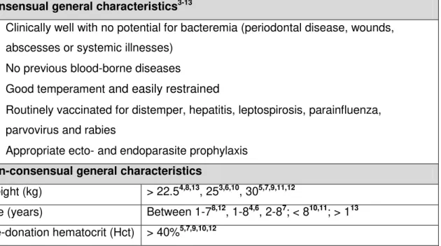
Before the admission of a dog to a blood donor program, it must be submitted to a careful evaluation, including an anamnesis that should review donor habits, living conditions, previous travelling, vaccination program, ectoparasite and endoparasite prophylaxis, clinical history and medical treatments. A complete physical exam should evaluate the donor ability to be safely submitted to blood withdrawal, and should inspect potential signs of infectious diseases able to be transmitted by blood transfusions. According to previous reports, there is a consensus regarding the ideal donor characteristics, described in Table 1.
Table 1. Blood donor requirements.
Consensual general characteristics3-13
Clinically well with no potential for bacteremia (periodontal disease, wounds, abscesses or systemic illnesses)
No previous blood-borne diseases
Good temperament and easily restrained
Routinely vaccinated for distemper, hepatitis, leptospirosis, parainfluenza, parvovirus and rabies
Appropriate ecto- and endoparasite prophylaxis
Non-consensual general characteristics
Weight (kg) > 22.54,8,13, 253,6,10, 305,7,9,11,12
Table 1. cont’d.
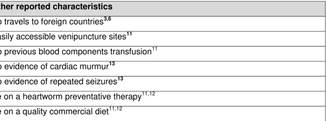
Other reported characteristics
No travels to foreign countries3,6 Easily accessible venipuncture sites11 No previous blood components transfusion11 No evidence of cardiac murmur13
No evidence of repeated seizures13 Be on a heartworm preventative therapy11,12 Be on a quality commercial diet11,12
Some authors advocate that female donors have to be nulliparous due to the risk of alloantibodies formation against RBCs (sensitization) by contacting with incompatible blood from puppies during pregnancy.6,11,12 However a recent study failed to document a significant difference between the frequencies of sensitized bitches with and without previous pregnancies, which suggest the absence of transplacental immunization in dogs, confirming previous reports by Young et al.14,15 Regarding breed selection, it is important to consider that Akitas present high physiological levels of intracellular potassium, which can induce hyperkalemia in the receptors, particularly by hemolyzed units or in cases of hemolytic transfusion reactions.7 Greyhounds are considered the best canine donors due to their athletic condition, calm temperament and lower frequency of RBC antigens able to induce hemolytic reactions.11
2.2
–
Pretransfusion screening tests
Table 2. Infectious agents with proved ability to be transmitted by blood transfusion.
Infectious agents Reported characteristics of the transfusion-related infection
Ehrlichia canis17 Transmission from infected donors may occur during the acute and chronic stages of the disease.
Babesia canis18 The donor, although clinically healthy, presented a serum antibody titer for Babesia canis > 1:5000. The patient exhibited hypotension, weakness and non-hemolytic pyrexia within hours after transfusion, and a low number of circulating Babesia canis was detected.
Babesia gibsoni19 A febrile hemolytic process was reported after transfusion with contaminated blood.
Leishmania infantum20,21
Three of seven dogs that received blood from infected donors developed serological titers and one of them developed severe disease more than two years after receiving pRBC transfusion. Both positive titers and clinical signs of visceral leishmaniasis were also reported in hamsters after administration of canine blood infected with Leishmania infantum.
Brucella canis22 After transfusion with contaminated blood, bacteria can persist for up to 2 years in infected dogs.
Diseases that are conditionally recommended to be tested and should be considered in a screening panel include: (1) diseases with documented experimental transmission but clinical transmission via transfusion is not described or (2) diseases that do not represent a threat to most receptors or are easily cleared by them. This group includes the following agents: Trypanosoma cruzi, which should be tested in dogs with history of travel to and from endemic areas, Bartonella vinsonii, because its potential transmission by blood products is unclear, and Mycoplasma haemocanis, which do not induce clinically evident disease in the majority of dogs.16
agents.24 In closed canine colonies and resident clinic donors the test should be done every 3-6 months, but for volunteer client-owned donors annual testing can be performed, provided that it is combined with careful anamnesis, proper physical examination and laboratorial tests including complete blood count, biochemical profile, urinalysis and fecal exam.4,8,11,12,16,24,25
One study in the United Kingdom reviewed the infectious agents screening results of 262 dogs enrolled in a blood donor program.3 These dogs were randomly selected and corresponded to 1-10% of the total program blood donors. The screening panel included PCR for the presence of hemotropic mycoplasmas, Bartonella, Babesia, Ehrlichia, Leishmania and Anaplasma spp.3 Two hundred and fifty eight (98.5%) dogs were PCR negative for all agents, and 4 dogs (1.5%) had positive or inconclusive PCR results (two tested for hemotropic mycoplasma and two for Leishmania).3 These dogs were retested 2-6 months later and negative PCR results were obtained, suggesting that they were either false positives or infected at the first sampling time but cleared the infection before the second PCR test.3 The possibility of false negative results on the second analysis was not postulated. According to authors, these results demonstrated that the prevalence of these infectious agents is low, hence the use of blood products is unlikely to represent a high risk for the transmission of infectious agents.3
Despite the many evidences of the transmission of infectious agents by blood transfusions and the publication of the ACVIM Consensus Statement,16 the guidelines are often not followed by clinicians and the donor selection is frequently based on physical examination of client or clinic-owned dogs, evidencing an absence of signs of infectious disease. One study reported that 36% of veterinary clinics did not evaluate the presence of infectious diseases among their donors, 24% searched only for circulating microfilaria, and 40% performed one or more tests generally recommended in screening panels.26 Moreover, crossmatching was performed in 40% of the clinics, donors blood type was determined in 32%, whereas receptors were not routinely typed by any evaluated practice.26 Economical reasons, unawareness of the potential risks and the absence of time required for laboratory processing in an emergency case may justify these clinical options that put receptors at a higher risk.
2.3
–
Blood collection
Collection systems, anticoagulants and additive solutions (ASs)
published studies that evaluated shelf-life and maximal expiration dates. Shelf-life is defined as the number of days after collection, assuming proper closed system collection and storage, at which 75% RBC viability is maintained in the recipient 24-hour after transfusion.27
Post-transfusion viability (PTV) studies are performed by infusion of radiolabeled or biotinylated RBCs followed by measurement of radionuclide activity and percentage of labeled cells, respectively, in blood samples drawn at frequent intervals after transfusion.28-30 After the maximum storage period, cell lesions may compromise the RBCs viability, which will not only significantly decrease their ability to carry oxygen, but also potentially trigger transfusion reactions, mainly associated with higher free-hemoglobin (Hb) concentrations and electrolyte imbalances.31,32
The collection systems, anticoagulant-preservative solutions and ASs that are more frequently used for canine blood collection are described in Table 3. The design of commercial collection devices ensure a closed system (with no air contact) from the collection to the administration, minimizing the risk for bacterial contamination.8,13 Special used plastic bags are not easily broken, facilitate components separation, avoid internal contamination, allow for gas exchange and hamper platelet and coagulation factors activation.32
The volume of blood to be drawn should be proportional to the amount of anticoagulant in the collection bag.13 Commercial collection bags have 63 mL of anticoagulant allowing for the collection of 405-495 mL of blood.8,9,33 Citrate-based anticoagulants are the most used and may include the following components:8,33
Citrate – prevents clotting and retards glycolysis;
Dextrose and adenine – ensure cell viability and function during storage (allow for continuation of the glycolytic pathway to help synthesis of adenosine triphosphate – ATP);
Phosphate – contributes to the adenosine phosphate pool and is a buffer for end products of RBC metabolism, such as lactate;
Citric acid – prevents glucose caramelization during autoclaving and provides optimal pH for RBCs.
such as dextrose, adenine, mannitol and sodium chloride, ensuring RBCs to maintain their function and viability during storage, preventing their lysis.9,36
Table 3. Commercial available blood collection systems, anticoagulant-preservative solutions and
ASs more frequently used in canine blood collections.
Blood collection systems
Single
One bag with anticoagulant-preservative solution.For WB storage only, with no components separation. Usually used for immediate administration.
Double
One bag with anticoagulant-preservative solution, connected bya plastic tube to a second empty transfer bag.
Allows preparing pRBC without additive and fresh frozen plasma (FFP), after centrifugation.
Triple
One bag with anticoagulant-preservative solution, one emptytransfer bag and one bag with AS, all connected by a plastic tube.
Used for the preparation of additive pRBC and FFP.
Quadruple
One bag with anticoagulant-preservative solution, two emptytransfer bags and one bag with AS, all connected by a plastic tube.
Used for the preparation of additive pRBC, FFP and platelet concentrate or cryoprecipitate.
Anticoagulant-preservative solutions
CPDA-1
Citrate, phosphate, dextrose, adenine and citric acid.Shelf - life: WB – 20 days37; 28 days10; 35 days35,38
ACD
Citrate, dextrose and citric acid.Shelf - life: WB – 21 days35,39; WB – 28 days40
CPD
Citrate, phosphate, dextrose and citric acid. Shelf - life: WB – 28 days41,42; 21 days35CP2D
Citrate, phosphate and double dextrose.Shelf - life: WB – 21 days35
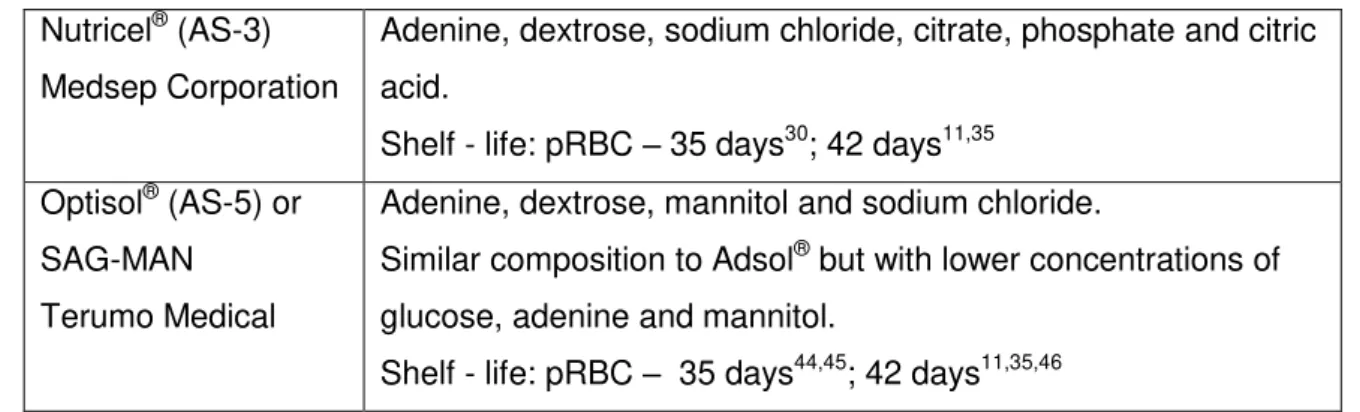
Additive solution
Adsol® (AS-1) Fenwal Laboratories
Table 3. cont’d.
Nutricel® (AS-3) Medsep Corporation
Adenine, dextrose, sodium chloride, citrate, phosphate and citric acid.
Shelf - life: pRBC – 35 days30; 42 days11,35 Optisol® (AS-5) or
SAG-MAN Terumo Medical
Adenine, dextrose, mannitol and sodium chloride.
Similar composition to Adsol® but with lower concentrations of glucose, adenine and mannitol.
Shelf - life: pRBC – 35 days44,45; 42 days11,35,46
Blood collection protocol
The collection technique is a pivotal step to optimize the final quality of blood products, but it is also crucial to ensure donor welfare. The process can be performed either by gravity or suction. A recent study compared both methods and concluded that suction-based blood collection is faster and safer for the donor, preserving blood components when a vacuum chamber is used with reasonable negative pressure (-101.6 mmHg).47 In fact, there was no difference in the final Hct or hemolysis index between the two methods. Although a post-donation decrease of the donors’ systolic blood pressure (SBP) was demonstrated, it remained within physiological values in all dogs with no significant differences between both methods.47
Although the suction method proved to be faster, reducing the collection time by almost 30%, it was also noisier, a fact that, by hampering the relaxation of some donors, may lead to more frequent movements.47 Though the authors considered this method as cost-effective, it certainly involves more expenses than the gravity method. Moreover, the suction device makes it impractical to use when blood collections are performed in different places.
The full technique for canine whole-blood collection should include:8,33,45 1. a pre-donation physical exam, including weight measurement;
2. to place the donor on the examination table in a comfortable either lateral or sternal recumbence;
3. to clip and surgically prepare the puncture site; 4. to place the collection bag on a gram scale;
5. to clamp the collection line at 15-20 cm distal to the needle, using a plastic hemostat;
6. to clearly identify the position of the vein, remove the needle cap and insert the needle into the jugular vein;
8. to perform careful agitation of the blood bag every 50-75 mL;
9. once the desired volume is collected, to clamp the line near the needle, remove the needle and apply digital pressure with a gauze over the phlebotomy site for 5 minutes;
10. to “strip” the blood left in the tube using a tube stripper, allowing it to mix with the anticoagulant in the blood bag, and return it into the tube;
11. to wrap the puncture site for a minimum of 30 minutes, avoiding bruising or hematoma formation;
12. to heat seal the collection line;
13. to label the collection bag with: donor identification, blood group, date, used anticoagulant and volume collected.
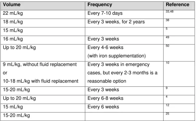
Total blood volume collection and donation frequency
Although the health and welfare of blood donors should be a major concern, it is often neglected or not mentioned in scientific studies. Therefore, there is currently no consensus on the ideal frequency of donations, the volume of blood that can safely be collected, or the need for nutritional supplementation of blood donors. In human transfusion medicine, the Council of Europe recommends up to four blood donations per year in men and up to three in women, with a maximum volume of 13% of TBV.23 Human recommendations are very different from those published in canine donors (Table 4).
Table 4. Blood volume and frequency of blood donations, according to different authors.
Volume Frequency Reference
22 mL/kg Every 7-10 days 33,48
18 mL/kg Every 3 weeks, for 2 years 38
15 mL/kg 5
16 mL/kg Every 3 weeks 49
Up to 20 mL/kg Every 4-6 weeks
(with iron supplementation)
50
9 mL/kg, without fluid replacement or
10-18 mL/kg with fluid replacement
Every 3 weeks in emergency cases, but every 2-3 months is a reasonable option
10
15-20 mL/kg Every 3 weeks 9
Up to 20 mL/kg Every 6-8 weeks 4
15 mL/kg Every 6 weeks 12
2.4
–
Donor reactions to blood collection
Few published reports described post-donation reactions in dogs. According to Gibson et al. (2012), adverse effects are uncommon but may include hypotension, bruising or bleeding from the venepuncture site and dermatitis, which can be detected within a few days after donation.44 Couto et al. (2005) also stated that, according to their experience, weakness or syncopal episodes after donation were exceedingly rare in dogs, reporting only one event in a Greyhound within 700 collections over ten years.51 A report describing a canine donor program evaluated 143 donations from 84 dogs and found acute donor reactions (e.g. pallor and light-headedness) in 2.8% of the donations, rebleeding in 2.1%, hematoma formation in 4.2% and skin irritation in 0.7%.4 The incidence of rebleedings and hematomas was reduced by bandaging the phlebotomy site and teaching owners to avoid putting the leash on the collar immediately after donation.4 Another study found no adverse effects (collapse, weakness or lethargy) on 19 Greyhound donors after collection of 17-22% of TBV, despite significant but transient decreases in SBP.51 Similar results were published in one study of thirteen dogs aiming to compare the two blood collection methods, gravity versus suction.47
3
–
Processing and storage of blood components
3.1
–
Blood components therapy
has resulted from the emergence of several new commercial blood banks, along with a better understanding of the components clinical advantages, is changing clinicians’ habits towards specific component hemotherapy.58,59
3.2
–
Processing methods
After blood collection, WB units must be immediately refrigerated in order to reduce the unit temperature to below 6°C.35,49 However, if platelets activity is to be preserved in WB, platelet-rich plasma or platelet concentrate units, those must be maintained at 22°C until administration.49,60-62 Plasma processing and freezing at -18°C ought to be performed within 8-hours after blood collection, in order to preserve the maximum activity of clot factors.35,49 However, a recent study reported that when WB units were processed within 24 hours after blood collection, the frozen plasma coagulation factor activities (II, V, VII, VIII, IX and X) and hemostatic protein contents (von Willebrand factor - vWF, antithrombin and fibrinogen) were maintained, making this a suitable method for the production of fresh frozen plasma.63 Whole blood processing allows for the preparation of three main components: pRBC, FFP and platelets concentrate. Additionally, cryoprecipitate (containing factor VIII, vWF and fibrinogen) and cryopoor plasma (with the FFP remaining components) can be prepared from a unit of FFP.49 Components separation techniques include:11,35,44,49
1. gentle stir of the WB unit was performed before processing;
2. proper packaging of the WB unit and remaining system bags into the centrifuge cups, avoiding plastic folds or empty spaces, and ensuring a symmetrical distribution of the weight between opposite cups;
3. centrifugation at 6°C (for RBC and protein preservation) or at 22°C (for additional platelet use). Described centrifugation protocols for canine WB processing include velocities ranging from 2000-5010 G, during 5-25 minutes, mainly with slow deceleration times, as sudden changes in centrifugation speed are associated with higher haemolysis;7,8,11,30,33,35,43,49,54,55,64-67
4. removal of the unit from the centrifuge without agitation and insertion in a plasma extractor in order to express the supernatant plasma into a transfer empty bag;
5. addition of the proper volume of AS to the primary pRBC bag, based on a proportion of 100 mL AS to 450 mL WB;
7. after separation of the components, tube sealing, with a thermal sealer, 10 cm away from the bag; additional seals along the tube filled with plasma or RBCs may be performed to sequester samples for future compatibility tests;
8. identification of each bag with donor identification, blood type, component, volume, date of collection, expiration date and storage temperature;
9. refrigeration of packed RBCs units as soon as possible at 1-6°C, and of FFP units at -80°C, followed by storage at -18°C.
Protocols for the production of canine platelet-rich plasma, platelet concentrate, cryoprecipitate and cryopoor plasma have been described by several authors despite the relatively low number of studies reporting their use.11,35,44,49,60-62
3.3
–
Packed RBCs units
As previously described, units of pRBC are obtained after separation of plasma content from WB. In addition to erythrocytes, pRBC units contain a small portion (approximately 10%) of the original plasma volume.11,68 If leukoreduction is not performed and platelets are not separated, non-functional leukocytes and platelets will also exist on pRBC units.11 Since triple collection systems with ASs are used, addition of 0.9% saline to RBCs reconstitution prior to transfusion is no longer necessary as some authors advocate.36,44 Instead, 100 mL of AS are added to erythrocytes, extending their shelf-life and reducing the unit viscosity, as previously described.8,34,35 Packed RBCs units with AS have a final packed cell volume (PCV) ranging between 55-65% and a volume of 200-300 mL.9,34,36,68,69
3.4
–
Storage lesions
2,3-Diphosphoglycerate (2,3-DPG)
Additive solutions have a pH of 5.7 that will decrease the pH of pRBC, avoiding the caramelization of glucose in heat-sterilized solutions.72 However, in such acid solutions 2,3-DPG is rapidly depleted from RBCs during storage.36,72 When 2,3-DPG combines with deoxyhemoglobin, it enhances the release of oxygen to the tissues, thus its decrease will shift the oxyhemoglobin dissociation curve to the left, hence reducing oxygen delivery.
36,72-75
In one study of canine pRBC units, preservation in ADSOL (AS-1) caused a significant decrease in 2,3-DPG during 44 days of storage (from 15.2 μmol/g Hb to 3.7 μmol/g Hb).28 Similar results were obtained with the use of other ASs.76 Fortunately, 2,3-DPG is regenerated in vivo after transfusion, reaching 50% of normal values within 7 hours, while its complete restoration takes 2-3 days.77
Red blood cells membrane
During storage the RBC membrane loses flexibility, compromising its normal function and hampering microcirculation.36,72,73 The cell morphology changes progressively, from biconcave disks to speculated or sphere-shaped cells (echinocytes or spherocytes, respectively), decreasing surface-to-lumen ratio.71,75,78 After transfusion, these RBCs will either be removed or they will assume the original biconcave shape within 24 hours.11 Additionally, RBCs develop microvesicles containing Hb that are released to the plasma, causing ongoing spontaneous hemolysis (degradation of RBCs), a phenomenon that contributes more to plasma Hb concentration rather than to free Hb during storage.78
ATP
ATP is progressively consumed during storage, especially if cell metabolic activity is not reduced by keeping units at low temperatures, usually 4°C.73 The reduction of RBCs ATP concentration may lead to protein denaturation and changes in the phospholipids composition of their membrane, decreasing its integrity and ability to deform.36,73,74 As RBCs rigidity stimulates their removal by the reticuloendothelial system after transfusion, ATP levels are directly related to the RBCs shelf-life.75 However, it seems that in dogs the RBCs ATP content may not be as vital as in humans.79
Accumulation of proinflammatory substances
responsible for the degradation of RBCs and transfusion reactions, e.g. febrile nonhemolytic transfusion reactions.71,80,81
pH
The pH of pRBC units decreases during storage.28,75,76 In spite of the left shift of the oxygen dissociation curve induced by acidemia, the administration of stored pRBC units has no consequences for the transfused patients because the alkaline by-products of citrate metabolization tend to reestablish the acid-base equilibrium.75
Other
Increased osmotic fragility, Hb oxidation and Hb denaturation have also been described as RBCs storage lesions that adversely affect their shelf life and function.71,82,83 Stored erythrocytes also evidenced increased aggregation, secondary to the loss of the negative charge in their surface, and increased adherence to endothelial cells, both phenomena contributing to a decreased microvascular flow.71
Fresh versus stored pRBC
Some canine studies evaluated the tissue oxygenation after the administration of stored pRBC, when compared to fresh pRBC, reporting either no differences or higher oxygenation levels with the use of fresh units, depending on the study.84,85 Although some human studies associated the administration of aged pRBC with higher incidence of infections, acute renal failure, longer hospitalizations and higher mortality,86-88 two large studies of critical anemic patients failed to reveal that morbidity or mortality were related to the age of transfused RBCs.89,90 However, some authors advocate that pRBC units with more than 28 days should be avoided in the following cases:
Patients with hepatic failure. As ammonia levels increase during storage, transfusion of stored units may induce or aggravate hyperammonemia and encephalopathy in those patients.94
There are no clinical reports documenting an increased morbidity or mortality associated with the transfusion of stored rather than fresh canine RBCs.73
4
–
Blood compatibility
4.1
–
Definition and genetics
Red blood cell membranes have species-specific antigens (glycolipids and glycoproteins) that constitute the basis for blood groups classification.95,96 These antigens may interact with autoantibodies (to self RBCs) or alloantibodies (to other dogs RBCs), causing an inflammatory reaction.96 A blood group is defined by the expression of two or more alleles in a givenlocus, responsible for the expression of a particular antigen on the RBCs surface.97 These genetic markers vary in immunogenicity and clinical significance, determined by three basic factors: 1) the prevalence of each antigen in the population; 2) the frequency and titer of natural or induced antibodies by previous transfusions; 3) the antibodies affinity to RBCs antigens and consequent induction of agglutination or complement activation.12,95,96,98
There are seven major blood groups in dogs identified with the acronym DEA (dog erythrocyte antigen) followed by the numbers 1, 3, 4, 5, 6, 7 and 8.95,99 In addition, 11 other antigens have been described, with no demonstrated clinical relevance.95 These antigens can be co-expressed by the same RBC and vary in frequency with breed, geographic location and antisera used.95,100 All groups seem to be transmitted by an autossomicdominant genetic mechanism.99 A single dog may or may not present each one of the antigens, thus being classified as positive or negative.99 However, the blood group DEA 1 presents three alleles sharing the same locus, therefore expressing one of three distinct antigens (or none of them): DEA 1.1, DEA 1.2 and DEA 1.3 (the latter recently described in Australia, where it seems to be common in German Shepherds101).102 The dominant order is DEA 1.1 > 1.2 > 1.3 > 1 negative.99,102,103 Thus, each animal can only be positive for one of the antigens or negative for all of them.95,99,100,103,104
and that 1.2 antibodies could be considered “weak” 1.1 antibodies, produced after exposure to a less antigenic DEA 1.105 Thus, these subgroups should be interpreted like subjective determinations of different antigenicity degrees of the same antigen, not separate blood types.105 The author also stated that additional studies are needed to describe the clinical importance of hemolytic transfusion reactions induced by weak DEA 1 antibodies (previously reported as 1.2 antibodies) but, while this information is not available, dogs typed as 1.2 positive should not be considered DEA 1.1 negative donors.105 Similar to this report, two previous studies suggested that 1.2 and 1.3 antigens may represent a weaker density of DEA 1.1 on the surface of canine RBCs.103,104
4.2
–
DEA system
Table 5 describes the main characteristics of each antigen, recognized as international standards.
Table 5. DEA system antigens description.
DEA 1 (1.1; 1.2; 1.3) Previous nomenclature: A102
Genetic: This group is a three factor (alleles Aa1, Aa2 and Aa3), four phenotypes system – 1.1, 1.2, 1.3 and null type, by order of inheritance.95,104
Incidence: In USA - DEA 1.1 42-45%;95,106 DEA 1.2 12-20%;95,106 DEA 1.3 identified only in Australia.104
Natural antibodies: No.95,102
Transfusion reactions: Delayed hemolysis in a first incompatible transfusion and acute hemolytic reactions in subsequent incompatible transfusions.95 It is the most clinically important blood group, and, as anti-DEA 1.1 antibodies are strong agglutinins and hemolysins, this antigen seems to be the major responsible for acute hemolytic reactions after an incompatible transfusion to a previous sensitized dog.95,100,107 Transfused 1.1 and 1.2 positive RBCs to sensitized DEA 1.1 and 1.2 negative dogs will be removed in less than 12 hours and 24 hours, respectively.95,100,102,108 No clinical transfusion reactions have ever been related to a 1.2 mismatch.109 No reports were published regarding the DEA 1.3 antigen incompatibilities.95,104
DEA 3 Previous nomenclature: B102
Table 5. cont’d.
Incidence: In USA – 6-7% DEA 3 positive;106,110 higher frequency in Greyhounds, with 23% DEA 3 positive.95 Over 60% positivity in native Japanese breeds.111
Natural antibodies: Yes. Up to 1.2-30% of DEA 3 negative dogs revealed the presence of anti-DEA 3 antibodies.95,112,113
Transfusion reactions: Delayed hemolytic reactions can occur in sensitized dogs, with removal of transfused RBCs within 5 days.95 Even though there are no published clinical studies to support it, some authors consider that severe acute reactions may also occur.95,111,114
DEA 4 Previous nomenclature: C102
Genetic: A one factor, two phenotype system – DEA 4 positive or DEA 4 negative, with dominance of the first one.102
Incidence: In USA – 98% DEA 4 positive.100,106,114 Lower frequency (25%) reported in Dobermans.95
Natural antibodies: No.102
Transfusion reactions: Contradictory reports. Sensitized DEA 4 negative dogs did not develop hemolytic reactions after transfusion with DEA 4 positive cells.114 One hemolytic reaction was described in a DEA 4 negative patient after multiple transfusions of DEA 4 positive blood.115
DEA 5 Previous nomenclature: D102
Genetic: A one factor, two phenotype system – DEA 5 positive or DEA 5 negative, with dominance of the first one.102
Incidence: Variable according to location.102 In USA, 11-23% DEA 5 positive,95,106 higher frequency in Greyhounds (up to 30%).95
Natural antibodies: Yes, in 0.8-10% of dogs from USA.112-114
Transfusion reactions: Delayed hemolytic reactions can occur in sensitized dogs, inducing premature extravascular hemolysis within 3 days.114
DEA 6 Previous nomenclature: F102
Genetic: A one factor, two phenotype system – DEA 6 positive or DEA 6 negative, with dominance of the first one.102
Incidence: Almost 100% in USA.110 Marked breed and geographic variation.116-118
Table 5. cont’d.
Transfusion reactions: Only one report of a moderately rapid extravascular hemolysis in a previously sensitized DEA 6 negative patient, after transfusion with DEA 6 positive blood.110
DEA 7 Previous nomenclature: Tr102
Genetic: A two factor, three phenotype system - Tr, O and null, respectively by order of dominance.102 Tr is not an integral erythrocyte membrane antigen, but secreted into plasma and adsorbed into the RBC surface after being produced in an unknown place.119
Incidence: Tr is present in 9.8-54% of dogs.106,113,118,120-122
Natural antibodies: Yes. In 20-50% of dogs a low titer (less than 1:8) of antigens was reported.95 No alloantibodies were found in another study.100
Transfusion reactions: Delayed hemolytic reactions can occur in sensitized dogs, with sequestration and loss of RBCs within 72 hours.95,110 However, no in vivo or in vitro hemolytic reaction was ever reported.99,100
DEA 8 Previous nomenclature: He102
Genetic: Unknown.
Incidence: 40-50% in USA.123
Natural antibodies: Unknown.
Transfusion reactions: Unknown.
4.3
–
Antibodies and hemolytic reactions
Blood groups that have proved to cause hemolytic reactions are DEA 1.1,100 DEA 4115 and, more recently, the presence of Dal antigen124. A hemolytic reaction following three different transfusions to the same dog was also reported, but an unknown alloantibody, other than DEA 1.1, 1.2, 3, 4, 5 and 7 should be related, according to the authors.125 Other non-consensual studies reported, without clinical case descriptions, both acute and delayed hemolytic reactions in incompatible transfusions for DEA 1.2,95 DEA 3,95,111,114 DEA 5,114 DEA 6110 and DEA 795. However, no acute hemolytic reaction was reported in a first incompatible transfusion, as no natural antibodies against the most antigenic RBCs markers are presented in dogs.7 Indeed, alloantibodies will only be produced 3 to 14 days after the first incompatible blood transfusion, inducing a delayed reaction that leads to premature and rapid destruction of transfused RBCs, decreasing the effectiveness of transfusion.95,98,100 It was estimated that 25% of random primary transfusions produce antibodies anti-DEA 1.1 and 1.2.75 In subsequent blood transfusions with incompatible blood, the sensitized receiver may immediately develop a serious acute hemolytic reaction.7,95,100 Despite the lack of case reports, DEA 1.1 antigen seems to be the most important in canine transfusion practice, due to its high antigenicity, high frequency in canine population and ability to induce antibodies that react as hemolysins, while all others react as hemagglutinins.96,100,125,126
4.4
–
New blood groups
antigen in the general population will be needed so that it may be classified as a high frequency RBC antigen.109 In a published case report, a DEA 1.2 and DEA 4 positive Whippet developed delayed hemolytic transfusion reactions after two RBCs transfusions and an acute form after the third transfusion.125 By a series of pre- and post-transfusion crossmatchings with 14 full typed bloods, authors conclude that an alloantibody, other than DEA 1.1, 1.2, 3, 4, 5 and 7, was responsible for such reactions, and postulated that it could be related to a high frequency unknown antigen.125 Further studies are needed to clarify the existence and clinical importance of these high frequency antigens in dogs. However, failure to obtain a negative crossmatching with several universal donors should indicate that a highly frequent undiscovered antigen can be related. In such cases, siblings may potentially be compatible donors, considering that they are more probable to miss such antigen.125
4.5
–
Universal donor definition
The definition of the canine universal donor is still not consensual.96,98 Considering that 98% of dogs are DEA 4 positive and that sensitized DEA 4 negative animals do not seem to develop acute hemolytic reactions after a second incompatible transfusion, some authors consider that DEA 4 positive and negative for every other DEA canine donors are universal donors.8,12,36,95,96 However there is a contradictory report of an acute hemolytic reaction in a dog that was DEA negative for all groups, after being transfused with a pRBC strictly DEA 4 positive.115 Other authors consider that, due to the low frequency of canine donors strictly DEA 4 positive (57.3% in Greyhounds and 28% in non-Greyhound dogs)122 and the lack of evidence that other antigens incompatibility increases the risk of hemolytic reactions, DEA 1.1, 1.2 and 7 negative dogs should be considered universal donors.31,36,75,100 These authors excluded DEA 7 positive dogs from the universal donor pool due to the presence of natural antibodies against this antigen in DEA 7 negative dogs, the evidence of delayed hemolytic reactions in incompatible DEA 7 transfusions and the important incidence of this antigen in the canine population, higher than the DEA 3 and 5 frequencies.95
Thus, attending to these reports, 1.1 negative dogs should be considered “evidence -based” universal donors in the first blood transfusion.95,97,109
4.6
–
Pretransfusion compatibility tests
Red blood cells compatibility tests are the only available canine pretransfusion tests, being currently impossible to determine incompatibilities against antigens from other blood components such as leucocytes, platelets or proteins. These tests will detect both RBC membrane antigens and the presence of alloantibodies against RBCs, which allow to minimize the risk for hemolytic reactions and to extend transfused RBCs survival times.13,98
Blood typing
Blood typing allows for the identification of a specific antigen in the RBCs surface.9,36 Both blood donors and patients should be typed for DEA 1.1, the most clinically important antigen in dogs, and only compatible blood should be transfused as it avoids:12,25,36,55,75,96,100,108,126
delayed hemolytic reactions, even in a first incompatible transfusion, which are responsible for early RBCs destruction, decreasing their lifespan to 2-5 days; this premature RBC extravascular destruction, although non-symptomatic, may increase the need for subsequent transfusions, of particular importance in non-regenerative anemias;
acute hemolytic anemias in pre-transfused and already sensitized patients with alloantibodies against DEA 1.1 antigen;
the risk of neonatal isoerythrolysis in puppies that have received DEA 1.1 alloantibodies from a sensitized mother.
some subjectivity, this test should be used by trained technicians from laboratories or blood bank programs.129
The Alvedia® Quick Test DEA 1.1® is an imumunochromatographic cartridge method that uses specific anti-DEA 1.1 monoclonal antibodies, incorporated in a membrane that will bind and retain DEA 1.1 positive RBCs during blood migration.129,130 Thus, the presence of a red line in the membrane indicates the presence of DEA 1.1 antigens, and the intensity of the color seems to directly correlate with the degree of expression of DEA 1.1 by RBCs and with the patient’s hematocrit.129,130 This method seems to be a suitable, easy to perform and to interpret rapid test.130 However, a false positive result in a dog with immune mediated hemolytic anemia (IMHA), typed by the gel column method (considered the “gold standard”109 but no longer commercially available) indicates the need for additional studies, particularly in animal affected by this disease.130 In opposition, another study indicates this test as the best option for typing samples with persistent autoagglutination, without the need for RBCs washing procedures, since only free non-agglutinated cells are allowed to migrate up the strip to bind the test antibodies, being less sensitive to agglutination interference than other methods.129 Thus, according to Seth el al. (2012), the high specificity and easy interpretation make this test appropriate for use in emergency situations by non-trained personnel.129
Recently, a new canine blood-typing method, the QuickVet®/RapidVet®, was developed by the Scandinavian Micro Biodevices Company. This system uses single use cartridges and an electronic analyzer, minimizing the user interpretation, but involves a higher initial investment.131 The system also uses a monoclonal antigen and agglutination is automatically interpreted by the analyzer. However, PCV of the samples have to be previously determined by the technician and values should be manually inserted in the electronic device, allowing for proper dilution with the reagent.131 Furthermore, samples have to be carefully evaluated, because severe hemolysis (leading to false-negative results) or agglutination (responsible by false-positive results) hampers the use of thismethod, according to the manufacturer’s instructions.131 One study compared the results obtained by this method with those from the gel column-base method, revealing a sensitivity of 96.2% and a specificity of 91.9%, concluding that, in spite of the discrepancies with the gel method (9/93, 9.7%), QuickVet®/ RapidVet® was suitable for in-clinic blood typing, provided that strict preanalytical guidelines are followed and samples with inconclusive results are tested by other methods.131
degree of agglutination or the intensity of the line on the Alvedia kits.105 Thus, the obtained results are essentially for DEA 1, and the named variables (1.1, 1.2 and 1.3) are simply a subjective determination of the agglutination reaction, not an identification of independent blood types.105
Commercial tests for in-clinic evaluation of antigens other than DEA 1.1 are not currently available, so these determinations can only be performed in a specialized laboratory in the USA (Animal Blood Resources International, Michigan).109 No typing serum is yet available for DEA 6 and DEA 8, hampering the blood typing of these groups by any method.98
Crossmatching
samples at a warm temperature, including the use of warm saline during the RBCs washing process.98
Traditional protocols for both slide (most reliable) or tube agglutination reactions, have been widely reported in the veterinary literature.9,11,12,25,31,44,57,132-134 The lack of standardization of the procedure, along with time consuming protocols and subjective operator-dependent interpretation are important limitations of this traditional method.25,133
Commercial in-clinic tests are recently available from two different companies. A gel crossmatch technology was applied in the Rapid Vet-H Companion animal crossmatch gel® test developed by DMS Laboratories, whereas the immuno-chromatography technique was used by Alvedia in the Lab test XM®. There are no published veterinary studies describing and comparing these methods but, considering the experience of the manufacturer laboratories, they should be considered faster and easier alternative methods when compared with traditional protocols. In addition, both techniques require smaller blood samples than a standard crossmatch (0.5 vs 2-3 mL) and can be used even with autoagglutinated samples.25 Besides, commercial tests do not require subjective interpretation of agglutination reactions and results can be reviewed at a later time.
Crossmatching should be performed in the second and subsequent blood transfusions, even if DEA 1.1 compatible blood is being used and even if blood from the same donor is used in both first and second transfusions.34,36,44,57,58,109,132 There are no natural antibodies against the most antigenic and clinical important RBCs antigens, thus, before the first blood transfusion, crossmatching should always be negative even between bloods from distinct groups, unless an IMHA is present.34,36,57,58,109,132 In subsequent transfusions, if ≥ 4 days from the first have passed, crossmatching should always be performed, even between DEA 1.1 typed dogs, due to the possibility of allosensitization for other antigens than DEA 1.1.5,57,109,132 If a transfusion reaction occurred or was suspected in the first transfusion, crossmatching should be performed in every subsequent transfusion from the first day.25,109
Despite its importance, crossmatching cannot substitute blood type determination because as there are no natural antibodies against DEA 1.1, in the presence of incompatible bloods crossmatching will always be negative in a first blood transfusion.98 Besides, crossmatching has some limitations, like false negative results, due to low antibodies titers in some sensitized dogs, and inherent subjectivity in qualitative evaluations of agglutination reactions by traditional methods.31
platelets or proteins, nor non-immunemediated reactions can be excluded with negative crossmatching tests, because only RBCs compatibilities are evaluated.44,98,132 Thus, it is extremely important to carefully monitorize transfused patients, even when RBCs compatibility tests are performed.98,132
Antibody screen
The antibody screening test aims to determine the presence of induced or natural alloantibodies in the receptor serum against RBCs antigens that may cause a hemolytic reaction.56,98,135 It is widely used in human medicine and requires the availability of RBCs of known phenotypes, able to react with the receptor serum.56,135 This pretransfusion test can be very useful in canine receptors with unknown previous history or at risk of possessing induced alloantibodies, allowing to select appropriate RBCs for transfusion that lack the antigen to which the receptor has developed antibodies.98 The main advantage of this test is that it avoids several crossmatchings with blood from potential donors in a pre-transfused sensitized receptor, as performed in some published clinical cases.124,125 Besides, it is impractical in the clinical setting to collect blood samples from a wide group of donors at the same time. Another potential application is the search of donors’ unexpected alloantibodies that could potentially react with the receptors RBCs after plasma or WB transfusion, although the clinical consequences of such reaction are debatable.13,56,98,113 Point-of-care tests are not available, and nowadays only one specialized laboratory in the USA is able to perform plasma antibody screening for anti-DEA antibodies (Animal Blood Resources International) and blood typing for antigens other than DEA 1.1.
5
–
Packed RBCs transfusion
5.1
–
Goals
Packed RBCs transfusion avoids damages caused by tissue hypoxia in anemic patients. The main goal is to improve tissues oxygen delivery, keeping the patient in a stable condition while diagnostic and etiologic therapeutic procedures are being performed.69 Oxygen delivered (DO2) to organs depends on the blood flow (cardiac output
- CO) and arterial oxygen content (CaO2), which is determined by the amount of Hb, its
oxygen saturation degree, and the partial pressure of oxygen in the blood (PaO2).36,71
the body, and only 2% is dissolved in plasma.36 The following formulas are used to address factors related to tissue oxygen delivery:54,71
DO2 = CO x CaO2, where CO = Heart rate (HR) x Stroke volume (SV)
CaO2 (mL/L) = [% SatO2 x 1.39 (mL/g) x Hb (g/L)] + [arterial PaO2 x 0.0031]
Thus, low cardiac output (stagnant hypoxia due to hypovolemia or cardiovascular compromise); low Hb saturation (hypoxic hypoxia, due to pulmonary disease); or low Hb concentration (anemic hypoxia) can be responsible for tissue hypoxia, and blood transfusions are indicated for the treatment of the last one, by improving Hb content.36,136
5.2
–
Indications
Globally, pRBC units are indicated for clinically symptomatic normovolemic anemias that do not require coagulation factors.36,54,68 Specific indications for pRBC transfusions vary among studies and may be affected by geographic and temporal variations. Pathologies that have been suggested to benefit from pRBC transfusion are detailed in Table 6.
Table 6. Reported pathologies that benefit from the administration of pRBC.5,9,11,34,36,55,137-146
Hemolysis Primary or secondary IMHA, pancreatitis, blood borne disease.
Blood loss Postoperative anemia, perioperative splenectomy (with or without hemoabdomen), gastrointestinal bleeding (e.g. ulceration, foreign body, hemorrhagic gastroenteritis, parvovirus infection, gastric dilatation-volvulus), coagulopathy (e.g. anticoagulant rodenticide ingestion, hepatopathy, disseminated intravascular coagulation -DIC, hemophilia A, immune-mediated thrombocytopenia, von Willebrand disease - vWD, trauma, idiopathic hemothorax, uterine hemorrhage secondary to pyometra, internal or external hemorrhage from bleeding neoplasia, severe dental disease.
Ineffective erythropoiesis
Chronic renal failure, primary bone marrow dysfunction (aplasia, hypoplasia, myelopthisis) or secondary to neoplasia (Sertoli cell tumor, leukemia, lymphoma and multiple myeloma).
trauma, surgery or bleeding tumors; anemia, most commonly due to IMHA or other hemolytic conditions; and bleeding due to coagulopathy associated with thrombocytopenia, DIC or vWD.5 A different study reviewed 163 pRBC transfusions administered to 131 dogs in 1989.55 Reasons to transfuse included anemia from blood loss in 70% of the cases; hemolysis in 22%; and bone marrow hypoplasia in 8%.55 In another retrospective study of 307 dogs submitted to RBCs transfusions, 72% where due to blood loss anemia (mostly related to trauma, surgery, neoplasia, thrombocytopenia, gastrointestinal bleeding and coagulopathy); 14% to hemolytic processes; and 14% due to ineffective erythropoiesis (including chronic renal failure, neoplasia and primary bone-marrow diseases).140 This study also reported lower mean pretransfusion PCVs (13%) in the hemolytic cases, comparing to hemorrhagic (21%) or nonregenerative processes (18%). This fact could be explained by the clinician’s hesitation to administer RBCs to dogs with ongoing hemolysis in IMHA or by difficulties in obtaining a clearly safe result in blood compatibility tests.140 On the other hand, acute blood losses do not allow for an effective activation of hypoxic compensatory mechanisms and anemia signs may develop with higher PCVs.140 In other studies, reported frequencies of anemic etiologies ranged from 12-63% for hemolysis, 29-74% for hemorrhage and 8-34% for ineffective erythropoiesis.138,141,143,147
5.3
–
Anemia compensatory mechanisms
In order to better define a transfusion trigger for pRBC administration, it is essential to understand the anemia compensatory mechanisms, including cardiovascular and metabolic responses, which allow patients to adapt and better tolerate low Hb concentrations. These include:
A decreased number of RBCs induces low blood viscosity that results in higher venous return and a decrease in the left ventricular afterload, responsible for increasing cardiac output.71 Hemodilution also improves blood flow in the microcirculation;54
The sympathetic stimulation increases ventricular inotropy and heart rate, which also contributes to increased cardiac output;54,136 however, severe tachycardia can be deleterious as it results in shorter diastolic times and subsequent reduction of myocardial perfusion by impaired coronary blood flow;136
Alterations in sympathetic tone induce peripheral vasoconstriction of the large veins, increasing blood return to the heart and consequently the SV. Additionally, a constriction of smaller arteries redirects blood flow to vital organs, thus facilitating coronary, pulmonary, renal and cerebral circulations;54,136
Vasoconstriction can also result in the mobilization of splenic blood reserves up to 50% of the sequestered blood volume.54
As these mechanisms take time to establish, an acute decrease in PCV is often associated with sudden onset of clinical signs, unlike chronic anemias that are well tolerated.68,73
5.4
–
Transfusion triggers
The transfusion trigger is the Hb level at which the decreased oxygen delivery is so reduced that anaerobic metabolism occurs, and it is used to describe a set of conditions under which transfusion is indicated.25,73 There are no established standardized guidelines for assessing whether humans or dogs benefit from a transfusion.44,147 However, several clinical and laboratory variables that may be useful in guiding the decision to transfuse have been described in dogs. Generally, RBCs replacement is necessary in the following conditions:
when PCV acutely drops to less than 20%;11,57,68,148
if PCV drops below 10%,36,57,68 as patients are at risk of multiple organ failure due to severe hypoxic injuries, or 20%,44,56 even in chronic anemias;
in cases of hemorrhage where more than 20%9 or 30%54,68,148,149 of TBV, according to different authors, is lost. Acute losses of 30-40% of TBV lead to shock with compromised organ function, and losses ≥ 50% lead to death if therapy is not instituted immediately;148
if a bleeding dog collapse;111,122
in ongoing non controlled hemorrhages;111,122
when PCV is < 18% with ongoing acute RBC destruction;9
in shock patients that are non-responsive to fluid therapy.68,148
maintained until PCV falls below 10%.151 However, it is possible that critically ill dogs with higher oxygen demands and/or compromise of either cardiovascular or respiratory systems may suffer from tissue hypoxia with higher PCVs. In fact, the decision to transfuse normovolemic patients, with no signs of organ dysfunction, will typically occur at much lower PCVs than to animals with hypovolemia, poor cardiac contractility, and/or respiratory compromise, because these patients require higher PCVs to maintain oxygen delivery.25,36,54 Other authors confirmed that PCV alone was not a predictive factor for transfusion requirements, as the overall clinical condition should be accessed and even considered the most important factor for the evaluation of transfusion needs.55,57,146 Furthermore, following acute hemorrhage PCV may be normal, due to the simultaneous loss of RBCs and plasma, or even elevated, after splenic contraction.55,152 In the latter cases, PCV only starts to decrease after 2 hours, when intravascular fluid shifts and other compensatory mechanisms (e.g. increased antidiuretic hormone secretion or renin-angiotensin-aldosterone system activation) result in plasma expansion and hemodilution.55,149
Table 7. Transfusion-need scale, suggested by Kerl et al. (1993).55
PCV Acute blood loss
<13% 5 15% decrease in PCV 1 13-15% 4 25% decrease in PCV 2
16-19% 3 Anesthesia 2
20-24% 2 Weakness 1
25-34% 1 Tachypnea 1
>34% 0 Tachycardia 1
This scale was constructed by the retrospective characterization of 131 dogs submitted to pRBC transfusion and allowing to determine criteria to predict the need for pRBC transfusion.55 However, this scale heavily relies on the PCV, an unreliable indicator of the need for transfusion.147 Indeed, some limitations were found in the ability to identify blood transfusion needs in dogs with acute blood loss, as pretransfusion PCVs were presumably falsely elevated.55 Furthermore, weakness, respiratory and cardiac rates were misevaluated in anesthetized dogs.55 Authors also suggested that the volume of blood loss and blood pressure should be included in subsequent prospective studies.55 Another recent study proposed an “anemic dog clinical assessment score” (ADCAS) to assess transfusion needs by clinical parameters in a more objective and standardized approach (Table 8).147
Table 8. Anemic dog clinical assessment score, suggested by Kisielewicz et al. (2014).147
Score 0 1 2 3
Mucous
membranes color
Salmon pink Slightly pale Moderately pale
Severely pale
Pulse quality Normal Bounding Weak Weak
Heart rate 65-109 b.p.m. 110-140 b.p.m. > 140 b.p.m. > 140 b.p.m.
Respiratory rate 15-24 r.p.m. 25-40 r.p.m. > 40 r.p.m. > 40 r.p.m.
Mentation,
exercise tolerance
Normal, walking
Quiet, able to walk
Lethargic, able to stand
Lethargic, Unable to stand