Analysis of Venom
in Cape Verde
Cone Snails
Jorge do Livramento Brito Neves
Tese de Doutoramento apresentada à
Faculdade de Ciências da Universidade do Porto,
Ciência e Tecnologia do Ambiente
2016
A na ly sis of V en om in C ap e Verde C on e S na ils Jorge d o Liv rament o Bri to Nev esPh
D
FCUP 2016 3.º CICLOD
D
D
Analysis of Venom in
Cape Verde Cone
Snails
Jorge do Livramento Brito Neves
Environmental Sciences and Technology
Department of Geosciences, Environment and Spatial Planning 2016
Supervisor
Vitor Manuel de Oliveira Vasconcelos, Cathedratic Professor, Faculty Of Sciences of University of Porto
Co-supervisor
Agostinho Antunes Pereira, Auxiliary Researches, Interdisciplinary Centre of Marine and Environmental Research of University of Porto
Acknowledgments
To Professor Vitor Vasconcelos for the extraordinary support, constant availability and the amazing encouragement. A special thanks for giving me the opportunity to learn and contribute to science.
To my co-supervisor Professor Agostinho Antunes for the availability, valuable suggestions and motivation.
To Professor Baldomero M. Olivera, for the extraordinary guidance and for giving me the opportunity to work and learn in his laboratory at University of Utah – USA; I would like to thank specially Julita S. Imperial for her daily guidance and encouragement. Thanks to all Olivera Lab. Staff, specially: My Huynh, Terry Merrit, Joseph W. Aman and Samuel S. Espino. Thanks to Zhenjian Lin, Eric W. Schmidt, David Morgenstern and Beatrix Ueberheide.
To all LEGE – Pedro Leão, Micaela Vale, João Morais, José Carlos, Vitor Ramos, Marisa Silva, Marisa Freitas, Ana Regueiras – and CIIMAR team members for the encouragement and incredible friendship over the years and also for the technical and administration support.
A special thanks to all my family, particularly to my parents, Maria Hirondina Brito Neves and João Baptista Neves because they always believed in me.
I am especially grateful to my wife, Ana Sofia, for the amazing and unconditional support. I dedicate this thesis to her and our son, David Neves.
This work was supported by Fundação para a Ciência e a Tecnologia (FCT) - PhD Scholarship (SFRH/BD/51477/2011); and the follow project: MARBIOTECH (NORTE-07-0124-FEDER-000047); NOVELMAR - Novel marine products with biotechnological applications (NORTE-01-0145-FEDER-000035); and FCT project UID/Multi/04423/2013.
ii FCUP
Preamble
Preamble
This thesis is based on the law (artigo 8º from Decreto Lei nº 388/70; República
Portuguesa), where most of the chapters are results of published works including work
in collaboration. The candidate participated on the acquisition, analysis, discussion and preparation of the work for publication.
The field work was done by the candidate through snorkeling and Scuba diving (until 20 meters deep), with the support of CIIMAR, Fundação Calouste Gulbenkian and University of Cape Verde (UniCV). The work presented here was conducted, in most part, in CIIMAR (Center of Marine and Environmental Research, University of Porto), but the candidate also conducted work for this dissertation during a 9-month stay at Department of Biology (University of Utah, Salt lake City, USA). The candidate has also established work partnerships with Institute of Molecular Pathology and Immunology of the University of Porto (IPATIMUP), Institute of Chemistry and Biology Technology (ITQB) at New University of Lisbon, and New York University Langone Medical Center, Department of Biochemistry and Molecular Pharmacology (New York, USA).
Thesis Organization
This dissertation was organized on five chapters. Chaper 1 presents a general introduction to the Cone Snail distribution, diversity, biology, ecology, structure and activity of toxins (conotoxins).
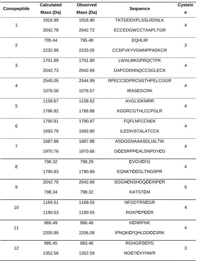
Chaper 2 describes the identification of 12 peptides from C. crotchii using MALDI-TOF and mass-matching. The number of molecular masses studied resembles the outputs from other studies performed in the general Conus enabling to validate the approach. It was identified several disulfide-rich conotoxins in C. crotchii venom duct samples that belongs to O1-superfamily (Eb6.18, Leo-O2, Bu2, PVIIA), A-superfamily (im23.3, Ai1.2, PnMGMR-02), T-superfamily (Ca5.1, TxVA) and O2- (Ec15a), O3- (VnMSGL-0123) and D-superfamilies (VxXXB).
In Chaper 3 it is described the collection and first toxinological characterization of the C.
ateralbus specie. An excitatory activity that acted on a majority of mouse lumbar dorsal
root ganglion neurons was purified and characterized from C. ateralbus venom. The 30AA venom peptide was named δ-conotoxin AtVIA. AtVIA has conserved sequence elements when compared to δ-conotoxins from fish-hunting Conus species, and from a peptide purified from Conus tessulatus, a species in the worm-hunting Indo-Pacific clade Tessiliconus.
In Chaper 4 it is identified a novel small-molecule guanine derivative with unprecedented features. Genuanine was isolated from the venom of two cone snail species (C. genuanus and C. imperialis). Genuanine causes paralysis in mice, indicating that small molecules and not just polypeptides may contribute to the activity of cone snail venom.
Finally, in Chapter 5 it is presented the overall discussion of results and main conclusions.
The structure of the manuscripts and the scientific paper was maintained according to the journal guidelines in which they were published or submitted, including the reference style.
iv FCUP
Abstract
Abstract
The cone snails (genus Conus), living in the tropical habitats, are highly venomous predatory gastropods that use peptide toxins (conotoxins) for major environmental interactions, such as prey capture, defense and competition. Approximately 800 species of Conus are known and each one can express more than 100 different peptides. Conotoxins (disulfide-rich peptides), can potentially target components of the neuromuscular system, mainly ligand- and voltage-gated ion channels.
The study of Conus crotchii venom duct revealed about 488 molecular masses between 700 and 3000 Da. Those masses were searched by matching with known peptide sequences from UniProtKB (UniProt Knowledgebase) protein sequence database. Through this method we were able to identify 12 conopeptides. For validation it was considered the error between the experimental molecular mass (monoisotopic) and the calculated mass of less than 0.5 Da. All conopeptides detected belong to the A-, O1-, O2-, O3-, T- and D-superfamilies, which can block Ca2+ channels, inhibit K+ channels and act on nicotinic acetylcholine receptors (nAChRs). Only a few of the detected peptides have a 100% UniProtKB database similarity, suggesting that several of them could be newly discovered marine drugs.
Conus ateralbus is an endemic cone snail that has been found only on the west side of
the island of Sal, in the Cape Verde Archipelago off West Africa. It was described the collection and first toxinological characterization of this species. An excitatory activity was purified and characterized from C. ateralbus venom that acted on a majority of mouse lumbar dorsal root ganglion neurons. This 30AA venom peptide, δ-conotoxin AtVIA, has conserved sequence elements when compared to δ-conotoxins from fish-hunting Conus species, and from a peptide purified from Conus tessulatus, a species in the worm-hunting Indo-Pacific clade Tessiliconus. In contrast, there is no comparable sequence similarity with δ-conotoxins from the venoms of molluscivorous Conus species. A rationale for the presence of δ-conotoxins that are potent in vertebrate systems in two different lineages of worm-hunting cone snails is discussed.
On the study of C. genuanus venom it should be noted that small molecules also contribute to the neuroactivity of the venoms. A novel guanine derivative, genuanine, causes paralysis in mice and is found in the venoms of at least two cone snail species.
The global results show that the venoms of Cape Verde cone species are rich source of powerful bioactive molecules in vertebrate systems and clearly present stronger pharmacology interest.
vi FCUP
Resumo
Resumo
Os caracóis marinhos (do gênero Conus) que se encontram em habitats tropicais são gastrópodes predadores altamente venenosos que utilizam toxinas peptídicas (conotoxinas) para as principais interações ambientais, como a captura de presas, defesa e competição. Cerca de 800 espécies de Conus são conhecidas e cada uma pode expressar mais de 100 péptidos diferentes no veneno. As conotoxinas (péptidos ricos em ligações dissulfureto) podem atingir potencialmente os componentes do sistema neuromuscular, principalmente os canais iónicos.
No estudo do ducto do veneno de Conus crotchii foram pesquisados cerca de 488 massas moleculares entre 700 Da e 3000 Da. Fez-se uma pesquisa dos peptídeos com os já conhecidos a partir do banco de dados UniProtKB (UniProt Knowledgebase). Através deste método, identificaram-se 12 conopéptidos. Para a validação foi considerado o erro (menos de 0,5 Da) entre a massa molecular experimental (monoisotópico) e a massa calculada. Todos os conopéptidos detectados pertencem às superfamílias A-, O1-, O2-, O3-, T e D-, que podem bloquear os canais de Ca2+, inibir canais de K+ e actuar sobre os receptores nicotínicos da acetilcolina (nAChRs). Apenas alguns dos péptidos detectados partilham 100% de similaridade com os dados da UniProtKB, sugerindo que vários compostos detectados podem ser novas toxinas marinhas.
Conus ateralbus é um cone caracol endémico que foi encontrado apenas no lado
oeste da ilha do Sal, no arquipélago de Cabo Verde (África Ocidental). Foi descrita a recolha e a primeira caracterização toxicológica desta espécie. Uma atividade excitatória foi caracterizada a partir do veneno purificado do C. ateralbus que atuou sobre a maioria dos neurónios dos ratos - dorsal root ganglian (DRG). O peptídeo caracterizado possui 30AA, designado de δ-conotoxin AtVIA, e conservou elementos da sequência quando comparado a δ-conotoxins de espécies Conus que se alimentam de peixes, e quando comparado a um peptídeo purificado a partir de Conus tessulatus, uma espécie do Indo-Pacífico que se alimenta de vermes (clade Tessiliconus). Em contraste, não há nenhuma similaridade de sequência comparável com δ-conotoxin AtVIA dos venenos de espécies Conus molluscivorous. É discutida a razão para a presença de δ-conotoxin AtVIA que são potentes em sistemas de vertebrados em duas linhagens diferentes dos Conus do grupo worm-hunting.
No estudo do veneno de C. genuanus apresenta-se que as moléculas pequenas também contribuem para a actividade neuronal dos venenos. Um derivado novo da guanina, genuanine, causa paralisia em ratos e é encontrado em venenos de pelo menos duas espécies de caracóis cone (C. genuanus e C. imperialis).
Os resultados na generalidade mostram que as espécies de Conus de Cabo Verde são importantes fontes de moléculas bioactivas que podem afectar os sistemas dos vertebrados, o que leva a um forte interesse farmacológico.
viii FCUP
Contents
Contents
Acknowlegments i
Preamble ii
Thesis organization iii
Abstract iv
Resumo vi
Contents viii
List of Tables ix
List of Figures xi
List of Abbreviations xvii
Chaper 1: Introduction 1
Chaper 2: Conopetides from Cape Verde Conus crotchii 23 Chaper 3: Characterization of a δ-Conotoxin from Conus ateralbus, a Vermivorous Cone Snail from the Cape Verde Archipelago 41 Chaper 4: Small Molecules in the Cone Snail Arsenal 64 Chaper 5: Discussion and Conclusions 95
List of Tables
Chapter 1: Introduction
Table 1: Post-translational modifications (PTM) of some Conus peptides. 9 Table 2: Conotoxin classification by gene superfamilies and pharmacological
families. For each gene superfamily the cysteine pattern and their target, the pharmacology families, and the reference are represented. 11
Chapter 2: Conopetides from Cape Verde Conus crotchii
Table 1: Peptide mass detected by MALDI-TOF-MS and putative sequence by mass-matching using MASCOT database research. 29 Table 2: Conotoxins identification by BLAST search on UniProtKB database; all
151 results have a 100% Max identity with the protein sequence. Signal, propeptide and mature peptide regions are shown in red, blue and black, respectively. Peptides identified in the venom by MALDI-TOF-MS— MASCOT database are underlined. 31 Table 3: Conotoxins isolated from different Conus species. 33
Chapter 3: Characterization of a
δ-Conotoxin from Conus ateralbus,
a Vermivorous Cone Snail from the Cape Verde
Archipelago
Table 1: Comparison of δ-conotoxin sequences from various cone snail species. 56
Chapter 4: Small Molecules in the Cone Snail Arsenal
Table S1. The mice test results for compounds 1-4. (See Figure S6 for data with crude venom fractions). All observed activities were reversible, and mice
x FCUP
List of Tables
Table S2. NMR data of natural 1 in D2O. 78
Chapter 5: Discussion and Conclusions
Table 1: Toxins isolated from Cape Verde cone snail venom. 98 Table 2: Toxicological venom effects from different Cape Verde cone snail using
List of Figures
Chapter 1: Introduction
Figure 1: Shells of some of the cone snails belonging to the CIIMAR collection. From left to right, top row: C. marmoreus; C. maldivus; C. viridulus. Second row: C. textile; C. miles; C. praelatus. Third row: C. tessulatus;
C. omaria; C. quercinus. Bottom row: C. terebra. The shells shown are
not represented to scale. 2
Figure 2: Cone species distribution per region (data adapted from the Illustrated Catalog of the Living Cone Shells 2013) [19]. 3 Figure 3: From left to right, Conus venulatus on egg capsules, 5-6 m depth, Boa
Vista Island; Conus ateralbus on egg capsules, 2-3 m depth, Sal Island;
Cape Verde arquipelago. 4
Figure 4: Conus venom apparatus of the C. striatus. Reproduced by personal permission from Baldomero M. Olivera [2]. This venom apparatus also represents the general cone snail venom apparatus. 5
Chapter 2: Conopetides from Cape Verde Conus crotchii
Figure 1: The endemic C. crotchii can be found only on Boa Vista Island - Santa
Mónica beach. 27
Figure 2: (A) Proteins separated by sodium dodecyl sulfate polyacrylamide gel electrophoresis (SDS-PAGE) from the venom duct of Conus crotchii. Mw: molecular weight, and S: sample; (B) Molecular mass range distribution of the 488 peptides detected by matrix-assisted laser desorption/ionization time-of-flight mass spectrometry (MALDI-TOF-MS)
analysis. 28
Figure 3: Distribution of peptide sequence identified as “Conoprotein”. 32 Figure 4: Shell and venom apparatus of C. crotchii: (a) Venom bulb; (b) Venom
xii FCUP
List of Figures
Chapter 3: Characterization of a
δ-Conotoxin from Conus ateralbus,
a Vermivorous Cone Snail from the Cape Verde
Archipelago
Figure 1: A phylogenetic tree showing the large clade of Conus encompassing all lineages that are fish hunting (underlined) and snail hunting (checked). Well established worm-hunting lineages are boxed. δ-conotoxins have been characterized from the 9 species figured, including the worm-hunting Conus tessulatus (red arrow). In this work, we investigated the venom of Conus ateralbus (shown by the thick red arrow), which is in the Kalloconus lineage. This tree was adapted from a figure from Puillandre et al. (2014). Species discussed in this study whose shells are figured are (from top to bottom): Conus bullatus (Textilia); Conus striatus (Pionoconus); Conus ermineus (l) and Conus
purpurascens (r) (Chelyconus); Conus tessulatus (l) and Conus eburneus (r) (Tesseliconus); Conus textile and Conus gloriamaris
(Cylinder); Conus ateralbus (Kalloconus). 45 Figure 2: Relationship of species in Kalloconus. Most of the species are
approximately the same size (ca 40 mm), except for Conus pulcher, which can be very much larger (up to 230 mm). 46 Figure 3: Conus ateralbus observed in the field feeding on a long worm, many
times the length of the predatory snail. 51 Figure 4: Fractionation of Conus ateralbus venom. The peptide characterized in
this study was first detected in the pool of fractions shown, 36-39 (Fig. 4A). Further purification of the active fraction (38) yielded the chromatogram shown in Figure 4B. The major peak (subfraction 38.6) was both biologically active and gave the results shown in Figure 4C upon analysis on an Orbitrap Elite mass spectrometer at 120,000
resolution. 52
Figure 5: Responses of 5 different cells to the purified at6a peptide (Fraction 38.6). Shown are the responses of 5 DRG neurons (see Methods); the size of each cell is indicated. A pulse of 25mM KCl was applied as described under Methods; the horizontal bar indicates when cells were incubated with the purified peptide. The first three cells (from top) show
a change in response upon depolarization with KCl. The fourth trace shows a cell that directly increased cytosolic Ca++ when the peptide was added, even without KCl depolarization (approximately 26% of cells responded in this manner) and the bottom trace shows a cell that did not respond (approximately 15% of DRG neurons were
non-responsive to the peptide). 53
Figure 6: Determination of the sequence of AtVIA by tandem mass spectrometry. MS/MS ETD spectrum of the (M + 5H)+5 ion of qCGADGQFCF(L/I)PG(L/I)G(L/I)NCCSG(L/I)C(L/I) (L/I)VCVPT after reduction and alkylation with 2-methylaziridine acquired on the Orbitrap Elite with 15,000 resolution (@ 400 m/z). N-terminal fragment ions (c-type ions) are indicated by and C-terminal fragment ions (z-type ions) are indicated by . Doubly charged ions are indicated with ++ and z ions resulting from cleavage at cysteine and loss of the cysteine side chain are indicated with *. [M+5H]+++++ and [M+5H]+++++ indicates quintuple charged precursor ions that captured 2 or 3 electrons, respectively, but have not dissociated in fragment ions. Due to space limitations, different charge states of already labeled peptide bond cleavages are not all indicated in the figure. The mass accuracy for all fragment ions is better than 15 ppm. The mass spectrometer used cannot differentiate between isoleucine or leucine and for simplicity leucine is used in the figure to indicate a fragment ion of mass 113.08406. 54 Figure 7: Synthesis of At6A[V28L;T30S]. (A) Crude pellet of At6A[V28L;T30S]
was suspended in high acetonitrile content HPLC buffer and injected on C18 RP-HPLC. No peak of desired peptide was observed. (B) MTS-ET treatment of the crude peptide led to a S-thiocholine modified peptide (as shown below the HPLC chromatogram); the peak of the temporary S-modified peptide is indicated with an asterisk (*) on the HPLC chromatogram. (C) HPLC profile of the folded and purified At6A[V28L;T30S], with an assumed disulfide bond pattern indicated
xiv FCUP
List of Figures
Chapter 4: Small Molecules in the Cone Snail Arsenal
Figure 1: Structure of genuanine. A) Key HMBC data used to determine the right ring substructure. Red arrows: 15N-HMBC; black arrows: 13C-HMBC.
B) Structure of genuanine. 66
Figure 2: Synthesis of genuanine (top)3 and synthetic isomers used in biological
assays (bottom). 67
Figure 3: Out of the ~800 known species of cone snails, C. imperialis (left) and C. genuanus (center) have the rare property of venom ducts containing red pigment at the distal end (right). 68 Figure 4: Venom components from C. genuanus. HPLC-UV traces show that the
distal and proximal venom ducts contain quite different components (top). Enumeration of major peaks in HPLC-MS spectra show that, in contrast to the proximal duct the distal duct is dominated by small
molecules (bottom). 69
Fig. S1A. 1H NMR spectrum of compound 1 in D2O in the crude H2O extract of
the venom. 79
Fig. S1B. HSQC spectrum of compound 1 in D2O in the crude H2O extract of the
venom. 80
Fig. S1C. HMBC spectrum of compound 1 in D2O in the crude H2O extract of the
venom. 80
Fig. S1D. 1H-1H COSY spectrum of compound 1 in D2O in the crude H2O extract
of the venom. 81
Fig. S1E. NOESY spectrum of compound 1 in DMSO-d6 in the crude H2O extract
of the venom. 81
Fig. S1F. 1H-15N HMBC spectrum of compound 1 in DMSO-d6 in the crude
venom. 82
Fig. S1G. 1H NMR spectrum of purified compound 1 in D2O. 82
Fig. S1H. HSQC spectrum of purified compound 1 in D2O. 83
Fig. S1J. HMBC spectrum of synthetic compound 1 in D2O. 84
Fig. S1K. 1H NMR spectrum of mixture of natural genuanine (0.4 mg) and synthetic genuanine (0.4 mg) in DCl 35% v/v in D2O. 84
Fig. S1L. 13C NMR spectrum of mixture of natural genuanine (0.4 mg) and synthetic genuanine (0.4 mg) in DCl 35% v/v in D2O. 85
Fig. S1M. 1H NMR spectrum of 2 in DCl 35% v/v in D2O. 85
Fig. S1N. 13C NMR spectrum of 2 in DCl 35% v/v in D2O. 86
Fig. S1O. 1H NMR spectrum of compound 2 in D2O. 86
Fig. S1P. HMBC spectrum of compound 2 in D2O. 87
Fig. S1Q. 1H NMR spectrum of compound 3 in D2O. 87
Fig. S1R. HMBC spectrum of compound 3 in D2O. 88
Fig. S1S. 1H NMR spectrum of compound 4 in D2O. 88
Fig. S1T. HMBC spectrum of compound 4 in D2O. 89
Fig. S2. Comparison of 1H NMR spectra of compound 1 in different conditions. 90 Fig. S3. HRESIMS spectrum of compounds 1 and 2. 91 Fig. S4. The unusual venom duct of C. genuanus. Normally, the entire venom
duct is just lightly pigmented as off-yellow. Here, only the distal part, closer to the venom bulb, is off-yellow. The proximal venom duct, closer to the proboscis of the snail, is unusual and is darkly pigmented. 92 Fig. S5. HPLC co-injection of natural and synthetic genuanine. 92 Fig. S6A. HPLC-UV of crude extracts from venom duct segments of 3 samples of
C. genuanus. Top 3 traces are distal venom duct (red) segments, while
bottom 3 are from proximal (yellow) venom ducts. 93 Fig. S6B. Activity of crude extracts in the mouse intracranial injection assay.
These initial assays were performed for the purpose of identifying active components. Activity level 0, no effect; 2, mice don’t move; 4, partial paralysis; 6-8, all limbs immobile, some stronger tremors; 10,
xvi FCUP
List of Figures
strong tremors followed by death within seconds. N=1 per condition for these initial experiments (12 total mice). 94
List of Abbreviations
2DE gel Two-dimensional gel electrophoresis AA Amino Acids
Ach Acetylcholine ACN Acetonitrile
BLAST Basic Local Alignment Search Tool
C. Conus
CIIMAR Center of Marine and Environmental Research of the University of Porto
13C NMR Carbon-13 Nuclear Magnetic Resonance Spectroscopy
COSY Correlation Spectroscopy DIPEA N,N-diisopropylethyl amine
DMF Dimethylformamide DRG Dorsal Root Ganglion DTT Dithiothreitol
ECD Eletron Capture Dissociation ESI Electrospray Ionization
ETD Electron Transfer Dissociation
FDA United States Food and Drug Administration
1
H NMR Proton Nuclear Magnetic Resonance Spectroscopy HMBC Heteronuclear Multiple Bond Correlation
HPLC High Performance Liquid Chromatography
HRESIMS High-resolution electrospray ionisation mass spectrometry HSQC Heteronuclear Single Quantum Coherence
xviii FCUP
List of Abbreviations
IACUC Institutional Animal Care and Use Committee IC Intracranial injection
IP Indo-Pacific region
IPATIMUP Institute of Molecular Pathology and Immunology of the University of Porto
ITQB Institute of Chemistry and Biology Technology LC Liquid Chromatography
LEGE Ecotoxicology, Genomics and Evolution Laboratory MALDI Matrix-assisted Laser Desorption Ionization
MASCOT Matrix Science protein identification program MS Mass Spectrometry
MTBE Methyl-tert-butyl ether
MTSET [2-(Trimethylammonium)ethyl] methane thiosulfonate bromide MTS-R Methanethiosulfonate reagents
nAChRs Nicotinic Acetylcholine Receptors
NCBI National Center for Biotechnology Information NOESY Nuclear Overhauser effect spectroscopy NMP N-methyl-2-pyrrolidone
NMR Nuclear Magnetic Resonance Spectroscopy NSS Normal Saline Solution
PyBOP Benzotriazol-1-yl-oxytripyrrolidinophosphonium hexafluorophosphate
RP Reversed-Phase
SDS Sodium Dodecyl Sulfate TFA Trifluoroacetic Acid TOF Time Of Flight
PTM Post-translational Modifications UniCV University of Cape Verde UniProtKB UniProt Knowledgebase UV Ultra-violet
Chapter 1
2 FCUP
Introduction
1.
Introduction
1.1.
Cone Snails (Conus): A brief history
Cone snails are tropical marine gastropods, belonging to the Conidae family (superfamily Conoidea) and the genus Conus (Figure 1). Although in recent taxonomic assessments marine gastropods have been separated into multiple families that belong to the Conoidea superfamily, they are close to other venomous marine gastropods like Terebridae and Turridae [1,2].
The Conoideans comprise a major and diverse group of gastropods with over 3000 species of venomous marine snails. Fossil evidence of cone snails (Conus) was known in Lower Eocene approximately 55 million years ago (first radiation), and in England and France (land flora indicates tropical climatic conditions). A second radiation occurred in Miocene, followed by a decrease in the Pliocene and then a very rapid expansion has continued until present [2].
Figure 1: Shells of some of the cone snails belonging to the CIIMAR collection. From left to right, top row: C.
marmoreus; C. maldivus; C. viridulus. Second row: C. textile; C. miles; C. praelatus. Third row: C. tessulatus; C. omaria; C. quercinus. Bottom row: C. terebra. The shells shown are not represented
Recent literature has suggested the existence of approximately 700 species of Conus [1–6,8–10]. In fact, Conus are one of the largest single genera of living marine invertebrates [11], and an important contributor to biodiversity in the sea. Cone snails are commonly found and extensively distributed (Figure 2) throughout all tropical oceans. They live frequently on stones with some sand and algae, in bays or harbors with shallow and clean waters. In the Indo-Pacific region (IP) approximately 60% of the
Conus species occur, being the location in the world where they predominate [12]. Only
in Papua New Guinea – known to be a biodiversity “hot spot” for marine benthic invertebrates [13] –, about 80 species are known, and there are roughly 70 species in China [14]. In Brazil (Western Atlantic) there are around 18 species of Conus [15], and in Southeast Africa 84 species are reported but only 18 are endemic [16]. All Conus are venomous and use this venom for self-defense and prey capture [1,17,18]. The biological activity of Conus venom shows great diversity, and molecular studies of venom components open a window for biomedicine investigation.
Figure 2: Cone species distribution per region (data adapted from the Illustrated Catalog of the Living Cone Shells 2013) [19].
1.2.
Cone Snails (Conus): Biology and Ecology
Cone snails are found in all tropical marine ecosystems, being prominent around coral reefs and shallow tropical marine habitats. In Indo-Pacific coral reefs it is possible to find over 30 different species of Conus [13]. The diversity of species outside the tropics
4 FCUP
Introduction
is poor, although some species have adapted to colder waters. The type of habitat is widely varied, including for example in sand or beneath and between the rocks and algae. Conus eggs (Figure 3) can be found under or over the rocks, and some species are collected only at depths around 150 meters [2,13]. Despite the fact that most cone snails are nocturnal and have two eyestalks, their vision is poor. However, Conus species have a great chemosensory prowess and this is used to effectively track prey [20].
Figure 3: From left to right, Conus venulatus on egg capsules (red arrow), 5-6 m depth, Boa Vista Island;
Conus ateralbus on egg capsules, 2-3 m depth, Sal Island; Cape Verde arquipelago.
Depending on its diet and other factors, each species of cone snails has a preferred environment. They are very slow moving species that will typically stay in the same location for most of its lifetime. One of the classifications of Conus is related to their major prey. Cone snails are classified into three groups: the (1) vermivorous species that eat polychaetes, hemichordates, and echiuroid worms (worm-hunting), being this the largest group 75%; (2) molluscivorous species that hunt other mollusks (snail-hunting); and (3) piscivorous cone snails (fish-hunting) [2,5,21–27] that hunt fish comprising around 10% of the species. The piscivorous Conus have the most potent toxins and are considered the most dangerous, with lethal effects on humans [18]. Few species with mixed diets, such as Conus californicus have been reported [28]. For prey capture the cone snails use their powerful venom, although the venom is also used for other environmental interactions such as a defense weapon, and competitor deterrence [20].
Some of the fish-hunting species (e.g. Conus geographus) are clearly dangerous to humans. In fact, the geography cone (Conus geographus) has been responsible for around three-dozen human fatalities [2].
1.2.1. Cone Snails Venom Apparatus
All cone snails use venom to capture prey and for other interactions such as defense from predators. As previously reported, cone snails are classified according to their type of prey (vermivorous, molluscivorous, and piscivorous), which is related to the biological diversity of the venom, usually a complex cocktail of potent and specifics peptides. Generally, each cone snail hunts only one kind of prey using its venom. The venom has more than 100 different peptides in each Conus species [29], showing species-specific differences in terms of prey, predators and competitors.
The biological system of the Conus venom apparatus (e.g. the venom apparatus of a fish-hunting C. striatus, Figure 4) consists of the venom bulb, the venom duct (where the venom is secreted), and lastly the radular sheath (radular teeth) followed with harpoon-like radula [2]. All cone snails have general anatomical similarities of the venom apparatus.
As opposed to the gastropod radula (usually with rows of chitinous teeth that scrape or bite during feeding), in cone snails the radular teeth are free and resemble barbed hypodermic needles very important for the venom injection into the prey, and also as a harpoon to tether fish [30]. When they look for prey, a single harpoon tooth is transferred into the lumen of a long distensible proboscis. When the extended proboscis feel that the prey is very close, the tooth is transferred from the radular sac to the proboscis. The venom is pressed by the muscle bulb (venom bulb) [31].
Figure 4: Conus venom apparatus of the C. striatus. Reproduced by personal permission from Baldomero M. Olivera [2]. This venom apparatus also represents the general cone snail venom apparatus.
In terms of the radular teeth, C. californicus for example, an atypical member of the genus Conus, is considered a generalized feeder with unusual radular teeth. The
6 FCUP
Introduction
anatomic radular teeth of Conus are so different for each hunting group that have been used for Conus taxonomy [2,30,32,33].
However, the venom duct, considered to be a very complex structure, has sparked discussion and some diverging interpretation. The venom duct produces the biologically active component (conotoxins) in epithelial cells lining the tubular venom duct. From production to venom it is considered the synthesis, processing and packaging of conotoxins; the production is followed by storage of radular teeth and the transfer to the tip of the proboscis; in the end the insertion of the tooth with ejection of venom [30,34]. The venom duct is divided into proximal and distal duct according to the influence in venom production. These general production steps are relatively invariable between Conus species, however there are clear differences in how some Conus species attack their prey. In the next sections we will analyze the closer relationship among proximal and distal duct, the venom production, and the influence into cone snails environmental behavior. We will also focus on conotoxins composition, envenomation strategies, developments and how this influences their molecular and pharmacological classification.
1.2.2. Conus: Envenomation Strategies
Here we will analyze the closer relationship among proximal and distal duct, the venom production and its influence into cone snails behavior. The successful prey capture depends on the venom potential, rapid physiological and biomechanical mechanisms [33]. However, there are some different ways for prey capture that are worth being discussed assome Conus attack quicker than others.
Contrary to the vermivorous and piscivorous Conus species that usually use a single radular tooth, the molluscivorous inject more than one tooth into a single prey [2].
Conus ammiralis (mollusc-hunting) has been observed to harpoon its prey more than
half a dozen times [1]. Also, molluscivorous cone snails, such as C. victorae injected five teeth and take about one hour to hunt a single prey snail (Cantharus
erythrostomus). Their behavior in complex feeding process was first described in detail
for vermivorous and piscivorous, between 1956 and 1971 [35]. The vermivorous C.
betulinus is described to eat large polychaetes, and cone snail took more than half an
hour to engulf the entire polychaete (may have in some cases over a meter long) [1]. Although the vermivorous Conus are considered the largest group, the venoms of
fish-hunting Conus when tested in vertebrates are much more lethal than those of mollusk- and vermivorous-hunting Conus [2].
The conotoxins potential of the fish-hunting Conus has been extensively studied and some different envenomation strategies are clearly noted. The Conus catus (fish-hunting), for example, take around 1 milliseconds to use their radular tooth to capture its prey in “one shot” [33]. The ecological behavior at feeding time led the fish-hunting cone snails into two classes: the “hook-and-line” fishing snails (e.g. Conus striatus) and “net-fishing” cone snails (e.g. Conus geographus) [36]. Olivera review (2002) shows these two distinct strategies for Conus striatus and Conus geographus when they hunt fish. The strategy of Conus striatus (mostly at night when fish are less active) is to bury itself in the sand, with only their fins extended to suddenly attack a fish by extending their long nose (proboscis) that acts like a harpoon line and strikes the fish. The prey moves freely within a small area, than is injected with venom through the hollow radular tooth (like as a hypodermic needle) [2]. After the fish jerks violently it is immobilized. The second strategy, used by Conus geographus, is to surround and completely cover the fish with its highly distensible false mouth like a large net. The fish are then stung within the rostrum one by one. Aquarium observations showed that Conus geographus never buries itself, it prefers to hide in crevices of rocks or coral rubble [2,37]. The group of toxins responsible for this behavior is called toxin “cabal” [37]. Listed at least two types of cabals: (a) the “lightning-strike cabal”: use e. g. by C. striatus, these conotoxins cause almost-immediate titanic paralysis causing immobilization in a short time; (b) the “nirvana cabal”: associated with the behavior of the C. geographus usually causes flaccid paralysis in fish [2]. The strategy for prey capture is species-specific and is related to aspects of venom biochemical and pharmacological diversity [20]. Ten years after approval of the pharmacological conotoxins ω-MVIIA (United States Food and Drug Administration, FDA, 2004), commercial name Prialt®, an interesting study was able to identify the distinctive use of Conus venom in response to predation or defensive situation and the relationship with the proximal (posterior) and distal portion of the venom duct (see Dutertre et al., 2014) [18]. Using two fish-hunting cone snails, C.
geographus and C.mamoreus, Dutertre et al. (2014) reported that the venom duct has
two distinguishable regions of toxin production, designated as defence-evoked (proximal sections, nearest the venom bulb) and predation-evoked (distal sections, nearest the pharynx) [18]. The two distinguished regions produce two different types of toxins. Notably, C. geographus contains high levels of paralytical toxins in the defence-evoked section, while the distinguished predation-defence-evoked venom, contains toxins
8 FCUP
Introduction
inactive at to human target, used usually for prey capturing. These results can explain the lethal effect of C. geographus in humans, by using the most potent toxins for defence and this results in the blockage of neuromuscular receptors [18,38,39]. However, the mechanism for “moving” the ‘defence-evoked’ venom is unclear, because the defense toxins use the same venom duct of the predation venom. The question is, how does the venom “travel” through the venom duct from proximal region to the proboscis? This important question requires further investigation.
The evolution of envenomation strategies has been demonstrated in snakes [40], as a classically predatory adaptation. However, the defense is very important for animal survival. Regionalization of toxins production was reported in other venomous animals such as sea anemone and scorpions [41,42]. Venom regionalization may explain the offense and defense action. Some venomous animal use the most toxic part of venom if their life is in danger [18,41–44]. Molecular evolution of predatory and defensive toxins showed evidence of positive selection, namely in A- and O-superfamily conotoxins [45,46].
1.3.
Conus Venom Peptides
Conus venoms – also termed conotoxins or conopeptides – have been referenced in
the literature as a large group of peptides. Every Conus species contains about 100-200 distinct toxins (small venom peptides) [2,47]. By definition, conotoxins are small peptides usually with 12–35 amino acids, in general connected through one to four disulfide bridges. But as is found in larger proteins, these compounds exhibit many motifs [16,22,29,48–56]. Conotoxins are usually defined bydisulfide-rich peptides (two
or more disulfide), but on the classification of Conus venom there are also disulfide-poor peptides (one or none disulfide bond) [28]. However, most of the scientific work developed has been on disulfide-rich conopeptides (conotoxins). A Pubmed search of conotoxins demonstrated the existence of about 2833 peer reviewed articles, but only 118 referring to conopeptides (http://www.ncbi.nlm.nih.gov/pubmed/?term=Conotoxins; on 2014 – 11 – 16).
On the chemical structures of the conotoxins, the cysteine frameworks are quite predictable although they have great sequence diversity. This is clear for example on ω-conotoxins MVIIA (isolated from the Conus magus, that targets the N-type Ca2+
channels) and GVIA (isolated from the Conus geographus, that targets the N-type Ca2+ channels) where in the mature toxin region, 16 of the 22 non cysteine residues are
different resulting in a 73% divergence [36]. The cysteine framework in general is given by the formula (X)n1CiCii(X)n2Ciii(X)n3Civ(X)n4 for the majority of the conotoxins, where the number of amino acids in n2 and n3 can have profound consequences in their selectivity toward a specific receptor class, and also this affects the conotoxin classification in terms of conotoxins family (bioactivity) [57]. For example, in the family α-conotoxins it is unambiguous that with a 3/5 cysteine framework (n2=3 and n3 = 5) the conotoxins will target muscle nAChRs, while conotoxins with a 4/7 framework (n2 = 4 and n3 = 7) will be specifically neuronal nAChRs [58–60].
Conotoxins contain numerous post-translational modifications (PTM). PTM of amino acids (AA), are covalent modifications characteristically pertaining to side chains or “R” group functions of the common AA. PTM (Table 1) are proteolytic processing of propeptide to mature peptide, including the following: C- and N-terminal modification, disulfide bridge formation, hydroxylation, carboxylation, bromination, epimerization, cyclization, sulfation and O-glycosylation [28,29,57,61–64].
Table 1: Post-translational modifications (PTM) of some Conus peptides
PTM Peptide Conus species
C- Terminal modification BtX C. betulinus
N- Terminal modification TxVIA C. textile
Hydroxylation µ-GIIIA C. geographus
Carboxylation Conatokin-G C. geographus
Bromination vc5c C. victoriae
Epimerization r11a C. radiatus
Sulfation PnIA C. pennaceus
O-glycosylation tx5a C. textile
Post-translational modifications on conotoxins molecular structure are responsible for a rich chemical diversity and may provide great pharmacology diversity (receptor selectivity) of the cone snail venom [57,62].
Overall, conotoxins have the following classification: (a) gene superfamily, classification center on evolution relationships between conopeptides; (b) cysteine framework, classification according to the arrangement of cysteines; and (c) pharmacological family, classification system that reflects the physiologically relevant protein target [28]. The predatory cone snails are very small animals that use their potent venoms as a major weapon. Conotoxins are considered underlying for discovery of ligands with
10 FCUP
Introduction
subtype selectivity for many classes and families of receptors and ion channels [18,57,65].
1.3.1. Conotoxins: Superfamilies and families
The classification of Cone snail venom (conotoxins or conopeptide) includes the gene superfamilies and also pharmacological families. Conopeptide precursors consisting of a highly conserved signal region (~20 amino-acids) followed by a protein precursor (more variable) and the formation of hypervariable mature toxin that have only a few conserved amino acids (disulfite-bonded cysteine) [66]. Peptides from Conus species share a highly conserved signal sequence in their precursors and a conserved arrangement of cysteine residues (Cysteine framework) they belong to the same superfamily [67]. In the last 10 years several superfamilies have been characterized [48]. So far, 26 conotoxin superfamilies were identified: A-,B-, C-, D-, I1-, I2-, I3-, J-, L-, M-, O1-, O2-, O3-, P-, S-, T-, V-, X1-, X2-, X3-, X4-, X5-, X6-, X7, G and Y- superfamilies. Some superfamilies have small amino acid variation and with that new branches, as the superfamily B (B1, B2 and B3), the superfamily I (I1, I2 and I3), the superfamily O (O1, O2 and O3) and X-superfamily (X1 to X7) [28,66,68]. The largest gene-superfamily in the recent data from Conoserver (www.conoserver.com, on December 2014) and belongs to the “top 4” (Table 2) are: O- superfamilies, being identified 575 protein precursors O1, 133 protein precursors O2 and 43 protein precursors O3; M- superfamily, with 443 protein precursors; A- superfamily, with 286 protein precursors and T- superfamily, with 239 protein precursors. The proteins identified in these four superfamilies represent 87% of the peptides present in the species that have been identified. On the other hand, the conotoxin families have an impressive diversity of targets ranging from voltage-gated ion channels (sodium, calcium, and potassium) to ligand-gated ion channels (such as nicotine receptors and serotonin receptors) [69].
Table 2: Conotoxin classification by gene superfamilies and pharmacological families. For each gene superfamily the cysteine pattern and their target, the pharmacology families, and the reference are represented
Superfamily Cysteine framework Pharmacological families Mode of action (target) Conus specie
(e.g. peptide) Sequence Reference
M-superfamily
CC-C-C-CC κ K+ channels C. radiatus (RIIIK) LOSCCSLNLRLCOVOACKRNOCCT* [25]
CC-C-C-CC µ Na+ channels C. geographus (GIIIA) RDCCTOOKKCKDRQCKOQRCCA* [70]
CC-C-C-CC ψ nAChR C. purpurascens
(PIIIE) HOOCCLYGKCRRYOGCSSASCCQR* [71]
A-superfamily
CC-C-C α nAChR C. aulicus (AuIB) GCCSYPPCFATNPDC* [20]
CC-C-C-C-C αA nAChR C. ermineus (EIVA) GCCGPYONAACHOCGCKVGROOYCDROSGG* [72]
CC-C-C-C-C ΚA K+ channels C. striatus (SIVA) ZKSLVPSVITTCCGYDOGTMCOOCRCTNS [28]
T-superfamily CC-CC χ NE transporter C. marmoreus (MrIA) NGVCCGYKLCHOC [73]
O-superfamily
C-C-CC-C-C ω Ca2+ Channels C. magus (MVIIA) CKGKGAKCSRLMYDCCTGSCRSGKC* [74]
C-C-CC-C-C Κ K+ channels C. purpurascens
(PVIIA) CRIONQKCFQHLDDCCSRKCNRFNKCV [75]
δ Na+ channels C. catus (CVIE) YGCSNAGAFCGIHPGLCCSELCLVWCT [76]
µO Na
+
and Ca2+
channels C. marmoreus (MrVIA) ACRKKWEYCIVPIIGFIYCCPGLICGPFVCV [31] * COOH-terminal group is amidated.
12 FCUP
Introduction
The superfamilies are subdivided into conotoxin families also previously termed ‘‘pharmacological families’’ according to their molecular targets [66]. The O-superfamily is considered a large and diverse group of peptides, sharing a cysteine pattern (C–C– CC–C–C, with three disulfide bridges) and appears as the most diverse in terms of pharmacological function. As pharmacological peptides family include µO- conotoxins that block voltage-gated sodium channels, ω-conotoxins that block voltage-sensitive calcium channels, κ-conotoxins that block voltage- gated potassium channels, and δ-conotoxins that delay the inactivation of voltage-sensitive sodium channels families as members [20,52,77,78]. The O-superfamily peptides can be found in all major feeding types of Conus but most typically in fish-hunting and mollusk-hunting Conus species [22,78]. Several peptides of O-superfamily are extensively used as molecular probes in neuroscience and pharmacology research where it belongs to the only peptide previously approved for the treatment of chronic pain by the FDA [9].
The M-superfamily of conotoxins is considered the most diverse of all the conotoxin superfamilies yet characterized [25], share Cys pattern as ‘‘–CC–C–C–CC–’’. Due to its great diversity, this superfamily can be divided into five branches as 1, 2, 3, M-4 and M-5, according to the different residue number in the last Cys loop, mean according to the number of amino acids that exist between the fourth and fifth cysteine. In terms of pharmacology family the biological activities include block voltage-gated sodium (µ) and potassium (κM) channels and acetylcholine receptors (ψ) [25]. Considered important because they allow for the development of therapeutic treatments, this superfamily is found in all of the hunting Conus species [79].
The A-superfamily peptides consists of four distinct pharmacological families, which are the κA-, ρ-, αA- and α–conotoxins. Alfa (α)-conotoxins have four cysteines (CC-C-C) and are nicotinic acetylcholine receptors (nAChRs) antagonists, αA-conotoxins also target nAChRs, but they have the CC-C-C-C-C cysteine framework. Rho (ρ)-conotoxins are preferential
a
1B-adrenoceptor antagonists while κA-conotoxins (CC-C-C-C-C) arethought to target K+ channels [48,77,79,80].
Finally, T-superfamily conotoxins have been identified in the venom ducts of different cone snails, including fish-hunting, snail-hunting, and worm-hunting Conus [81], and are extremely small peptides (10–17 amino acids in length). The T-superfamily, includes µ-, χ-, and ϵ-conotoxins families are members [76,81–83]. The widespread distribution of T-superfamily conotoxins suggests that they might have important physiological functions for the genus. For example, the highly post-translationally modified (ԑ-conotoxin TxIX) of Tx5a from C. textile elicits hyperactivity and spasticity in
mice when injected intracranially [84]. The T-superfamily conotoxins includes two types of peptides, which share two common cysteine pattern; (a) T-1-conotoxins group with cysteine pattern CC–C–C (also named τ-CTX) and (b) cysteine pattern CC–C–C (χ-CTX) [85,86].
Since the ω-conotoxin MVIIA, purified from Conus magus was approved by FDA in December 2004, the investigation of these beautiful and intriguing animals increased significantly. The Conus peptides that share a highly conserved signal sequence have been organized into gene superfamilies, for example, O- M- A- T- superfamilies. Also, peptide toxins form a large family (according to the biologic activity) from cone snail venoms that act on a broad spectrum of ion channels and receptors. Due to their enormous diversity and target specificity, cone snails peptides have become inestimable tools in molecular pharmacology and as therapeutic agents. Despite the clear pharmacological potential, the production and the biology envenomation mechanism remains unclear, this reinforces the need to increase the level of research in this subject. Most of the venom studies belong to the piscivorous species. However, vermivorous and molluscivorous species show an interesting role in pharmacology and have more species belonging to these two groups.
1.4.
Aims of the study
Of the 57 Conus species described in Cape Verde archipelago, 54 are endemic, of which only three are non-endemics [87].In Cape Verde archipelago the great variety of
Conus species represents about 10% of species diversity throughout the world. Most of
the studies conducted so far concern the morphology, phylogeny and philogeography [88]. In this context it was projected the analysis and identification of conotoxins. To achieve this goal the specific aims proposed were: (1) identification of toxins (e.g. conopeptides); (2) analyze the diversity of toxins; and (3) search for potential bioactive substances produced by Conus from Cape Verde.
With this work, the specific proposed aims were achieved mainly with the comprehensive study of three species, C. crotchii (Chapter 2), C. ateralbus (Chapter 3) and C. genuanus (Chapter 4).
14 FCUP
Introduction
1.5.
References
1. Olivera, B. M.; Showers Corneli, P.; Watkins, M.; Fedosov, A. Biodiversity of Cone Snails and Other Venomous Marine Gastropods: Evolutionary Success Through Neuropharmacology. Annu. Rev. Anim. Biosci. 2014, 2, 487–513.
2. Olivera, B. M. CONUS VENOM PEPTIDES: Reflections from the Biology of Clades and Species. Annu. Rev. Ecol. Syst. 2002, 33, 25–47.
3. Peng, C.; Han, Y.; Sanders, T.; Chew, G.; Liu, J.; Hawrot, E.; Chi, C.; Wang, C. Alpha4/7-conotoxin Lp1.1 is a novel antagonist of neuronal nicotinic acetylcholine receptors. Peptides 2008, 29, 1700–1707.
4. Dobson, R.; Collodoro, M.; Gilles, N.; Turtoi, A.; De Pauw, E.; Quinton, L. Secretion and maturation of conotoxins in the venom ducts of Conus textile. Toxicon 2012, 60, 1370–1379.
5. Möller, C.; Vanderweit, N.; Bubis, J.; Marí, F. Comparative analysis of proteases in the injected and dissected venom of cone snail species. Toxicon 2013, 65, 59–67. 6. Heralde, F. M.; Imperial, J.; Bandyopadhyay, P. K.; Olivera, B. M.; Concepcion, G. P.; Santos, A. D. A rapidly diverging superfamily of peptide toxins in venomous Gemmula species. Toxicon 2008, 51, 890–897.
7. Holford, M.; Zhang, M.-M.; Gowd, K. H.; Azam, L.; Green, B. R.; Watkins, M.; Ownby, J.-P.; Yoshikami, D.; Bulaj, G.; Olivera, B. M. Pruning nature: Biodiversity-derived discovery of novel sodium channel blocking conotoxins from Conus bullatus. Toxicon
2009, 53, 90–98.
8. Wu, X.-C.; Zhou, M.; Peng, C.; Shao, X.-X.; Guo, Z.-Y.; Chi, C.-W. Novel conopeptides in a form of disulfide-crosslinked dimer. Peptides 2010, 31, 1001–1006. 9. Neves, J.; Campos, A.; Osório, H.; Antunes, A.; Vasconcelos, V. Conopeptides from Cape Verde Conus crotchii. Mar. Drugs 2013, 11, 2203–2215.
10. Holford, M.; Zhang, M.-M.; Gowd, K. H.; Azam, L.; Green, B. R.; Watkins, M.; Ownby, J.-P.; Yoshikami, D.; Bulaj, G.; Olivera, B. M. Pruning nature: Biodiversity-derived discovery of novel sodium channel blocking conotoxins from Conus bullatus.
11. Van Der Haegen, A.; Peigneur, S.; Dyubankova, N.; Möller, C.; Marí, F.; Diego-García, E.; Naudé, R.; Lescrinier, E.; Herdewijn, P.; Tytgat, J. Pc16a, the first characterized peptide from Conus pictus venom, shows a novel disulfide connectivity.
Peptides 2012, 34, 106–13.
12. Duda, T. F.; Kohn, A. J. Species-level phylogeography and evolutionary history of the hyperdiverse marine gastropod genus Conus. Mol. Phylogenet. Evol. 2005, 34, 257–72.
13. Kohn, A.J. Maximal species richness in Conus: diversity, diet and habitat on reefs of northeast Papua New Guinea. Coral Reefs 2001, 20, 25–38.
14. Xian-dong, D. A. I.; Chong-xu, F. A. N.; Ying, C. A. O.; Hui, J.; Shang-yi, L. I. U.; Ji-sheng, C. Isolation and Characterization of Conotoxin bt5a from Conus betulinus. Chin.
J. Nat. Med. 2010, 8, 132–136.
15. Vianna Braga, M. C.; Konno, K.; Portaro, F. C. V; de Freitas, J. C.; Yamane, T.; Olivera, B. M.; Pimenta, D. C. Mass spectrometric and high performance liquid chromatography profiling of the venom of the Brazilian vermivorous mollusk Conus
regius: feeding behavior and identification of one novel conotoxin. Toxicon 2005, 45,
113–22.
16. Kauferstein, S.; Porth, C.; Kendel, Y.; Wunder, C.; Nicke, A.; Kordis, D.; Favreau, P.; Koua, D.; Stöcklin, R.; Mebs, D. Venomic study on cone snails (Conus spp.) from South Africa. Toxicon 2011, 57, 28–34.
17. Imperial, J. S.; Cabang, A. B.; Song, J.; Raghuraman, S.; Gajewiak, J.; Watkins, M.; Showers-Corneli, P.; Fedosov, A.; Concepcion, G. P.; Terlau, H.; Teichert, R. W.; Olivera, B. M. A family of excitatory peptide toxins from venomous crassispirine snails: Using Constellation Pharmacology to assess bioactivity. Toxicon 2014, 89, 45–54. 18. Dutertre, S.; Jin, A.-H.; Vetter, I.; Hamilton, B.; Sunagar, K.; Lavergne, V.; Dutertre, V.; Fry, B. G.; Antunes, A.; Venter, D. J.; Alewood, P. F.; Lewis, R. J. Evolution of separate predation- and defence-evoked venoms in carnivorous cone snails. Nat.
Commun. 2014, 5.
19. John, K. T.; Manuel, J. T. Illustrated Catalog of the Living Cone Shells. MdM Publishing, Wellington, FL. 2013; pp. 5.
16 FCUP
Introduction
20. Terlau, H.; Olivera, B. M. Conus venoms: a rich source of novel ion channel-targeted peptides. Physiol. Rev. 2004, 84, 41–68.
21. Aguilar, M. B.; Flores-Torres, A.; Batista, C. V. F.; Falcón, A.; López-Vera, E.; de la Cotera, E. P. H. Structural characterization of five post-translationally modified isomorphs of a novel putative delta-conotoxin from the vermivorous snail Conus
delessertii from the Mexican Caribbean Sea. Peptides 2009, 30, 458–466.
22. Jiang, H.; Wang, C.-Z.; Xu, C.-Q.; Fan, C.-X.; Dai, X.-D.; Chen, J.-S.; Chi, C.-W. A novel M-superfamily conotoxin with a unique motif from Conus vexillum. Peptides 2006,
27, 682–689.
23. Liu, Z.; Li, H.; Liu, N.; Wu, C.; Jiang, J.; Yue, J.; Jing, Y.; Dai, Q. Diversity and evolution of conotoxins in Conus virgo, Conus eburneus, Conus imperialis and Conus
marmoreus from the South China Sea. Toxicon 2012, 60, 982–989.
24. Mo, C.; Rahmankhah, S.; Lauer-fields, J.; Bubis, J.; Fields, G. B.; Marı, F. A Novel Conotoxin Framework with a Helix-Loop-Helix (Cs R/R) Fold †. Pharmacia 2005, 15986–15996.
25. Jacob, R. B.; McDougal, O. M. The M-superfamily of conotoxins: a review. Cell. Mol.
Life Sci. 2010, 67, 17–27.
26. Olivera, B. M. Conus venom peptides: correlating chemistry and behavior. J. Comp.
Physiol. A. 1999, 185, 353–359.
27. Pi, C.; Liu, J.; Peng, C.; Liu, Y.; Jiang, X.; Zhao, Y.; Tang, S.; Wang, L.; Dong, M.; Chen, S.; Xu, A. Diversity and evolution of conotoxins based on gene expression profiling of Conus litteratus. Genomics 2006, 88, 809–819.
28. Kaas, Q.; Westermann, J.-C.; Craik, D. J. Conopeptide characterization and classifications: an analysis using ConoServer. Toxicon 2010, 55, 1491–1509.
29. Aguilar, M. B.; Zugasti-Cruz, A.; Falcón, A.; Batista, C. V. F.; Olivera, B. M.; de la Cotera, E. P. H. A novel arrangement of Cys residues in a paralytic peptide of Conus
cancellatus (jr. syn.: Conus austini), a worm-hunting snail from the Gulf of Mexico. Peptides 2013, 41, 38–44.
30. Marshall, J.; Kelley, W. P.; Rubakhin, S. S.; Bingham, J.-P.; Sweedler, J. V; Gilly, W. F. Anatomical correlates of venom production in Conus californicus. Biol. Bull. 2002,
31. Norton, R. S.; Olivera, B. M. Conotoxins down under. Toxicon 2006, 48, 780–798. 32. Schulz, J. R.; Norton, A. G.; Gilly, W. F. The Projectile Tooth of a Fish-Hunting Cone Snail : Conus catus Injects Venom Into Fish Prey Using a High-Speed Ballistic Mechanism. 2004, 77–79.
33. Salisbury, S. M.; Martin, G. G.; Kier, W. M.; Schulz, J. R. Venom kinematics during prey capture in Conus: the biomechanics of a rapid injection system. J. Exp. Biol. 2010,
213, 673–682.
34. Kantor, Y. I.; Taylor, J. D. Formation of marginal radular teeth in Conoidea (Neogastropoda) and the evolution of the hypodermic envenomation mechanism. Nat.
Hist. 2000.
35. Kohn, A. J. The feeding process in Conus victoriae. Flora 2003.
36. Olivera, B. M. E.E. Just Lecture, 1996. Conus venom peptides, receptor and ion channel targets, and drug design: 50 million years of neuropharmacology. Mol. Biol.
Cell 1997, 8, 2101–2109.
37. Olivera, B. M.; Cruz, L. J. Conotoxins, in retrospect. Toxicon 2001, 39, 7–14.
38. Olivera, B. M.; McIntosh, J. M.; Clark, C.; Middlemas, D.; Gray, W. R.; Cruz, L. J. A sleep-inducing peptide from Conus geographus venom. Toxicon 1985, 23, 277–282. 39. Olivera, B. M.; Gray, W. R.; Zeikus, R.; McIntosh, J. M.; Varga, J.; Rivier, J.; de Santos, V.; Cruz, L. J. Peptide neurotoxins from fish-hunting cone snails. Science 1985,
230, 1338–1343.
40. Tsetlin, V.; Hucho, F. Snake and snail toxins acting on nicotinic acetylcholine receptors: fundamental aspects and medical applications. FEBS Lett. 2004, 557, 9–13. 41. Moran, Y.; Praher, D.; Schlesinger, A.; Ayalon, A.; Tal, Y.; Technau, U. Analysis of soluble protein contents from the nematocysts of a model sea anemone sheds light on venom evolution. Mar. Biotechnol. (NY). 2013, 15, 329–339.
42. Inceoglu, B.; Lango, J.; Jing, J.; Chen, L.; Doymaz, F.; Pessah, I. N.; Hammock, B. D. One scorpion, two venoms: prevenom of Parabuthus transvaalicus acts as an alternative type of venom with distinct mechanism of action. Proc. Natl. Acad. Sci.
18 FCUP
Introduction
43. Bohlen, C. J.; Chesler, A. T.; Sharif-Naeini, R.; Medzihradszky, K. F.; Zhou, S.; King, D.; Sánchez, E. E.; Burlingame, A. L.; Basbaum, A. I.; Julius, D. A heteromeric Texas coral snake toxin targets acid-sensing ion channels to produce pain. Nature
2011, 479, 410–414.
44. Morgenstern, D.; King, G. F. The venom optimization hypothesis revisited. Toxicon
2013, 63, 120–128.
45. Remigio, E. a; Duda, T. F. Evolution of ecological specialization and venom of a predatory marine gastropod. Mol. Ecol. 2008, 17, 1156–1162.
46. Puillandre, N.; Watkins, M.; Olivera, B. M. Evolution of Conus Peptide Genes: Duplication and Positive Selection in the A-Superfamily. J. Mol. Evol. 2010, 190–202. 47. Schmidtko, A.; Lötsch, J.; Freynhagen, R.; Geisslinger, G. Ziconotide for treatment of severe chronic pain. Lancet 2010, 375, 1569–1577.
48. Santos, A. D.; McIntosh, J. M.; Hillyard, D. R.; Cruz, L. J.; Olivera, B. M. The A-superfamily of conotoxins: structural and functional divergence. J. Biol. Chem. 2004,
279, 17596–606.
49. Stevens, M.; Peigneur, S.; Dyubankova, N.; Lescrinier, E.; Herdewijn, P.; Tytgat, J. Design of bioactive peptides from naturally occurring μ-conotoxin structures. J. Biol.
Chem. 2012, 287, 31382–31392.
50. Corpuz, G. P.; Jacobsen, R. B.; Jimenez, E. C.; Watkins, M.; Walker, C.; Colledge, C.; Garrett, J. E.; McDougal, O.; Li, W.; Gray, W. R.; Hillyard, D. R.; Rivier, J.; McIntosh, J. M.; Cruz, L. J.; Olivera, B. M. Definition of the M-conotoxin superfamily: characterization of novel peptides from molluscivorous Conus venoms. Biochemistry
2005, 44, 8176–8186.
51. McIntosh, M.; Cruz, L. J.; Hunkapiller, M. W.; Gray, W. R.; Olivera, B. M. Isolation and structure of a peptide toxin from the marine snail Conus magus. Arch. Biochem.
Biophys. 1982, 218, 329–334.
52. Wen, L.; Yang, S.; Qiao, H.; Liu, Z.; Zhou, W.; Zhang, Y.; Huang, P. SO-3, a new O-superfamily conopeptide derived from Conus striatus, selectively inhibits N-type calcium currents in cultured hippocampal neurons. Br. J. Pharmacol. 2005, 145, 728– 739.
53. Daly, N. L.; Callaghan, B.; Clark, R. J.; Nevin, S. T.; Adams, D. J.; Craik, D. J. Structure and activity of alpha-conotoxin PeIA at nicotinic acetylcholine receptor subtypes and GABA(B) receptor-coupled N-type calcium channels. J. Biol. Chem. 2011,
286, 10233–10237.
54. Remigio, E. a; Duda, T. F. Evolution of ecological specialization and venom of a predatory marine gastropod. Mol. Ecol. 2008, 17, 1156–1162.
55. Kauferstein, S.; Kendel, Y.; Nicke, A.; Coronas, F. I. V; Possani, L. D.; Favreau, P.; Krizaj, I.; Wunder, C.; Kauert, G.; Mebs, D. New conopeptides of the D-superfamily selectively inhibiting neuronal nicotinic acetylcholine receptors. Toxicon 2009, 54, 295– 301.
56. López-Vera, E.; Heimer de la Cotera, E. P.; Maillo, M.; Riesgo-Escovar, J. R.; Olivera, B. M.; Aguilar, M. B. A novel structural class of toxins: the methionine-rich peptides from the venoms of turrid marine snails (Mollusca, Conoidea). Toxicon 2004,
43, 365–374.
57. Thapa, P.; Espiritu, M. J.; Cabalteja, C. C.; Bingham, J.-P. Conotoxins and their regulatory considerations. Regul. Toxicol. Pharmacol. 2014, 70, 197–202.
58. Gray, W. R.; Luque, a; Olivera, B. M.; Barrett, J.; Cruz, L. J. Peptide toxins from Conus geographus venom. J. Biol. Chem. 1981, 256, 4734–4740.
59. Bingham, J.-P.; Mitsunaga, E.; Bergeron, Z. L. Drugs from slugs–past, present and future perspectives of omega-conotoxin research. Chem. Biol. Interact. 2010, 183, 1– 18.
60. Bingham, J.-P.; Baker, M. R.; Chun, J. B. Analysis of a cone snail’s killer cocktail - The milked venom of Conus geographus. Toxicon 2012, 60, 1166–1170.
61. Espiritu, M. J.; Cabalteja, C. C.; Sugai, C. K.; Bingham, J.-P. Incorporation of post-translational modified amino acids as an approach to increase both chemical and biological diversity of conotoxins and conopeptides. Amino Acids 2014, 46, 125–51. 62. Bergeron, Z. L.; Chun, J. B.; Baker, M. R.; Sandall, D. W.; Peigneur, S.; Yu, P. Y. C.; Thapa, P.; Milisen, J. W.; Tytgat, J.; Livett, B. G.; Bingham, J.-P. A “conovenomic” analysis of the milked venom from the mollusk-hunting cone snail Conus textile-The pharmacological importance of post-translational modifications. Peptides 2013, 49, 145–158.

![Figure 2: Cone species distribution per region (data adapted from the Illustrated Catalog of the Living Cone Shells 2013) [19]](https://thumb-eu.123doks.com/thumbv2/123dok_br/19178437.944217/28.893.161.721.564.924/figure-species-distribution-adapted-illustrated-catalog-living-shells.webp)