Printed version ISSN 0001-3765 / Online version ISSN 1678-2690 http://dx.doi.org/10.1590/0001-3765201520140619
www.scielo.br/aabc
Mesenchymal stem cells for the treatment of neurodegenerative and psychiatric disorders
Gabriela D. Colpo1
, bruna M. asColi2,4
, bianCa Wollenhaupt-aGuiar2,5 , bianCa pfaffenseller2,5
, eMily G. silva2,4
,elizabeth o. Cirne-liMa3 , JoÃo QueveDo1,6, flávio KapCzinsKi2,4 and aDriane r. rosa2,4
1
Center for Translational Psychiatry, Department of Psychiatry and Behavioral Sciences, The University of Texas Health Center at Houston, 1941 East Road, 77054 Houston, TX, USA
2
Laboratório de Psiquiatria Molecular, Hospital de Clínicas de Porto Alegre,
Universidade Federal do Rio Grande do Sul, Rua Ramiro Barcelos, 2350, 90035-903 Porto Alegre, RS, Brasil
3
Laboratório de Embriologia e Diferenciação Celular, Hospital de Clínicas de Porto Alegre, Universidade Federal do Rio Grande do Sul, Rua Ramiro Barcelos, 2350, 90035-903 Porto Alegre, RS, Brasil
4
Programa de Pós-Graduação em Ciências Médicas: Psiquiatria,
Universidade Federal do Rio Grande do Sul, Rua Ramiro Barcelos, 2350, 90035-903 Porto Alegre, RS, Brasil
5
Programa de Pós-Graduação em Ciências Biológicas: Bioquímica,
Universidade Federal do Rio Grande do Sul, Rua Ramiro Barcelos, 2600, 90035-903 Porto Alegre, RS, Brasil,
6
Programa de Pós-Graduação em Ciências da Saúde, Laboratório de Neurociências, Unidade Acadêmica de Ciências da Saúde, Universidade do Extremo Sul Catarinense, Av. Universitária, 1105, 88806-000 Criciúma, SC, Brasil
Manuscript received on December 11, 2014; accepted for publication on May 13, 2015
abstraCt
Mesenchymal stem cells (MSCs) are multipotent progenitor cells that have the capacity to differentiate into all lineages of mesodermal origin, e.g., cartilage, bone, and adipocytes. MSCs have been identified at different stages of development, including adulthood, and in different tissues, such as bone marrow, adipose tissue and umbilical cord. Recent studies have shown that MSCs have the ability to migrate to injured sites. In this regard, an important characteristic of MSCs is their immunomodulatory and anti-inflammatory effects. For instance, there is evidence that MSCs can regulate the immune system by inhibiting proliferation of T and B cells. Clinical interest in the use of MSCs has increased considerably over the past few years, especially because of the ideal characteristics of these cells for regenerative medicine. Therapies with MSCs have shown promising results neurodegenerative diseases, in addition to regulating inflammation, they can promote other beneficial effects, such as neuronal growth, decrease free radicals, and reduce apoptosis. Notwithstanding, despite the vast amount of research into MSCs in neurodegenerative diseases, the mechanism of action of MSCs are still not completely clarified, hindering the development of effective treatments. Conversely, studies in models of psychiatric disorders are scarce, despite the promising results of MSCs therapies in this field as well.
Key words: Mesenchymal stem cells, treatment, neurodegenerative disease, psychiatric disorders.
Correspondence to: Gabriela Delevati Colpo E-mail: [email protected]
introDuCtion
Mesenchymal stem cells were first described by Friedensteinas “colony forming units-fibroblastic”
due to their ability to generate single cell-derived
colonies (Friedenstein et al. 1976). Subsequently,
authors have used different names to refer to these
structures, and only in the 2000s did the committee
mesenchymal stem cells (MSCs) (Dominici et al. 2006).
MSCs are multipotent progenitor cells that have the capacity to differentiating into all lineages of mesodermal origin, e.g., cartilage, bone, and adipocytes (Pittenger et al. 1999).Recent studies have shown that MSCs are also able to differentiate into cells from sources other than mesodermal, such as neurons and hepatocytes (Dezawa et al. 2004, Hermann et al. 2004, Perrier et al. 2004, Sato et al. 2005). MSCs have been identified at different stages of development, including adulthood, and in different tissues, such as bone marrow, adipose tissue, bone, lung, liver, teeth, skeletal muscle, amniotic fluid, umbilical cord, and cord blood (Campagnoli et al. 2001, da Silva Meirelles et al. 2006, Erices et al. 2000, Lee et al. 2004).
Three basic criteria have been established by the International Society of Cellular Therapy to determine whether ex vivo expanded cells could be considered MSCs; namely: 1) ability to adhere to plastic in cell culture; 2) capacity to differentiate into osteoblasts, adipocytes, and chondrocytes in vitro; and 3) expression of specific surface membrane molecules (CD73, CD90, CD105), simultaneous lack of expression of hematopoietic markers (CD14, CD34, CD45) and human leukocyte antigen DR (HLA-DR) (Dominici et al. 2006).
Due to their differentiation, self-renewal, and immune-suppressive abilities, MSCs have received increasing attention from investigators with regard to their potential use as cell therapy in different conditions, including ischemia, diabetes, and even neurological diseases (Drela et al. 2013, Ezquer et al. 2008, Honma et al. 2006, Horwitz et al. 1999, Phinney Prockop 2007). Therapies can consist of the implantation of exogenous cells to the injured site, or the release of trophic factors that will assist in the endogenous regeneration of the injured region (Sohni and Verfaillie 2013).
MiGration of MsCs
Recent studies have shown that MSCs have the ability to migrate to injured sites (Chapel et al. 2003, Wang et al. 2002). MSCs migration mechanisms involve the expression of specific receptors to facilitate reaching the target site, adhering to and infiltrating the damaged organ or tissue. In this line, an important issue in MSCs therapy is the way that cells will reach the site of damage, a process denominated “homing.” Therapy effectiveness depend directly on the cells’ ability to produce trophic factors, such as growth factors and cytokines that will assist in the regeneration of endogenous cells. For this to occur, correct migration of cells to the injured tissue is of utmost importance. Important factors such as age, number of cell passages, number of cells, and the protocols used for cell delivery are crucial for the migration and homing processes to be successful (Ries et al. 2007, Rombouts and Ploemacher 2003).
So far, the gold standard delivery method in the administration of MSCs is intravenous infusion (Akiyama et al. 2002, Nomura et al. 2005). Notwithstanding, research continues to try to improve homing and migration, with the goal of increasing the number of MSCs capable of reaching the target site and consequently improving MSCs transplantation protocols for specific clinical applications (Cheng et al. 2008, Choi et al. 2010, Gerrits et al. 2010, Grayson et al. 2007, Maijenburg et al. 2011).
MsCs seCretoMe anD paraCrine aCtivity
have been investigated with a focus on potential clinical applications.
The MSCs secretome includes the molecules released by MSCs in response to injury that directly or indirectly promote repair, e.g., growth factors, cytokines, antioxidants, and extracellular matrix proteins (Chan and Lam 2013, Chen et al. 2008). Some conditions that lead to tissue damage, such as pro-inflammatory or hypoxic stimuli and exposure to apoptotic factors, increase the secretion of specific factors by MSCs, which act on angiogenesis, neurogenesis and regulate neural niche environment, mediating protection and repair processes (Chen et al. 2003, 2002, Rosova et al. 2008).
Evidence has suggested that MSCs have the potential to show paracrine activity even without direct cell contact. For instance, studies have dem-onstrated that MSCs conditioned medium was able to protect neurons from inflammation in the ab -sence of engraftment, suggesting a neuroprotec-tive effect through secretion of neurotrophic factors even at a distance from the damaged organ (Bai et al. 2012, Uccelli and Prockop 2010). Some of these factors with protective effects have been identi-fied, namely: stem cell-secreted hepatocyte growth factor (HGF), fibroblast growth factor (FGF)-II, brain-derived neurotrophic factor (BDNF), and platelet-derived growth factor (PDGF)-AB (Bai et al. 2012, Constantin et al. 2009, Voulgari-Kokota et al. 2012). MSCs-secreted BDNF and nerve growth factor beta (β-NGF), for instance, have promoted cell resilience and neuritogenesis in co-culture ex-periments (Crigler et al. 2006). The characteriza-tion of MSCs condicharacteriza-tioned media has pointed to insulin-like growth factor 1 (IGF-1), HGF, vascu-lar endothelial growth factor (VEGF), and trans-forming growth factor beta (TGF-β) (Nakano et al. 2010), but other factors involved in the MSCs secretome remain to be identified.
MSCs can also secrete vesicles containing important molecules such as cytokines, in addition
to isolated paracrine soluble factors. These vesicles act via paracrine or endocrine signaling, however, their composition and role remain to be established (Biancone et al. 2012, Camussi et al. 2013). Both the soluble factors and these vesicles seem to be essential to the paracrine activity associated with MSCs.
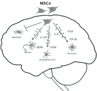
The paracrine effects of MSCs have been studied in different animal models of neurological disorders, e.g., stroke, Parkinson’s, Alzheimer’s, and Huntington’s diseases, and amyotrophic lateral sclerosis, as will be discussed below. The paracrine activity of MSCs could also be related to the immunomodulatory properties of these cells – two functions acting together for brain protection and regeneration (Fig. 1).
MsCs seCretoMe anD iMMunoMoDulation
An important characteristic of MSCs is their sig-nificant immunomodulatory and anti-inflammato -ry effects. For instance, evidence has shown that MSCs can regulate the immune system by inhibit-ing the proliferation of T and B cells (Duffy et al. 2011, Franquesa et al. 2012), natural killer (NK) cells (Di Nicola et al. 2002, Spaggiari et al. 2008) and neutrophil apoptosis (Raffaghello et al. 2008). Via this mechanism, MSCs influence the produc -tion and secre-tion of antibodies by B cells, cyto-kine secretion, and NK cytotoxicity (Aggarwal and Pittenger 2005, Spaggiari et al. 2008). MSCs have also been suggested to inhibit the differentiation of monocytes into dendritic cells and in addition to influencing the roles of these cells (Aggarwal and Pittenger 2005, Ivanova-Todorova et al. 2009).
TGF-β1, HGF (Di Nicola et al. 2002), interleu-kin-10 (IL-10), prostaglandin E2 (pGe2)
(Aggarw-al and Pittenger 2005), heme oxygenase-1 (HO1),
IL-6 (Kogler et al. 2005) and soluble HLA-G5
(Sel-mani et al. 2008). Several studies have suggested that MSCs inhibit inflammatory processes in dif -ferent disease states (Lee et al. 2009a, Sanchez et
al. 2011, van Koppen et al. 2012), contributing to
the regeneration of damaged tissues, probably by modulating the immune response.
In summary, the paracrine and
immunomodu-latory factors present in the MSCs secretome seem
to play important roles in establishing an appro-priate tissue microenvironment to promote repair
in damaged situations justifying research into the
therapeutic potential of these cells.
MsCs seCretoMe: CliniCal appliCation
Clinical interest in the use of MSCs has increased significantly over the past few years, especially because of the ideal characteristics of these cells for regenerative medicine. Specifically, MSCs can be obtained from tissues commonly present in clinical situations (e.g., bone marrow, adipose tissue, and umbilical cord blood), they can be expanded in culture for testing purposes and for clinical use, and have low immunogenicity, which is very useful for potential clinical applications (Le Blanc et al. 2003).
However, before MSCs can be used in regenerative medicine, it is essential to understand the biology of these cells and investigate the most appropriate ways to culture and handle them.
figure 1 - Schematic figure to show the mechanism of action of MSCs in the CNS. The MSCs has been described to release neurotrophic factors
and anti-inflammatories cytokines (BDNF, GDNF, VEGF, TGF-β1,
In addition, it is important to know in detail the properties of the molecules secreted by MSCs, through the characterization of their secretome. In this sense, gene expression, proteomics, and metabolomics have been applied to investigate the potential therapeutic action of new and already known soluble factors secreted by MSCs.
MsCs appliCation in neuroDeGenerative Diseases
Despite the vast amount of research into neurodegenerative diseases over the past few years, their etiology and pathophysiology are still not completely understood. Moreover, the complexity of these conditions pose difficulties to the development of effective treatments. Inflammation has been identified as a key factor in the pathophysiology of degenerative diseases affecting the central nervous system (CNS). In these pathologies, the primary insult evokes a local inflammation, with reactive astrogliosis, macrophage influx, and cell death generating tissue damage and glial scar formation (Drela et al. 2013). Therefore, immunomodulatory therapies may become a good therapeutic strategy in these cases.
MSCs therapies have shown promising results in neurodegenerative diseases. In addition to regulating inflammation, they can also promote other beneficial effects, such as neuronal growth, a decrease in free radical levels, and reduce apoptosis (Dharmasaroja 2009). Once again, application of MSCs therapies in these scenarios may help reduce all the adverse pathological events caused by neurodegenerative diseases (Table I).
alzheiMer’s Disease
Alzheimer’s disease (AD) is a devastating and the most common form of dementia, characterized by extracellular amyloid plaques, neurofibrillary tangles, and a progressive loss of neurons and synapses in different brain regions. Patients
with AD present memory deficits and cognitive impairment (Ballard et al. 2011). To date, the treatment of AD is only palliative, and involves mainly drugs to increase cerebral acetylcholine levels. MSCs therapy seems to be a very attractive option in this condition (Drela et al. 2013).
Several studies have shown promising results with the use of MSCs in animal models of AD. One study in particular showed that bone marrow-derived MSCs injected intracerebral were effective in reducing accumulation of amyloid-β (Aβ) in the brain of an animal model of AD prepared via direct Aβ injection in the hippocampal dentate gyrus (Lee et al. 2010b). The same group showed that intracerebral transplantation of bone marrow-derived MSCs to amyloid precursor protein and presenilin in double transgenic mice ameliorated cognitive function. Furthermore, mice treated with MSCs showed a decrease in hyperphosphorylated tau protein levels (Lee et al. 2010b). In another study, Lee et al. (2010a) reported that intracerebral injection of human umbilical cord blood-derived MSCs in an acute model of AD, improved cognitive function, reduced levels of neuronal apoptosis, and decreased activation of astrocytes and microglia (Lee et al. 2010a). Injection intracerebral of bone marrow-derived MSCs has also been shown to improve learning and memory in a chemically and age-induced rat model of AD (Babaei et al. 2012). Kim et al. (2012) in turn, evaluated the mechanisms involved in Aβ degradation induced by MSCs. According to that author, soluble intracellular adhesion molecule-1 (sICAM-1) is released by human umbilical cord blood-derived MSCs and acts on microglial cells, inducing the expression of the Aβ-degrading enzyme (Kim et al. 2012).
increased in the brains of AD animals treated with MSCs (Bae et al. 2013).
Even though the results obtained with animal models have been encouraging, findings from clinical studies are not yet available.
parKinson’s Disease
Parkinson’s disease (PD) is an extremely common neurodegenerative illness. This condition is char-acterized by the progressive loss of dopaminergic neurons in the substantia nigra and a severe de-crease in striatal dopamine contents (Jenner 2008). The clinical symptoms of PD include tremor, mus-cle rigidity, bradykinesia, and postural instability. Existing pharmacological therapies and surgeries can improve clinical symptoms at early stages, but become less effective as the disease progresses (Glavaski-Joksimovic and Bohn 2013). Thus, it is clear that new therapeutic strategies are needed to decrease neuronal loss and slow progression of dis-ease.
MSCs therapy has been considered in PD to replace lost neurons in the substantia nigra with healthy dopaminergic neurons and to avoid neuron loss (Huang et al. 2012). Over the past years, an increasing number of reports have described promising results of MSCs therapy in experimental models of PD. Animals treated with MSCs had the capacity to protect and decrease damage in dopaminergic neurons (Blandini et al. 2010, Danielyan et al. 2011, Li et al. 2001).
Improved behavior has been observed in a 1-methyl-4-phenyl-1,2,3,6-tetrahydropyridine (MPTP) animal model of PD after bone marrow-derived MSCs transplantation (Li et al. 2001). In addition, increased viability and migration of transplanted bone marrow-derived MSCs was observed in lost dopaminergic neurons after the administration of 6-hydroxydopamine (6-OHDA) in an animal model (Hellmann et al. 2006).
MSCs have also been used as a vector for gene delivery in a rat model of PD: cells were transfected with the gene coding for tyrosine hidroxylase (TH), which is the limiting-rate enzyme for dopamine synthesis. The authors reported successful transfer and expression of the TH gene in the rat striatum, as well as clinical improvement (Lu et al. 2005). Experimental studies have shown that MSCs conditioned medium promotes the survival of grafted xenogeneic dopaminergic neurons in affected mice (Shintani et al. 2007). Another study using MSCs intravenously showed a significant decrease in the loss of dopaminergic neurons in rats treated with MG-132, which causes a neurodegenerative disease similar to PD (Park et al. 2008). Furthermore, intravenous injection of MSCs reduced the loss of dopaminergic neurons in models of disease induced by injection of lipopolysaccharide (LPS) into the substantia nigra or by intraperitoneal injection of MPTP (Kim et al. 2009). In another study, treatment with MSCs and pretreatment with glial cell line-derived neurotrophic factor (GDNF) increased the proportion of TH-positive and dopamine-producing cells, resulting in clinical improvement in a 6-OHDA rat model of PD (Dezawa et al. 2004). Yet another study of the same group of authors showed that administration of dopaminergic neuron-like MSCs to the striatum, ameliorated motor function in parkinsonian macaques – a finding that was associated with restoration of the dopaminergic function (Hayashi et al. 2013). Finally, in the same line, intra-striatal injection of MSCs cultured in favorable conditions for neuronal differentiation has been shown to improve clinical symptoms in murine models of PD (Bouchez et al. 2008, Levy et al. 2008). One of the latter studies found comparable efficacy when using undifferentiated MSCs (Bouchez et al. 2008).
body of evidence suggesting that inflammation and microglial proliferation are involved in the pathophysiology of PD. One study has demonstrated that MSCs have the capacity to protect dopaminergic neurons from LPS-induced microglial activation and from the production of nitric oxide and tumor necrosis factor alpha (TNF-α) (Kim et al. 2009). Chao et al. (2009) observed that intravenous administration of mouse MSCs, protected dopaminergic neurons from MPTP toxicity and decreased microglial activation (Chao et al. 2009). Those studies reinforce the potential importance of the immunomodulatory effects of MSCs for the treatment of PD.
In humans, only one clinical trial has been conducted, consisting of seven PD patients aged between 22 and 62 years, followed up for a period that ranged from 10 to 36 months. The patients received a single dose of autologous bone marrow-derived MSCs transplanted to the subventricular zone using stereotaxic surgery. Three of the seven patients showed an improvement in symptoms, with a decrease in off/on periods measured using Unified Parkinson’s Disease Rating Scale. Two patients also reported subjective improvement of symptoms and reduction in drug dosage (Venkataramana et al. 2010). Further investigation is necessary to confirm the efficacy of this therapy.
Multiple sClerosis
Multiple sclerosis (MS) is a chronic inflammatory autoimmune disease that affects the CNS, characterized by recurrent episodes of axonal lesion and demyelination (Compston and Coles 2002, Hernandez-Pedro et al. 2013). MS is the most common cause of neurological disability in young adults (Chandran et al. 2008). Treatments currently available focus on the immune system, aiming to control the inflammatory process that leads to demyelination (Karussis and Kassis 2008). These therapies are only partially effective, as they are
not capable of reversing neuronal damage. Because neurodegeneration is thought to be the cause of the gradual worsening observed in patients with MS, new approaches that promote neuronal repair are needed (Cohen 2013).
Over the last decade, several preclinical studies have demonstrated a great potential of MSCs in the treatment of MS (Gerdoni et al. 2007, Karussis et al. 2010, Zappia et al. 2005, Zhang et al. 2005). The most common animal model of MS is experimental autoimmune encephalomyelitis (EAE), in which immunization with neural antigens derived mainly from myelin, in combination with adjuvants, leads to demyelination, inflammation, and axonal damage in the CNS (Cohen 2013). Zappia et al. (2005) have shown that intravenous injection of MSCs in mice with chronic EAE leads to reduction of demyelination and CNS infiltration by inflammatory cells (Zappia et al. 2005). In that study, MSCs also improved the clinical severity of MS. MSCs were effective when administered at disease onset and peak, but not after disease stabilization. In a study conducted by Zhang et al. (2005), MSCs also caused significant functional improvement when injected intravenously in EAE mice, with some level of engraftment in the CNS. Demyelination significantly decreased, and BDNF cells significantly increased in treated mice, compared to controls (Zhang et al. 2005).
antigens. Bai et al. (2009) showed that treatment with human bone marrow-derived MSCs, reduced inflammatory T-cells and associated cytokines, and concomitantly increased IL-4-producing type 2 helper (Th2) cells and anti-inflammatory cytokines in treated EAE mice (Bai et al. 2009).
Based on the evidence provided by preclinical studies, a few clinical trials have attempted to demonstrate the safety and efficacy of MSCs in patients with MS (Bonab et al. 2012, Connick et al. 2012, Karussis et al. 2010, Yamout et al. 2010). All those studies were open-label and employed autologous MSCs. Bonab et al. (2012) studied 25 patients with progressive MS unresponsive to conventional treatment recruited to receive a single intrathecal injection of autologous bone marrow-derived MSCs. Therapeutic response was followed for 12 months, and the authors showed that the clinical course of the disease could be stabilized with no serious adverse effects (Bonab et al. 2012). Another recent clinical trial assessed the neuroprotective effects of intravenous MSCs on optic nerve function and reported improvement after MSCs injection (Connick et al. 2012).
Currently, there are a upcoming clinical trials registered in clinicaltrials.gov aiming to assess the efficacy of MSCs therapies in MS. Although this approach has shown promising results, larger randomized controlled clinical trials are needed to determine treatment feasibility and to elucidate the mechanisms by which this tool can be useful.
huntinGton’s Disease
Huntington’s disease (HD) is an autosomal dominant neurodegenerative disorder caused by a mutation in the huntingtin (htt) gene. HD is characterized by neuronal cell loss leading to intellectual decline, movement disorders, and behavioral changes (Lin et al. 2011, Maucksch et al. 2013). The neuropathology of HD manifests in the progressive degeneration of striatal GABAergic
neurons. Currently, there is no therapy available capable of interrupting disease progression (Lin et al. 2011, Maucksch et al. 2013).
There is a large interest in the use of MSCs to treat HD, and several preclinical studies have investigated the potential applications of this therapy in animal models. Lee et al. (2009b) showed that human adipose-derived MSCs transplanted into the ipsilateral striatal border of mice transgenic for HD, increased survival, attenuated the loss of striatal neurons, and reduced htt aggregates (Lee et al. 2009b). The same study also investigated the effects of human adipose-derived MSCs in cell culture. The authors demonstrated that the cells secreted multiple growth factors, e.g., BDNF, IGF-1, and NGF, among others.
A recent study investigated the effect of the extract of adipose-derived MSCs in a HD mouse model and in neuronal cells. Intraperitoneal injection of the extract improved performance in the rotarod test, used to evaluate motor coordination in rodents and especially sensitive to detect cerebellar dysfunction (Im et al. 2013, Shiotsuki et al. 2010) . Treatment also ameliorated atrophy and mutant htt aggregation in the striatum. Neuro2A neuroblastoma cells treated with the same extract showed increased expression of cAMP response element-binding protein (p-CREB) and peroxisome proliferator-activated receptor γ coactivator-1α (PGC1α), which could modify HD progression (Im et al. 2013).
Another two studies have investigated the effects of human MSCs in different mouse models of HD (Lin et al. 2011, Snyder et al. 2010). Both studies demonstrated reduced striatal atrophy after intrastriatal transplantation of human MSCs, suggesting a neuroprotective effect associated with the neurotrophic factors secreted by these cells (Maucksch et al. 2013).
table i
summary of the main results using MsCs application in neurodegenerative and psychiatric diseases.
Disease Model route of
delivery
Main findings references
alzheimer’s disease
Animal mode Intracerebral Reducing accumulation of amyloid-β (Aβ) in the hippocampal dentate gyrus
Lee et al. 2010b
Animal model Intracerebral Improved cognitive function, reduced levels of neuronal apoptosis, and decreased activation of astrocytes and microglia
Lee et al. 2010a
Animal model Intracerebral Improved learning and memory Babaei et al. 2012 Animal model Intracerebral Decreased cerebral Aβ deposition Bae et al. 2013
parkinson’s disease
Animal model Intracerebral Increased viability and migration of dopaminergic neurons
Hellmann et al. 2006
Animal model Intracerebral Increased expression of the TH gene in the rat striatum, as well as clinical improvement
Lu et al. 2005
Animal model Intravenous Reduced the loss of dopaminergic neurons in models of disease induced by injection intraperitoneal of MPTP
Kim et al. 2009
Animal model Intravenous Protected dopaminergic neurons from MPTP toxicity and decreased microglial activation
Chao et al. 2009
Clinical trial Intra-ventricular
Three of the seven patients showed an improvement in symptoms, with a decrease in off/on periods measured
using Unified Parkinson’s Disease Rating Scale
Venkataramana et al. 2010
Multiple sclerosis
Animal model Intravenous Reduction of demyelination and CNS infiltration by
inflammatory cells and improved the clinical severity
of MS
Zappia et al. 2005
Animal model Intravenous Functional improvement with some level of
engraftment in the CNS and decreased demyelination and increased BDNF
Zhang et al. 2005
Animal model Intravenous Reduced inflammatory T-cells and associated cytokines, and concomitantly increased IL-4-producing type 2 helper (Th2) cells and
anti-inflammatory cytokines
Bai et al. 2009
Clinical trial Intrathecal Clinical course of the disease was stabilized with no serious adverse effects
Bonab et al. 2012
Clinical trial Intravenous Improvement in visual acuity and visual evoked response latency
Connick et al. 2012
Clinical trial Intrathecal + Intravenous
Functional improvement Karussis et al. 2010
huntington’s disease
Animal model Intracerebral Increased survival, attenuated the loss of striatal neurons, and reduced htt aggregates
Lee et al. 2009b
Animal model Intraperitoneal Improved performance in the rotarod test, ameliorated atrophy and mutant htt aggregation in the striatum.
Im et al. 2013
Animal model Intracerebral Demonstrated reduced striatal atrophy after intrastriatal transplantation of human MSCs,
suggesting a neuroprotective effect associated with the neurotrophic factors secreted by these cells
Lin et al. 2011
Depression Animal model Intracerebral Increased hippocampal neurogenesis and improved depressive behavior
safety and effectiveness of using MSCs in the treatment of HD.
MsCs appliCation in psyChiatriC DisorDers
Over the past few years, many studies have focused on immunological abnormalities and the decrease of neurotrophic factors that characterize the pathophysiology of psychiatric disorders. Several studies have shown increased levels of pro-inflammatory cytokines, e.g., TNF-α, 6, and IL-2, as well as decreased levels of BDNF, an important neurotrophin for CNS, in severe mental illnesses, such as bipolar disorder, schizophrenia, and major depression (Asevedo et al. 2013, Kapczinski et al. 2011, Kunz et al. 2011, Patas et al. 2013). Furthermore, many studies have shown important cognitive impairment, neuroanatomical alterations and decreased neurogenesis in the hippocampus of patients with affective disorders (Caletti et al. 2013, Dranovsky and Hen 2006, Thomas et al. 2007, Torrent et al. 2010, Trivedi and Greer 2013).
In fact, despite the promising contributions of MSCs therapies in psychiatry, few studies have evaluated the effects of MSCs in models of psychiatric disorders. MSCs have the ability to promote neurogenesis and the survival and differentiation of neural cells by expressing neurotrophic factors, e.g., BDNF, NGF, and IGF. Moreover, as a result of their immunomodulatory properties, they can prevent apoptosis and decrease inflammation (Crigler et al. 2006, Yoo et al. 2008). Tfilin et al. (2010) showed that treatment of an animal model of depression with MSCs, increased hippocampal neurogenesis and improved depressive behavior (Tfilin et al. 2010). Another study evidenced that intra-hippocampal transplantation of MSCs enhanced neurogenesis and did not impair behavioral functions in rats (Coquery et al. 2012). These results are promising and may lead to a novel modality for the treatment of psychiatric disorders. However, more studies are
necessary to elucidate the precise mechanisms of action of MSCs in mental illness.
ConClusions
There are numerous preclinical studies using MSCs transplantation for diseases on the CNS that show promising results. These studies suggest that MSCs act through release of different neurotrophic factors, anti-inflammatories and antiapoptotic factors that can promote recover the injured area and prevent damage in neurodegenerative disorder. However, more clinical studies are necessary to understand the exact mechanism of action of MSCs in neurodegenerative disease and evaluate if the treatment with MSCs could cause side effects in patients. On the other hands, there are a few studies using the MSCs in psychiatric disease, but these studies have demonstrated promising results in depression and suggest that the MSCs can be a new strategy for the treatment of mental disorder. Future studies should be developed to evaluate the most effective routes of administration, dose and source of MSCs for each disease and their therapeutics effects. Thus, this new treatment may became an important therapeutic option for psychiatric patientes that do not respond to conventional treatment.
aCKnoWleDGMents
B Pffafenseller, EG da Silva have schoorship from CNPq and CAPES.
resuMo
Células tronco mesenquimais (CTMs) são células progenitoras multipotentes que têm a capacidade de se diferenciar em todas as linhagens de origem mesodérmica, como, cartilagem, ossos, e adipócitos. CTMs têm sido identificadas em diferentes fases do desenvolvimento, incluindo a idade adulta e em diferentes tecidos, tais como medula óssea, tecido adipose e cordão umbilical. Estudos recentes têm mostrado que as CTMs possuem a capacidade de migrar para locais de lesões. Nesse sentido, uma característica importante das CTMs são os seus efeitos imunomodulatórios e anti-inflamatórios. Por exemplo, há evidências que as CTMs podem regular o sistema imune por inibição da proliferação de células T e B. O interesse clínico no uso das CTMs tem aumentado consideravelmente nos últimos anos, especialmente devido às características ideais destas células para a medicina regenerativa. Terapias com CTMs têm mostrado resultados promissores em doenças neurodegenerativas, além de regular a inflamação, elas podem promover outros efeitos benéficos, tais como, crescimento neuronal, diminuição de radicais livres e reduzir a apoptose. No entanto, apesar de muitas pesquisas das CTMs em doenças neurodegenerativas, o mecanismo de ação das CTMs ainda não estão completamente esclarecidos, o que dificulta o desenvolvimento de tratamentos eficazes. Por outro lado, estudos em modelos de doenças psiquiátricas são escassos, apesar dos resultados promissores utilizando terapias com CTMs nesta área.
palavras-chave: Células-tronco mesenquimais, tra-tamento, doença neurodegenerativa, transtornos psiquiátricos.
referenCes
AGGARWAL S AND PITTENGER MF. 2005. Human mesenchymal stem cells modulate allogeneic immune cell responses. Blood 105: 1815-1822.
AKIYAMA Y, RADTKE C AND KOCSIS JD. 2002. Remyelination of the rat spinal cord by transplantation of identified bone marrow stromal cells. J Neurosci 22: 6623-6630.
ASEVEDO E ET AL. 2013. Impact of peripheral levels of chemokines, BDNF and oxidative markers on cognition
in individuals with schizophrenia. J Psychiatr Res 7: 1376-1382.
BABAEI P, SOLTANI TEHRANI B AND ALIZADEH A. 2012. Transplanted bone marrow mesenchymal stem cells improve memory in rat models of Alzheimer’s disease. Stem Cells Int, p. 369-417.
BAE JS, JIN HK, LEE JK, RICHARDSON JC AND CARTER JE. 2013. Bone marrow-derived mesenchymal stem cells contribute to the reduction of amyloid-beta deposits and the improvement of synaptic transmission in a mouse model of pre-dementia Alzheimer’s disease. Curr Alzheimer Res 10: 524-531.
BAI L, LENNON DP, CAPLAN AI, DECHANT A, HECKER
J, KRANSO J, ZAREMBA A AND MILLER RH. 2012. Hepatocyte growth factor mediates mesenchymal stem cell-induced recovery in multiple sclerosis models. Nat Neurosci 15: 862-870.
BAI L, LENNON DP, EATON V, MAIER K, CAPLAN AI, MILLER
SD AND MILLER RH. 2009. Human bone marrow-derived mesenchymal stem cells induce Th2-polarized immune response and promote endogenous repair in animal models of multiple sclerosis. Glia 57: 1192-1203.
BALLARD C, GAUTHIER S, CORBETT A, BRAYNE C, AARSLAND
D AND JONES E. 2011. Alzheimer’s disease. Lancet 377: 1019-1031.
BIANCONE L, BRUNO S, DEREGIBUS MC, TETTA C AND
CAMUSSI G. 2012. Therapeutic potential of mesenchymal stem cell-derived microvesicles. Nephrol Dial Transplant 27: 3037-3042.
BLANDINI F ET AL. 2010. Transplantation of undifferentiated human mesenchymal stem cells protects against 6-hydroxydopamine neurotoxicity in the rat. Cell Transplant 19: 203-217.
BONAB MM ET AL. 2012. Autologous mesenchymal stem cell therapy in progressive multiple sclerosis: an open label study. Curr Stem Cell Res Ther 7: 407-414.
BOUCHEZ G, SENSEBE L, VOURC’H P, GARREAU L, BODARD S, RICO A, GUILLOTEAU D, CHARBORD P, BESNARD JC AND
CHALON S. 2008. Partial recovery of dopaminergic pathway after graft of adult mesenchymal stem cells in a rat model of Parkinson’s disease. Neurochem Int 52: 1332-1342. CALETTI E ET AL. 2013. Neuropsychology, social cognition
and global functioning among bipolar, schizophrenic patients and healthy controls: preliminary data. Front Hum Neurosci 7: 661.
CAMPAGNOLI C, ROBERTS IA, KUMAR S, BENNETT PR,
BELLANTUONO I AND FISK NM. 2001. Identification of mesenchymal stem/progenitor cells in human first-trimester fetal blood, liver, and bone marrow. Blood 98: 2396-2402.
CHAN JK AND LAM PY. 2013. Human mesenchymal stem cells and their paracrine factors for the treatment of brain tumors. Cancer Gene Ther 20: 539-543.
CHANDRAN S, HUNT D, JOANNIDES A, ZHAO C, COMPSTON A
AND FRANKLIN RJ. 2008. Myelin repair: the role of stem and precursor cells in multiple sclerosis. Philos Trans R Soc Lond B Biol Sci 363: 171-183.
CHAO YX, HE BP AND TAY SS. 2009. Mesenchymal stem cell transplantation attenuates blood brain barrier damage and neuroinflammation and protects dopaminergic neurons against MPTP toxicity in the substantia nigra in a model of Parkinson’s disease. J Neuroimmunol 216: 39-50.
CHAPEL A ET AL. 2003. Mesenchymal stem cells home to injured tissues when co-infused with hematopoietic cells to treat a radiation-induced multi-organ failure syndrome. J Gene Med 5: 1028-1038.
CHEN J, ZHANG ZG, LI Y, WANG L, XU YX, GAUTAM SC, LU
M, ZHU Z AND CHOPP M. 2003. Intravenous administration of human bone marrow stromal cells induces angiogenesis in the ischemic boundary zone after stroke in rats. Circ Res 92: 692-699.
CHEN L, TREDGET EE, WU PY AND WU Y. 2008. Paracrine factors of mesenchymal stem cells recruit macrophages and endothelial lineage cells and enhance wound healing. PLoS One 3: e1886.
CHEN X, LI Y, WANG L, KATAKOWSKI M, ZHANG L, CHEN
J, XU Y, GAUTAM SC AND CHOPP M. 2002. Ischemic rat brain extracts induce human marrow stromal cell growth factor production. Neuropathology 22: 275-279.
CHENG Z ET AL. 2008. Targeted migration of mesenchymal stem cells modified with CXCR4 gene to infarcted myocardium improves cardiac performance. Mol Ther 16: 571-579.
CHOI YJ, LI WY, MOON GJ, LEE PH, AHN YH, LEE G
AND BANG OY. 2010. Enhancing trophic support of mesenchymal stem cells by ex vivo treatment with trophic factors. J Neurol Sci 298: 28-34.
COHEN JA. 2013. Mesenchymal stem cell transplantation in multiple sclerosis. J Neurol Sci 333: 43-49.
COMPSTON A AND COLES A. 2002. Multiple sclerosis. Lancet 359: 1221-1231.
CONNICK P ET AL. 2012. Autologous mesenchymal stem
cells for the treatment of secondary progressive multiple sclerosis: an open-label phase 2a proof-of-concept study. Lancet Neurol 11: 150-156.
CONSTANTIN G ET AL. 2009. Adipose-derived mesenchymal stem cells ameliorate chronic experimental autoimmune encephalomyelitis. Stem Cells 27: 2624-2635.
COQUERY N, BLESCH A, STROH A, FERNANDEZ-KLETT F,
KLEIN J, WINTER C AND PRILLER J. 2012. Intrahippocampal transplantation of mesenchymal stromal cells promotes neuroplasticity. Cytotherapy 14: 1041-1053.
CRIGLER L, ROBEY RC, ASAWACHAICHARN A, GAUPP D
AND PHINNEY DG. 2006. Human mesenchymal stem
cell subpopulations express a variety of neuro-regulatory molecules and promote neuronal cell survival and neuritogenesis. Exp Neurol 198: 54-64.
DA SILVA MEIRELLES L, CHAGASTELLES PC AND NARDI NB. 2006. Mesenchymal stem cells reside in virtually all post-natal organs and tissues. J Cell Sci 119: 2204-2213. DANIELYAN L ET AL. 2011. Therapeutic efficacy of intranasally
delivered mesenchymal stem cells in a rat model of Parkinson disease. Rejuvenation Res 14: 3-16.
DEZAWA M ET AL. 2004. Specific induction of neuronal cells from bone marrow stromal cells and application for autologous transplantation. J Clin Invest 113:12 1701-1710.
DHARMASAROJA P. 2009. Bone marrow-derived mesenchymal stem cells for the treatment of ischemic stroke. J Clin Neurosci 16: 12-20.
DI NICOLA M, CARLO-STELLA C, MAGNI M, MILANESI M, LONGONI PD, MATTEUCCI P, GRISANTI S AND GIANNI
AM. 2002. Human bone marrow stromal cells suppress T-lymphocyte proliferation induced by cellular or nonspecific mitogenic stimuli. Blood 99: 3838-3843. DOMINICI M, LE BLANC K, MUELLER I, SLAPER-CORTENBACH
I, MARINI F, KRAUSE D, DEANS R, KEATING A, PROCKOP
D AND HORWITZ E. 2006. Minimal criteria for defining multipotent mesenchymal stromal cells. The International Society for Cellular Therapy position statement. Cytotherapy 8: 315-317.
DRANOVSKY A AND HEN R. 2006. Hippocampal neurogenesis: regulation by stress and antidepressants. Biol Psychiatry 59: 1136-1143.
DRELA K, SIEDLECKA P, SARNOWSKA A AND DOMANSKA -JANIK K. 2013. Human mesenchymal stem cells in the treatment of neurological diseases. Acta Neurobiol Exp (Wars) 73: 38-56.
DUFFY MM, RITTER T, CEREDIG R AND GRIFFIN MD. 2011. Mesenchymal stem cell effects on T-cell effector pathways. Stem Cell Res Ther 2: 34.
ERICES A, CONGET P AND MINGUELL JJ. 2000. Mesenchymal progenitor cells in human umbilical cord blood. Br J Haematol 109: 235-242.
EZQUER FE, EZQUER ME, PARRAU DB, CARPIO D, YANEZ
AJ AND CONGET PA. 2008. Systemic administration of multipotent mesenchymal stromal cells reverts hyperglycemia and prevents nephropathy in type 1 diabetic mice. Biol Blood Marrow Transplant 14: 631-640.
FRANQUESA M, HOOGDUIJN MJ, BESTARD O AND GRINYO
JM. 2012. Immunomodulatory effect of mesenchymal stem cells on B cells. Front Immunol 3: 212.
FRIEDENSTEIN AJ, GORSKAJA JF AND KULAGINA NN. 1976. Fibroblast precursors in normal and irradiated mouse hematopoietic organs. Exp Hematol 4: 267-274.
GERRITS A, DYKSTRA B, KALMYKOWA OJ, KLAUKE K,
VEROVSKAYA E, BROEKHUIS MJ, DE HAAN G AND
BYSTRYKH LV. 2010. Cellular barcoding tool for clonal analysis in the hematopoietic system. Blood 115: 2610-2618.
GLAVASKI-JOKSIMOVIC A AND BOHN MC. 2013. Mesenchymal stem cells and neuroregeneration in Parkinson’s disease. Exp Neurol 247: 25-38.
GRAYSON WL, ZHAO F, BUNNELL B AND MA T. 2007. Hypoxia enhances proliferation and tissue formation of human mesenchymal stem cells. Biochem Biophys Res Commun 358: 948-953.
HAYASHI T ET AL. 2013. Autologous mesenchymal stem cell-derived dopaminergic neurons function in parkinsonian macaques. J Clin Invest 123: 272-284.
HELLMANN MA, PANET H, BARHUM Y, MELAMED E AND
OFFEN D. 2006. Increased survival and migration of engrafted mesenchymal bone marrow stem cells in 6-hydroxydopamine-lesioned rodents. Neurosci Lett 395: 124-128.
HERMANN A ET AL. 2004. Efficient generation of neural stem cell-like cells from adult human bone marrow stromal cells. J Cell Sci 117: 4411-4422.
HERNANDEZ-PEDRO NY, ESPINOSA-RAMIREZ G, DE LA
CRUZ VP, PINEDA B AND SOTELO J. 2013. Initial Immunopathogenesis of Multiple Sclerosis: Innate Immune Response. Clin Dev Immunol 413-465.
HONMA T, HONMOU O, IIHOSHI S, HARADA K, HOUKIN K, HAMADA H AND KOCSIS JD. 2006. Intravenous infusion of immortalized human mesenchymal stem cells protects against injury in a cerebral ischemia model in adult rat. Exp Neurol 199: 56-66.
HORWITZ EM ET AL. 1999. Transplantability and therapeutic effects of bone marrow-derived mesenchymal cells in children with osteogenesis imperfecta. Nat Med 5: 309-313. HUANG B, TABATA Y AND GAO JQ. 2012. Mesenchymal stem
cells as therapeutic agents and potential targeted gene delivery vehicle for brain diseases. J Control Release 162: 464-473.
IM W, BAN J, LIM J, LEE M, LEE ST, CHU K AND KIM M. 2013. Extracts of adipose derived stem cells slows progression in the R6/2 model of Huntington’s disease. PLoS One 8: e59438.
IVANOVA-TODOROVA E, BOCHEV I, MOURDJEVA M, DIMITROV
R, BUKAREV D, KYURKCHIEV S, TIVCHEV P, ALTUNKOVA
I AND KYURKCHIEV DS. 2009. Adipose tissue-derived mesenchymal stem cells are more potent suppressors of dendritic cells differentiation compared to bone marrow-derived mesenchymal stem cells. Immunol Lett 126: 37-42. JENNER P. 2008. Functional models of Parkinson’s disease: a
valuable tool in the development of novel therapies. Ann Neurol 64: S16-29.
KAPCZINSKI F ET AL. 2011. Peripheral biomarkers and illness activity in bipolar disorder. J Psychiatr Res 45: 156-161.
KARUSSIS D ET AL. 2010. Safety and immunological effects of mesenchymal stem cell transplantation in patients with multiple sclerosis and amyotrophic lateral sclerosis. Arch Neurol 67: 1187-1194.
KARUSSIS D AND KASSIS I. 2008. The potential use of stem cells in multiple sclerosis: an overview of the preclinical experience. Clin Neurol Neurosurg 110: 889-896.
KIM JY ET AL. 2012. Soluble intracellular adhesion molecule-1 secreted by human umbilical cord blood-derived mesenchymal stem cell reduces amyloid-beta plaques. Cell Death Differ 19: 680-691.
KIM YJ, PARK HJ, LEE G, BANG OY, AHN YH, JOE E, KIM
HO AND LEE PH. 2009. Neuroprotective effects of human mesenchymal stem cells on dopaminergic neurons through anti-inflammatory action. Glia 57: 13-23.
KOGLER G, RADKE TF, LEFORT A, SENSKEN S, FISCHER J, SORG RV AND WERNET P. 2005. Cytokine production and hematopoiesis supporting activity of cord blood-derived unrestricted somatic stem cells. Exp Hematol 33: 573-583. KUNZ M ET AL. 2011. Serum levels of IL-6, IL-10 and TNF-alpha in patients with bipolar disorder and schizophrenia: differences in pro- and anti-inflammatory balance. Rev Bras Psiquiatr 33: 268-74.
LE BLANC K, TAMMIK C, ROSENDAHL K, ZETTERBERG E AND
RINGDEN O. 2003. HLA expression and immunologic
properties of differentiated and undifferentiated mesenchymal stem cells. Exp Hematol 31: 890-896. LEE HJ, LEE JK, LEE H, SHIN JW, CARTER JE, SAKAMOTO T,
JIN HK AND BAE JS. 2010a. The therapeutic potential of human umbilical cord blood-derived mesenchymal stem cells in Alzheimer’s disease. Neurosci Lett 481: 30-35. LEE JK, JIN HK, ENDO S, SCHUCHMAN EH, CARTER JE
AND BAE JS. 2010b. Intracerebral transplantation of bone marrow-derived mesenchymal stem cells reduces amyloid-beta deposition and rescues memory deficits in Alzheimer’s disease mice by modulation of immune responses. Stem Cells 28: 329-343.
LEE OK, KUO TK, CHEN WM, LEE KD, HSIEH SL AND CHEN
TH. 2004. Isolation of multipotent mesenchymal stem cells from umbilical cord blood. Blood 103: 1669-1675. LEE RH, PULIN AA, SEO MJ, KOTA DJ, YLOSTALO J, LARSON
BL, SEMPRUN-PRIETO L, DELAFONTAINE P AND PROCKOP
DJ. 2009a. Intravenous hMSCs improve myocardial infarction in mice because cells embolized in lung are activated to secrete the anti-inflammatory protein TSG-6. Cell Stem Cell 5: 54-63.
LEE ST ET AL. 2009b. Slowed progression in models of Huntington disease by adipose stem cell transplantation. Ann Neurol 66: 671-681.
LEVY YS, BAHAT-STROOMZA M, BARZILAY R, BURSHTEIN A, BULVIK S, BARHUM Y, PANET H, MELAMED E AND OFFEN
LI Y, CHEN J, WANG L, ZHANG L, LU M AND CHOPP M. 2001. Intracerebral transplantation of bone marrow stromal cells in a 1-methyl-4-phenyl-1,2,3,6-tetrahydropyridine mouse model of Parkinson’s disease. Neurosci Lett 316: 67-70. LIN YT, CHERN Y, SHEN CK, WEN HL, CHANG YC, LI H,
CHENG TH AND HSIEH-LI HM. 2011. Human mesenchymal stem cells prolong survival and ameliorate motor deficit through trophic support in Huntington’s disease mouse models. PLoS One 6: e22924.
LU L, ZHAO, C, LIU Y, SUN X, DUA C, JI M, ZHAO H, XU Q AND YANG H. 2005. Therapeutic benefit of TH-engineered mesenchymal stem cells for Parkinson’s disease. Brain Res Brain Res Protoc 15: 46-51.
MAFI R, HINDOCHA S, MAFI P, GRIFFIN M AND KHAN WS 2011. Sources of adult mesenchymal stem cells applicable for musculoskeletal applications - a systematic review of the literature. Open Orthop J 5: 242-248.
MAIJENBURG MW ET AL. 2011. Nuclear receptors Nur77 and Nurr1 modulate mesenchymal stromal cell migration. Stem Cells Dev 21: 228-238.
MAUCKSCH C, VAZEY EM, GORDON RJ AND CONNOR B. 2013. Stem cell-based therapy for Huntington’s disease. J Cell Biochem 114: 754-763.
MEISEL R, ZIBERT A, LARYEA M, GOBEL U, DAUBENER
W AND DILLOO D. 2004. Human bone marrow stromal cells inhibit allogeneic T-cell responses by indoleamine 2,3-dioxygenase-mediated tryptophan degradation. Blood 103: 4619-4621.
MENARD C AND TARTE K. 2013. Immunoregulatory properties of clinical grade mesenchymal stromal cells: evidence, uncertainties, and clinical application. Stem Cell Res Ther 4: 64.
MORANDO S, VIGO T, ESPOSITO M, CASAZZA S, NOVI G, PRINCIPATO MC, FURLAN R AND UCCELLI A. 2012. The therapeutic effect of mesenchymal stem cell transplantation in experimental autoimmune encephalomyelitis is mediated by peripheral and central mechanisms. Stem Cell Res Ther 3: 3. MURELI S, GANS CP, BARE DJ, GEENEN DL, KUMAR NM
AND BANACH K. 2013. Mesenchymal stem cells improve cardiac conduction by upregulation of connexin 43 through paracrine signaling. Am J Physiol Heart Circ Physiol 304: H600-609.
NAKANO N ET AL. 2010. Characterization of conditioned medium of cultured bone marrow stromal cells. Neurosci Lett 483: 57-61.
NOMURA T, HONMOU O, HARADA K, HOUKIN K, HAMADA
H AND KOCSIS JD. 2005. I.V. infusion of brain-derived neurotrophic factor gene-modified human mesenchymal stem cells protects against injury in a cerebral ischemia model in adult rat. Neuroscience 136: 161-169.
PARK HJ, LEE PH, BANG OY, LEE G AND AHN YH. 2008. Mesenchymal stem cells therapy exerts neuroprotection in a progressive animal model of Parkinson’s disease. J Neurochem 107: 141-151.
PATAS K, PENNINX BW, BUS BA, VOGELZANGS N, MOLENDIJK
ML, ELZINGA BM, BOSKER FJ AND OUDE VOSHAAR
RC. 2013. Association between serum brain-derived neurotrophic factor and plasma interleukin-6 in major depressive disorder with melancholic features. Brain Behav Immun 36: 71-79.
PERRIER AL, TABAR V, BARBERI T, RUBIO ME, BRUSES J, TOPF N, HARRISON NL AND STUDER L. 2004. Derivation of midbrain dopamine neurons from human embryonic stem cells. Proc Natl Acad Sci USA 101: 12543-12548.
PHINNEY DG AND PROCKOP DJ. 2007. Concise review:
mesenchymal stem/multipotent stromal cells: the state of transdifferentiation and modes of tissue repair current views. Stem Cells 25: 2896-2902.
PITTENGER MF, MACKAY AM, BECK SC, JAISWAL RK,
DOUGLAS R, MOSCA JD, MOORMAN MA, SIMONETTI DW, CRAIG S AND MARSHAK DR. 1999. Multilineage potential of adult human mesenchymal stem cells. Science 284: 143-147.
QUEVEDO HC ET AL. 2009. Allogeneic mesenchymal stem cells restore cardiac function in chronic ischemic cardiomyopathy via trilineage differentiating capacity. Proc Natl Acad Sci USA 106: 14022-14027.
RAFFAGHELLO L, BIANCH G, BERTOLOTTO M, MONTECUCCO
F, BUSCA A, DALLEGRI F, OTTONELLO L AND PISTOIA V. 2008. Human mesenchymal stem cells inhibit neutrophil apoptosis: a model for neutrophil preservation in the bone marrow niche. Stem Cells 26: 151-162.
RIES C, EGEA V, KAROW M, KOLB H, JOCHUM M AND NETH
P. 2007. MMP-2, MT1-MMP, and TIMP-2 are essential for the invasive capacity of human mesenchymal stem cells: differential regulation by inflammatory cytokines. Blood 109: 4055-4063.
ROMBOUTS WJ AND PLOEMACHER RE. 2003. Primary murine MSC show highly efficient homing to the bone marrow but lose homing ability following culture. Leukemia 17: 160-170. ROSOVA I, DAO M, CAPOCCIA B, LINK D AND NOLTA JA. 2008.
Hypoxic preconditioning results in increased motility and improved therapeutic potential of human mesenchymal stem cells. Stem Cells 26: 2173-2182.
SANCHEZ L ET AL. 2011. Enrichment of human
ESC-derived multipotent mesenchymal stem cells with immunosuppressive and anti-inflammatory properties capable to protect against experimental inflammatory bowel disease. Stem Cells 29: 251-262.
SATO K, OZAKI K, OH I, MEGURO A, HATANAKA K, NAGAI T, MUROI K AND OZAWA K. 2007. Nitric oxide plays a critical role in suppression of T-cell proliferation by mesenchymal stem cells. Blood 109: 228-234.
SATO Y ET AL. 2005. Human mesenchymal stem cells
xenografted directly to rat liver are differentiated into human hepatocytes without fusion. Blood 106: 756-763.
SELMANI Z ET AL. 2008. Human leukocyte antigen-G5
suppress T lymphocyte and natural killer function and to induce CD4+CD25highFOXP3+ regulatory T cells. Stem Cells 26: 212-222.
SHINTANI A, NAKAO N, KAKISHITA K AND ITAKURA T. 2007. Protection of dopamine neurons by bone marrow stromal cells. Brain Res 1186: 48-55.
SHIOTSUKI H, YOSHIMI K, SHIMO Y, FUNAYAMA M,
TAKAMATSU Y, IKEDA K, TAKAHASHI R, KITAZAWA S AND
HATTORI N. 2010. A rotarod test for evaluation of motor skill learning. J Neurosci Methods 189: 180-185.
SNYDER BR, CHIU AM, PROCKOP DJ AND CHAN AW. 2010. Human multipotent stromal cells (MSCs) increase neurogenesis and decrease atrophy of the striatum in a transgenic mouse model for Huntington’s disease. PLoS One 5: e9347.
SOHNI A AND VERFAILLIE CM. 2013. Mesenchymal Stem Cells Migration Homing and Tracking. Stem Cells Int 2013: 130763.
SPAGGIARI GM, CAPOBIANCO A, ABDELRAZIK H, BECCHETTI
F, MINGARI MC AND MORETTA L. 2008. Mesenchymal stem cells inhibit natural killer-cell proliferation, cytotoxicity, and cytokine production: role of indoleamine 2,3-dioxygenase and prostaglandin E2. Blood 111: 1327-1333.
TFILIN M, SUDAI E, MERENLENDER A, GISPAN I, YADID G AND
TURGEMAN G. 2010. Mesenchymal stem cells increase hippocampal neurogenesis and counteract depressive-like behavior. Mol Psychiatry 15: 1164-1175.
THOMAS RM, HOTSENPILLER G AND PETERSON DA. 2007. Acute psychosocial stress reduces cell survival in adult hippocampal neurogenesis without altering proliferation. J Neurosci 27: 2734-2743.
TORRENT C ET AL. 2010. Long-term outcome of cognitive impairment in bipolar disorder. J Clin Psychiatry 73: e899-905.
TRIVEDI MH AND GREER TL. 2013. Cognitive dysfunction in unipolar depression: Implications for treatment. J Affect Disord 152: 19-27.
UCCELLI A AND PROCKOP DJ. 2010. Why should mesenchymal stem cells (MSCs) cure autoimmune diseases? Curr Opin Immunol 22: 768-774.
VAN KOPPEN A, JOLES JA, VAN BALKOM BW, LIM SK, DE
KLEIJN D, GILES RH AND VERHAAR MC. 2012. Human embryonic mesenchymal stem cell-derived conditioned medium rescues kidney function in rats with established chronic kidney disease. PLoS One 7: e38746.
VENKATARAMANA NK ET AL. 2010. Open-labeled study of unilateral autologous bone-marrow-derived mesenchymal stem cell transplantation in Parkinson’s disease. Transl Res 155: 62-70.
VOULGARI-KOKOTA A, FAIRLESS R, KARAMITA M,
KYRARGYRI V, TSEVELEKI V, EVANGELIDOU M, DELORME
B, CHARBORD P, DIEM R AND PROBERT L. 2012.
Mesenchymal stem cells protect CNS neurons against glutamate excitotoxicity by inhibiting glutamate receptor expression and function. Exp Neurol 236: 161-170. WANG L, LI Y, CHEN X, CHEN J, GAUTAM SC, XU Y AND
CHOPP M. 2002. MCP-1, MIP-1, IL-8 and ischemic cerebral tissue enhance human bone marrow stromal cell migration in interface culture. Hematology 7: 113-117. WASZAK P, ALPHONSE R, VADIVEL A, IONESCU L, EATON F
AND THEBAUD B. 2012. Preconditioning enhances the paracrine effect of mesenchymal stem cells in preventing oxygen-induced neonatal lung injury in rats. Stem Cells Dev 21: 2789-2797.
YAMOUT B ET AL. 2010. Bone marrow mesenchymal stem cell transplantation in patients with multiple sclerosis: a pilot study. J Neuroimmunol 227: 185-189.
YOO SW, KIM SS, LEE SY, LEE HS, KIM HS, LEE YD AND SUH -KIM H. 2008. Mesenchymal stem cells promote proliferation of endogenous neural stem cells and survival of newborn cells in a rat stroke model. Exp Mol Med 40: 387-397. ZAPPIA E ET AL. 2005. Mesenchymal stem cells ameliorate
experimental autoimmune encephalomyelitis inducing T-cell anergy. Blood 106: 1755-1761.
ZHANG J, LI Y, CHEN J, CUI Y, LU M, ELIAS SB, MITCHELL