1
UNIVERSIDADE DA BEIRA INTERIOR
Ciências da Saúde
Modelos de co-culturas de células in vitro para
desenvolvimento de novos sistemas de entrega de
fármacos
Elisabete Cristina da Rocha Costa
Dissertação para obtenção do Grau de Mestre em
Ciências Biomédicas
(2
ociclo de estudos)
Orientador: Professor Doutor Ilídio Joaquim Sobreira Correia
Coorientador: Mestre Vítor Manuel Abreu Gaspar
In vitro cell co-culture models for development of new drug delivery systems
In vitro cell co-culture models for development of new drug delivery systems
iii
List of publications
Articles in peer reviewed international journals:
Costa, C. E., Gaspar. V. M., Marques, J.G., Coutinho, P., Correia, I. J. (2013). “Evaluation of Nanoparticle Uptake in Co-culture Cancer Models.”PloS one. Article in press.
Poster communications:
Costa, C. E., Gaspar. V. M., Marques, J.G., Coutinho, P., Correia, I. J., Mimicking Breast Cancer Microenvironment with In Vitro Co-culture Models, Instituto Politécnico da Guarda (IPG), 3rd of May 2013, Guarda, Portugal.
Best poster award:
Costa, C. E., Gaspar. V. M., Marques, J.G., Coutinho, P., Correia, I. J., Mimicking Breast Cancer Microenvironment with In Vitro Co-culture Models, Instituto Politécnico da Guarda (IPG), 3rd of May 2013, Guarda, Portugal.
In vitro cell co-culture models for development of new drug delivery systems
In vitro cell co-culture models for development of new drug delivery systems v “ “TThheerree’’ssaapphhoottoooonnmmyywwaallllooffaawwoommaannII’’vveenneevveerrmmeett,,iittsslleeffttccoorrnneerrttoorrnnaannddppaattcchheeddttooggeetthheerrwwiitthh t taappee.. SShhee llooookkss ssttrraaiigghhtt iinnttoo tthhee ccaammeerraa aanndd ssmmiilleess,, hhaannddss oonn hhiippss,, ddrreessss ssuuiitt nneeaattllyypprreesssseedd,, lliippss p paaiinntteeddddeeeepprreedd..IItt’’sstthheellaattee11994400ssaannddsshheehhaassnn’’ttyyeettrreeaacchheeddtthheeaaggeeoofftthhiirrttyy..HHeerrlliigghhttbbrroowwnnsskkiinn i iss ssmmooootthh,,hheerreeyyeess ssttiillllyyoouunnggaannddppllaayyffuull,,oobblliivviioouussttootthhee ttuummoorrggrroowwiinnggiinnssiiddeehheerr-- aattuummoorrtthhaatt w woouullddlleeaavveehheerrffiivveecchhiillddrreennmmootthheerrlleessssaannddcchhaannggeetthheeffuuttuurreeooffmmeeddiicciinnee””..
In vitro cell co-culture models for development of new drug delivery systems
vi
Dedication
The completion of this work has not possible without the familiar support. Thus, I would like to dedicate this last year of my academic path to parents and to my little sister which always understood my distance.
Moreover, I would like to dedicate this work to all persons with cancer around the world. I wish that the investigation in tumor biology continues to open new windows for new anti-cancer therapies for better quality of life of cancer patients.
In vitro cell co-culture models for development of new drug delivery systems
In vitro cell co-culture models for development of new drug delivery systems
viii
Acknowledgments
Firstly, I would like to thank to my supervisor Professor Ilídio Correia for the opportunity of work in his group, and for all the time and orientation provided during the aquision of my master degree.
Specially, I would like to thanks my co-supervisor, Vítor Gaspar (Ph.D. student), for supporting me in my instabilities and doubts. Furthermore, for spend great part of his time in helping me, putting in second place his own work. In fact, without his orientation I would not be the person academically and personally that I am today.
To João Marques, my co-worker and friend, I would like to thanks him for all accomplices and honesty. To him, I would like to wish a wonderful future like he deserves.
To another great influence and orientation to me, Ph.D. students and M.Sc. of the group, I thank for good working environment, entrepreneurship, good humour and in last for the friendship. We are a family.
I acknowledge to my friends and housemates which always listen my confidences. In last, I thank to me for surpass myself.
In vitro cell co-culture models for development of new drug delivery systems
In vitro cell co-culture models for development of new drug delivery systems
x
Abstract
The demanding applications of nanocarriers in cancer biology require the existence of testing platforms that mimic the in vivo tumor microenvironment and its unique biological features. For this, highly informative methodologies such as animal experimentation are the current gold-standard. However, very recent reports issued by regulatory agencies appeal for the reduction of the used animal research models due to economical and ethical issues, thus evidencing the urgent necessity for novel alternatives. Co-culture cell models have the potential to bridge the gap between the required reduction of animal use and the existence of suitable models that closely reproduce in vivo tumors. This is a novel type of in vitro cell culture that is mainly characterized by the culture of cancer cells in contact with stromal cells, mimicking the tumor microenvironment in vitro trough the establishment of cancer-stroma synergic interactions. However, this evaluation was until now limited to co-culture systems established with precise cell ratios, not addressing the natural heterogeneity commonly found in tumors of different patients. The research work presented in this thesis describes the development and optimization of novel 2D co-culture models of breast and cervical cancers with various cell-to-cell ratios, in order to unravel the influence of heterogeneous conditions on the evaluation of nanocarrier biological performance and ultimately in the therapeutic outcome. As a proof of concept these novel platforms were used to evaluate a multifunctional gene delivery system designed for cancer therapy and revealed that in fact different co-culture ratios may influence the overall assessment of nanocarrier targeting specificity. In addition, since recent reports demonstrate the high influence of the 3D architecture of tumor masses in the response to anti-cancer drugs or delivery systems, the engineering and optimization of suitable substrates for generation of organotypic 3D co-culture models with various cancer-fibroblast cell ratios was also investigated. The 3D multicelular spheroid models of breast and cervix cancer produced at various time points, possess all the major characteristics of in vivo tumors including the structural rearrangement, the diffusional limit of oxygen or nutrients and most importantly, the distinctive necrotic core of solid tumors. Overall, these newly developed co-culture and 3D models assume crucial importance for the future design and optimization of new drug delivery providing a new level of in vitro reproducibility of in vivo tumors.
Keywords
2D co-cultures; 3D multicellular spheroids; Nanosized delivery systems; Tumor microenvironment.
In vitro cell co-culture models for development of new drug delivery systems
In vitro cell co-culture models for development of new drug delivery systems
xii
Resumo alargado
Os recentes avanços na área da Nanotecnologia abriram novas oportunidades para o desenvolvimento de novos nano-sistemas como as nanopartículas para entrega de fármacos ou de informação genética com potencial para serem usadas futuramente na terapia do cancro. Todavia, para que as suas aplicações terapêuticas sejam significativas num contexto clínico, estes sistemas devem ser testados em modelos que representem o mais aproximadamente possível as propriedades únicas que o microambiente tumoral tem in vivo. Por forma a atingir este objetivo, vários protocolos experimentais usam modelos animais para avaliar a atividade biológica de nano transportadores. No entanto, recentemente, as diferentes agências regulatórias têm apelado pela aplicação da regra dos 3Rs (Reduzir, Reutilizar, Reciclar) em relação ao uso de animais como modelos para estudos experimentais em fases pré-clinicas, não só devido aos problemas económicos e legais associados ao seu uso, mas também devido a inconvenientes éticos, e à variabilidade dos resultados obtidos quando comparados com aqueles adquiridos em estudos clínicos com humanos. Neste contexto, tem-se procurado desenvolver novas alternativas que permitam reproduzir o que ocorre in vivo.
A cultura in vitro de células tumorais em co-cultura com outras células presentes no microambiente tumoral surge como uma abordagem muito promissora no que diz respeito a mimetizar as caraterísticas dos variados tipos tumores. Esta metodologia permite estudar de uma forma abrangente a biologia dos tumores, sob variadas condições e até mesmo testar, de uma forma rápida, novos fármacos ou sistemas de entrega direcionada. O contacto direto entre as células cancerígenas e as células do estroma, reproduzem as interações sinergéticas que ocorrem no microambiente tumoral. Contudo, estes sistemas de culturas celulares são usualmente desenvolvidos tendo por base um número fixo de células tumorais em relação às células do estroma. De facto, até à data apenas foram descritos testes de novos agentes anti-tumorais ou de novos sistemas de entrega em co-culturas com apenas um rácio de células, sendo que esta abordagem não permite assim analisar a heterogeneidade natural dos tumores.
Desta forma, o trabalho de investigação desenvolvido nesta tese descreve desenvolvimento e otimização de novos modelos 2D de co-culturas do cancro da mama e do colo do útero. Para tal, foram usados vários rácios de células cancerígenas células normais do estroma, com o intuito de representar a distribuição celular em diferentes tumores. Adicionalmente, pretende-se também verificar de que forma estas diferentes condições influenciam a atividade dos novos sistemas de entrega de drogas e consequentemente a sua eficácia terapêutica.
Os resultados obtidos demonstraram uma influência evidente dos fibroblastos, sobre o comportamento das células cancerígenas. Inicialmente, foi possível verificar uma alteração na organização estrutural das co-culturas, quando comparadas com as monoculturas de
In vitro cell co-culture models for development of new drug delivery systems
xiii controlo. Além disto, foi também observado um aumento na viabilidade celular na presença de fibroblastos, tendo sido obtida uma correlação entre o tempo de co-cultura e um aumento de proliferação celular. Após a demonstração do sucesso da otimização destas plataformas, foram testadas nanopartículas funcionalizadas nas várias co-culturas desenvolvidas. Os resultados obtidos na microscopia confocal e citometria de fluxo demonstraram que o uso de diferentes rácios celulares pode de facto influenciar a avaliação da especificidade de nano transportadores. Estes resultados evidenciam assim que, os mesmos sistemas de entrega podem atuar de forma diferente, de paciente para paciente.
Para além do desenvolvimento destes sistemas de co-culturas, foi também otimizada a produção de novos modelos de culturas tridimensionais. Estes modelos 3D, mais comumente chamados de esferoides, conseguem mimetizar os tumores sólidos, pois são constituídos por vários tipos de células e com o decorrer do tempo adquirirem propriedades únicas, nomeadamente uma superfície constituída por células com elevada proliferação, que mimetizam as zonas do tumor irrigadas por vasos sanguíneos e um núcleo necrótico, que corresponde às zinas do tumor com baixa densidade de vasos. Para promover a formação destes modelos foi utilizada a técnica de cultura celular com sobreposição líquida em conjugação com agitação horizontal. Esta nova abordagem permitiu evitar a adesão das células e promoveu a formação de esferoides 3D com morfologias bem definidas e reprodutíveis.
Para a formação destes modelos fibroblastos revelaram um papel fundamental visto que as interações que se estabelecem entre os dois tipos celulares são essenciais para formar esferoides coesos e com um gradiente de densidades celulares da periferia para o núcleo. Em geral, os novos modelos de co-culturas desenvolvidos assumem um papel crucial no futuro desenvolvimento e investigação de novos nano transportadores, já que estes serão na prática direcionados para os tumores. Por outro lado, os modelos 3D permitem reproduzir com exatidão o que acontece in vivo, criando um conjunto de ferramentas que poderão contribuir para aperfeiçoar as terapias anti-tumorais.
Palavras-chave
Co-culturas 2D; Esferoides multicelulares; Microambiente tumoral; Nano sistemas de entrega direcionada.
In vitro cell co-culture models for development of new drug delivery systems
In vitro cell co-culture models for development of new drug delivery systems
xv
Table of Contents
1. Introduction
11.1. Tumor microenvironment: The driving force for cancer evolution 2
1.1.1. ECM in the tumor microenvironment 5
1.1.2. Stromal cells 5
1.1.2.1. Vascular and lymphatic endothelial cells 5
1.1.2.2. Endothelial cells 5
1.1.2.3. Perycites 5
1.1.2.4. Adipocytes 6
1.1.2.5. Immune System cell 6
1.1.2.6. Fibroblasts 7
1.2. Nanosized delivery systems as novel therapeutic approaches for cancer
therapy 11
1.3. Experimental models to evaluate nanoparticulated delivery systems for
application in cancer therapy 14
1.3.1. In vivo models 14
1.3.2. In vitro models 16
1.3.2.1. Co-cultures 16
1.3.2.2. 3D cell cultures: Spheroids 17
1.4. Objectives 21
2. Methods
222.1. Materials 23
2.2. Breast cancer and cervical cancer 2D and 3D in vitro co-culture models
optimization 23
2.2.1. Cell lines maintenance 23
2.2.2. Optimization of 2D in vitro cell co-culture models of breast cancer
In vitro cell co-culture models for development of new drug delivery systems
xvi
2.2.2.1. Optical microscopy analysis of the distribution and morphology of 2D
in vitro cell co-culture models of breast cancer (MCF-7:hFIB) and cervical cancer (HeLa:hFIB)
24
2.2.2.2. Resazurin assay for analysis of cell viability of 2D in vitro cell
co-culture models of breast cancer (MCF-7:hFIB) 24
2.2.3. Optimization of 3D in vitro cell co-culture models of breast cancer
(MCF-7:hFIB) and cervical cancer (HeLa:hFIB) 25
2.2.3.1. Optical microscopy analysis of the distribution and morphology of the
3D in vitro cell co-culture models of breast cancer (MCF-7:hFIB) and cervical cancer (HeLa:hFIB)
25
2.2.3.2. Scanning electron microscopy (SEM) analysis of the 3D in vitro cell
co-culture models of breast cancer (MCF-7:hFIB) 26
2.2.3.3. Confocal laser scanning microscopy analysis of 3D in vitro cell
co-culture models of cervical cancer (HeLa:hFIB) 26
2.3. Evaluation of Chitosan-Histidine-Arginine/pDNA nanoparticles in 2D
breast cancer co-culture models (MCF-7:hFIB) 26
2.3.1. CLSM of CH-H-R/pDNA nanoparticles cell uptake analysis in 2D
breast cancer co-culture models 26
2.3.2. Flow cytometry of CH-H-R/pDNA nanoparticles cell uptake analysis
in 2D breast cancer co-culture models 27
3. Results and Discussion
293.1. Breast cancer and cervical cancer 2D and 3D in vitro co-culture models 30
3.1.1. Development and optimization of 2D in vitro cell co-culture models
of breast cancer (MCF-7:hFIB) 30
3.1.2. Development and optimization of 2D in vitro cell co-culture models
of cervical cancer (HeLa:hFIB) 37
3.1.3. Evaluation of CH-H-R/pDNA nanoparticles cellular uptake in 2D
breast cancer co-culture models (MCF-7:hFIB) 40
3.1.3.1. CLSM analysis of CH-H-A/pDNA nanoparticles cellular uptake analysis
in 2D cell co-culture models of breast cancer with different MCF-7 to hFIB ratios
44
3.1.3.2. Flow cytometry analysis of CH-H-A/pDNA nanoparticles cellular
uptake analysis in 2D cell co-culture models of breast cancer with different MCF-7 to hFIB ratios
44
3.2. 3D in vitro cell co-culture models of breast cancer and cervical cancer 48
In vitro cell co-culture models for development of new drug delivery systems
xvii 3.2.2. 3D in vitro cell co-culture models of cervical cancer (HeLa: hFIB)
3.2.2.1. SEM analysis of the 3D in vitro cell co-culture models of breast cancer
(MCF-7:hFIB) 53
3.2.3. 3D in vitro cell co-culture models of cervical cancer (HeLa: hFIB) 53
3.2.3.1. CLSM analysis of 3D in vitro 3D in vitro cell co-culture models of
cervical cancer (HeLa:hFIB) 55
4. Conclusions and Future Perspectives
59In vitro cell co-culture models for development of new drug delivery systems
In vitro cell co-culture models for development of new drug delivery systems
xix
List of Figures
Chapter I - Introduction
Figure 1 – Representation of carcinogenesis. 2
Figure 2 – Representation of tumor microenvironment. 4
Figure 3 – General interactions established between tumour cells and stromal
cells in human tumor tissue. 4
Figure 4 – Immune system behaviour in tumor microenvironment. 6
Figure 5 – Comparison of normal and cancer associated fibroblasts (CAF). 7
Figure 6 – Influence of fibroblasts in tumor microenvironment. 8
Figure 7 – Representation of the mechanism of epithelial-to-mesenchymal
transition (EMT). 10
Figure 8 – Relation between drug anti-tumoral effect and toxic effect. 11
Figure 9 – Representation of the different types of nanoparticles available. 12
Figure 10 – Steps needed for nanoparticle development. 14
Figure 11 - Absolute bioavailability of various drugs in dogs, primates and rodents versus the absolute bioavailability reported for humans. 15
Figure 12 – Representation of the vascularisation in normal and in cancer tissue. 18
Figure 13 – Representation of the process of vascularisation in solid tumors. 19
Figure 14 – Characterization of the similarities between the original tumor and
the respective spheroids. 20
Chapter III – Results and Discussion
Figure 14 - Light Microscope images of 2D MCF-7 and hFIB co-cultures during 10
days of culture. 32
Figure 15 - Light Microscope images of 2D MCF-7 and hFIB monocultures during
10 days of culture (controls). 33
Figure 16 – Cell viability of 2D MCF-7 and hFIB co-culture models with different
In vitro cell co-culture models for development of new drug delivery systems
xx Figure 17 – Optical contrast microscopy images of 1MCF-7:3hFIB 2D co-cultures
after 9 and 10 days of culture. 35
Figure 18 – Optical contrast microscopy images of 1MCF-7:5hFIB 2D co-cultures
after 9 and 10 days of culture. 35
Figure 19 – Microscope images of H&E histological sections of human breast healthy and carcinoma tissue in different stages of tumor evolution. 36 Figure 20 - Light Microscope images of 2D HeLa and hFIB co-cultures during 10
days of culture. 38
Figure 21 - Light Microscope images of 2D HeLa and hFIB monocultures during 10
days of culture (controls). 39
Figure 22 – Inverted Light Microscope images of fillopodium structures (arrows)
of HeLa and Fibroblasts cells after 10 days in co-culture. 39
Figure 23 - Confocal Laser Scanning Microscopy images of nanoparticles cellular uptake, after 4 h of incubation in MCF-7:hFIB 2D co-cultures models. 41 Figure 24 - CLSMimages of nanoparticles cellular uptake, after 4h of incubation
in MCF-7 and hFIB 2D monocultures (controls). 42
Figure 25 - CLSM images co-cultures at 1MCF-7:1hFIB ratio for nanoparticles
cellular localization analysis. 43
Figure 26 – Representative dot plots of nanoparticles cellular uptake analysis by flow cytometry after 4 h of incubation with RITC-labelled pDNA/CH-H-R nanoparticles in MCF-7:hFIB 2D co-cultures models.
45
Figure 27 – Representative histograms of nanoparticle uptake in MCF-7 (A) and hFIB (B) monocultures after 4h of incubation with RITC-labelled pDNA/CH-H-R nanoparticles.
46
Figure 28 - Flow cytometry analysis of mono and co-cultures non- stained with
GFP and non- incubated with nanoparticles. 46
Figure 29 - Representative histograms of flow cytometry analysis of nanoparticles cellular uptake in MCF-7 and hFIB cell populations after 4h of incubation with RITC-labelled pDNA/CH-H-R nanoparticles.
47
Figure 30 – Light Microscope images of HeLa agglomerates formed using a flat
hydrophobic bottom culture plate. 49
Figure 31 – Scheme of spheroids formation protocol. 49
Figure 32 – Contrast microscopy images of 3D MCTS of MCF-7 and hFIB in mono and co-cultures grown during 12 days at various initial cell numbers and ratios. 51 Figure 33 – Representative high resolution micrograph of a 3D MCTS produced
In vitro cell co-culture models for development of new drug delivery systems
xxi Figure 34 – Scanning Electron Microscope (SEM) images representation of 3D
MCF-7:hFIB MCTS. 54
Figure 35 - Light Microscope images of 3D MCTS of HeLa and hFIB mono and co-cultures grown during 12 days at various initial cell numbers and ratios. 54 Figure 36 – CLSM images of 3D reconstruction and dept coding of 2HeLa:1hFIB
MTCS. 56
Figure 37 - CLSM of 2HeLa:1hFIB MCTS Z-stack slice representation orthogonal sectioning in xy axis and high resolution images of the spheroid surface. 57
In vitro cell co-culture models for development of new drug delivery systems
In vitro cell co-culture models for development of new drug delivery systems
xxiii
List of Tables
Chapter I – Introduction
Table 1 – Interactions between cancer cells and tumor stromal fibroblasts. 9
Table 2 - The influence of 3D cell organization in cell behaviour and signalling. 18
Chapter II – Methods
Table 3 - MCF-7 to hFIB cell ratios used in vitro to mimic the breast cancer
microenvironment. 24
Table 4 - HeLa to hFIB cell ratios used in vitro to mimic the cervical cancer
In vitro cell co-culture models for development of new drug delivery systems
In vitro cell co-culture models for development of new drug delivery systems
xxv
List of Acronyms
-SMA -Smooth muscle actin
2D Two-dimensional
3D Three-dimensional
ADH Atypical ductal hyperplasia
ALH Atypical lobular hyperplasia
bFGF Basic fibroblast growth factor
CAF Cancer associated fibroblasts
CAM Cell adhesion molecule
CH-H-R/pDNA Chitosan-Histidine-Arginine/plasmid deoxyribonucleic acid
CLSM Confocal laser scanning microscopy
CO2 Carbon dioxide
CXCL12 (SDF-1) Chemokine stromal derived factor-1
DC Dendritic cell
DCIS Ductal carcinoma in situ
DEMEM-HG Dulbecco’s Modified Eagle’s Medium High Glucose
DMEM-F12 Dulbecco’s Modified Eagle’s Medium F-12
DMSO Dimethyl sulfoxide
DNA Deoxyribonucleic acid
EC European Commission
ECM Extracellular matrix
EDTA Ethylenediamine tetraacetic acid
EGF Endothelial growth factor
EMEA European medicines agency
EMT Epithelial-to-mesenchymal transition
EtOH Ethanol
FBS Fetal bovine serum
FDA Food and Drug Administration
FL Fluorescence
GFP Green fluorescent protein
In vitro cell co-culture models for development of new drug delivery systems
xxvi
HeLa Human cervix adenocarcinoma
hFIB Primary normal human dermal fibroblasts
HGF Hepatocyte growth factor
HIF-1 Tumor hypoxia-inducible factor-1
IBC Invasive breast cancer
IGF Insulin-like growth factor
IL Interleukin
ISO Organization for Standardization
KGF Keratinocyte growth factor
LOX Lysyl oxidase
MCF-7 Oestrogen-dependent human breast adenocarcinoma
MCP Monocyte chemotatic protein
MCTS Multicellular tumor spheroids
MDR Acquired multidrug resistance
MDSC Myeloid-derived suppressive cell
MMP Metalloproteinase
NK Natural killer cells
NO Nitrogen species
O2 Oxygen
PBS Phosphate-buffered saline
PDGF Platelet-derived growth factor
pDNA Plasmid deoxyribonucleic acid
PFA Paraformaldehyde
PPFS Patient progression free survival
RB Retinoblastoma protein
RITC Rhodamine B isothiocianate
ROI Region of interest
ROS Reactive oxygen species
RT Room temperature
SEM Scanning electron microscopy
T reg Regulatory T cells
TGF-β Transforming growth factor-β
In vitro cell co-culture models for development of new drug delivery systems
xxvii
TP53 Tumor protein p53
VEGF Vascular endothelial growth factor
Chapter I
Introduction
Chapter I - Introduction
2
1. Introduction
1.1. Tumor microenvironment: The driving force for cancer
evolution
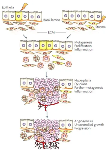
Cancer is nowadays a major public health problem, being the second main cause of death in developing countries (Siegel et al., 2012). It is estimated that in 2020 the world population will reach a total of 7.5 billion people and a total of 15 million new cancer cases will arise (Bray and Møller, 2005). These impairing statistics contribute for the tremendous efforts put forward from behalf of the medical and scientific community to develop more effective therapeutic approaches and also to understand the mechanisms responsible for cancer development. The complex transformation of a normal cell into a malignant phenotype is generally dependent on the accumulation of multiple changes in gene expression patterns in healthy cells (Ruddon, 2007). This cascade of events termed carcinogenesis (Figure 1) encompasses all the complex intracellular signalling pathways responsible for the initiation, stimuli, and progression of cancer cells in an extended time scale (Farber, 1984; Hanahan and Weinberg, 2000).
Chapter I - Introduction
3
Cancer development, starts with changes in structure and function of deoxyribonucleic acid (DNA), usually known as mutations (Mbeunkui and Johann, 2009). These mutations usually lead to the loss of tumor-suppressor genes (tumor protein p53 (TP53), retinoblastoma protein (RB)), or activation of oncogenes (myc, RAS, AKT) (Gaspar et al., 2011; Janssen and Medema, 2012), which propels malignant cells to escape from senescence signals and accept the proliferative stimuli, respectively (Levine and Puzio-Kuter, 2010). Thus, cancer cells acquire unlimited replicative potential, losing their characteristic non-dividing state (Hanahan and Weinberg, 2011). In addition, the deregulations of growth-promoting signals lead to an uncontrolled proliferation of cancer cells (Witsch et al., 2010; Zhang et al., 2010). In the last stages of cancer development, in order to sustain this dynamic growth and high metabolic activity, extensive neovascularisation is promoted by malignant cells in order to assure the necessary uptake of nutrients and oxygen (Annibaldi and Widmann, 2010). The angiogenesis in association with mechanisms that underlie the extravasion of cancer cells through the extracellular matrix (ECM) facilitating tumor spread into healthy tissues (Poste and Fidler, 1980; Zetter, 1998; Mbeunkui and Johann, 2009).Recent reports demonstrate that these abilities acquired by cancer cells, i.e., sustained proliferation, cell death resistance, immortality, angiogenesis, invasion and metastization are a consequence of the unique conditions of the tumor niche. In fact, the tumor microenvironment is of critical importance for the success of cancer progression (Straussman
et al., 2012). Already in 1984, Dolberg and Bissel described an impaired tumor development
in chicken embryos infected with an oncogene expressing Rous sarcoma virus, and hypothesized that these results were probably correlated with the absence of some additional cellular components (Dolberg and Bissell, 1984). Recent studies demonstrated that phenotypic and genotypic abnormalities of cancer cells are insufficient to induce the malignant phenotype (Ma et al., 2003; Weigelt et al., 2003). The influence of the tumor microenvironment in cancer development was then postulated. So far different therapies specifically directed against the tumor microenvironment have been produced (McMillin et
al., 2013; Sounni and Noel, 2013).
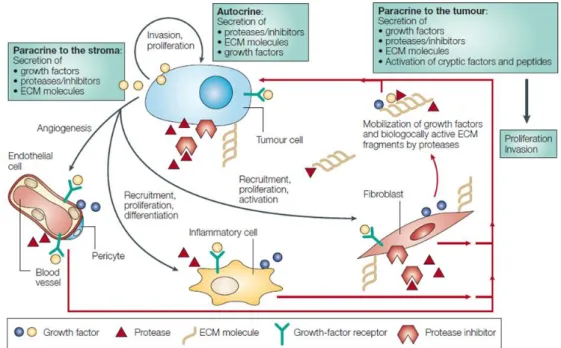
The tumor niche is characterized by the establishment of interactions between tumor cells and stromal cells. The stroma is composed of various cell types such as: i) fibroblasts; ii) endothelial cells; iii) pericytes; iv) adipocytes and v) immune cells (Figure 2), enclosed in a complex ECM (Swartz et al., 2012). The closely regulated interactions between cancer cells and the surrounding healthy stromal cells originate an exclusive environment that propels tumor progression (Hogan, 2012). Moreover, tumor cells are capable of persistently shape their microenvironment according to an extracellular stimuli such as chemotherapy, thereby always maintaining an abnormal ecosystem that assures cancer cell survival (Merlo et al., 2006).
Chapter I - Introduction
4
Figure 2 – Representation of tumor microenvironment (Adapted from Coussens and Werb, 2002).
The mechanisms that modulate the tumor microenvironment are highly dependent on cancer-host cells communications, and are generally formed by dynamic autocrine and paracrine signalling events (Alderton, 2012). The main mediators of these signalling cascades are soluble biomolecules, adhesion molecules, cytokines, chemokines, proteases and other degradative enzymes that are secreted by cancer cells, as well as, by stromal cells (Figure 3).
Figure 3 – General interactions established between tumour cells and stromal cells in human tumor
Chapter I - Introduction
5
1.1.1. ECM in the tumor microenvironment
ECM is a complex three-dimensional (3D) network formed by macromolecular fibers (collagen, elastin, etc) as well as nonfibrous proteins (like proteoglycans and glicoproteins). This 3D matrix is present in the stroma of all tissues, either healthy or malignant (Noguera et al., 2012). The ECM regulates almost all cellular behaviour (Brizzi et al., 2012), and provides structural support and strength, thus allowing cellular communication, adhesion and migration (Noguera et al., 2012). In tumor, the deregulation of ECM is essential for the angiogenesis and invasion of cancer cells into normal tissues (Shuman Moss et al., 2012). Abnormal ECM can promote an increase in collagen deposition or stiffness. As a consequence, the integrin signalization is increased and stimulates cytoskeletal remodeling to regulate cell behaviour, thus inducing cell survival and proliferation (Lu et al., 2012). The ECM also regulates the activity of immune system cells. ECM components function as chemoattractans playing an essential role in infiltration, differentiation, and functional activation of immune systems cells. In turn, these immune cells produce degradative enzymes such as metalloproteinases (MMPs), which are responsible for the degradation of ECM anti-apoptotic activities (Wang et al., 2012).
1.1.2. Stromal cells
1.1.2.1.
Vascular and lymphatic endothelial cells
Tumor proliferation and metastasis are dependent on the growth of blood and lymphatic vessels into the tumor mass (Weis and Cheresh, 2011; Balkwill et al., 2012). The major constituent of these vessels are the endothelial cells. Particularly, in the case of blood vessels, endothelial cells are disorganized, loosely connected, branched and form a defective cellular lining on the vessel wall (Hashizume et al., 2000). These cells are able to recruit immune cells due to the expression of cell adhesion molecules (CAMs) on their surface (Kobayashi et al., 2007; Hanahan and Coussens, 2012). In turn, these immune cells in association with other stromal cells and cancer cells, will promote the formation of new vessels due to the activation of endothelial cells by the secretion of the main growth factor involved in angiogenesis, the vascular endothelial growth factor (VEGF) (Weis and Cheresh, 2011). Once endothelial cells become activated, they secrete their own VEGF and hepatocyte growth factor (HGF) (Li et al., 2007).
The lymphatic endothelial cells are also responsible for secretion of angiogenic factors (VEGF) and for modulating the host immune cells in the tumor microenvironment (Skobe et al., 2001; Balkwill et al., 2012).
1.1.2.2.
Perycites
Another important component of vascular vessels is the pericytes cells, which are specialized smooth-muscle cells that are present outside the vessel wall (Pietras and Östman, 2010). These cells have a potential influence on endothelial cells due to the direct contact and paracrine signalling with them, through platelet-derived growth factor subunit-B (PDGF-B),
Chapter I - Introduction
6
which is a potential mitogenic factor of fibroblasts (Minami et al., 2013). In addition, pericytes stabilize endothelial cells, mediating their survival, i.e., indirectly, pericytes control the formation, maturation, remodeling, stabilization and function of vascular vessels (Minami et al., 2013).1.1.2.3. Adipocytes
The influence of adipocytes in the tumor microenvironment is characterized by their capacity to secret of adipokines that promote the growth of malignant cells by providing them fatty acids (Nieman et al., 2011).
1.1.2.4.
Immune System cells
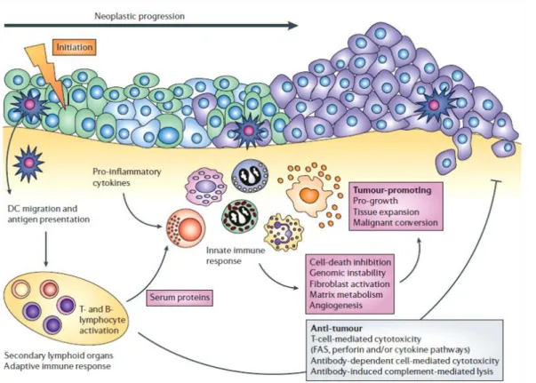
Normally, inflammation serves to protect a defined region of infected or damaged tissue by recruiting cells necessary to resolve the insult, while also isolating the area to prevent the spread of infection and subsequently re-establish the normal tissue function and homeostasis (Janeway et al., 2001; Sherwood, 2012). However, a non-resolved inflammation can promote genomic instability, growth, angiogenesis, survival and escape of tumor cells from immune surveillance (Figure 4) (Zamarron and Chen, 2011).
Figure 4 – Immune system behaviour in tumor microenvironment (Adapted from de Visser et al., 2006).
In cancer, myeloid-derived suppressive cells (MDSCs), macrophages, mast cells, dendritic cells (DCs), eosinophils, neutrophils, lymphocytes and natural killer cells (NK) are generally recruited by malignant tumors to support their development trough signalling events
Chapter I - Introduction
7
mediated by chemokines, cytokines, cytotoxic mediators and soluble mediators of cell death and proliferation (Tlsty and Coussens, 2006; Kerkar and Restifo, 2012).The cells from the immune system induce DNA damage by production of reactive oxygen species (ROS) and nitrogen species (NO) (Alexander and Friedl, 2012). In addition, immune cells promote angiogenesis and tissue remodelling by producing growth factors such as HGF, transforming growth factor-β (TGF-β), VEGF, cytokines (tumour necrosis factor- (TNF-), interleukins (IL-6, IL-10) and MMPs (MMP-7, MMP-9) (Stockmann et al., 2008; See et al., 2012), Moreover, tumor growth can be promoted by regulatory T cells (T reg) that suppress cytotoxic T cell responses (Kerkar and Restifo, 2012), and also by humoral immune responses that increase chronic inflammation in the tumor microenvironment (Grivennikov et al., 2010). All these processes are summarized in Figure 4.
1.1.2.5. Fibroblasts
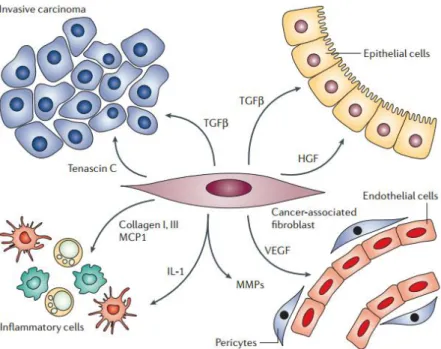
It is now becoming clear that among host cells present in the tumor microenvironment, fibroblasts play crucial roles during various steps of cancer development (Strell et al., 2012). These stromal constituents are elongated cells with extended and fusiform or spindle-like shapes. They are the non-vascular, non-epithelial and non-inflammatory cells of the connective tissue (Kalluri and Zeisberg, 2006). Normal stroma in most organs contains a reduced number of fibroblasts (Kalluri and Zeisberg, 2006), but in the tumor microenvironment, these cells are highly prevalent (Micke and Östman, 2005). Interestingly, the fibroblasts within the tumour stroma acquire a dynamic phenotype, similar to that of fibroblasts associated with wound healing (Cirri and Chiarugi, 2012), with high expression of
-smooth muscle actin (-SMA) and vimentin (mesenchymal marker)(Horimoto et al., 2012). These activated fibroblasts also termed cancer associated fibroblasts (CAF) (Figure 5) have been observed repeatedly in the stroma of the majority of aggressive and invasive human breast cancers (Sappino et al., 2006).
Figure 5 – Comparison of normal and cancer associated fibroblasts (CAF) (Adapted from Kalluri and
Chapter I - Introduction
8
The great influence of fibroblasts in cancer evolution is proved by their capability to induce the growth and activation of immortalized prostate epithelial cells in vitro and in vivo (Olumiet al., 1999). Parrott and co-workers, showed in 2001, that ovarian cancer cells recruit
adjacent fibroblasts from normal tissue in order to from the stroma of the tumor, demonstrating the need of other cell types to form tissue primary cancer. Furthermore, it has been also shown that, therapies against stromal fibroblasts obtained high anti-tumoral effect (Parrott et al., 2001; Strell et al., 2012; Mertens et al., 2013).
In the tumor mass, fibroblasts interact with all other elements of the tumor microenvironment (Figure 6), affecting their action.
Figure 6 – Influence of fibroblasts in tumor microenvironment (Adapted from Kalluri and Zeisberg,
2006).
The interactions between fibroblasts and cancer cells are essentially mediated by growth factors (Table 1), that are secreted by these two cells to stimulate both of them and induce tumor progression. In brief, cancer cells produce growth factors such as PDGF, TGF-β, endothelial growth factor (EGF) and basic fibroblast growth factor (bFGF), in order to activate fibroblasts. Subsequently, these fibroblasts secrete growth factors, such HGF, keratinocyte growth factor (KGF), insulin-like growth factor 1 and 2 (IGF-1 and -2), chemokines, like stromal cell-derived factor-1 (CXCL12) and glycoproteins (tenascin C) to stimulate cancer cells (Bhowmick et al., 2004; Cirri and Chiarugi, 2012).
Chapter I - Introduction
9
Table 1 – Interactions between cancer cells and tumor stromal fibroblasts.
Interaction Factor Role Ref.
Cancer cells towards Fibroblasts
PDGF Fibroblasts activation to CAF. (Kalluri and Zeisberg, 2006; Cirri and Chiarugi, 2012)
TGF-β Fibroblasts activation to CAF. (Bierie and Moses, 2006; Yin et al., 2012)
EGF Stimulates fibroblasts to produce VEGF and HGF. (Yu et al., 2012)
bFGF Fibroblasts proliferation. (Yu et al., 2012)
Fibroblasts towards Cancer cells HGF Proliferation; Drug resistance. (Bhowmick et al., 2004; Straussman et al., 2012) KGF Migration; Metastasis.
(Faria et al.; Leyva-Illades et al., 2012)
IGF-1, -2 Survival of tumor cells. Hanahan and Coussens, (LeBedis et al., 2002; 2012)
CXCL12
Growth and survival of malignant cells; Stimulate the migration of stromal cell
types into the tumor microenvironment (T cells, B cells and monocyts).
(Balkwill et al., 2012)
Tenascin C Influence the capacity of tumor cells to adhere and spread through the ECM. (Brellier and Chiquet-Ehrismann, 2012)
Besides the fact that fibroblasts induce tumor proliferation, these stromal cells also generate ROS in environments with low pH and oxygen concentration (Xing et al., 2010). ROS are responsible for multiple mutations in malignance surrounding cells.
In terms of cancer cell invasion through the ECM, fibroblasts have a potential influence. CAFs retain a major role in ECM remodelling since they are mainly responsible for the production of ECM proteins (collagens and fibronectins) as well as proteases and other enzymes involved in the post-transcriptional modification of ECM proteins (Vanharanta and Massagué, 2012). In tumor, fibroblasts are responsible for the increase of matrix deposition and ECM stiffening (Cirri and Chiarugi, 2012). This stiffening is responsible for an increase in cross-linking between collagen molecules induced by lysyl oxidase (LOX) expressed by fibroblasts. These enzymes are responsible for ECM remodelling and as consequence the tumor cell migration and invasion (Cirri and Chiarugi, 2012). In addition, cancer cells induce CAF to produce MMP
Chapter I - Introduction
10
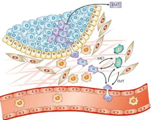
(Talmadge and Fidler, 2010; Xing et al., 2010), which binds to the cancer cells and it is used for degradation and invasion of malignance cells through the ECM (Pavlaki and Zucker 2003). Two other ECM components produced by fibroblasts are fibronectin and hyaluronan. Fibronectin is associated with integrin receptors and MMP secretion, thus affecting cell adhesion, migration. Within tumors, fibroblasts, secrete high concentrations of hyaluronan that are responsible for macrophage recruitment, which have also an essential role in tumor progression, as previously mentioned (Lu et al., 2012; Raz and Erez, 2013). In addition to attract macrophages, fibroblasts are also responsible for mediating the inflammatory response, by secreting chemokines (monocyte chemotactic protein-1 (MCP-1)), interleukins (IL-1) and inducing immune suppression by expression of TGF-β (McClellan et al., 2012; Raz and Erez, 2013). This transforming growth factor is also responsible for the orchestration of the epithelial-to-mesenchymal transition (EMT) (Figure 7) (Chaffer and Weinberg, 2011). Different studies showed the crucial role of EMT in tumor progression, since it promotes the invasion and metastasis of cancer cells (Alexander and Friedl, 2012).Figure 7 – Representation of the mechanism of epithelial-to-mesenchymal transition (EMT) (Adapted
from (Peinado et al., 2007).
EMT is the mechanism responsible for the acquisition of mesenchymal like properties of epithelial cells, in result of disruption of intercellular adhesion (adherens junctions) and the enhancement of cell motility (Alexander and Friedl, 2012).
In last, fibroblasts stromal cells also interfere in tumor vascularization. These stromal cells express VEGF, which have a potential angiogenic effect in tumor mass (Strell et al., 2012).
Chapter I - Introduction
11
1.2. Nanosized delivery systems as novel therapeutic
approaches for cancer therapy
Advanced oncologic diseases have been for long associated with limited patient progression free survival (PPFS) (Chabner and Roberts, 2005). Over the last two decades, physicians have been focused in increasing PPFS rate through the application of multimodal treatment approaches that involve surgery, chemo- and radiotherapy, in an attempt to tackle the above mentioned adaptive and multi-resistant profile of cancer cells (Riehemann et al., 2009). The anti-cancer chemical drugs are commonly administered intravenously, being partitioned in both the tumor and major organs (e.g. liver, lungs, kidneys) due to their inability to differentiate healthy from malignant cells (Sinha et al., 2006).
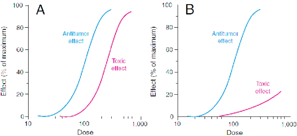
Figure 8 – Relation between drug anti-tumoral effect and toxic effect. A) Hypothetical dose-response
curves for conventional chemotherapeutic drugs. In conventional therapies the curves of antitumor
effect and toxic effect are parallel, revealing that higher doses induce higher adverse effects.
B) Hypothetical dose-response curves for new drug delivery systems. The drug delivery systems increase
the specificity for cancer cells, reducing the toxic adverse effects. (Adapted from Fox et al., 2002).
This pharmacokinetic profile accounts for the adverse cytotoxicity effect attained with the majority of chemotherapeutic agents (Fox et al., 2002). In addition, due to an inherent limited aqueous solubility, anti-cancer drugs present a short circulation time and sometimes their concentration in blood remains below the therapeutic window concentrations, rendering this therapy rather ineffective (Danhier et al., 2012). To overcome these issues, physicians administer chemotherapy through longer time periods (usually every 21 days, with a total of 5 to 8 sessions) (Hamilton and Hortobagyi, 2005). Although, this approach has a limited success, since tumor drug accumulation remains low and cancer cells develop resistance against the effect of chemotherapeutics (Krishna and Mayer, 2000; Raguz and Yagüe, 2008). This acquired multidrug resistance (MDR) is nowadays one of the most serious problems associated with chemotherapy, having a negative impact in PPFS.
Similarly, radiotherapy-based treatments are also impaired by the lack of cell specificity (Sorensen et al., 2012; Allen et al., 2013). In fact, ionizing radiation penetrates within tissues indiscriminately and although it triggers cancer cell death, it also damages the surrounding
Chapter I - Introduction
12
tissues and organs in such a way that new cancer cells may arise from radiation-induced mutations (Allen et al., 2013; Zimmermann et al., 2013).Radiation as an anti-cancer therapy in young woman can affect reproductive organs, and as a consequence induce fertility problems (Metzger et al., 2013). Another major disadvantage of these conventional therapies is their inability to eradicate circulating cancer cells that may lay dormant for a refractory period and form new metastatic cancers in other organs (Chabner and Roberts, 2005; Alexander and Friedl, 2012). Metastatic cancer cells are markedly more aggressive than their parent cells and these conventional therapies are still ineffective (Alexander and Friedl, 2012).
Thus, despite having increased expectations for cancer treatment, these traditional therapies still lack the necessary specificity and effectives to eliminate cancer cells. Therefore, recently the advent of Nanotechnology was unlocked a whole new range of opportunities to develop safe and highly effective anti-cancer treatments. In reality, gathering the unique capacity of nanoscale materials and directed it to the manufacture of novel medical devices brings forth the potential to change the effectiveness of cancer therapies (Zhang et al., 2013). Moreover, nano-based platforms also make an important contribution in cancer prevention, detection, diagnosis and imaging (Yang et al., 2013).
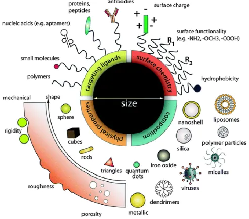
In contrast to conventional therapies, nanomedicine attempts to use sophisticated approaches to either kill specific cells or repair them, one cell at a time (Zhang et al., 2013), like nanoparticles (Figure 9).
Figure 9 – Representation of the different types of nanoparticles available (Adapted from Chou et al.,
Chapter I - Introduction
13
These nanosized particles were developed for the first time around 1970 (Couvreur, 2012) and include numerous architectural designs in terms of: i) size (from a few to several hundred nanometers), ii) shape (porosity, roughness, rigidity), iii) structure (sphere, cubes, tubes), iv) materials (polymers, metal, ceramic) and v) functionalization (hydrophobic character, surface charge, ligands) (Chou et al., 2011). The different proprieties presented by each particle are responsible for different drug loading capacity, particle and drug stability, drug release profile and capacity of targeted delivery. For anti-cancer therapy, different nanocarriers have been already developed (Wang and Thanou, 2010), such as, polymeric nanoparticles, micelles, dendrimers, lipossomes, carbon nanotubes, quantum dots, magnetic nanoparticles, among others, which are able to entrap, encapsulate or attach various anti-cancer agents such as: i) Drugs, that include Doxorubicin, Paclitaxel and Cysplatin; or ii) tumor suppressor genes, that include P53, TNF-α which could induce cell death (Gaspar et al., 2011).These nanocarriers offer numerous advantages compared to conventional therapies. Particle size and surface characteristics of nanoparticles can be easily tailored (Yun et al., 2012), and the sub-cellular size of these nanosystems increases the overall surface area, thereby increasing the rate of drug dissolution (Riehemann et al., 2009). Nanocarriers also encapsulate several hydrophilic and hydrophobic compounds simultaneously (Kamaly et al., 2012). This is a highly advantageous characteristic since a nano delivery system could co-delivery a therapeutic agent with an imaging agent, allowing physicians to track cancer cells and at the same time treat them, these systems are also known as theragnostic (Farokhzad and Langer, 2009). On the other hand, the use of multiple drugs acting in a synergistic mode could be highly useful to surpass the various defence mechanisms of cancer cells (Han et al., 2013).
In addition, these nanocarriers improve the absorption of insoluble compounds and macromolecules, also improving the bioavailability and release rates, potentially reducing the amount of dose required and increasing safety by decreasing the toxic side effects (Figure 8 B) (Riehemann et al., 2009). Also, the drug release could be sustained during a period of few hours to several days or weeks (Panyam and Labhasetwar, 2012), controlling in this way the therapeutic effect of the drug as intended.
In last, the best upgrade of these nano drug delivery system is their combination with cell- or tissue-specific ligands, for targeted drug delivery (Figure 9) (Farokhzad and Langer, 2009). The association of nanoparticles with these specific ligands (antibodies, molecules or nucleic acids) allows the specific recognition of cancer cells, avoiding the negative effect of the drug in normal cells (Brannon-Peppas and Blanchette, 2004).
In conclusion, with the appearance of nanomedicine new drug delivery systems have been developed, namely nanoscale particles/molecules, that improve the bioavailability and pharmacokinetics of therapeutics. Thus, these systems could improve the treatment by increasing the therapeutic efficacy and decreasing the side effects, improving patient compliance.
Chapter I - Introduction
14
1.3. Experimental models to evaluate nanoparticulated
delivery systems for application in cancer therapy
Nowadays, tremendous resources about the application of nanoparticles in prevention, diagnosis, and treatment of cancer are being investigated (Zhang et al., 2007). The discovery and development of anti-cancer agents are the main objectives of important pharmaceutical companies (Narang and Desai, 2009). However, the introduction of new nanotechnology based pharmaceuticals creates new challenges for regulatory institutions (Lövestam et al., 2010). In Europe, the European Commission (EC) and European Medicines Agency (EMEA) already define the need that all products based on nanomaterials used in humans must comply with the high level of public health and safety regulations (Hellsten, 2005). In addition, internationally, the new nanodevices need to be in agreement with the basic rules from regulatory agencies such as Food and Drug Administration (FDA) and Organization for Standardization (ISO) (Grieger et
al., 2009).
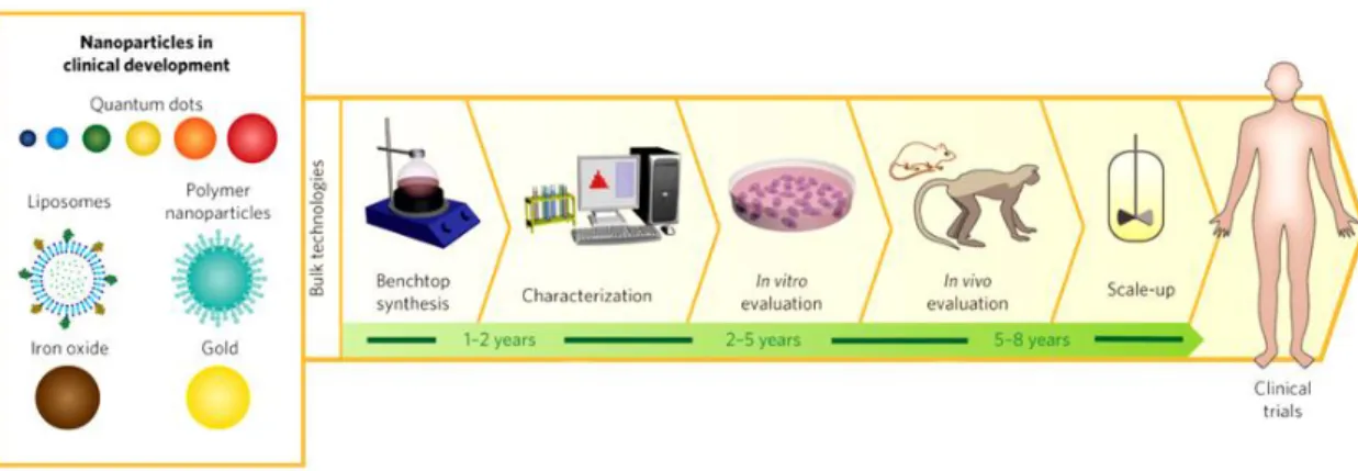
Clinical trials, which are prospective tests to evaluate the effect of medical devices and pharmaceutics in humans under specified conditions, have become a standard and integral part for the development and approval of new cancer therapies (Meinert, 2012). Clinical trials are divided into two main stages (pre-clinical and clinical stage). At each step, from the point of discovery and design, through the demonstration of safety and efficacy in humans, drug and nanoparticle candidates are closely scrutinized.
Figure 10 – Steps needed for nanoparticle development (Adapted from Valencia et al., 2012).
The pre-clinical stage includes basic physicochemical studies that allow the identification and characterization of the synthesis of compounds. In this stage, in vitro testing, using cell lines and also in vivo assays, in animal models, are performed in order to characterize the pharmacokinetic/pharmacodynamic and toxic profile of the drug delivery system in biologic systems (Lee, 2007; Eifler and Thaxton, 2011).
1.3.1.
In vivo models
International efforts have been made in other to decrease or avoid the use of animals in laboratory, as regulated by the European Directive 86/609/EEC (Stacey, 2012). Futhermore,
Chapter I - Introduction
15
there are different countries where animal experimentation is not allowed (Milstein et al., 1996; Stacey, 2012). Thus, nowadays the scientists are using the 3 Rs rule (Replacement, Reduction, and Refinement) in what concerns research animal experimentations (Ranganatha and Kuppast, 2012; Wolfensohn et al., 2013). As a consequence, the experiments are performed only with the essential animals needed to validate the results, with fewer repetitions and during limited periods of a time (Madden et al., 2012). In addition, in order to reduce maintenance costs, the studies tend to use young animals, whereas, the most difficult patients to treat are the elderly ones (Hartung, 2008).Another major disadvantage of animal use is the difference between species (Astashkina et
al., 2012). At the laboratory rodent species are usually used. As demonstrated in Figure 11, it
is clear that there is no apparent relationship between animal bioavailability and human bioavailability. In fact, there are several cases with high drug bioavailability in animals and low bioavailability in humans (false positives). Moreover, also low drug bioavailability in animals and high bioavailability in humans (false negatives) is also reported.
Figure 11 - Absolute bioavailability of various drugs in dogs (red triangles), primates (blue squares) and
rodents (green circles) versus the absolute bioavailability reported for humans (Adapted from Sietsema, 1989).
Like these examples, there are others where the predictions offered by in vivo models fail categorically in humans (Johnson et al., 2001; Kelland, 2004). In order to avoid this issue, in vivo studies could be performed with different species, but that would involve the use of more animals (Hartung, 2008).
Furthermore, some of the studies described in the literature are restricted just to one sex, and both sexes have different sensitivities to drugs, as previously reported in the literature (Hartung, 2008). So, studies should be performed in both sexes in order to obtain more realistic results. The stress caused by the experimental procedures in animals is also responsible for the variability and should be carefully addressed in these studies (Hartung, 2008).
Chapter I - Introduction
16
1.3.2.
In vitro models
Cell cultures emerged in 19th century as a valuable solution for challenging issues of in vivo models. The first culture was performed by Wilhem Roux in 1885, by maintaining in vitro chicken embryo tissue during several days (Langdon, 2004). However, only in 1922 the culture of epithelial cells was performed and in 1951 the first continuous human cancer cell line (HeLa) was obtained and cultured by George Gey (Langdon, 2004).
These in vitro techniques allow the growth of cells outside of their natural environment, under controlled conditions (Lindl and Steubing, 2013). These types of assays become the main force of biopharmaceutical investigation due to their few limitations, namely, bacterial and fungal contaminations (Stacey, 2011). Cell culture offers a unique testing platform to investigate the effects of different drug formulations and nanoparticle designs during pre-clinical development, under highly controlled and reproducible conditions (HogenEsch and Nikitin, 2012), such as temperature, pH, osmotic pressure, oxygen (O2) and carbon dioxide (CO2) tension. Moreover, cell cultures provide an easy way to manipulate numerous experimental variables in order to mimic some in vivo conditions, whilst avoiding ethical and legal issues associated with animal handling and experimentation (Duell et al., 2011). However, up till now, the cell-cell interactions were commonly disregarded in the majority of the studies reported so far (Duell et al., 2011).
1.3.2.1. Co-cultures
One of the main goals of the in vitro systems has been the reproduction of original tissue characteristics and its cell–cell interactions, to simulate, as close as possible, the in vivo environment. To overcome such limitations a new category of cell cultures, termed co-cultures, is currently being developed (Miki et al., 2012). This cell culture system arises as a quite interesting alternative that allows a better method to mimic the tumor microenvironment (Duell et al., 2011).
A co-culture consists of at least two different types of cells (i.e., cancer/fibroblast, epithelial cell/lymphocyte, etc.) (Miki et al., 2012) and they provide a more sophisticated and sensitive system that allows to reproduce in vitro the in vivo tumor niche. By using co-cultures it is possible to recreate some of the cell inter-relationship (Tumarkin et al., 2011), since heterotypic cell-cell interactions are established in close contact. These direct physical connections between cells play a pivotal role in the mechanisms of cancer invasion through actions of adhesion molecules, such as Cadherins (De Wever and Mareel, 2003; Miki et al., 2012). In addition, different cells in co-culture could release cytokines, interleukins, chemokines and growth factors that are essential for the establishment of cell morphology, phenotype, metabolism and proliferation, features that are always present in vivo (Streuli et
al., 1991; Krause et al., 2010; Purpura et al., 2011; Miki et al., 2012). In addition, co-cultures
can also influence the drug resistance presented by cancer cells. In fact, Martinez-Outschoorn and his team, 2011, (Martinez-Outschoorn et al., 2011) analyzed de apoptotic effect of Tamoxifen in monocultures of breast cancer cells and co-cultures of breast cancer cells and
Chapter I - Introduction
17
fibroblasts. As a conclusion, they verified a markedly reduction in apoptosis of breast cancer cells when they were in co-culture, highlighting the importance of these models.1.3.2.2. 3D cell cultures: Spheroids
In addition to the presence of various cell types encountered in the tumor microenvironment, the inclusion of ECM, which is 3D, complex and dynamic network is crucial for mimic the in
vitro tumor (Cukierman et al., 2002). So, in 1972, researchers began to explore the
differences between cells grown on a flat surface versus 3D supports (Elsdale and Bard, 1972). They concluded that, ECM is not just a random mix of secreted components, but it has a specific composition of biomolecules entrapped in a very well defined geometrical structure, that stimulates specific cell responses, such as differentiation. Thus, in addition to co-localization of different cell types with cell–cell interactions and the exchange of bioactive molecules, a 3D architecture that mimics cell organization in tissues and organs is demanded to mimic tissue native proprieties using in vitro conditions (Kim et al., 2004). Such, is the main disadvantage of two-dimensional (2D) in vitro models. Such models rely on the migration of cells on flat substrates, like glass or polystyrene surfaces (Freshney et al., 2006), in which cells grow in two dimensions (Burdett et al., 2010), which leads to the loss of spatial organization of cells and a deregulation of cell metabolism and functionality as consequence (Lee et al., 2008).
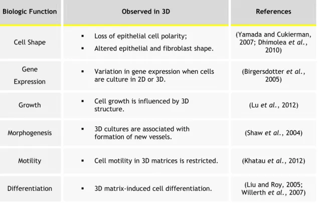
To overcome such disadvantage, the 3D cell cultures have emerged as a viable option. One of the most widely used 3D in vitro models are the tumor spheroids (Burdett et al., 2010). This unique culture is based in a small, tightly bound cellular aggregate that tends to form when cancer cells are cultured in non adherent surfaces (Trédan et al., 2007; Burdett et al., 2010; Fennema et al., 2013). Spheroids have been used in cancer research, because the 3D architecture and extensive cell–cell interactions provided by spheroid growth closely mimic the in vivo cellular environment. Recently, 3D culture gene expression profiles, have been shown to reflect the clinical expression profiles, in comparison with those observed for 2D cultures (Hirschhaeuser et al., 2010). In fact, the 3D cell organization can influence cell shape, gene expression, growth, morphogenesis, motility and differentiation (Table 2) (Yamada and Cukierman, 2007). In addition, cells cultured in 2D monolayers typically exhibit a lower resistance to therapy than those of in vivo tumors, a critical factor if novel therapeutic approaches are under evaluation (Phung et al., 2011). Spheroids have also been considered an essential cell culture technique to study solid tumors. Some cancer types (sarcomas, carcinomas and lymphomas) tend to form solid tumors (Gavhane et al., 2011). These tumors have a unique 3D architecture characterized by hypoxia, low pH and low levels of glucose (Box et al., 2010; Yeom et al., 2012).
Chapter I - Introduction
18
Table 2 - The influence of 3D cell organization in cell behaviour and signalling.
Biologic Function Observed in 3D References
Cell Shape Loss of epithelial cell polarity;
Altered epithelial and fibroblast shape.
(Yamada and Cukierman, 2007; Dhimolea et al.,
2010) Gene
Expression
Variation in gene expression when cells are culture in 2D or 3D.
(Birgersdotter et al., 2005)
Growth Cell growth is influenced by 3D structure. (Lu et al., 2012)
Morphogenesis 3D cultures are associated with formation of new vessels. (Shaw et al., 2004)
Motility Cell motility in 3D matrices is restricted. (Khatau et al., 2012)
Differentiation 3D matrix-induced cell differentiation. Willerth et al., 2007) (Liu and Roy, 2005;
Due to these characteristics, instead of normal arterioles, capillaries, and venules (Figure 12 A), the tumor vessels in solid tumors often have irregular diameters, abnormal branching patterns, and a defective wall structure (Figure 12 B) (Trédan et al., 2007; De Bock et al., 2011).
Figure 12 – Representation of the vascularization in normal (A) and in cancer (B) tissue (Adapted from
Trédan et al., 2007).
These irregular vessels could be responsible for the formation of hypoxic regions that are characterized by a defective supply of oxygen and nutrients (glucose and essential amino acids) (Jones and Thompson, 2009). In response, cancer cells are characterized by using lactate to obtain their energy instead of glucose - Warburg effect - (Warburg, 1956;