Article
Inhalable Antitubercular Therapy Mediated by
Locust Bean Gum Microparticles
Ana D. Alves1, Joana S. Cavaco1, Filipa Guerreiro1,2, João P. Lourenço3,4, Ana M. Rosa da Costa4and Ana Grenha1,2,*
1 Center for Biomedical Research (CBMR), Faculty of Sciences and Technology, University of Algarve, 8005-139 Faro, Portugal; [email protected] (A.D.A.); [email protected] (J.S.C.); [email protected] (F.G.)
2 Centre for Marine Sciences (CCMar), University of Algarve, 8005-139 Faro, Portugal
3 Centro de Química Estrutural (CQE), Instituto Superior Técnico, University of Lisbon, 1049-001 Lisbon, Portugal; [email protected]
4 Algarve Chemistry Research Center (CIQA) and Department of Chemistry and Pharmacy,
Faculty of Sciences and Technology, University of Algarve, 8005-139 Faro, Portugal; [email protected] * Correspondence: [email protected]; Tel.: +351-289-800-100 (ext. 7441); Fax: +351-289-800-066
Academic Editor: Peter J. Rutledge
Received: 13 February 2016; Accepted: 19 May 2016; Published: 28 May 2016
Abstract:Tuberculosis remains a major global health problem and alternative therapeutic approaches are needed. Considering the high prevalence of lung tuberculosis (80% of cases), the pulmonary delivery of antitubercular drugs in a carrier system capable of reaching the alveoli, being recognised and phagocytosed by alveolar macrophages (mycobacterium hosts), would be a significant improvement to current oral drug regimens. Locust bean gum (LBG) is a polysaccharide composed of galactose and mannose residues, which may favour specific recognition by macrophages and potentiate phagocytosis. LBG microparticles produced by spray-drying are reported herein for the first time, incorporating either isoniazid or rifabutin, first-line antitubercular drugs (association efficiencies >82%). Microparticles have adequate theoretical properties for deep lung delivery (aerodynamic diameters between 1.15 and 1.67 µm). The cytotoxic evaluation in lung epithelial cells (A549 cells) and macrophages (THP-1 cells) revealed a toxic effect from rifabutin-loaded microparticles at the highest concentrations, but we may consider that these were very high comparing with in vivo conditions. LBG microparticles further evidenced strong ability to be captured by macrophages (percentage of phagocytosis >94%). Overall, the obtained data indicated the potential of the proposed system for tuberculosis therapy.
Keywords: alveolar macrophages; antitubercular drugs; inhalable therapy; locust bean gum; microparticles; spray-drying; tuberculosis therapy
1. Introduction
Although there is an effective treatment for tuberculosis (TB), the disease remains one of the major health problems worldwide [1]. In 2014, the World Health Organization reported 9.6 million new cases of TB [2], infection caused by inhalation of aerosol particles containing Mycobacterium tuberculosis (MTB) bacilli [1]. The inhaled bacilli are phagocytosed by alveolar macrophages, which triggers a series of events that can lead to either control of the infection, i.e., latent TB, or progression to an active form of the disease [1].
Conventional TB therapy involves oral co-administration of several antitubercular drugs over a period of time that may exceed two years in some cases [3]. Severe side effects are frequently reported that, in many cases, lead to therapeutic noncompliance. Therefore, other alternatives are being actively
searched to shorten the duration, and perhaps the modality, of treatment [2]. Shortening treatment duration would permit minimising possible side effects in organs such as the liver and kidneys, and avoiding the emergence of resistant TB species. Considering that pulmonary TB represents 80% of cases, the design of antitubercular inhalable formulations is considered an adequate therapeutic approach. Inhalable therapy allows the co-localisation of drugs and pathogens and is thought to enable increased drug concentration in the lungs, along with favoured lung-to-plasma ratio. Overall, the approach is expected to permit reducing doses and frequency of administration, possibly shortening treatment periods, resulting in general therapeutic improvement [3]. Importantly, it has been referred that inhalable TB therapy can either be used to replace oral antibiotherapy, or as add-on therapy [4].
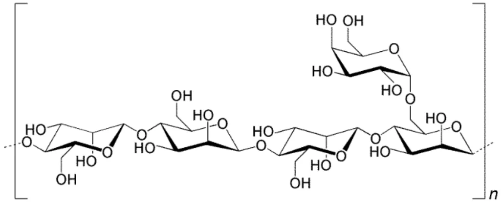
Applying this approach requires that the inhalable antitubercular formulations exhibit suitable aerodynamic properties to reach the alveolar zone, where the alveolar macrophages hosting MTB are located [3]. Microparticles have been considered adequate for this end, ensuring not only the protection of the drugs until they reach the site of action, but also providing the necessary aerodynamic characteristics. To reach the alveoli, the aerodynamic diameter should be within 1–3 µm [5], but macrophage uptake is reported to be maximal for particles of 1–2 µm [6,7], thus identifying this size as a target in formulation design. Spray-drying has revealed appropriate to produce polymeric microparticles for inhalation purposes, being a versatile technique that permits tailoring microparticles to the desired characteristics, i.e., size, density and morphology [8]. Different materials have been used in lung drug delivery systems. Polysaccharides are one of the most popular classes, mainly due to their flexible chemical structures, involving many hydroxyl groups available for functionalisation, apart from the high probability of biocompatibility and biodegradability, and availability at a relatively low price [9]. Locust bean gum (LBG) is a neutral polysaccharide of the galactomannan family, with reported pharmaceutical application mainly in tablet production. It is extracted from the seeds of the carob tree (Ceratonia siliqua) [10] and consists of a linear chain of (1–4)-linked β-D-mannopyranosyl units with (1–6)-linked side chains of α-D-galactose, in a mannose/galactose ratio of 4:1, as depicted in Figure1. LBG molecular weight is estimated to be in the range of 50 to 3000 kDa [11]. The application of LBG as a matrix material of microparticles designed to carry and deliver antitubercular drugs can be of great interest owing to the ability of macrophages to recognize and phagocytose with preference materials having structural units such as mannose and galactose [7].
Molecules 2016, 21, 702 2 of 21
duration would permit minimising possible side effects in organs such as the liver and kidneys, and avoiding the emergence of resistant TB species. Considering that pulmonary TB represents 80% of cases, the design of antitubercular inhalable formulations is considered an adequate therapeutic approach. Inhalable therapy allows the co-localisation of drugs and pathogens and is thought to enable increased drug concentration in the lungs, along with favoured lung-to-plasma ratio. Overall, the approach is expected to permit reducing doses and frequency of administration, possibly shortening treatment periods, resulting in general therapeutic improvement [3]. Importantly, it has been referred that inhalable TB therapy can either be used to replace oral antibiotherapy, or as add-on therapy [4].
Applying this approach requires that the inhalable antitubercular formulations exhibit suitable aerodynamic properties to reach the alveolar zone, where the alveolar macrophages hosting MTB are located [3]. Microparticles have been considered adequate for this end, ensuring not only the protection of the drugs until they reach the site of action, but also providing the necessary aerodynamic characteristics. To reach the alveoli, the aerodynamic diameter should be within 1–3 μm [5], but macrophage uptake is reported to be maximal for particles of 1–2 μm [6,7], thus identifying this size as a target in formulation design. Spray-drying has revealed appropriate to produce polymeric microparticles for inhalation purposes, being a versatile technique that permits tailoring microparticles to the desired characteristics, i.e., size, density and morphology [8]. Different materials have been used in lung drug delivery systems. Polysaccharides are one of the most popular classes, mainly due to their flexible chemical structures, involving many hydroxyl groups available for functionalisation, apart from the high probability of biocompatibility and biodegradability, and availability at a relatively low price [9]. Locust bean gum (LBG) is a neutral polysaccharide of the galactomannan family, with reported pharmaceutical application mainly in tablet production. It is extracted from the seeds of the carob tree (Ceratonia siliqua) [10] and consists of a linear chain of (1–4)-linked β-D-mannopyranosyl units with (1–6)-linked side chains of α-D-galactose, in a mannose/galactose ratio of 4:1, as depicted in Figure 1. LBG molecular weight is estimated to be in the range of 50 to 3000 kDa [11]. The application of LBG as a matrix material of microparticles designed to carry and deliver antitubercular drugs can be of great interest owing to the ability of macrophages to recognize and phagocytose with preference materials having structural units such as mannose and galactose [7].
Figure 1. Structure of locust bean gum showing a linear polysaccharide (1–4)-β-linked backbone of mannose units with single (1–6)-α-D-galactose units attached.
In this work we have prepared spray-dried LBG microparticles for inhalable antitubercular therapy. Microparticles were tailored to exhibit adequate aerodynamic properties to reach the alveolar zone and favour phagocytosis. To the best of our knowledge, LBG microparticles prepared by this method are not described in the literature. The ability to associate two first-line antitubercular drugs, isoniazid (INH) and rifabutin (RFB), was demonstrated. The cytotoxicity of drug formulations, free drugs and unloaded microparticles was tested in relevant respiratory cell lines, i.e., A549 and macrophage-like THP-1 cells, and the ability of macrophages to capture LBG microparticles was established.
Figure 1.Structure of locust bean gum showing a linear polysaccharide (1–4)-β-linked backbone of mannose units with single (1–6)-α-D-galactose units attached.
In this work we have prepared spray-dried LBG microparticles for inhalable antitubercular therapy. Microparticles were tailored to exhibit adequate aerodynamic properties to reach the alveolar zone and favour phagocytosis. To the best of our knowledge, LBG microparticles prepared by this method are not described in the literature. The ability to associate two first-line antitubercular drugs, isoniazid (INH) and rifabutin (RFB), was demonstrated. The cytotoxicity of drug formulations, free drugs and unloaded microparticles was tested in relevant respiratory cell lines, i.e., A549
and macrophage-like THP-1 cells, and the ability of macrophages to capture LBG microparticles was established.
2. Results and Discussion
2.1. Preparation of Locust Bean Gum (LBG) Microparticles by Spray-Drying
Obtaining a polymeric dispersion adequate for spray-drying required a great optimisation of each step, due to the high viscosity of LBG in solution. LBG is described to adopt an extended rod-like conformation in solution, occupying a large volume of gyration, which results in high viscosity [12]. A LBG dispersion at 2% (w/v) exhibits such a high viscosity that spray-drying is hampered. For the specific LBG polymer being used, Sigma-Aldrich (the provider) reports a viscosity of 2100–3750 cps for a 1% (w/v) solution, which is much greater than the ideal for an operation with the spray-drying equipment, reported by Buchi (Flawil, Switzerland) to be of 300 cps [13]. The high viscosity of LBG solutions at relatively low concentrations is reported, which is only slightly affected by pH, salts or temperature [14]. Nevertheless, in this work the addition of hydrochloric acid (HCl) 0.1 M was found to provide the adequate viscosity for spray-drying. Additionally, the grinding steps that are described in Section3.3and the use of hot water during LBG solubilisation were also very relevant. Several (unsuccessful) attempts were performed to use less concentrated HCl for LBG solubilisation (0.001 M and 0.01 M). The more significant reduction of viscosity in presence of HCl 0.1 M is attributed to a high concentration of H+in solution, which causes a weak protonation of the hydroxyl groups of water and galactomannan molecules, resulting in reduced number of hydrogen bonds. With less bonds being established between water and LBG, and between LBG molecules, there is less expansion of the LBG chain and, therefore, fewer interactions of galactomannans occur, causing a reduction of viscosity [15].
After obtaining a polymeric dispersion with adequate viscosity, LBG microparticles were successfully prepared in a one-step spray-drying process, exhibiting the characteristics that are detailed in the following section.
2.2. Association of Drugs and Characterisation of Microparticles
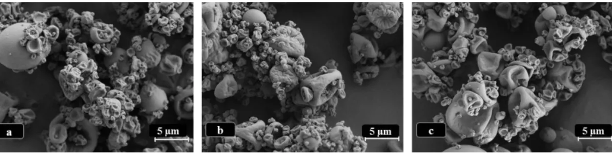
LBG microparticles, either in presence or absence of drug, presented a convoluted surface, as depicted in Figure2. No morphological alterations were perceived after drug incorporation, even when testing different concentrations of RFB, although these photographs are not shown. Many particles presented a spherical or approximately spherical shape, although strongly convoluted, while some others displayed rather irregular morphology.
Molecules 2016, 21, 702 3 of 21
2. Results and Discussion
2.1. Preparation of Locust Bean Gum (LBG) Microparticles by Spray-Drying
Obtaining a polymeric dispersion adequate for spray-drying required a great optimisation of each step, due to the high viscosity of LBG in solution. LBG is described to adopt an extended rod-like conformation in solution, occupying a large volume of gyration, which results in high viscosity [12]. A LBG dispersion at 2% (w/v) exhibits such a high viscosity that spray-drying is hampered. For the specific LBG polymer being used, Sigma-Aldrich (the provider) reports a viscosity of 2100–3750 cps for a 1% (w/v) solution, which is much greater than the ideal for an operation with the spray-drying equipment, reported by Buchi (Flawil, Switzerland) to be of 300 cps [13]. The high viscosity of LBG solutions at relatively low concentrations is reported, which is only slightly affected by pH, salts or temperature [14]. Nevertheless, in this work the addition of hydrochloric acid (HCl) 0.1 M was found to provide the adequate viscosity for spray-drying. Additionally, the grinding steps that are described in Section 3.3 and the use of hot water during LBG solubilisation were also very relevant. Several (unsuccessful) attempts were performed to use less concentrated HCl for LBG solubilisation (0.001 M and 0.01 M).The more significant reduction of viscosity in presence of HCl 0.1 M is attributed to a high concentration of H+ in solution, which causes a weak protonation of the hydroxyl groups of
water and galactomannan molecules, resulting in reduced number of hydrogen bonds. With less bonds being established between water and LBG, and between LBG molecules, there is less expansion of the LBG chain and, therefore, fewer interactions of galactomannans occur, causing a reduction of viscosity [15].
After obtaining a polymeric dispersion with adequate viscosity, LBG microparticles were successfully prepared in a one-step spray-drying process, exhibiting the characteristics that are detailed in the following section.
2.2. Association of Drugs and Characterisation of Microparticles
LBG microparticles, either in presence or absence of drug, presented a convoluted surface, as depicted in Figure 2. No morphological alterations were perceived after drug incorporation, even when testing different concentrations of RFB, although these photographs are not shown. Many particles presented a spherical or approximately spherical shape, although strongly convoluted, while some others displayed rather irregular morphology.
Figure 2. Microphotographs of LBG-based microparticles viewed by scanning electron microscopy. (a) Unloaded LBG microparticles; (b) LBG:INH = 10:1 (w/w) microparticles; (c) LBG:RFB = 10:0.5 (w/w) microparticles, the latter representative of formulations containing different amounts of RFB; INH: isoniazid, LBG: locust bean gum, RFB: rifabutin.
The observed morphologies are similar to those reported for other spray-dried polysaccharide microparticles including matrixes of hyaluronic acid-hydroxypropyl cellulose [16] and chitosan-gelatin [17]. Nevertheless, spherical shapes have also been reported for many polysaccharide-based spray-dried microparticles [18–20], evidencing the important role of the matrix material in this regard. The morphology of spray-dried microparticles is reported to strongly reflect the affinity between the polymer being sprayed and the solvent. In fact, when the droplets of polymer dispersion start drying, a film of polymer is first formed on the surface that might interfere with diffusion of water from the
Figure 2.Microphotographs of LBG-based microparticles viewed by scanning electron microscopy. (a) Unloaded LBG microparticles; (b) LBG:INH = 10:1 (w/w) microparticles; (c) LBG:RFB = 10:0.5 (w/w) microparticles, the latter representative of formulations containing different amounts of RFB; INH: isoniazid, LBG: locust bean gum, RFB: rifabutin.
The observed morphologies are similar to those reported for other spray-dried polysaccharide microparticles including matrixes of hyaluronic acid-hydroxypropyl cellulose [16] and chitosan-gelatin [17]. Nevertheless, spherical shapes have also been reported for many polysaccharide-based spray-dried
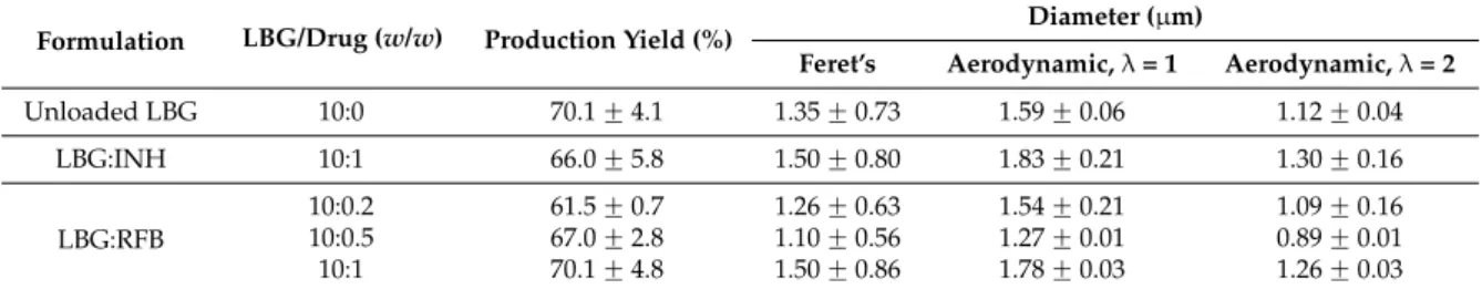
microparticles [18–20], evidencing the important role of the matrix material in this regard. The morphology of spray-dried microparticles is reported to strongly reflect the affinity between the polymer being sprayed and the solvent. In fact, when the droplets of polymer dispersion start drying, a film of polymer is first formed on the surface that might interfere with diffusion of water from the inside out. This leads to an increase of the internal pressure that might reach a critical point where the particle deforms or even disintegrates [21]. The irregular morphology observed in the microparticles of this work may improve the dispersibility and flow properties of dry powders, as surface irregularities will reduce the contact between microparticles and the possibility of establishing van der Waals forces that lead to agglomeration [22]. It is important to highlight that, from an eye observation, the presence of RFB in microparticles seemed to improve dispersibility, an effect that was concentration-dependent, as it was particularly visible for formulations LBG:RFB = 10:1 and 10:0.5 (w/w). An experimental determination of dispersibility and aerosolisation properties would be very important to reinforce this observation. Tables 1and 2display the physical and aerodynamic characteristics of LBG microparticles, along with the spray-drying yields. The former were appropriate for the intended application, with aerodynamic diameters varying between 0.89 and 1.83 µm, and Feret’s diameters ranging from 1.10 to 1.50 µm. The latter were considered good, varying between 60% and 70%, thus indicating a low loss of materials and the effectiveness of the technique. The use of the high performance cyclone instead of the conventional cyclone separator strongly contributed to the high yields, an effect that was previously reported [23].
Table 1. Spray-drying production yields and microparticle Feret’s and aerodynamic diameters (mean ˘ SD, n = 3).
Formulation LBG/Drug (w/w) Production Yield (%) Diameter (µm)
Feret’s Aerodynamic, λ = 1 Aerodynamic, λ = 2
Unloaded LBG 10:0 70.1 ˘ 4.1 1.35 ˘ 0.73 1.59 ˘ 0.06 1.12 ˘ 0.04 LBG:INH 10:1 66.0 ˘ 5.8 1.50 ˘ 0.80 1.83 ˘ 0.21 1.30 ˘ 0.16 LBG:RFB 10:0.2 61.5 ˘ 0.7 1.26 ˘ 0.63 1.54 ˘ 0.21 1.09 ˘ 0.16 10:0.5 67.0 ˘ 2.8 1.10 ˘ 0.56 1.27 ˘ 0.01 0.89 ˘ 0.01 10:1 70.1 ˘ 4.8 1.50 ˘ 0.86 1.78 ˘ 0.03 1.26 ˘ 0.03 INH: Isoniazid; LBG: Locust Bean Gum; RFB: Rifabutin; λ: shape factor (1: spherical shape; 2: irregular shape).
Table 2.Microparticle real, bulk and tap densities (mean ˘ SD, n = 3).
Formulation LBG/Drug (w/w) Density (g/cm
3)
Real Bulk Tap
Unloaded LBG 10:0 1.39 ˘ 0.01 0.24 ˘ 0.06 0.37 ˘ 0.08 LBG:INH 10:1 1.41 ˘0.02 0.24 ˘ 0.01 0.36 ˘ 0.00 LBG:RFB 10:0.2 1.41 ˘ 0.03 0.20 ˘ 0.01 0.32 ˘ 0.05 10:0.5 1.33 ˘ 0.03 0.15 ˘ 0.04 0.25 ˘ 0.07 10:1 1.39 ˘ 0.02 0.14 ˘ 0.02 0.25 ˘ 0.02
INH: Isoniazid; LBG: Locust Bean Gum; RFB: Rifabutin.
The particle size and the density of inhalable dry powders are prominent factors in the success of the formulations, because they strongly influence the sedimentation and dispersion properties [24]. Among the microparticle formulations developed in this work, no relevant differences were found on the Feret’s diameter, which varied from 1.10 µm to 1.50 µm (Table1). As expected, the inclusion of drugs had no effect on size, given the relatively low loading. Real, bulk and tap densities were also very similar amongst the formulations, with real density varying within 1.3–1.4 g/cm3(Table2), very common values for spray-dried powders [18,25]. Bulk densities varied between 0.14 and 0.25 g/cm3 and tap densities had slightly increased values in comparison with the former, as a consequence of the
method used for the determinations. The real density values, along with Feret’s diameters, resulted in theoretical aerodynamic diameters between 0.9 and 1.8 µm (Table1). While some microparticles were spherical, the majority was strongly convoluted, as referred above. When microparticles have a clear spherical shape, a shape factor of 1 should be considered [26], but for irregular morphologies the shape factor to be used should be 2 [27]. Therefore, the calculation was performed using a shape factor of either 1 or 2, in order to find an interval of expected aerodynamic diameters [27]. The aerodynamic diameters determined for antitubercular drug-loaded microparticles appear suitable for deep lung deposition upon inhalation.
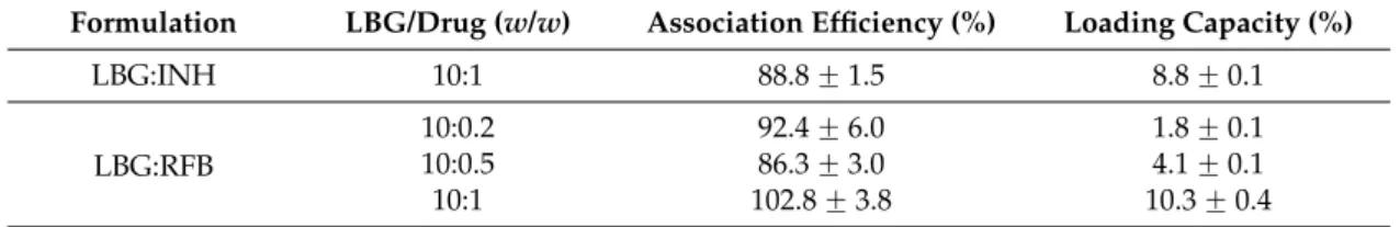
Obtaining efficient association of antitubercular drugs was one of the objectives of this work. Spray-drying is a technique usually providing high association efficiencies [28], which was reinforced in this study, where encapsulation efficiencies up to 100% were observed. Association efficiency and loading capacity of LBG microparticles are displayed in Table3.
Table 3. Drug association efficiency and microparticle loading capacity determined for LBG microparticles (mean ˘ SD, n = 3).
Formulation LBG/Drug (w/w) Association Efficiency (%) Loading Capacity (%)
LBG:INH 10:1 88.8 ˘ 1.5 8.8 ˘ 0.1
LBG:RFB
10:0.2 92.4 ˘ 6.0 1.8 ˘ 0.1
10:0.5 86.3 ˘ 3.0 4.1 ˘ 0.1
10:1 102.8 ˘ 3.8 10.3 ˘ 0.4
INH: isoniazid, LBG: locust bean gum, RFB: rifabutin.
INH and RFB were associated with approximately equal efficiency, demonstrating that the process was independent of the aqueous solubility of the drugs (125 mg/mL for INH and 0.19 mg/mL for RFB). Owing to its hydrophilic nature, INH was easily dissolved in the LBG solution and, thus, the association efficiency of 89% was not surprising, being similar to that reported for other INH-loaded spray-dried microparticles [29,30]. Such association efficiency resulted in a loading capacity around 9%. Contrary to INH, RFB is a lipophilic drug and its association to the microparticles required an optimisation of the solubilisation conditions. The presence of HCl in the dissolving medium, included to decrease the viscosity of LBG solution (Section2.1), induced the protonation of the drug that allows an increase of the solubility in water and the adequate incorporation in the LBG dispersion. The concomitant trituration of LBG and RFB powders further contributed to the successful association of RFB, demonstrating the importance of improving solid dispersions. RFB-loaded microparticles were produced with different LBG:RFB mass ratios (10:1, 10:0.5; 10:0.2) after verifying the strong cell toxicity induced by RFB. This was not observed for INH, justifying that only the formulation LBG:INH (10:1, w/w) was produced. As shown in Table3, RFB association efficiency was very high in all cases (86%–100%). The highest association efficiency (100%, p < 0.05) was observed for the formulation with the highest theoretical loading, that is, the formulation LBG:RFB = 10:1 (w/w), which also showed the highest loading capacity (10%). Although the association efficiency of the other two formulations was not significantly different (86%–92%), the loading capacity of LBG:RFB = 10:0.5 microparticles (4%) was significantly higher than that of LBG:RFB 10:0.2 microparticles (2%) because the former had a higher amount initially added of RFB (5% vs. 2%). Overall, the loading capacities are in line with the theoretical loadings initially established for the preparation of microparticles, owing to the high association efficiencies.
2.3. Cristallinity of LBG-Based Microparticles
X-ray diffraction (XRD) was used to assess LBG and unloaded LBG microparticles, drugs before and after spray-drying, and drug-loaded LBG microparticles. Diffractograms resulting from drug assessment are presented in Figures S1 and S2 (Supplementary Materials), for INH and RFB, respectively. INH was prepared as aqueous solution and RFB as acidic solution. INH (commercial
sample) displays a XRD pattern with sharp and intense peaks, denoting a highly crystalline structure. The pattern matches that described in the literature [31] and no other peaks than those belonging to the INH crystalline phase were found. The pattern recorded after the spray-drying process indicates the presence of crystalline INH. However, the different relative peak intensities, when compared with parent INH (Figure S1), suggest a different preferential orientation probably caused by drug recrystallization. The XRD pattern of RFB recorded before the spray-drying process (Figure S2) shows the typical diffraction peaks of the crystalline drug [32]. These peaks completely vanish after the spray-drying process, indicating substantial reduction of crystallinity. In this case, the spray-drying process seems to promote the formation of amorphous RFB, although the presence of small-sized crystals of RBF (not detectable by XRD), formed either by recrystallization or dehydration, cannot be discarded.
Diffractograms of LBG, unloaded LBG microparticles and drug-loaded LBG microparticles are presented in Figure 3. For the formulation containing RFB, the diffractogram of LBG:RFB 10:1 is presented, which is considered representative of the other ratios.
Molecules 2016, 21, 702 6 of 21
process seems to promote the formation of amorphous RFB, although the presence of small-sized crystals of RBF (not detectable by XRD), formed either by recrystallization or dehydration, cannot be discarded.
Diffractograms of LBG, unloaded LBG microparticles and drug-loaded LBG microparticles are presented in Figure 3. For the formulation containing RFB, the diffractogram of LBG:RFB 10:1 is presented, which is considered representative of the other ratios.
Figure 3. XRD diffractograms of locust bean gum (LBG) raw material and spray-dried formulations:
LBG raw material (brown line), unloaded LBG microparticles (green line), LBG:INH 10:1 microparticles (blue line), LBG:RFB 10:1 microparticles (red line); INH: isoniazid; LBG: locust bean gum; RFB: rifabutin.
Locust bean gum does not show diffraction peaks, in agreement with its non-crystalline nature. The diffraction patterns obtained after and before spray-drying are similar to each other, and are also similar to those described in the literature [33]. Spray-dried INH retains high crystallinity when compared with RFB, as shown in Figures S1 and S2 (Supplementary Materials). Therefore, at least the most intense diffraction peaks from INH were expected to be observed in microparticle formulations (these peaks were clearly visible in the diffractogram of a physical mixture 10:1 of LBG and INH— Figure S3). Nevertheless, in the diffractogram of LBG:INH microparticles no diffraction peaks were identified. The patterns are identical among themselves and also similar to that of the polymer. This observation is common to other spray-dried microparticles [4,30], where INH peaks disappear in INH-loaded microspheres, only peaks from the matrix material being observed. This absence of INH peaks might be due to the fact that very small crystals are formed after INH recrystallization on the matrix of LBG, which are below the detection limit of the equipment [34]. It may also be justified by the rapid process of drying occurring in spray-drying, which affects crystal rearrangement, leading to the formation of an amorphous phase [28]. On the other hand, high inlet temperatures used in the process have also been referred in the literature as possibly affecting the final structure [35]. In this regard, the production of INH-loaded microparticles was also performed at 120 °C (instead of 160 °C), but no differences in the crystallinity were found (data not shown), indicating an absence of the effect of inlet temperature in this case. The diffractogram of LBG:RFB does not show any additional diffraction peak when compared with that of unloaded LBG. This was somewhat expected, because the spray-drying process itself promotes the loss of crystallinity of RFB, as described above.
2.4. In Vitro Drug Release
The medium selected to evaluate the release of the drugs consisted of phosphate buffered saline (PBS) pH 7.4 added of 1% Tween 80®. The latter, apart from contributing to the resemblance with the
surfactant present in the lung lining fluid, is essential to enable the dissolution of RFB, which is not soluble in PBS. The presence of Tween 80® at the used concentration was reported not to affect
spectrophotometric measurements at the considered wavelength [36].
5 10 15 20 25 30 35 40 45 50 55 60
2θ (degrees)
LBG polymer Unloaded LBG LBG.INH LBG.RFB
Figure 3. XRD diffractograms of locust bean gum (LBG) raw material and spray-dried formulations: LBG raw material (brown line), unloaded LBG microparticles (green line), LBG:INH 10:1 microparticles (blue line), LBG:RFB 10:1 microparticles (red line); INH: isoniazid; LBG: locust bean gum; RFB: rifabutin.
Locust bean gum does not show diffraction peaks, in agreement with its non-crystalline nature. The diffraction patterns obtained after and before spray-drying are similar to each other, and are also similar to those described in the literature [33]. Spray-dried INH retains high crystallinity when compared with RFB, as shown in Figures S1 and S2 (Supplementary Materials). Therefore, at least the most intense diffraction peaks from INH were expected to be observed in microparticle formulations (these peaks were clearly visible in the diffractogram of a physical mixture 10:1 of LBG and INH—Figure S3). Nevertheless, in the diffractogram of LBG:INH microparticles no diffraction peaks were identified. The patterns are identical among themselves and also similar to that of the polymer. This observation is common to other spray-dried microparticles [4,30], where INH peaks disappear in INH-loaded microspheres, only peaks from the matrix material being observed. This absence of INH peaks might be due to the fact that very small crystals are formed after INH recrystallization on the matrix of LBG, which are below the detection limit of the equipment [34]. It may also be justified by the rapid process of drying occurring in spray-drying, which affects crystal rearrangement, leading to the formation of an amorphous phase [28]. On the other hand, high inlet temperatures used in the process have also been referred in the literature as possibly affecting the final structure [35]. In this regard, the production of INH-loaded microparticles was also performed at 120˝C (instead of 160˝C), but no differences in the crystallinity were found (data not shown), indicating an absence of the effect of inlet temperature in this case. The diffractogram of LBG:RFB does not show any additional diffraction peak
when compared with that of unloaded LBG. This was somewhat expected, because the spray-drying process itself promotes the loss of crystallinity of RFB, as described above.
2.4. In Vitro Drug Release
The medium selected to evaluate the release of the drugs consisted of phosphate buffered saline (PBS) pH 7.4 added of 1% Tween 80®. The latter, apart from contributing to the resemblance with the surfactant present in the lung lining fluid, is essential to enable the dissolution of RFB, which is not soluble in PBS. The presence of Tween 80®at the used concentration was reported not to affect spectrophotometric measurements at the considered wavelength [36].
The release profile of both INH and RFB from the respective microparticle formulations is plotted in Figure4. INH-loaded microparticles have the fastest release (86% in 20 min), which was expected owing to the high solubility of INH. In general, this profile is identical to that obtained from INH-loaded polylactic acid microparticles in PBS pH 7.4 [29].
Molecules 2016, 21, 702 7 of 21
The release profile of both INH and RFB from the respective microparticle formulations is plotted in Figure 4. INH-loaded microparticles have the fastest release (86% in 20 min), which was expected owing to the high solubility of INH. In general, this profile is identical to that obtained from INH-loaded polylactic acid microparticles in PBS pH 7.4 [29].
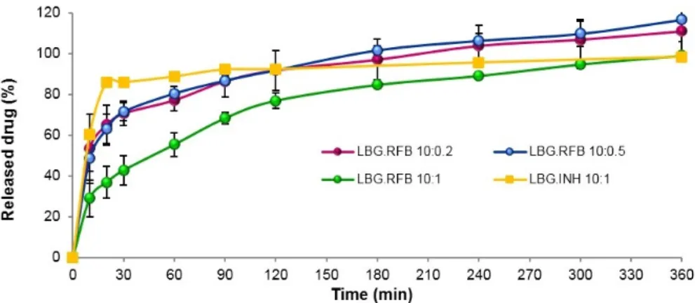
Figure 4. In vitro release of isoniazid (INH) from LBG:INH (10:1, w/w) microparticles and of rifabutin
(RFB) from LBG:RFB microparticles at mass ratios 10:0.2, 10:0.5 and 10:1, in PBS pH 7.4.Tween 80®
(LBG: locust bean gum; mean ± SD, n = 3).
The release of RFB was somewhat slower at initial time points, particularly for microparticles with the highest amount of the drug (LBG:RFB 10:1, w/w), although it still corresponds to a rapid release profile. This is the formulation permitting the most direct comparison with INH-loaded microparticles, because the same amount of drug is associated. In the same interval where LBG:INH microparticles released 86% (20 min), LBG:RFB 10:1 (w/w) microparticles released 37% (p < 0.05). This difference is certainly a consequence of the different water solubility of each drug. As said, the other two formulations containing RFB (LBG:RFB 10:0.5 and 10:0.2, w/w) comparatively showed a faster release (63% in the same period, p < 0.05), with a similar release pattern during the whole assay. Additionally, these reached 100% release at 240 min, while at that time LBG:RFB 10:1 (w/w) microparticles reached only 80%.
In spite of the slight differences detailed above, the release was generally rapid for all formulations, which is possibly due to two factors. First, the high specific surface area of these convoluted microparticles, which provides improved contact with the release medium. Moreover, a second reason can be the apparent absence or very low crystallinity pattern of both INH- and RFB-loaded microparticles that potentiates a rapid dissolution in the release medium. Nevertheless, a certain sustained release effect is observed for RFB-loaded microparticles, although it lasts for a short time, which is attributed to the gelling ability of LBG along with the lower aqueous solubility of RFB. When the assay starts and microparticles initiate the contact with the aqueous medium, the polymer matrix gradually begins to hydrate from periphery to centre, forming a gelatinous swollen mass, which progressively allows the release of drugs into the medium [37]. It is also important to stress that, although drug release is fast and it could be thought to occur before microparticle internalization by cells, real in vivo conditions of the lung are not being simulated. The alveolar zone has a much lower amount of liquid comparing with that involved in the assay and, therefore, the release rate is expected to be slower than that reported. Therefore, this experimental setup is more useful and accurate at providing a comparison between formulations than at predicting in vivo occurrences.
2.5. Cytotoxic Evaluation
The cytotoxic evaluation of LBG microparticles was performed in two cell lines of the alveolar environment, epithelial A549 cells and macrophage-differentiated THP-1 cells. The differentiation is performed with phorbol 12-myristate 13-acetate (PMA) and is reported to induce a phenotype having characteristics of alveolar macrophages [38]. Evidence of this process is presented as Supplementary Materials (Figure S4). Different in vitro tests were employed, allowing the evaluation of the cell Figure 4. In vitrorelease of isoniazid (INH) from LBG:INH (10:1, w/w) microparticles and of rifabutin (RFB) from LBG:RFB microparticles at mass ratios 10:0.2, 10:0.5 and 10:1, in PBS pH 7.4.Tween 80® (LBG: locust bean gum; mean ˘ SD, n = 3).
The release of RFB was somewhat slower at initial time points, particularly for microparticles with the highest amount of the drug (LBG:RFB 10:1, w/w), although it still corresponds to a rapid release profile. This is the formulation permitting the most direct comparison with INH-loaded microparticles, because the same amount of drug is associated. In the same interval where LBG:INH microparticles released 86% (20 min), LBG:RFB 10:1 (w/w) microparticles released 37% (p < 0.05). This difference is certainly a consequence of the different water solubility of each drug. As said, the other two formulations containing RFB (LBG:RFB 10:0.5 and 10:0.2, w/w) comparatively showed a faster release (63% in the same period, p < 0.05), with a similar release pattern during the whole assay. Additionally, these reached 100% release at 240 min, while at that time LBG:RFB 10:1 (w/w) microparticles reached only 80%.
In spite of the slight differences detailed above, the release was generally rapid for all formulations, which is possibly due to two factors. First, the high specific surface area of these convoluted microparticles, which provides improved contact with the release medium. Moreover, a second reason can be the apparent absence or very low crystallinity pattern of both INH- and RFB-loaded microparticles that potentiates a rapid dissolution in the release medium. Nevertheless, a certain sustained release effect is observed for RFB-loaded microparticles, although it lasts for a short time, which is attributed to the gelling ability of LBG along with the lower aqueous solubility of RFB. When the assay starts and microparticles initiate the contact with the aqueous medium, the polymer matrix gradually begins to hydrate from periphery to centre, forming a gelatinous swollen mass, which progressively allows the release of drugs into the medium [37]. It is also important to stress that, although drug release is fast and it could be thought to occur before microparticle internalization by
cells, real in vivo conditions of the lung are not being simulated. The alveolar zone has a much lower amount of liquid comparing with that involved in the assay and, therefore, the release rate is expected to be slower than that reported. Therefore, this experimental setup is more useful and accurate at providing a comparison between formulations than at predicting in vivo occurrences.
2.5. Cytotoxic Evaluation
The cytotoxic evaluation of LBG microparticles was performed in two cell lines of the alveolar environment, epithelial A549 cells and macrophage-differentiated THP-1 cells. The differentiation is performed with phorbol 12-myristate 13-acetate (PMA) and is reported to induce a phenotype having characteristics of alveolar macrophages [38]. Evidence of this process is presented as Supplementary Materials (Figure S4). Different in vitro tests were employed, allowing the evaluation of the cell metabolic activity (thiazolyl blue tetrazolium bromide (MTT) assay) and membrane integrity (lactate dehydrogenase (LDH) release assay) after exposure to drug formulations.
2.5.1. Evaluation of Metabolic Activity
Free drugs (INH and RFB), raw material (LBG) as obtained commercially, unloaded LBG microparticles and drug-loaded LBG microparticles were incubated with cultures of the two cell lines. A shorter (3 h) and a more prolonged period of exposure (24 h) were tested, and three concentrations of materials were assessed (0.1, 0.5 and 1 mg/mL). Considering that the theoretical drug loading of microparticles was 10%, free drugs were evaluated at 10x lower concentrations comparing with the other materials (0.01, 0.05 and 0.1 mg/mL). In all cases samples were presented as solutions/suspensions prepared in pre-warmed cell culture medium (CCM). For the discussion of results, it was considered that a material has cytotoxic potential when cell viability after exposure to the material decreases below 70% (indicated with a dashed line in all figures), as designated by the ISO 10993-5 [39].
The exposure of cells to the free drugs revealed two important aspects. INH did not show any detrimental effect on cell viability, which remained around 90%–100% in all cases, irrespective of the cell line, tested concentration and time of exposure (Figure S5). On the contrary, time- and concentration-dependent effects were observed for RFB (p < 0.05). After 3 h of exposure to this drug, cell viabilities remained above 80% (Figure S6), but a strong decrease was observed after 24 h (Figure5). This decrease was particularly noticeable for the highest concentration tested (0.1 mg/mL). There was also a trend indicating lower viabilities obtained in A549 cells, suggesting higher sensitivity of this cell line comparing with differentiated THP-1 cells upon contact with the free drugs.
Molecules 2016, 21, 702 8 of 21
metabolic activity (thiazolyl blue tetrazolium bromide (MTT) assay) and membrane integrity (lactate dehydrogenase (LDH) release assay) after exposure to drug formulations.
2.5.1. Evaluation of Metabolic Activity
Free drugs (INH and RFB), raw material (LBG) as obtained commercially, unloaded LBG microparticles and drug-loaded LBG microparticles were incubated with cultures of the two cell lines. A shorter (3 h) and a more prolonged period of exposure (24 h) were tested, and three concentrations of materials were assessed (0.1, 0.5 and 1 mg/mL). Considering that the theoretical drug loading of microparticles was 10%, free drugs were evaluated at 10x lower concentrations comparing with the other materials (0.01, 0.05 and 0.1 mg/mL). In all cases samples were presented as solutions/suspensions prepared in pre-warmed cell culture medium (CCM). For the discussion of results, it was considered that a material has cytotoxic potential when cell viability after exposure to the material decreases below 70% (indicated with a dashed line in all figures), as designated by the ISO 10993-5 [39].
The exposure of cells to the free drugs revealed two important aspects. INH did not show any detrimental effect on cell viability, which remained around 90%–100% in all cases, irrespective of the cell line, tested concentration and time of exposure (Figure S5). On the contrary, time- and concentration-dependent effects were observed for RFB (p < 0.05). After 3 h of exposure to this drug, cell viabilities remained above 80% (Figure S6), but a strong decrease was observed after 24 h (Figure 5). This decrease was particularly noticeable for the highest concentration tested (0.1 mg/mL). There was also a trend indicating lower viabilities obtained in A549 cells, suggesting higher sensitivity of this cell line comparing with differentiated THP-1 cells upon contact with the free drugs.
LBG and LBG-based microparticles were evaluated separately, in order to disclose an effect from the carrier structure [40]. Testing LBG is not only a relevant control of the work, but also contributes to the state of the art, as no similar evaluation is, to our knowledge, described in the literature.
Figure 5. A549 (lighter colour) and macrophage-differentiated THP-1 (darker colour) cell viabilities
after 24 h of exposure to free drugs, isoniazid (blue) and rifabutin (red). Results are expressed as mean ± SEM (n = 3, six replicates per experiment at each concentration). Dashed line represents 70% cell viability.
Again, while no overt toxicity was found in THP-1 cells (cell viability >85% in all conditions), A549 cells demonstrated to be more sensitive, showing 40% cell viability after 24 h of exposure independently of the concentration (Figure S7). Interestingly, this detrimental effect completely reverted after spray-drying, as unloaded LBG microparticles exhibited a very mild effect even after 24 h (A549 cell viability >75% in all cases, Figure 6).
Figure 5.A549 (lighter colour) and macrophage-differentiated THP-1 (darker colour) cell viabilities after 24 h of exposure to free drugs, isoniazid (blue) and rifabutin (red). Results are expressed as mean ˘ SEM (n = 3, six replicates per experiment at each concentration). Dashed line represents 70% cell viability.
LBG and LBG-based microparticles were evaluated separately, in order to disclose an effect from the carrier structure [40]. Testing LBG is not only a relevant control of the work, but also contributes to the state of the art, as no similar evaluation is, to our knowledge, described in the literature.
Again, while no overt toxicity was found in THP-1 cells (cell viability >85% in all conditions), A549 cells demonstrated to be more sensitive, showing 40% cell viability after 24 h of exposure independently of the concentration (Figure S7). Interestingly, this detrimental effect completely reverted after spray-drying, as unloaded LBG microparticles exhibited a very mild effect even after 24 h (A549 cell viability >75% in all cases, Figure6).
Molecules 2016, 21, 702 9 of 21
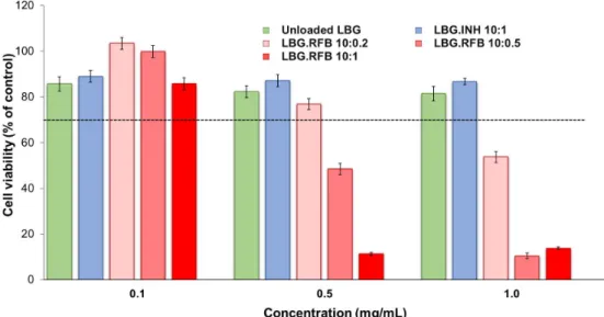
Figure 6. A549 cell viabilities after 24 h of exposure to LBG-based microparticle formulations. Results are expressed as mean ± SEM (n = 3, six replicates per experiment at each concentration). Dashed line represents 70% cell viability (INH: isoniazid, LBG: locust bean gum, RFB: rifabutin).
The described effect of LBG as raw material after the contact with A549 cells could be attributed to either of two reasons: (i) the occurrence of a partial hydrolysis of commercial LBG in the acidic solution either prior to spray-drying or potentiated by the heat and shear forces of the process, leading to lower molecular weight polymer chains; or (ii) a slower solubilisation of the microparticles when compared to the commercial powder, which delays the increase of viscosity in the solution to be tested. This translates directly to an effect at the level of the viscosity of dispersions prepared at the same concentration using LBG commercial powder and unloaded LBG microparticles. As the assay is performed by incubating a dispersion of polymer/microparticles with cells, higher viscosity of the dispersion possibly makes gaseous exchanges between cells, medium and air a difficult task. This could lead to higher cell death. In fact, the dispersion obtained from LBG microparticles was perceived as more fluid than that prepared from commercial LBG polymer.
Concerning macrophage-like THP-1 cells, no relevant variation of cell viability was observed upon exposure to either a dispersion of commercial LBG (Figure S7) or unloaded LBG microparticles (Figure 7). This observation is again in line with a higher sensitivity of A549 cells to the materials, comparing with THP-1 cells.
Figure 7. THP-1 cell viabilities after 24 h of exposure to LBG-based microparticle formulations. Results are expressed as mean ± SEM (n = 3, six replicates per experiment at each concentration). Dashed line represents 70% cell viability (INH: isoniazid, LBG: locust bean gum, RFB: rifabutin).
Figure 6.A549 cell viabilities after 24 h of exposure to LBG-based microparticle formulations. Results are expressed as mean ˘ SEM (n = 3, six replicates per experiment at each concentration). Dashed line represents 70% cell viability (INH: isoniazid, LBG: locust bean gum, RFB: rifabutin).
The described effect of LBG as raw material after the contact with A549 cells could be attributed to either of two reasons: (i) the occurrence of a partial hydrolysis of commercial LBG in the acidic solution either prior to spray-drying or potentiated by the heat and shear forces of the process, leading to lower molecular weight polymer chains; or (ii) a slower solubilisation of the microparticles when compared to the commercial powder, which delays the increase of viscosity in the solution to be tested. This translates directly to an effect at the level of the viscosity of dispersions prepared at the same concentration using LBG commercial powder and unloaded LBG microparticles. As the assay is performed by incubating a dispersion of polymer/microparticles with cells, higher viscosity of the dispersion possibly makes gaseous exchanges between cells, medium and air a difficult task. This could lead to higher cell death. In fact, the dispersion obtained from LBG microparticles was perceived as more fluid than that prepared from commercial LBG polymer.
Concerning macrophage-like THP-1 cells, no relevant variation of cell viability was observed upon exposure to either a dispersion of commercial LBG (Figure S7) or unloaded LBG microparticles (Figure7). This observation is again in line with a higher sensitivity of A549 cells to the materials, comparing with THP-1 cells.
The results of the cytotoxic evaluation of drug-loaded LBG microparticles are presented in Figures6and7representing the 24 h exposure of A549 and macrophage-differentiated THP-1 cells, respectively. The results obtained after a short contact time (3 h) are available as Supplementary Materials (Figures S8 and S9) and show similar tendencies, although in some cases the determined cell viabilities were higher than those at 24 h, indicating a time-dependent effect.
The general trend indicates that both cell lines responded to the presence of drug-loaded formulations according to a similar pattern, that is, INH-loaded microparticles induced low effect
Molecules 2016, 21, 702 10 of 22
on cell viability and RFB-loaded microparticles elicited considerable cytotoxicity. More specifically, INH-loaded microparticles induced cell viabilities above 70% in all tested conditions (exposure times, concentrations and cell lines) with the exception of the 24 h exposure of A549 cells to concentrations of 0.5 and 1 mg/mL, which resulted in viabilities of 66%–68% (Figure6). It was also observed an absence of concentration-dependent effect for this formulation, at 3 h and 24 h. The only exception occurs in A549 cells after 24 h exposure, where 0.1 mg/mL of INH-loaded microparticles induced 92% cell viability, contrasting with 66%–68% when testing 0.5 and 1 mg/mL (p < 0.05). Worth mentioning is the fact that responses to INH-loaded microparticles were very similar to those generated by unloaded LBG microparticles in both cell lines, giving a clear indication on the absence of toxicity of INH itself. No overt cytotoxic effect was, thus, considered to occur for INH-loaded microparticles. These observations are in line with the literature, as the IC50of INH is reported as 1000 mg/mL in alveolar macrophages (isolated from albino rats) [41]. The referred study was performed in primary cells, thus different from those used in this study, but they mimic in vivo conditions in a closer way and are supposedly more sensitive than established cell lines [38].
Figure 6. A549 cell viabilities after 24 h of exposure to LBG-based microparticle formulations. Results are expressed as mean ± SEM (n = 3, six replicates per experiment at each concentration). Dashed line represents 70% cell viability (INH: isoniazid, LBG: locust bean gum, RFB: rifabutin).
The described effect of LBG as raw material after the contact with A549 cells could be attributed to either of two reasons: (i) the occurrence of a partial hydrolysis of commercial LBG in the acidic solution either prior to spray-drying or potentiated by the heat and shear forces of the process, leading to lower molecular weight polymer chains; or (ii) a slower solubilisation of the microparticles when compared to the commercial powder, which delays the increase of viscosity in the solution to be tested. This translates directly to an effect at the level of the viscosity of dispersions prepared at the same concentration using LBG commercial powder and unloaded LBG microparticles. As the assay is performed by incubating a dispersion of polymer/microparticles with cells, higher viscosity of the dispersion possibly makes gaseous exchanges between cells, medium and air a difficult task. This could lead to higher cell death. In fact, the dispersion obtained from LBG microparticles was perceived as more fluid than that prepared from commercial LBG polymer.
Concerning macrophage-like THP-1 cells, no relevant variation of cell viability was observed upon exposure to either a dispersion of commercial LBG (Figure S7) or unloaded LBG microparticles (Figure 7). This observation is again in line with a higher sensitivity of A549 cells to the materials, comparing with THP-1 cells.
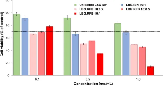
Figure 7. THP-1 cell viabilities after 24 h of exposure to LBG-based microparticle formulations. Results are expressed as mean ± SEM (n = 3, six replicates per experiment at each concentration). Dashed line represents 70% cell viability (INH: isoniazid, LBG: locust bean gum, RFB: rifabutin).
Figure 7.THP-1 cell viabilities after 24 h of exposure to LBG-based microparticle formulations. Results are expressed as mean ˘ SEM (n = 3, six replicates per experiment at each concentration). Dashed line represents 70% cell viability (INH: isoniazid, LBG: locust bean gum, RFB: rifabutin).
The response to RFB-loaded microparticles contrasted well with the previous results. After a contact of 3 h, a reduction of cell viability in both lines to values around 40%–50% and even 20% (THP-1 cells), particularly for the concentrations of 0.5 and 1.0 mg/mL (Figures S8 and S9), was observed. After 24 h, cell viabilities were generally not very different from those at 3 h, with the relevant exception corresponding to the highest concentration of microparticles (1 mg/mL), namely for LBG:RFB = 10:1 (w/w) microparticles (Figures6and7). In these conditions, cell viabilities of 10%–15% were determined (p < 0.05). In A549 cells, a time-dependent effect was observed as a whole (p < 0.05), although it was more pronounced for microparticles LBG:RFB 10:1 (w/w). A significant difference was also perceived between the various RFB-loaded microparticles (p < 0.05), in the order LBG:RFB = 10:1 > 10:0.5~10:0.2, reinforcing that the cytotoxic effect is due to RFB. THP-1 cells also revealed a time-dependent effect (p < 0.05), which was particularly visible for microparticles LBG:RFB = 10:1 and 10:0.5 (w/w) and for the two highest concentrations tested (0.5 and 1 mg/mL). A dose-dependent effect was also visible for RFB-loaded microparticles (p < 0.05), which was more pronounced than that observed for A549 cells.
The above mentioned trend, indicating higher susceptibility of A549 cells when compared with THP-1 cells, was thus not followed when testing microparticles, where relatively similar responses are
observed between both cell lines. The literature reports opposite effects, either demonstrating higher resistance of differentiated THP-1 cells [42,43] or establishing lower susceptibility for A549 cells [44], clearly indicating that the generated responses are strongly dependent on the assessed materials. What is clearly seen in our study is that there was a difference of susceptibilities when testing free drugs and polymer, which are exposed as solutions, comparing with microparticles. This different outcome is probably related with specific endocytic–exocytic mechanisms of phagocytic and non-phagocytic cells, along with the specialized physiological role of each particular cell type. THP-1 cells are phagocytes with significant endocytic and exocytic activity and natural ability to uptake particulate matter [43]. Therefore, they possibly respond with higher intensity to the ingestion of particulates [44]. On the contrary, epithelial cells possibly have more intimate contact with dissolved solutes than with the corresponding particulates.
2.5.2. Evaluation of Cell Membrane Integrity
As a complementary study, cell membrane integrity was evaluated upon exposure to the different materials. Taking into account the results obtained in the MTT assay, released LDH was determined after 24 h of exposure to the highest concentration tested, 1 mg/mL. For RFB-loaded microparticles, the concentration of 0.5 mg/mL was also tested. RFB was tested as free drug at the concentrations of 0.05 and 0.1 mg/mL while free INH was only tested at 0.1 mg/mL.
The results of free drugs are available as Supplementary Materials (Figure S10), being in agreement with those of the MTT assay for both cell lines and reinforcing the observations of RFB cytotoxicity. While the contact with INH did not increase significantly the release of LDH, RFB induced an increase to 150%–170%, which was particularly noticeable for the highest concentration of the drug (0.1 mg/mL). The assessment of the effect of LBG microparticles was also in line with MTT results. Figures8and9 show the observations performed in A549 and macrophage-like THP-1 cells, respectively. The first remarkable observation was related with the effect of LBG as raw material. The MTT assay had shown very different outcomes between the raw material and the unloaded microparticles. However, these differences did not appear at the level of LDH release, as the amount of released enzyme was similar in both cases, for both cell lines. Therefore, although cell death was observed after the MTT assay, it was not related with events at the level of membrane integrity. Additionally, it is important to mention that the amount of enzyme that was released was comparable or even lower than that observed for the control (cells incubated with culture medium).
Molecules 2016, 21, 702 11 of 21
after 24 h of exposure to the highest concentration tested, 1 mg/mL. For RFB-loaded microparticles, the concentration of 0.5 mg/mL was also tested. RFB was tested as free drug at the concentrations of 0.05 and 0.1 mg/mL while free INH was only tested at 0.1 mg/mL.
The results of free drugs are available as Supplementary Materials (Figure S10), being in agreement with those of the MTT assay for both cell lines and reinforcing the observations of RFB cytotoxicity. While the contact with INH did not increase significantly the release of LDH, RFB induced an increase to 150%–170%, which was particularly noticeable for the highest concentration of the drug (0.1 mg/mL). The assessment of the effect of LBG microparticles was also in line with MTT results. Figures 8 and 9 show the observations performed in A549 and macrophage-like THP-1 cells, respectively. The first remarkable observation was related with the effect of LBG as raw material. The MTT assay had shown very different outcomes between the raw material and the unloaded microparticles. However, these differences did not appear at the level of LDH release, as the amount of released enzyme was similar in both cases, for both cell lines. Therefore, although cell death was observed after the MTT assay, it was not related with events at the level of membrane integrity. Additionally, it is important to mention that the amount of enzyme that was released was comparable or even lower than that observed for the control (cells incubated with culture medium).
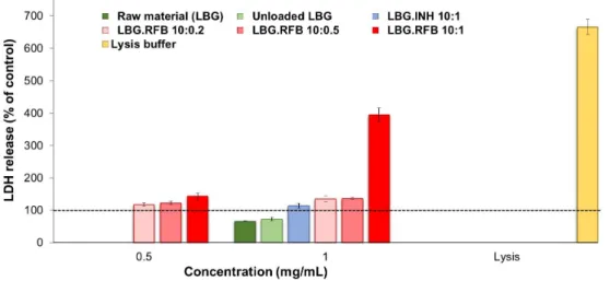
Figure 8. LDH released from A549 cells after 24 h exposure to LBG polymer, LBG microparticles and lysis buffer. Amount of LDH released from cells incubated with cell culture medium is assumed as 100% (dashed line). Results are expressed as mean ± SEM (n = 3, six replicates per experiment at each concentration; INH: isoniazid; LBG: locust bean gum; RFB: rifabutin).
Figure 9. LDH released from macrophage-differentiated THP-1 cells after 24 h exposure to LBG polymer, LBG microparticles and lysis buffer. Amount of LDH released from cells incubated with cell culture medium is assumed as 100% (dashed line). Results are expressed as mean ± SEM (n = 3, six replicates per experiment at each concentration; INH: isoniazid; LBG: locust bean gum. RFB: rifabutin). Figure 8.LDH released from A549 cells after 24 h exposure to LBG polymer, LBG microparticles and lysis buffer. Amount of LDH released from cells incubated with cell culture medium is assumed as 100% (dashed line). Results are expressed as mean ˘ SEM (n = 3, six replicates per experiment at each concentration; INH: isoniazid; LBG: locust bean gum; RFB: rifabutin).
Molecules 2016, 21, 702 12 of 22 after 24 h of exposure to the highest concentration tested, 1 mg/mL. For RFB-loaded microparticles, the concentration of 0.5 mg/mL was also tested. RFB was tested as free drug at the concentrations of 0.05 and 0.1 mg/mL while free INH was only tested at 0.1 mg/mL.
The results of free drugs are available as Supplementary Materials (Figure S10), being in agreement with those of the MTT assay for both cell lines and reinforcing the observations of RFB cytotoxicity. While the contact with INH did not increase significantly the release of LDH, RFB induced an increase to 150%–170%, which was particularly noticeable for the highest concentration of the drug (0.1 mg/mL). The assessment of the effect of LBG microparticles was also in line with MTT results. Figures 8 and 9 show the observations performed in A549 and macrophage-like THP-1 cells, respectively. The first remarkable observation was related with the effect of LBG as raw material. The MTT assay had shown very different outcomes between the raw material and the unloaded microparticles. However, these differences did not appear at the level of LDH release, as the amount of released enzyme was similar in both cases, for both cell lines. Therefore, although cell death was observed after the MTT assay, it was not related with events at the level of membrane integrity. Additionally, it is important to mention that the amount of enzyme that was released was comparable or even lower than that observed for the control (cells incubated with culture medium).
Figure 8. LDH released from A549 cells after 24 h exposure to LBG polymer, LBG microparticles and lysis buffer. Amount of LDH released from cells incubated with cell culture medium is assumed as 100% (dashed line). Results are expressed as mean ± SEM (n = 3, six replicates per experiment at each concentration; INH: isoniazid; LBG: locust bean gum; RFB: rifabutin).
Figure 9. LDH released from macrophage-differentiated THP-1 cells after 24 h exposure to LBG polymer, LBG microparticles and lysis buffer. Amount of LDH released from cells incubated with cell culture medium is assumed as 100% (dashed line). Results are expressed as mean ± SEM (n = 3, six replicates per experiment at each concentration; INH: isoniazid; LBG: locust bean gum. RFB: rifabutin). Figure 9. LDH released from macrophage-differentiated THP-1 cells after 24 h exposure to LBG polymer, LBG microparticles and lysis buffer. Amount of LDH released from cells incubated with cell culture medium is assumed as 100% (dashed line). Results are expressed as mean ˘ SEM (n = 3, six replicates per experiment at each concentration; INH: isoniazid; LBG: locust bean gum. RFB: rifabutin).
As such, it was also verified that the amount of released LDH induced by the contact with INH-loaded microparticles was, in both cell lines, similar to that induced by unloaded microparticles, raw material and the control, again evidencing an absence of toxicity of INH.
The observations were very different for RFB-loaded microparticles, justifying the assessment of two concentrations of these microparticles. In A549 cells, the three RFB-loaded microparticles induced similar effect when tested at the lower concentration (0.5 mg/mL), with only a slight increase in LDH release to 110%–140%. Interestingly, the cells responded in a similar manner to a doubled concentration (1 mg/mL) of LBG:RFB 10:0.5 and 10:0.2 (w/w) microparticles, with no significant alterations in LDH release. However, LBG:RFB 10:1 (w/w) microparticles elicited a stronger increase in LDH release (p < 0.05) to approximately 400%, indicating a clear concentration-dependent effect. The exposure to the lysis buffer, indicating the highest LDH amount possibly released, induced 666%.
The trend was approximately similar in THP-1 cells. LBG:RFB 10:0.2 (w/w) microparticles induced 125%–150% LDH release at both concentrations tested. A clear concentration-dependent effect was observed for the formulation 10:0.5 (w/w), with LDH release increasing from 139% to 223% with the increase of concentration (p < 0.05). LBG:RFB 10:1 (w/w) microparticles induced similar release at both concentrations (220%–230%). The lysis buffer induced a value around 350%.
The general observation from the whole set of results of cytotoxicity is that an increased toxic effect is seen when RFB is included in the microparticles. The high in vivo toxicity of RFB is well reported [45]. Although the mechanism is still not well established, the cytotoxic behavior might be due in part to the lipophilic character of RFB. It presents a high membrane lipid tropism, resulting in high penetration into the cells, which can impose increased toxicity [46].
Notwithstanding the determinations that were made in this study, there is the expectation that the toxicity does not translate to such a severe level in vivo. This belief is based on the assumption of a relatively even distribution of the dry powder in the alveolar zone upon inhalation. The highest dose tested in the described assays (1 mg/mL) corresponds to 303.03 µg/cm2. The area of the epithelial surface of alveolar zone is about 70 m2(700,000 cm2) [47]. If an even distribution of microparticles is assumed and it is considered that a nominal proportion (e.g., a third) of an inhaled dose (considering the approximate 160 mg of powder delivered by the TOBI® Podhaler in one dose) deposits in the alveolar region, the dose estimated across this area is 0.08 µg/cm2. This dose is remarkably lower than the 303.03 µg/cm2used in our study. However, one should bear in mind that the lung of tuberculosis patients possibly has a much lower area, apart from certainly being variable among patients. Still, even
if only 10% of the alveolar area is considered functional (7 m2), the dose will be 0.76 µg/cm2, very far from that used in this study. Taking this into account, the effects will be much closer to those of the lower dose of 0.1 mg/mL than to those of the higher dose or even the 0.5 mg/mL. Unfortunately, we could not meet the conditions permitting weighing such a low amount of dry powder that could resemble in a better way the in vivo conditions. Another reason contributing to a possible decrease of in vivo toxicological effects associated with the microparticles is their capacity to form complexes with polar heads of groups of the phospholipids present in the pulmonary surfactant, allowing reaching high concentrations without damaging the epithelium [48].
Several other issues related to the toxicity need to be discussed and addressed experimentally in the near future in order to verify the real possibilities of using LBG microparticles for the proposed application. LBG has been clearly referred as biodegradable when administered orally, owing to the presence of β-mannosidase in the human intestine [49]. Importantly, the enzyme has also been detected in the lung, although in lower concentration comparing with other organs [50]. As long-term dosing is needed in the application focused in this work, it is very important to ensure the biodegradability of the microparticles and, although the presence of β-mannosidase is a promising indication, more studies are needed in this regard. In parallel, it is important to unveil the immunological effects of these microparticles, as LBG is a novel polymer in lung delivery and polysaccharides are particularly susceptible in this regard. We are currently performing in vivo assays to verify the immunological response to the administration of LBG microparticles by inhalation.
2.6. Preliminary Evaluation of Macrophage Ability to Uptake LBG Microparticles
The ability of LBG microparticles to be taken up by alveolar macrophages is crucial to allow the co-localisation of microparticles with the tuberculosis pathogenic agent. Microparticle uptake was evaluated in two macrophage cell lines (human macrophage-differentiated THP-1 cells and rat alveolar macrophages NR8383). The assay was performed by flow cytometry, requiring the use of fluorescently-labelled LBG microparticles. LBG was labelled with fluorescein (fluorescein sodium salt was activated by N-(3-dimethylaminopropyl)-N1-ethylcarbodiimide hydrochloride (EDAC), at pH 4, and reacted with the nucleophilic hydroxyl groups of LBG, resulting in a fluorescent polymer). This was then used to produce microparticles specifically for this assay, with Feret’s diameter similar to that of unloaded LBG microparticles. In the analysis, cells exhibiting fluorescence were assumed to have phagocytosed microparticles. Two different doses were tested (50 and 220 µg/cm2) and the contact with the cells was allowed for 2 h. Phagocytosis is a fast process, usually 50%–75% of the particles are phagocytosed in 2–3 h, 90% or more in 10 h, and nearly 100% at 24 h after particle deposition [51]. The time of 2 h, selected to perform the assay, is deemed adequate for the occurrence of phagocytosis and was also used by other authors [52].
Macrophage-differentiated THP-1 cells were exposed to the two microparticle concentrations referred above. The percentage of macrophages taking up LBG microparticles was very high in both cases (99.6 ˘ 0.2% for 220 µg/cm2and 99.5 ˘ 0.4% for 50 µg/cm2). This evidenced an absence of effect of concentration and suggests a high affinity of macrophages for LBG microparticles. Using such different concentrations has been reported to permit the observation of dose-dependent effects on phagocytosis of microparticles of other materials [53], but this did not occur for LBG. Considering these results, NR8383 cells were exposed only to the lowest concentration of microparticles and results are depicted in Figure10.
As indicated above for differentiated THP-1 cells, rat alveolar macrophages also showed high affinity for LBG microparticles, as 94.4% of the macrophages of the population exhibited fluorescent signal, indicating the occurrence of phagocytosis. No significant differences were found between the uptake by both cells. The graphics depicting the analysis of the populations corresponding to cells of each line not exposed to fluorescently-labelled LBG microparticles (control, incubated with CCM) and cells exposed to fluorescently labelled microparticles, are available as Supplementary Materials