UNIVERSIDADE DA BEIRA INTERIOR
Ciências da Saúde
Efeito do Consumo Diário de Chá Branco no
Córtex Cerebral de Ratos Pré-diabéticos
Ana Raquel Mendes da Paula Nunes
Dissertação para a Obtenção de Grau de Mestre em
Ciências Biomédicas
(2º Ciclo de Estudos)
Orientadora: Prof. Doutora Branca M. Silva (CICS-UBI)
Co-Orientador: Prof. Doutor Marco G. Alves (CICS-UBI)
Co-Orientadora: Prof. Doutora Paula I. Moreira (CNC-UC)
O conteúdo do presente trabalho é da exclusiva responsabilidade do autor: __________________________________________________
“Acredita no melhor... tem um objetivo para o melhor, nunca fiques satisfeito com menos que o teu melhor, dá o teu melhor, e a longo prazo as coisas correrão pelo melhor.” (Henry Ford)
Agradecimentos
A realização desta Dissertação de Mestrado foi possível graças ao contributo das mais diversas formas, de todos os que, direta ou indiretamente, me ajudaram a cumprir os objetivos desta etapa da minha formação académica. O meu sincero obrigado!
À minha orientadora, Professora Doutora Branca M. Silva, pela sua competência científica, acompanhamento e empenho no desenvolvimento deste trabalho, assim como pelas críticas, correções e sugestões relevantes feitas durante a sua orientação.
Ao meu co-orientador, Professor Doutor Marco G. Alves, por toda a disponibilidade, apoio, palavras de incentivo, correções e sugestões feitas ao longo deste ano. Obrigado por mostrar que os obstáculos ultrapassam-se com determinação, espírito de sacrífico, dedicação e atitude, e que com humildade e competência, se pode chegar muito longe.
À minha co-orientadora, Professora Doutora Paula I. Moreira, pela competência científica, sugestões, correções, disponibilidade e simpatia sempre demonstradas.
Ao Professor Doutor Pedro F. Oliveira, pela disponibilidade demonstrada, correções, sugestões, e pelo trabalho incansável sobretudo no tratamento de resultados.
À Professora Doutora Ana Clara Cristóvão, pela disponibilidade e ajuda na dissecação dos cérebros de rato e recolha das amostras, e pelas importantes elucidações sobre a anatomia e fisiologia cerebral.
Ao Gonçalo Tomás, por toda a ajuda e apoio que teve desde o início do trabalho prático. Aos meus restantes colegas de doutoramento: Tânia Dias, Raquel Bernardino, Tito Jesus, Luís Pedro Rato e Ana Martins por toda a ajuda, disponibilidade e acompanhamento no laboratório, e pelos bons e agradáveis momentos que passámos juntos. Aos meus restantes colegas de laboratório pela ajuda sempre que necessária e pelo bom ambiente de trabalho.
À Vanessa Conde e à Cátia Rocha, pelo suporte, amizade, carinho, compreensão e ajuda que sempre demonstraram durante estes anos da nossa vida académica. Foram, sem dúvida, imprescindíveis em todos os momentos, principalmente nos mais difíceis, tanto profissional como pessoalmente. Obrigado pelos fantásticos momentos que passámos juntas e por fazerem com que cada dia valesse a pena.
Aos meus amigos, pela amizade, preocupação, ajuda e pelos bons momentos que me proporcionaram. Um especial obrigado à Inês Serra, pela amizade sempre demonstrada ao longo de todos estes anos e por todos os momentos que vivemos. Sem o teu apoio não seria possível ultrapassar os momentos menos bons.
À Helena, ao Nelson e à Joana, por serem como uma segunda família e um apoio constante a cada dia.
Ao João Dias, pelo exemplo de competência, esforço e dedicação. Obrigado por todo o amor, compreensão, suporte, paciência e palavras de encorajamento e confiança. Em parte, devo-te o que hoje sou.
À minha família, particular e especialmente, ao meus pais, ao meu irmão e aos meus avós, pelo suporte financeiro e emocional, pelo amor e paciência que sempre tiveram comigo. Obrigado por nunca terem deixado de acreditar em mim.
Resumo
A Diabetes Mellitus (DM) é um problema de saúde pública e a sua incidência está a aumentar drasticamente. O cérebro, especialmente o córtex cerebral, é muito suscetível a flutuações dos níveis de glucose e ao stress oxidativo induzido pela hiperglicémia. O chá (Camellia sinensis (L.)) é amplamente consumido, porém as propriedades antidiabéticas e neuroprotetoras do chá branco permanecem por explorar. Neste trabalho investigámos os efeitos do consumo diário de chá branco no córtex cerebral de ratos pré-diabéticos. Os animais foram divididos aleatoriamente em três grupos: grupo controlo, grupo de ratos pré-diabéticos que ingeriram água e grupo de ratos pré-pré-diabéticos que ingeriram chá branco. O perfil metabólico do córtex foi avaliado e a expressão dos níveis dos transportadores de glucose (GLUTs), da fosfofrutoquinase-1, da lactato desidrogenase (LDH) e do transportador de monocarboxilato 4 foram também determinados. O perfil oxidativo do córtex foi obtido através da avaliação do seu poder antioxidante (ensaio FRAP) e dos níveis de peroxidação lipídica (ensaio TBARS) e de oxidação proteica (ensaio dos grupos carbonilo). A catalase e a glutationa, bem como o conteúdo em glutamato, N-acetilaspartato, aspartato, colina, ácido gama-aminobutírico, taurina e valina foram também determinados. Embora o consumo diário de chá branco não tenha diminuído os níveis de glicémia no sangue, melhorou a tolerância à glucose e a sensibilidade à insulina dos ratos pré-diabéticos. Além disso, o consumo diário do chá branco alterou o perfil glicolítico do córtex cerebral dos ratos pré-diabéticos, modulando a expressão dos GLUTs e o conteúdo em lactato e alanina. O consumo de chá branco também foi capaz de restabelecer os níveis de oxidação proteica e de peroxidação lipídica para valores controlo, no córtex de ratos pré-diabéticos. No geral, o córtex cerebral de ratos pré-diabéticos que consumiram chá branco apresentou uma maior capacidade antioxidante e uma expressão normalizada da catalase. Concluindo, o consumo diário de chá branco por ratos pré-diabéticos melhora o perfil metabólico e oxidativo do seu córtex cerebral, sugerindo que a ingestão de chá branco pode ser uma estratégia boa, segura e económica para evitar os efeitos relacionados com a DM no córtex cerebral.
Palavras-chave
Resumo Alargado
A Diabetes Mellitus (DM) representa uma das maiores ameaças à saúde pública nas sociedades modernas. A sua incidência tem vindo a aumentar drasticamente e, de acordo com a Organização Mundial de Saúde, estima-se que esta doença vá afetar cerca de 300 milhões de pessoas em 2025. Estes números tendem a agravar-se devido a alguns fatores de risco relacionados com o estilo de vida, tais como o excesso de peso, a dieta desequilibrada ou o hábito de fumar. A DM é uma doença metabólica caracterizada por hiperglicémia resultante de defeitos na secreção e/ou ação da insulina. A doença pode ser dividida em dois tipos: a DM tipo 1 (T1DM) e a DM tipo 2 (T2DM). A T1DM tem geralmente o seu desenvolvimento numa idade jovem, e é causada pela destruição autoimune das células beta pancreáticas, pelo que requer uma terapia diária de reposição de insulina. Por sua vez, a T2DM é a forma mais comum da doença, sendo responsável por 95% de todos os casos de DM. Atualmente, afeta crianças, adolescentes e adultos jovens que irão enfrentar o fardo da doença por períodos prolongados. A T2DM ocorre quando as células beta pancreáticas não conseguem produzir insulina suficiente para manter os níveis de glucose no sangue dentro dos parâmetros normais.
O diagnóstico da DM é complexo, o que levou a que fosse estabelecido um estado intermédio denominado “pré-diabetes”. O estado pré-diabético antecede o aparecimento da doença propriamente dita, e a sua prevalência está a aumentar entre os jovens. Contudo, pode ser reversível. A intolerância à glucose, bem como a resistência à insulina e os níveis de glucose no sangue acima do normal estão associados a este estado. Indivíduos pré-diabéticos têm importantes alterações metabólicas que propiciam a progressão para T2DM. A transição do estado pré-diabético para T2DM ocorre quando a capacidade secretora das células beta pancreáticas não é capaz de compensar a resistência à insulina.
O comprometimento do metabolismo da glucose, tanto numa fase inicial como numa fase mais tardia da diabetes, afeta vários órgãos, incluindo o cérebro. Sabe-se que o cérebro utiliza a glucose como a sua principal fonte de energia e, assim, é expectável que a disfunção no metabolismo da glucose leve a danos cerebrais graves. Na verdade, a hiperglicémia, mesmo que transitória, pode provocar efeitos deletérios sobre a função cerebral. Elevados níveis de glucose sanguínea afetam, de modo diferente, várias regiões do cérebro, sendo o córtex cerebral particularmente sensível. Tem sido descrito que os neurónios corticais e os astrócitos são mais vulneráveis à desregulação do metabolismo glicolítico do que as células do corpo estriado ou do hipocampo. Por outro lado, o córtex apresenta reduzidas defesas antioxidantes, tornando-o muito suscetível ao stress oxidativo
(OS). O OS ocorre quando existe um desequilíbrio entre a formação de espécies reativas de oxigénio (ROS) e a proteção contra as mesmas. Embora as ROS sejam importantes em diversos processos biológicos, quando presentes em excesso, causam danos severos nos ácidos nucleicos, nos lípidos, nas membranas celulares e nas proteínas. Assim, é importante manter os níveis de ROS controlados. Além disso, o córtex cerebral é afetado em várias doenças neurodegenerativas, pelo que a hiperglicémia pode desempenhar um papel importante no desenvolvimento e progressão das mesmas.
A DM é uma doença incurável e a limitação das terapias existentes para o seu tratamento e das suas complicações, tem encorajado a procura de alternativas mais eficientes, associadas a alterações na dieta e estilos de vida. Nos últimos anos, é notório o aumento crescente do interesse pelos produtos nutracêuticos, como complemento ou até mesmo substituição de terapias atuais para a prevenção e tratamento das mais diversas patologias. As plantas medicinais (e respetivos extratos) são usadas, desde os tempos mais antigos, na redução dos níveis de glicémia. Contudo, os mecanismos através dos quais exercem esse efeito no organismo ainda não foram totalmente elucidados.
O chá (Camellia sinensis (L.)) é uma das bebidas mais consumidas a nível mundial, e é composto por vários componentes bioativos, que têm sido descritos como promotores de benefícios para a saúde. Propriedades antioxidantes, antidiabéticas e neuroprotetoras têm sido atribuídas aos compostos fenólicos encontrados no chá, especialmente às catequinas. Por outro lado, tem sido descrito que o consumo de chá interage com várias vias metabólicas, suprimindo a resistência à insulina e melhorando a sensibilidade à mesma. Dos vários tipos de chá existentes, o chá branco é dos menos estudados. Este chá é preparado a partir dos rebentos e folhas imaturas da C. sinensis, contendo níveis de antioxidantes mais elevados comparativamente aos outros tipos de chá.
O objetivo a que nos propusemos neste trabalho foi o de estudar os efeitos do consumo diário de chá branco no córtex cerebral de um modelo animal de pré-diabetes induzido por estreptozotocina (STZ). Pretendeu-se avaliar de que modo a ingestão deste tipo de chá influencia o perfil metabólico e oxidativo do córtex cerebral. Para o efeito foram usados três grupos experimentais de ratos macho Wistar (Rattus norvegicus): um grupo controlo, um grupo de pré-diabetes induzida por STZ com consumo diário de água, e um grupo de pré-diabetes induzida por STZ mas com consumo diário de chá branco em vez de água. Foi avaliado o perfil metabólico e os níveis de expressão dos transportadores de glucose (GLUTs), da fosfofrutoquinase-1, da lactato desidrogenase (LDH) e do transportador de monocarboxilatos do tipo 4. A atividade da LDH foi também avaliada. O perfil oxidativo do córtex cerebral foi determinado através da avaliação do seu poder antioxidante (ensaio FRAP) e dos níveis de peroxidação lipídica (TBARS) e oxidação proteica (ensaio dos grupos carbonilo). A catalase e a glutationa, bem como o conteúdo em glutamato,
N-acetilaspartato, aspartato, colina, ácido gama-aminobutírico, taurina e valina foram também determinados.
Os nossos resultados mostram que foi possível desenvolver um modelo de pré-diabetes adequado, com as características que caracterizam esta situação. Embora o consumo diário de chá branco não tenha diminuído os níveis de glicémia no sangue, foi capaz de melhorar a tolerância à glucose e a sensibilidade à insulina dos ratos pré-diabéticos. Por outro lado, a ingestão deste tipo de chá alterou o perfil glicolítico do córtex cerebral de ratos pré-diabéticos, através da modulação da expressão dos GLUTs e do conteúdo em lactato e alanina. Os níveis de oxidação proteica e de peroxidação lipídica foram restaurados para os valores controlo. O córtex de ratos pré-diabéticos que consumiram o chá branco apresentaram uma maior capacidade antioxidante e uma expressão normalizada da catalase.
Em conclusão, o consumo diário de chá branco por ratos pré-diabéticos melhora o perfil metabólico e oxidativo do seu córtex cerebral. Deste modo, a ingestão regular deste tipo de chá parece ser uma estratégia segura e económica de evitar os efeitos relacionados com a DM no cérebro, nomeadamente a nível do córtex cerebral.
Abstract
Diabetes Mellitus (DM) is a major public health problem and its incidence is dramatically rising. The brain, particularly the cerebral cortex, is very susceptible to glucose fluctuations and hyperglycemia-induced oxidative stress. Even though tea (Camellia
sinensis (L.)) is widely consumed, white tea antidiabetic properties remain largely
unexplored. Herein, we investigated the effects of white tea daily consumption on the cerebral cortex of prediabetic rats. Animals were randomly divided in 3 groups: control and prediabetic rats drinking water or white tea. Cortex metabolic profile was evaluated and expression of glucose transporters (GLUTs), phosphofructokinase-1, lactate dehydrogenase (LDH), monocarboxylate transporter 4 levels was assessed. LDH activity was also determined. Cortex oxidative profile was determined by evaluating its antioxidant power, lipid peroxidation and protein oxidation levels. Catalase, glutathione, glutamate, N-acetylaspartate, aspartate, choline, gamma-aminobutyric acid, taurine and valine contents were determined. Daily white tea consumption ameliorated glucose tolerance and insulin sensitivity and altered the cortex glycolytic profile of prediabetic rats by modulating GLUTs expression and lactate and alanine contents. It also restored protein peroxidation levels, catalase expression and improved antioxidant capacity of the cortex of prediabetic rats. In conclusion, daily white tea consumption by prediabetic rats improves cerebral cortex metabolic and oxidative profile suggesting that it can be a good, safe and inexpensive strategy to prevent DM-related effects in the cerebral cortex.
Keywords
Table of Contents
Agradecimentos ... iii Resumo ... vi Palavras-chave ... vii Resumo Alargado ... ix Abstract ... xiii Keywords ... xiii Table of Contents ... xvList of Figures ... xix
List of Tables ... xxi
List of Abbreviations ... xxiii
I. Introduction ... 1
1. Diabetes Mellitus at a Glance ... 2
1.1 Prediabetes: A Prodromal Stage of DM ... 3
2. General Effects of Hyperglycemia on the Brain ... 4
2.1 Hyperglycemia and Brain Metabolism ... 5
3. Brain Oxidative Stress and Hyperglycemia ... 8
4. Tea ... 12
4.1 Types of Tea ... 12
4.2 White Tea ... 14
4.2.1.1 Phenolic compounds ... 14
4.2.1.2 Methylxanthines ... 17
4.2.1.3 L-theanine ... 18
5. White Tea Potential and Health Benefits ... 19
5.1 Antioxidant Potential ... 19
5.2 Antidiabetic Potential ... 22
5.3 Neuroprotective Potential ... 23
II. Aims of Project ... 25
III. Methods ... 27
1. Chemicals ... 28
2. White Tea Infusion ... 28
3. Rats ... 28
4. Rat Model and Experimental Design ... 28
5. Insulin and Glucose Tolerance Test ... 29
6. Ferric Reducing Antioxidant Power Assay ... 29
7. Thiobarbituric Acid Reactive Species Assay ... 30
8. Analysis of Carbonyl Groups ... 30
9. Gluthatione Assay ... 31
10. Western Blot ... 31
11. Lactate Dehydrogenase Activity Assay ... 32
12. Proton Nuclear Magnetic Resonance ... 32
IV. Results ... 34
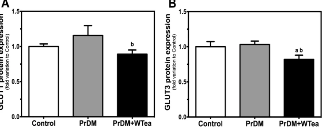
1. Daily white tea consumption ameliorates glucose and insulin tolerance in prediabetic rats 35 2. Daily white tea consumption decreases GLUTs expression and lactate accumulation in the cerebral cortex of prediabetic rats ... 36
3. Daily white tea consumption decreases alanine content but was not able to restore lactate/alanine ratio in cerebral cortex of prediabetic rats ... 38
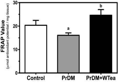
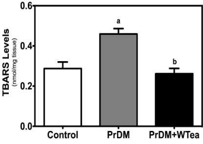
4. Daily white tea consumption increases the antioxidant capacity and catalase expression, preventing lipid peroxidation and protein oxidation in the cerebral cortex of prediabetic rats ... 40
5. Daily white tea consumption restores valine content in the cerebral cortex of prediabetic rats ... 44
V. Discussion ... 46
VI. Conclusions ... 52
VII. References ... 54
List of Figures
Figure 1 - Mitochondrial dysfunction and sustained activation of nicotinamide adenine
dinucleotide phosphate (NADPH) oxidase lead to insulin resistance, reactive oxygen species (ROS) production and impaired antioxidant defenses. ... 10
Figure 2 - Schematic representation of tea processing. ... 13 Figure 3 - Chemical structures of the main tea catechins. ... 16 Figure 4 - Effect of daily white tea consumption in protein expression of glucose
transporters (GLUTs) in the cerebral cortex of prediabetic rats. ... 36
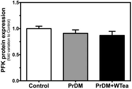
Figure 5 - Effect of daily white tea consumption in protein expression of
phosphofructokinase-1 (PFK-1) in the cerebral cortex of prediabetic rats. ... 37
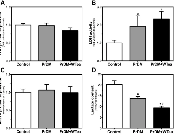
Figure 6 - Effect of daily white tea consumption in lactate dehydrogenase (LDH) and
monocarboxylate transporter 4 (MCT4) protein expression, LDH activity and lactate content in cerebral cortex of prediabetic rats. ... 38
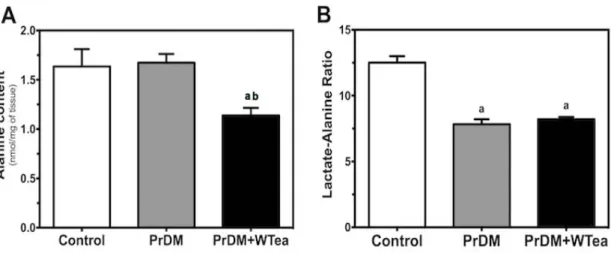
Figure 7 - Effect of daily white tea consumption in alanine content and lactate-alanine
ratio in the cerebral cortex of prediabetic rats. ... 39
Figure 8 - Effect of daily white tea consumption by prediabetic rats in antioxidant power of
cerebral cortex. ... 40
Figure 9 - Effect of daily white tea consumption by prediabetic rats in lipid peroxidation of
cerebral cortex. ... 41
Figure 10 - Effect of daily white tea consumption by prediabetic rats in protein oxidation
of cerebral cortex. ... 42
Figure 11 - Effect of daily white tea consumption in catalase protein expression and
List of Tables
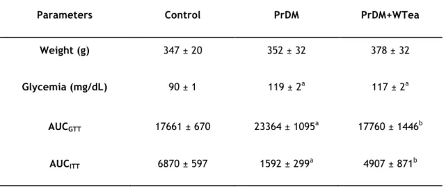
Table 1 - Average values of the rats weight, blood glycemia, area under the curve for
glucose tolerance (AUCGTT) and insulin tolerance (AUCITT) tests in rats from the control,
prediabetic rats drinking water (PrDM) and prediabetic rats drinking white tea (PrDM+WTea) groups after 60 days of treatment. ... 35
Table 2 - Levels of some metabolites in the cortex of control, prediabetic (PrDM) rats
List of Abbreviations
1H NMR - Proton Nuclear Magnetic Resonance
Acetyl-CoA – Acetyl coenzyme A AD – Alzheimer’s disease
ADA – American Diabetes Association AGEs – Advanced glycation end-products ALT – Alanine aminotransferase
ATP – Adenosine triphosphate AUC – Area under the curve
AUCGTT - Area under the curve for glucose tolerance test
AUCITT - Area under the curve for insulin tolerance test
BBB – Blood-brain barrier BSA - Bovine serum albumin CNS – Central nervous system DM – Diabetes Mellitus DNP - 2,4-Dinitrophenol DPPH - Diphenylpicrylhydrazyl
DTNB - 5,5’-dithiobis-(2-nitrobenzoic acid) EC – (-)epicatechin
ECG – (-)-epicatechin-3-gallate EGC – (-)-epigallocatechin
EGCG – (-)-epigallocatechin-3-gallate ETC – Electron transport chain
FRAP - Ferric reducing antioxidant power GABA - Gamma-aminobutyric acid GLUT1 – Glucose transporter 1 GLUT3 – Glucose transporter 3 GLUTs - Glucose transporters GSH - Reduced glutathione GSSH - Oxidized glutathione GTT - Glucose tolerance test HK – Hexokinase
IFG – Impaired fasting glucose
IgG-AP - Alkaline phosphatase linked immunoglobulin G IGT – Impaired glucose tolerance
INT - Tetrazolium salt IP - Intraperitoneal
ITT - Insulin tolerance test LDH – Lactate dehydrogenase
MCT4 – Monocarboxylate transporter 4 MCTs – Monocarboxylate transporters
MDA - Malondialdehyde NAA - N-Acetylaspartate
NAD+ - Oxidized nicotinamide adenine dinucleotide
NADH - Reduced nicotinamide adenine dinucleotide NADPH – Nicotinamide adenine dinucleotide phosphate OADs – Oral antidiabetic drugs
OS – Oxidative stress
PBS - Phosphate buffered solution PD – Parkinson’s disease
PFK-1 – Phosphofructokinase 1 PO – Polyphenol oxidase PrDM - Prediabetes
PSMF - Phenylmethylsulfonyl fluoride RNS – Reactive nitrogen species ROS – Reactive oxygen species SSA - 5-sulfosalicylic acid STZ - Streptozotocin
T1DM – Type 1 diabetes mellitus T2DM – Type 2 diabetes mellitus TBA - Thiobarbituric acid
TBARS - Thiobarbituric acid reactive species TCA – Tricarboxylic acid
TNB - 2-nitro-5-mercaptobenzoic acid TP – Tea polyphenols
TPTZ - 2,4,6-Tripyridyl-s-Triazine WHO – World Health Organization WTEA - White tea
1. Diabetes Mellitus at a Glance
Diabetes Mellitus (DM) represents one of the greatest threats to modern global health and its incidence is rapidly increasing. The World Health Organization (WHO) estimated that about 300 million of people will develop DM in 2025 (Agbaje, I. M. et al., 2007). These numbers tend to aggravate due to some risk factors related to lifestyle, such as being overweight, having an unhealthy diet or smoking. Nowadays, this disease is considered one of leading causes of morbidity and mortality in both developed and under development countries. It is potentially devastating, and although treatable, it is a lifelong disease (Al-Attar, A. M. and Zari, T. A. 2010). Thus, the healthcare costs associated with DM are enormous (Zhang, P. et al., 2010).
DM is described as a metabolic disorder of multiple etiologies, characterized by chronic hyperglycemia that can result from defects in insulin secretion and/or insulin action (Association, A. D. 2010). Moreover, there is a severe alteration in carbohydrate, lipid, protein metabolism (Association, A. D. 2010) and defects in reactive species of oxygen (ROS) scavenging enzymes (Kesavulu, M. M. et al., 2000), which results in increased oxidative stress (OS) and impairment of the pancreatic beta cells (Kahn, S. E. 2001). The DM may be classified as Type 1 Diabetes Mellitus (T1DM) or Type 2 Diabetes Mellitus (T2DM). T1DM is responsible for only 5-10% of those with DM and generally develops at young age with the great majority of the patients being diagnosed before the age of 30 (Agbaje, I. M. et al., 2007). It results from the autoimmune destruction of the insulin-producing beta pancreatic cells, and therefore there is a complete lack of insulin that leads to the increase of glucose levels in blood and urine (Association, A. D. 2010). Thus, T1DM patients need exogenous insulin administration and are insulin dependent. Untreated T1DM is characterized by hyperglycemia, hypoinsulinemia, ketonuria, and hyperlipidemia, resulting from a general metabolic failure (for review see (Emilien, G. et al., 1999)). In turn, T2DM is the most common type of DM, accounting for up to 90-95% of all cases diagnosed (Association, A. D. 2010). Among other features, T2DM is characterized by insulin resistance and/or insufficient insulin secretion. As a result, body glucose metabolism becomes compromised. The risk of developing T2DM increases with age, obesity, cardiovascular diseases and lack of physical activity (for review see (Golay, A. and Ybarra, J. 2005)). Noteworthy, the clinical symptoms are frequently detected only in an advanced phase of the disease, allowing the progression of functional changes in cells and tissues that may not be reverted. Diabetic patients possess a higher risk of death, together with lower survival rates and lower life expectancy than non-diabetic persons (Gu, K. et al., 1998).
DM is an incurable disease but there are many strategies available for its treatment such as the stimulation of endogenous insulin secretion, enhancement of insulin action at
the target tissues, inhibition of dietary starch lipid degradation, and pharmacological treatment with oral antidiabetic drugs (OADs) like biguanides (e.g. metformin) and sulfonylureas (e.g. clorpropamid) (Birari, R. B. and Bhutani, K. K. 2007, García-Pérez, L. E. et al., 2013)). However, these OADs can cause side effects like major and minor hypoglycemia, gastrointestinal problems, peripheral edema, body weight gain, liver diseases and, over time, they lose their efficacy (Dilla, T. et al., 2008, Donnelly, L. A. et al., 2009, García-Pérez, L. E. et al., 2013).
The limitation of existing therapies for the treatment of diabetes has encouraged the search of more efficient and cost-effective alternatives, recurring to dietary and lifestyle changes. In recent years, there is an increased interest in functional and nutraceutical food for pharmacological purposes, in order to complement or replace current therapies. It has been reported that numerous extracts obtained from plants can be used in DM treatment to reduce glycemia (Gupta, R. K. et al., 2005, Abolfathi, A. A. et al., 2012). However, little is known about the molecular mechanisms involved when using such extracts.
1.1 Prediabetes: A Prodromal Stage of DM
The complexity of DM diagnosis, especially in obese patients, led to the establishment of an intermediate state known as “prediabetes”. Prediabetes is defined as elevated blood glucose levels, although not sufficient to meet the criteria for established diabetes (Association, A. D. 2010). The American Diabetes Association (ADA) defined prediabetes as either impaired fasting glucose (IFG) (100-110 mg/dL) and/or impaired glucose tolerance (IGT) (140-199 mg/dL) (Association, A. D. 2010). The prediabetic state is characterized by resistance to insulin-mediated glucose disposal and compensatory hyperinsulinemia (Reed, M. J. et al., 2000, Alves, M. G. et al., 2013a) and its prevalence is increasing among young people (Association, A. D. 2010). This intermediate state is commonly associated with the metabolic syndrome which represents a group of abnormalities, including overweight (visceral abdominal fat distribution), dyslipidaemia, hypertension, and impaired glucose metabolism, with insulin resistance as the postulated underlying pathogenic mechanism (for review see (Kasturi, S. S. and Tannir, J. 2008)). Prediabetic patients have important metabolic alterations that increase the risk for the development of T2DM (for review see (Engelgau, M. M. et al., 2000)). The transition from prediabetes to T2DM occurs when the secretory capacity of the pancreatic beta cells is no longer able to compensate insulin resistance. This progression occurs over many years and compelling evidence support that intervention delay the progression from prediabetes to DM (Weyer, C. et al., 1999).
Glycemic levels are rapidly increasing in developed and developing countries, which increases the prevalence of prediabetes and it is projected that more than 470 million people will have prediabetes in 2030 (for review see ((Tabák, A. G. et al., 2012)). Every year, about 5–10% of the individuals with prediabetes become diabetic (Nathan, D. M. et al., 2007) and population habits may increase these rates.
The prediabetic state is not only related to an increased risk of DM development and its complications. Damage on kidney and nerves occurs in these individuals (Fox, C. S. et al., 2005). Besides, prediabetes can lead to complications such as nephropathies and chronic kidney disease, neuropathies, diabetic retinopathy, and macrovascular diseases (for review see (Tabák, A. G. et al., 2012)). Along with these complications, the risk of cognitive decline and neurodegeneration are increased in these patients (Luchsinger, J. A. et al., 2004). The exact pathophysiology of alterations that occur in the brain of prediabetic subjects is not completely understood, but it is likely that abnormal levels of blood glucose and insulin resistance play significant roles (for review see (Kodl, C. T. and Seaquist, E. R. 2008)).
2. General Effects of Hyperglycemia on the Brain
Blood glucose concentrations alter the function of several organs and tissues. As discussed above, DM is a complex metabolic disorder and hyperglycemia is a hallmark of this disease as a consequence of impaired insulin synthesis and/or insulin resistance. Consequently, glucose is not efficiently transported and metabolized in the target organs. Chronic hyperglycemia is associated with long-term injury and dysfunction of several organs, including a slow progressive brain damage (Diaz-Parejo, P. et al., 2003, Biessels, G. J. and Gispen, W. H. 2005).
The mammalian brain depends upon glucose as its main source of energy. Thus, a tight regulation of glucose metabolism is critical for brain physiology. In recent years, significantly more interest has been dedicated to the effect of hyperglycemia on the brain. DM is implicated in the development of cerebrovascular disease and other neurological co-morbidities, such as cognitive dysfunction and dementia (for review see (Roriz-Filho, J. S. et al., 2009)). A study conducted by Luchsinger and collaborators (2004) suggested that the risk of cognitive decline and neurodegeneration are increased in prediabetic patients. The exact pathophysiology of brain damage caused by DM is not completely understood, but it is likely that hyperglycemia and insulin resistance play a significant role (for review see (Roriz-Filho, J. S. et al., 2009)). Changes in peripheral insulin and glucose homeostasis may affect the action of insulin on the brain and its receptors functions (for review see (Gasparini, L. and Xu, H. 2003)). Moreover, insulin resistance leads to formation of
advanced glycation end-products (AGEs) and, consequently, oxidative stress-related events (for review see (Smith, M. A. et al., 1995)).
Hyperglycemia is known to differently affect different brain regions being that the cerebral cortex is highly sensitive to glucose fluctuations (Serpa, Jesus et al. 2006, Cardoso, Santos et al. 2010). Indeed, it has been reported that cortical neurons and astrocytes are more vulnerable to glucose metabolism deregulation than cells from striatum or hippocampus (Xu, L. et al., 2001). Moreover, it has also been reported that DM increases the vulnerability of specific brain areas to neuronal damage being the cerebral cortex particularly sensitive (Bree, A. J. et al., 2009). Furthermore, the cerebral tissues such as the cortex are quite vulnerable to OS due to its high consumption of oxygen, the abundance of easily oxidizable fatty acids (for review see (Wang, X. and Michaelis, E. K. 2010)), and the relative low presence of antioxidant defenses in comparison with other tissues. For example, the brain has 10% less antioxidant defenses than the liver (for review see (Uttara, B. et al., 2009)). Moreover, brain has higher levels of iron in certain regions and in general has high levels of ascorbate. Therefore, neural cells are considered to be more susceptible to oxidative damage as compared to other body tissues (Floyd, R. A. and Carney, J. M. 1992).
DM besides increasing the probability of occurrence of a stroke (Baird, T. A. et al., 2002), also increases the risk of cognitive impairments and dementia (for review see (Biessels, G. J. and Gispen, W. H. 2005)). Furthermore, it is becoming evident that diabetics have a higher risk for developing neurodegenerative diseases. Cerebral cortex is greatly affected by Alzheimer’s disease (AD), and several studies have demonstrated that AD and DM are connected (for review see (Moreira, P. I. 2012)). Insulin resistance and its signaling impairment, mitochondrial abnormalities, OS, are some of the relevant events in both disorders (Santos, R. X. et al., 2014b). Therefore, hyperglycemia and other DM-related alterations have several negative effects on brain function and structure.
2.1 Hyperglycemia and Brain Metabolism
The brain depends on glucose as its main source of energy. In the adult brain, neurons have the highest energy demand (Howarth, C. et al., 2012), requiring continuous delivery of glucose from blood. This organ is metabolically very active, consuming 20% of the total body’s oxygen and receiving 15% of the cardiac output (for review see (McCall, A. L. 2004)), in resting state. Energy consumption by the brain is largely needed to maintain and restore ionic gradients associated with synaptic transmission (for review see (McCall, A. L. 2004)), and the chemical energy within the brain exists primarily in the form of
high-The largest proportion of energy in the brain is used for neuronal computation and information processing; for example, the generation of action potentials and postsynaptic potentials generated after synaptic events and the maintenance of ion gradients and neuronal resting potential (Howarth, C. et al., 2012). Moreover, glucose metabolism provides the energy and precursors for the biosynthesis of neurotransmitters (for review see (Dienel, G. A. 2012)).
Dependence of the brain on glucose as its obligatory fuel derives mainly from the blood-brain barrier (BBB) and its selective permeability for glucose. Glucose cannot be replaced as an energy source, but it can be supplemented, as during strenuous physical activity when blood lactate levels are elevated (van Hall, G. et al., 2009) or during prolonged starvation (Lutas, A. and Yellen, G. 2013) when blood levels of ketone bodies are elevated. Cerebral metabolism of glucose requires transport through the BBB, glycolytic conversion to pyruvate, metabolism via the tricarboxylic acid (TCA) cycle and ultimately oxidation to carbon dioxide and water for full provision of ATP and its high-energy equivalents. Any disturbance in glucose metabolism compromises the brain normal functioning.
Brain is compromised by metabolic changes associated to DM, leading to cognitive deficits and to an increased risk of brain vascular complications (for review see (Biessels, G. J. and Gispen, W. H. 2005)). Thus, it is expectable that glucose metabolism dysfunction promoted by DM, particularly T2DM, is responsible for severe brain damage (for review see (McCall, A. L. 2004)). High blood glucose levels, obesity, increased blood triacylglycerol’s concentration and insulin resistance, are some risk factors that, individually or collectively, increase the probability of neurodegeneration or even neuronal death (Duarte, A. I. et al., 2013).
Hyperglycemia has a variety of adverse effects upon brain metabolism and function (Diaz-Parejo, P. et al., 2003, Alves, M. G. et al., 2012). Glucose is a primary fuel for the brain, but lactate also plays an important role (Pellerin, L. et al., 2002). Animal studies have shown that there is a reduced overall glucose metabolism and regional changes in glucose metabolism in individuals with poorly controlled DM (Jakobsen, J. et al., 1990). The brain glucose metabolism is crucial to normal cerebral functioning. Under normal conditions, glucose crosses BBB through specific glucose transporters (GLUTs), glucose transporter 1 (GLUT1) and glucose transporter 3 (GLUT3). The rate of entry of glucose into the cell is limited by the number of GLUTs on the cell surface and the affinity of the transporters for glucose. Glucose is phosphorylated by hexokinase (HK) to produce glucose-6-phosphate that is then converted to pyruvate by phosphofructokinase-1 (PFK-1). The reaction catalyzed by this enzyme is usually described as one of the most important regulatory steps in glycolysis. PFK-1 activity is essential since it is responsible for the
conversion of fructose 6-phosphate to fructose 1,6-bisphosphate, the first irreversible step of glycolysis, and a limiting step of glycolytic flux (Underwood, A. H. and Newsholme, E. A. 1965). The pyruvate can be processed into three main metabolic pathways: a) transported into the mitochondrial matrix to form acetyl coenzyme A (acetyl-CoA); b) converted to lactate by lactate dehydrogenase (LDH); or converted to alanine by alanine aminotransferase (ALT). The lactate produced can be released in the extracellular space through monocarboxylate transporters (MCTs), mainly monocarboxylate transporter 4 (MCT4) (for review see (Oliveira, P. F. et al., 2014)).
The deleterious effects of hyperglycemia are mediated through an increased flux of glucose through the polyol and hexosamine pathways, disturbances of intracellular second messenger pathways, an imbalance in the generation and scavengers of ROS, and by AGEs (for review see (Brownlee, M. 2001)).
3. Brain Oxidative Stress and Hyperglycemia
OS may be defined as a measure of the steady-state level of reactive oxygen or oxygen radicals in biological systems. DM has been linked to ROS since the early 90s (for review see (Baynes, J. W. 1991)). OS is present in the early (prediabetes state) and late phase of DM (Su, Y. et al., 2008). Nowadays, it is widely accepted that OS is important in the development and progression of DM (for review see (Ceriello, A. 2000)). It has been reported that DM-related hyperglycemia and the glycemia fluctuations can amplify OS (for review see (Brownlee, M. 2001)) by increasing the production of free radicals and/or by impairing antioxidant defenses (for review see (Bloch-Damti, A. and Bashan, N. 2005)).
Excessively high levels of free radicals cause damage to cellular proteins, membrane lipids and nucleic acids, and eventually cell death. Various mechanisms have been suggested to contribute to the formation of ROS. Glucose oxidation, non-enzymatic glycation of proteins, oxidative degradation of glycated proteins and the mitochondrial respiratory system form free radicals in diabetic individuals (for review see (Maritim, A. C. et al., 2003)). Nevertheless, moderate amounts of ROS are important for various biological processes (for review see (Juranek, I. et al., 2013)).
OS occurs when there is an overproduction of ROS and/or a decreased efficiency of radical scavengers, such as glutathione (Bravi, M. C. et al., 2006). Glutathione is one of the most important intracellular antioxidants (for review see (Niedowicz, D. M. and Daleke, D. L. 2005)) and the relative amount of intracellular reduced and oxidized glutathione (GSSH) is a measure of the cellular redox status. Experimental and clinical studies have shown that glutathione levels are altered in diabetic patients (Dinçer, Y. et al., 2002).
It has been shown that ROS are produced in various tissues under diabetic conditions (for review see (Baynes, J. W. and Thorpe, S. R. 1999)), including the brain tissue. The hyperglycemia-related increase in ROS and reactive nitrogen species (RNS) can be due to several factors, such as mitochondrial respiratory system (for review see (Nishikawa, T. and Araki, E. 2007)), nicotinamide adenine dinucleotide phosphate (NADPH) oxidase (for review see (Gao, L. and Mann, G. E. 2009)), formation of AGEs (for review see (Brownlee, M. 2001)), and imbalance of glutathione redox status (Bravi, M. C. et al., 2006). As discussed above, under normal conditions, glucose enters the glycolytic pathway, through GLUTs, it is metabolized to pyruvate that is transported to the mitochondrial matrix, oxidized and decarboxylated by the pyruvate dehydrogenase forming the two carbon intermediate acetyl-CoA, which can enter the TCA cycle. However, the mitochondrial electron transport chain (ETC) is the main ROS producer (mainly the mitochondrial complex I and III) (for review see (Martin, S. D. and McGee, S. L. 2014)), and abnormal levels of blood glucose leads to production of larger amounts of ROS, leading to
OS and contributing to peripheral insulin resistance (Figure 1). Higher levels of blood glucose lead the increased supply of energy substrates and the inflammatory environment is thought to result in excessive mitochondrial ROS generation (Loh, K. et al., 2009). Thus, is possible that certain signaling pathways are activated and consequently induce insulin resistance (for review see (Kim, J. A. et al., 2008, Tiganis, T. 2011)). Increased ROS production associated with a reduction in plasma antioxidants, particularly glutathione, may have toxic effects on the plasma membrane structure/activity of the pancreatic beta-cells, contributing to the impaired insulin secretion (for review see (Paolisso, G. and Giugliano, D. 1996)). Furthermore, direct evidence for the involvement of hyperglycemia-induced ROS in promoting insulin resistance has been provided using mouse and rat animal models (Haber, C. A. et al., 2003, Anderson, E. J. et al., 2009). However, is important to note that ROS might be both good and bad, promoting insulin sensitivity early in disease progression, and contributing to the development of insulin resistance later, when hyperglycemia prevails (for review see (Tiganis, T. 2011)). On the other hand, sustained activation of NADPH oxidase leads to impaired antioxidant defenses due to superoxide production (for review see (Gao and Mann 2009)). If the level of NADPH decreases, the recycling of reduced glutathione (GSH) is limited (glutathione reductase uses NADPH to regenerate GSH). Particularly, increased NADPH oxidase activity contributes to a large number of pathologies such as DM, cardiovascular diseases and neurodegeneration. Noteworthy, it has been proposed that sustained activation of NADPH oxidase in DM leads to ROS production and impaired antioxidant defenses (for review see (Gao and Mann 2009)).
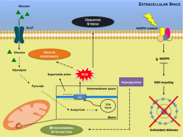
Figure 1 - Mitochondrial dysfunction and sustained activation of nicotinamide adenine dinucleotide phosphate (NADPH) oxidase lead to insulin resistance, reactive oxygen species (ROS) production and impaired antioxidant defenses. Mitochondria are the main generators of ROS within
electron transport chain (ETC). In normal conditions, glucose breakdown starts by glycolysis, generating among other compounds, pyruvate. Pyruvate is then converted to acetyl coenzyme A (acetyl-CoA) that enters the tricarboxylic acid (TCA) cycle. The produced electrons are stored in molecules that are then injected into the ETC, to generate the electrochemical gradient. When an abnormal increase in the electrochemical potential difference in the inner membrane of the
mitochondria occurs leads to the overproduction of O2-. This is particularly important since ROS may
contribute to insulin resistance. Sustained activation of NADPH oxidase leads to decreased intracellular levels of NADPH and therefore the recycling of reduced glutathione (GSH) is limited, impairing antioxidant defenses.
Lipid peroxidation and protein carbonyls are biomarkers of OS. The abnormal enhancement of free radicals and the decline of antioxidant defense mechanisms lead to the damage of cellular organelles and enzymes, the increase in lipids peroxidation and the increase of insulin resistance (for review see (Maritim, A. C. et al., 2003)). The nonradical oxidants such as hydrogen peroxide, hypochlorous acid, singlet oxygen and radicals like superoxide anion and hydroxyl anion, can attack the double bound of unsaturated fatty acids promoting the formation of lipid peroxides (for review see (Lipinski, B. 2001)). On the other hand, protein oxidation originates carbonyl groups and their level in tissues and plasma is a stable marker of OS (Odetti, P. et al., 1999).
New ways to reduce the brain damage caused by DM may arise by modifying lifestyles, particularly by changes in diet. There is a large interest in finding an effective
therapy for DM-associated brain dysfunction and white tea seems to be a good candidate, with interesting properties such as antidiabetic (Dieren, S. v. et al., 2009), neuroprotective (Unno, K. et al., 2007, López, V. and Calvo, M. I. 2011) and antioxidant (Almajano, M. P. et al., 2008) properties. Thus, tea and its phytochemicals properties may be important and will be discussed below.
4. Tea
Since ancient times, medicinal plants have been used to prevent and treat a wide range of diseases. Camellia sinensis (L.), commonly known as the tea plant, is an evergreen shrub of the Theaceae family, native to Southeast China and is now cultivated in over 30 countries across the world (López, V. and Calvo, M. I. 2011), including S. Miguel Island (Azores Archipelago, Portugal).
Tea is one of the most widely consumed beverages in the world, surpassed only by the water (Cheng, T. O. 2006), with a per capita consumption of approximately 120 mL/day (Mckay, D. L. and Blumberg, J. B. 2002). The popularity of tea consumption is probably related with its sensorial properties, relatively low retail price, stimulating effects and potential health benefits (Moderno, P. M. et al., 2009, Dias, T. R. et al., 2013, Dias, T. R. et al., 2014, Martins, A. D. et al., 2014).
The origins of tea are mythological. The “Father of Tea”, Eisai, said: “Tea is a miraculous medicine for the maintenance of health. Tea has an extraordinary power to prolong life.” (Wheeler, D. and Wheeler, W. 2004). In fact, tea has been extensively used by traditional Chinese medicine for centuries to prevent and treat several diseases, such as DM (Wheeler, D. and Wheeler, W. 2004, Moderno, P. M. et al., 2009).
4.1 Types of Tea
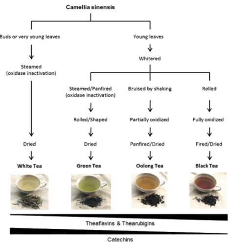
Tea is an infusion prepared from the leaves of C. sinensis, but each type of tea has a different composition, which depends on the type of processing, growing conditions, botanical variety, and geographical origin (for review see (de Mejia, E. G. et al., 2009)). According to processing and collection, tea can be classified into black tea (completely fermented), oolong tea (semi-fermented), green tea and white tea (not fermented). After collection, the leaves gain a darker color, which means that chlorophylls are breaking down and tannins are being released. Upon harvesting, the leaves suffer oxidation, commonly called “fermentation”, which occurs with the exposure to air and is a reaction catalyzed by the enzyme polyphenol oxidase (PO) (for review see (Mckay, D. L. and Blumberg, J. B. 2002)). Depending upon the level of “fermentation”, all types of tea have different chemical compositions (phenolic profiles) and organoleptic properties (appearances and tastes).
To produce green tea, the leaves are rolled and steamed to minimize the oxidation by inactivation of the PO before drying (for review see (Mckay, D. L. and Blumberg, J. B. 2002)). Thus, the chemical composition of green tea remains similar to that of the C.
sinensis fresh leaves. In black tea, the most consumed type of tea in the western countries
(Li, S. et al., 2013), the leaves are rolled and cellular compartmentalization is disrupted bringing the phenolic compounds to contact with PO and then they undergo oxidation for 90 to 120 minutes (Rusak, G. et al., 2008). Oolong tea is produced with a shorter oxidation period than black tea and has a taste and color somewhere between green tea and black tea (Rio, D. D. et al., 2004) (Figure 2). Finally, white tea is the rarest and most expensive tea, and how it is produced and its chemical composition will be discussed below.
Figure 2 - Schematic representation of tea processing (adapted from (Dias, T. R. et al., 2013)).
All four types of tea are significant sources of antioxidant (Costa, R. M. et al., 2009, Moderno, P. M. et al., 2009), antidiabetic (Song, E. K. et al., 2003, Abolfathi, A. A. et al., 2012) and neuroprotective (Unno, K. et al., 2007, López, V. and Calvo, M. I. 2011) compounds.
4.2 White Tea
White tea is prepared from very young tea leaves or buds covered with tiny, silvery hair, which are harvested only once a year in the early spring (Rusak, G. et al., 2008). To prevent oxidation, white tea is steamed and dried immediately after harvest. The buds may be shielded from sunlight during growth to reduce the formation of chlorophylls, giving the young leaves a white appearance (Alcázar, A. et al., 2007). It is one of the less studies teas but its flavor is more accepted in Europe than that of green tea (Almajano, M. P. et al., 2008).
Many health benefits have been attributed to tea consumption. However, scientific investigations of this beverage and its constituents have been underway for less than three decades. In spite of numerous data about the phenolic constituents, antioxidant activity and ameliorating effects of green and black tea on human health, little is known in this sense about white tea, which is the rarest and the least processed tea (Rusak, G. et al., 2008). The possible beneficial health effects of white tea are being extensively investigated and have received a great deal of attention in recent years by our research group.
4.2.1 Chemical Composition
Tea is composed by a complex mixture of about 2000 chemical compounds, including proteins, polysaccharides, minerals and trace elements, organic acids, lignins, polyphenols, methylxanthines and amino acids (Seeram, N. P. et al., 2006, Moderno, P. M. et al., 2009). Several of these compounds are bioactive and are believed to possess health benefits (Carvalho, M. et al., 2010, Dias, T. R. et al., 2014, Martins, A. D. et al., 2014). Phenolic compounds, methylxanthines (mainly caffeine) and L-theanine have received particular attention among tea phytochemicals.
4.2.1.1 Phenolic compounds
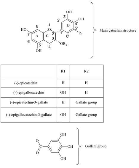
Polyphenols are secondary plant metabolites, widely distributed in nature, and are the most abundant and active group of compounds present in tea. Catechins (also known as flavan-3-ols) and their derivatives are the main class of phenolic compounds present in tea leaves, constituting about 30% of their dry weight. The major catechins are (-)-epicatechin (EC), (-)-epigallocatechin (EGC) collectively known as flavanol monomers, (-)-epicatechin-3-gallate (ECG), (-)-epigallocatechin-(-)-epicatechin-3-gallate (EGCG) which are also called flavanol gallates (de Mejia, E. G. et al., 2009, Dias, T. R. et al., 2014). The health benefits attributed to catechins are mainly due to its chemical structure. The main catechins are
composed by two aromatic rings (A and B) linked to a dihydropyran heterocyclic ring (C) and are characterized by the presence of several hydroxyl groups (for review see (Braicu, C. et al., 2013)) (Figure 3). Their chemical differences are due to the presence of different groups attached to those rings. In EC, we can find an ortho-di-hydroxil group in the B ring (at carbons 3’ and 4’) and a hydroxyl group in the C ring (at carbon 3); EGC, which is an ester derivative of EC, additionally contains a gallate moiety esterified in the C ring, at carbon 3. EGC, on the other hand, possesses a trihydroxil group on the B ring (at carbons 3’, 4’ and 5’), and EGCG differs in this structure by additionally possessing an esterified gallate at the carbon 3 of the C ring. Green and white teas are the types of tea with higher catechin content, while oolong and black tea possess other phenolic compounds, in addition to lower catechins levels (for review see (Lin, Y. S. et al., 2003, Dias, T. R. et al., 2013, Li, S. et al., 2013)). Tea composition is affected by the oxidation process, a reaction catalyzed by PO that is released during the crushing of the leaves in the production of black and oolong tea, and catalyzes the oxidation and polymerization of the catechins EC, ECG, EGC and EGCG, producing theaflavins and thearubigins (for review see (Lin, Y. S. et al., 2003, Li, S. et al., 2013)). These oligomers/polymers are responsible for black tea bitter taste and dark color (Wheeler, D. and Wheeler, W. 2004). Theaflavins possess a basic chemical skeleton comprised of the bicyclic benzotropolone ring and are the result of main catechins dimerization (for review (Li, S. et al., 2013)). Thearubigins are produced subsequently to a series of complex reactions that form its oligo-polymeric structures.
EGCG is the most abundant catechin in tea leaves and has been extensively studied by several authors (for review see (Yang, C. S. et al., 2004, Seeram, N. P. et al., 2006)). It represents 50–80% of the total catechins, and is thought to contribute to the beneficial effects ascribed to tea (for review see (Khan, N. and Mukhtar, H. 2007)).
Figure 3 - Chemical structures of the main tea catechins. The figure illustrates two aromatic rings
(A, B) and a dihydropyran heterocyclic ring (C), which is the basic structure of flavonoids. The (-)-epicatechin (EC) is constituted by an ortho-di-hydroxil group in the B ring (at carbons 3’ and 4’) and a hydroxyl group in the C ring (at carbon 3), and its ester derivative (-)-epicatechin 3-gallate (ECG) differs in this structure by possessing an additional gallate moiety esterified in the C ring, at carbon 3. On the other hand, (-)-epigallocatechin (EGC) contains a trihydroxil group on the B ring (at carbons 3’, 4’ and 5’) and its ester derivative (-)-epigallocatechin-3-gallate (EGCG) additionally possesses an esterified gallate at the carbon 3 of the C ring.
The redox properties of phenolic compounds are in the basis of the tea antioxidant properties, which can be very useful if its consumption is adopted as a natural health practice (Atoui, A. K. et al., 2005). Several reports have shown that tea catechins and other polyphenols are effective scavengers of ROS and RNS (Guo, Q. et al., 1999, Paquay, J. B. et al., 2000). This is of extreme relevance since OS is known to induce neuronal death and to be involved in neurodegenerative diseases (for review see (Agostinho, P. et al., 2010, Dumont, M. et al., 2010). Thus, there is a growing interest in the possible neuronal tea benefits for DM patients.
4.2.1.2 Methylxanthines
Methylxanthines are purine bases derivatives present in tea, 2-4% as caffeine and small amounts of theophylline and theobromine (Hara, Y. et al., 1995). Caffeine (1,3,7-trimethylxanthine) is one of the most consumed substances in the world (Hashimoto, T. et al., 2004) and due to its chemical stability, the oxidation process does not affect caffeine levels in tea (for review see (Li, S. et al., 2013)). However, some researchers found that black and oolong tea have greater caffeine content than green tea (Lin, Y. S. et al., 2003) and that white tea has also a higher content than green tea (Unachukwu, U. J. et al., 2010, Dias, T. R. et al., 2014). These discrepancies may be due to different extraction conditions (solvents, temperatures, times of extraction and ratio leaves/water) and of distinct analytical methods. Besides, the natural variability of plants caused by edapho-climatic factors, harvesting techniques or agricultural practices may contribute to these differences.
The excessive consumption of caffeine can cause many adverse effects (for review see (Nawrot, P. et al., 2003)), such as nervousness, irritability, insomnia, diuresis, arrhythmia, tachycardia and gastrointestinal disturbances. Death provoked by excessive intake of caffeine, although rare, has also been reported (for review see (Nawrot, P. et al., 2003)). Some authors argue that the lowest caffeine content in green tea contributes to its beneficial health properties, mainly attributed to its phenolic compounds (Lee, L.-S. et al., 2013). Nevertheless, there are also some studies that highlight the potentially beneficial action of caffeine. Similarly to tea catechins, caffeine also has different effects at cellular and metabolic levels (for review see (Mandel, H. G. 2002)). The most important mechanism by which caffeine can act in Central Nervous System (CNS) is by selectively blocking the adenosine receptors and competitively inhibiting the action of adenosine in the cells, which results in an increased release of hormones such as norepinephrine, dopamine and serotonin (for review see (Nawrot, P. et al., 2003)). Caffeine is also a likely candidate against memory loss (for review see (Cunha, R. A. 2008)) and with a great neuroprotective potential (Cunha, R. A. 2005, Duarte, J. M. et al., 2009). Accordingly, studies in rats have shown that this methylxanthine can interact with GLUTs in adipocytes and act as an antagonist of adenosine receptors (Steinfelder, H. J. and Pethö-Schramm, S. 1990). Also, intravenous administration of caffeine to healthy human subjects resulted in decreased whole-body glucose uptake along with a decrease in carbohydrate storage (Greer, F. et al., 2001), as well as an increase in insulin insensitivity, resulting from the caffeine-induced release of the insulin-antagonistic hormone epinephrine (Keijzers, G. B. et al., 2002). Increases in blood pressure have also been reported (Keijzers, G. B. et al., 2002). Interestingly, the consumption of caffeine-containing beverages, in particular tea, is associated with a lower risk of developing T2DM (Dieren, S. v. et al., 2009, Sartorelli, D. S. et al., 2010). Some authors have also reported that caffeine intake is inversely associated
with body weight increase and satiety (Westerterp-Plantenga, M. S. et al., 2005, Lopez-Garcia, E. et al., 2006). Caffeine and theophylline are also involved in the stimulation of pancreatic beta cells (Johnston, K. L. et al., 2003). In the brain it has been shown that increased levels of caffeine are associated with decreased risk of neurodegenerative diseases (Chen, J.-F. et al., 2001). However, data on the role of caffeine on tea-associated health benefits are scarce and much work needs to be done.
4.2.1.3 L-theanine
L-theanine is a free amino acid which presents structural similarity to glutamate, an important neurotransmitter related to memory (for review see (Kakuda, T. 2011)). L-theanine constitutes between 1 and 3% of the dry weight of tea, but this percentage may vary according to growing location and method of cultivation, tea grade, variety, processing and collection time (Vuong, Q. V. et al., 2011). Green tea contains lower or similar levels of L-theanine as compared to black and oolong tea (Ekborg-Ott, K. H. et al., 1997). This amino acid is considered as a relaxing agent with antioxidant (Nishida, K. et al., 2008, Patti, M. E. and Corvera, S. 2010) and neuroprotective effects (Egashira, N. et al., 2007, Cho, H. S. et al., 2008, Kakuda, T. 2011). However, its pharmacology is relatively unknown and human studies are inconclusive (Lu, K. et al., 2004). Metabolically, it is easily absorbed from the gastrointestinal tract and peak plasma concentrations are detected 0.5 hour after administration (Kakuda, T. 2011). According to Yokogoshi and collaborators (1998), L-theanine is partially transported to the brain via a leucine-preferring transporter system and can cross the BBB, exercising protector effects in the brain and a preventive effect on neuronal cell death. The benefits of L-theanine for health are reported to be associated with regulation of blood pressure, effective prophylaxis and treatment of neurodegenerative diseases, improvement of the immune system, among others (Yokogoshi, H. and Kobayashi, M. 1998, Rogers, P. J. et al., 2008, Di, X. et al., 2010, Takagi, Y. et al., 2010).
5.
White Tea Potential and Health Benefits
Bioactive components of plants have served as sources of inspiration for generations of medicinal and organic chemists, and will continue to provide humankind with valuable agents of potential use in research, prevention, and treatment of several diseases, like DM. Medicinal plants used in pharmaceutical products to treat diabetic conditions have aroused considerable interest in recent years (for review see (Ayyanar, M. et al., 2008)), including white tea (Islam, M. S. 2011).
Conventionally, DM is treated with OADs in the case of T2DM, or with exogenous insulin in case of T1DM or T2DM uncontrolled on OADs. However, these drugs are not completely effective and have adverse effects. Natural compounds are considered to be less toxic and relatively cheaper than synthetic ones and large amounts can be consumed in everyday diet (for review see (Saxena, A. and Vikram, N. K. 2004)). Scientific papers concerning the health benefits of tea consumption are relatively recent, and the studies are not as conclusive as we could expect. Thus, the search for evaluating the efficacy and safety of tea, particularly white tea and its phytochemicals, has become one important area of research.
5.1 Antioxidant Potential
In the last few years, antioxidant components have aroused great interest because of their ability to scavenge free radicals, reducing the harmful effects of ROS and RNS, thereby inhibiting oxidation (Alarcón, E. et al., 2008). The majority of living organisms possess efficient enzymatic and nonenzymatic defense systems against excessive production of ROS. Nevertheless, factors such as lifestyle (smoke, diet, alcohol, some drugs, among others) and internal factors (such as aging) decrease the efficiency of endogenous antioxidant defenses, creating an impairment in the redox equilibrium that is established in healthy conditions (for review see (Rietveld, A. and Wiseman, S. 2003)). Chronic exposure to ROS can damage DNA, membrane lipids, lipoproteins, and functional and structural proteins (Halliwell, B. 1997). Increased OS has been proposed to be one of the major causes of the hyperglycemia-induced diabetic complications (Valko, M. et al., 2007). Due to these events, the cellular balance between radical formation and protection against them is disturbed. The elimination of ROS to decrease the oxidative damage is seen as beneficial to public health. Therefore, antioxidants that scavenge ROS may be of great value in preventing the onset and/or the progression of oxidative diseases (for review see (Willett, W. C. 1994)).
It is well known that hyperglycemia increases the formation of ROS and decreases antioxidant endogenous mechanisms (Rahimi, R. et al., 2005). Several studies have reported that tea phenolic compounds, mainly catechins, are potent antioxidant agents, scavenging ROS (Nakagawa, T. and Yokozawa, T. 2002) and metal chelators (Atoui, A. K. et al., 2005). Numerous studies have demonstrated that tea catechins and polyphenols are effective scavengers of physiologically relevant ROS and RNS in vitro, including superoxide (Nanjo, F. et al., 1993, Nakagawa, T. and Yokozawa, T. 2002), peroxyl radicals and singlet oxygen (Guo, Q. et al., 1999). The chemical structure of tea components is associated with its antioxidant properties. In this context, a relationship has been suggested between the content of pyrogallol and hydroxyl groups and the superoxide anion scavenging ability, as well as between the presence of galloyl moieties and the ability to quench hydroxyl radicals (Nanjo, F. et al., 1999, Moderno, P. M. et al., 2009). Several structures appear to be important for these antioxidant activities of tea polyphenols (TP), including the ortho-3’,4’-dihydroxyl (catechol) group in the B-ring, that promotes the formation of a stable phenoxyl radical due to effective electron delocalization (Wiseman, S. A. et al., 1997) or the 3’,4’,5’-trihydroxyl (gallate) group in the B-ring, a gallate group esterified at the 3 position of the C-ring, and hydroxyl groups at the 5 and 7 positions of the A-ring (Rice-Evans, C. A. et al., 1996). It is known that the number and position of the hydroxyl groups on the molecules greatly influence the antioxidant ability of flavonoids (for review see (Braicu, C. et al., 2013)). Particularly, tea catechins such as EGCG lack a 2, 3 double bond and a carbonyl group at the 4-position, a combination that is known to strengthen the antioxidant activity (for review see (Moderno, P. M. et al., 2009). The structure of catechins has a major influence in their antioxidant properties, such as radical scavenging, transition-metal chelation, inhibition of redox sensitive transcription factors, inhibition of pro-oxidant enzymes and induction of antioxidant and phase II detoxification enzymes (for review see (Aboul-Enein, H. Y. et al., 2013, Braicu, C. et al., 2013)). Catechins antioxidant activities are related to their ability to enter in several chemical reactions of hydrogen atom and single electron transfers, involving hydroxyl groups. Studies have reported that the antioxidant properties of tea catechins are only observed in vivo when the animals are under OS, contrary to in vitro studies, where these activities can nearly always be observed. Studies have also demonstrated that catechins influence the levels of endogenous antioxidants. A study performed on rats by Srividhya and collaborators (2008) showed that continuous administration of EGCG for 30 days was able to significantly improve the animals’ antioxidant defenses, ameliorating the age-induced OS in their brains. After analysis of the enzymatic and non-enzymatic antioxidants, lipid peroxidation and protein carbonyl groups on the animals brain tissue, the authors reported that EGCG successfully induced a rising in the activity of the antioxidant enzymes superoxide dismutase, catalase, glutathione peroxidase, glutathione reductase and glucose-6-phosphate dehydrogenase, as well as in the levels of non-enzymatic antioxidants such as L-ascorbic acid, α-tocopherol and glutathione. Also, lipid peroxidation and levels of protein