Full paper published online: November 30, 2009
IDENTIFICATION OF BRADYKININ-RELATED PEPTIDES FROM Phyllomedusa
nordestina SKIN SECRETION USING ELECTROSPRAY IONIZATION TANDEM
MASS SPECTROMETRY AFTER A SINGLE-STEP LIQUID CHROMATOGRAPHY
ConceiçãoK (1), Bruni FM (1), Sciani JM (3), Konno K (1), Melo RL (1), Antoniazzi MM (2), Jared C (2), Lopes-Ferreira M (1), Pimenta DC (3)
(1) Special Laboratory of Applied Toxinology, Center for Applied Toxinology, CAT/CEPID, Butantan Institute, São Paulo, São Paulo State, Brazil; (2) Laboratory of Cell Biology, Center for Applied Toxinology, CAT/CEPID, Butantan Institute, São Paulo, São Paulo State, Brazil; (3) Laboratory of Biochemistry and Biophysics, Butantan Institute, São Paulo, São Paulo State, Brazil.
ABSTRACT: Amphibian skin secretions are a source of potential new drugs with medical and biotechnological applications. Rich in peptides produced by holocrine-type serous glands in the integument, these secretions play different roles, either in the regulation of physiological skin functions or in the defense against predators or microorganisms. The aim of the present work was to identify novel peptides with bradykinin-like structure and/or activity present in the skin of Phyllomedusa nordestina. In order to achieve this goal, the crude skin secretion of this frog was pre-fractionated by solid phase extraction and separated by reversed-phase chromatography. The fractions were screened for low-molecular-mass peptides and sequenced by mass spectrometry. It was possible to identify three novel bradykinin-related peptides, namely: KPLWRL-NH2 (Pnor 3), RPLSWLPK (Pnor 5) and
VPPKGVSM (Pnor 7) presenting vascular activities as assessed by intravital microscopy. Pnor 3 and Pnor 7 were able to induce vasodilation. On the other hand, Pnor 5 was a potent vasoconstrictor. These effects were reproduced by their synthetic analogues.
KEY WORDS: Phyllomedusa nordestina, bradykinin, mass spectrometry, natural peptides.
CONFLICTS OF INTEREST: There is no conflict.
FINANCIAL SOURCE: FAPESP, CAPES and CNPq.
CORRESPONDENCE TO:
INTRODUCTION
Amphibian skin secretions contain a large number of biologically active molecules
that function not only as a protection against microorganisms, but also as an
anti-predator system (1, 2-4). Moreover, some amphibian secretions contain a number of
small proteins and a large number of peptide components whose biological functions
are still undetermined. Several peptides from skin secretions of amphibians, more
particularly tree-frogs, have been purified in recent years, including antimicrobial
peptides, bradykinin related peptides and one bradykinin-potentiating peptide (BPP)
(5-9).
The generation of bradykinin (BK) in the blood by the action of the kallikrein–kinin
system has been studied intensively in mammals, but the system has received
relatively little attention in non-mammalian vertebrates. Amphibian skin has proven a
remarkably rich storehouse of regulatory peptides including kinins, while the
occurrence of BK-related peptides in skin secretions of frogs has been investigated
by several groups (1, 10-12). Yet, the search for the existence of at least one
component of the kallikrein–kinin system in anuran plasma has been unsuccessful,
for instance one in the form of a biosynthetic precursor (e.g., pre-pro-BK) (13).
The hylid genus Phyllomedusa contains 32 species distributed throughout southern
Central America and much of South America (14, 15). Phyllomedusa nordestina,
formely comprised into Phyllomedusa hypochondrialis, is a typical Brazilian genus,
inhabiting the semi-arid region of northeastern Brazilian (15-17). Peptides weighing
less than 5 kDa are the predominant molecules in the secretions of many tree frogs
of the sub-family Phyllomedusinae, whereas bradykinin and bradykinin-related
peptides (BRP) have been identified in the skin secretions of these animals. The fact
that such analogues have been found in these skin secretions supports the idea that
BRP are produced as part of a diverse defense system.
The aim of the present work was to investigate directly by RP-HPLC and mass
spectrometry the low-molecular-mass peptides from P. nordestina skin secretion. A
single chromatographic step was chosen in order to avoid the loss of the minor
components. Matrix assisted laser desorption ionization time-of-flight mass
spectrometry (MALDI-TOF/MS) and orthogonal quadrupole time-of-flight electrospray
ionization tandem mass spectrometry (ESI-Q-TOF-MS/MS) de novo sequencing
intravital microscopy, that were reproduced by the synthetic analogues. Taken
together, these results indicate the usefulness of this sole chromatographic step
approach that, combined with specific biological assays, was able to successfully
identify low-abundance small bioactive peptides from complex samples such as this
anuran skin secretion.
MATERIALS AND METHODS
Animals
Groups of five male Swiss mice weighing 18 to 22 g were used throughout. The
animals, provided by Instituto Butantan animal house, were kept in temperature and
humidity-controlled rooms, and received food and water ad libitum. All the
procedures involving mice were in accordance with the guidelines provided by Ethics
Committee on Animal Use of Butantan Institute (n. 378/07).
Drugs and Reagents
Sep-Pak C18® cartridges were purchased from Waters Corporation (USA);
Fmoc-amino acids from Calbiochem-Novabiochem Corporation (USA); and acetonitrile
(HPLC grade) was purchased from J. T. Baker (USA).
Alpha-cyano-4-hydroxycinnamic acid, iodoacetamide, NaI, molecular mass standards (ProteoMass
Kit®) and acetylcholine hydrochloride were all obtained from Sigma Co. (USA).
Sodium pentobarbital and sodium heparin were acquired from Roche Laboratories
(Brazil).
Collection of Specimens
Specimens (n = 12; non-sexed) of Phyllomedusa nordestina were collected at
Angicos, in the state of Rio Grande do Norte, Brazil. The tree frogs were kept alive in
the animal house of the Department of Cellular Biology, Butantan Institute, São
Paulo, Brazil. The tree frogs were collected according to the Brazilian environmental
agency (Brazilian Institute of Environment and Renewable Natural Resources –
Purification Procedures
Skin secretion obtainment and solid phase extraction
Glandular secretions were obtained from adult specimens of P. nordestina,
submerged in a beaker containing deionized water, that were manually and gently compressed. This solution was lyophilized and stored at –20°C. Pooled lyophilized
secretions from twelve animals were dissolved in deionized water and centrifuged at
5000 x g for 20 minutes (room temperature). The supernatant was pre-purified by
solid phase extraction (SPE) using Sep-Pak C18® cartridges (Waters Corporation,
USA). A single aliquot of 2 mg diluted in 2 mL of 0.1% TFA was loaded; then 80%
acetonitrile containing 0.1% TFA was eluted. This sample was lyophilized to prior
analysis.
Reversed phase chromatography
A reversed-phase binary HPLC system (Äkta, Sweden) was used for sample
separation. The SPE-eluted fraction was loaded in a Shimadzu C18 column
(Shim-Pack® 5µ, 4.6 x 250 mm, Japan) in a two-solvent system: (A) trifluoroacetic acid
(TFA)/H2O (1:1000) and (B) TFA/acetonitrile (ACN)/H2O (1:900:100). The column
was eluted at a flow rate of 1.0 mL/min with a 10 to 80% gradient of solvent B over
60 minutes. The HPLC column eluents were monitored by their UV absorbance at
214 nm.
Mass spectrometry analyses
Peptide mass analyses were performed on a Q-TOF Ultima API® (Micromass, UK),
under positive ionization mode and/or by MALDI-TOF mass spectrometry on an Ettan
MALDI-TOF/Pro system® (Amersham Biosciences, Sweden) using α
-cyano-4-hydroxycinnamic acid as matrix.
Mass spectrometric de novo peptide sequencing was carried out in positive ionization
mode on a Q-TOF Ultima API® fitted with an electrospray ion source (Micromass,
UK). Samples were dissolved into a mobile phase of 50% acetonitrile, containing
0.1% formic acid, and directly injected (10 µL) using a Rheodyne 7010® sample loop
(USA) coupled to a LC-10A VP® Shimadzu (Japan) pump at a constant 20
µL/minute flow rate. The instrument control and data acquisition were conducted by
scanning from a mass-to-charge ratio (m/z) of 50 to 1800 using a scan time of 2
seconds applied during the whole infusion. The mass spectra corresponding to each
signal from the total ion current (TIC) chromatogram were averaged, allowing an
accurate molecular mass determination. External calibration of the mass scale was
performed with NaI. For the MS/MS analysis, collision energy ranged from 18 to 45
eV and the precursor ions were selected under a 1-m/z window. All MS/MS spectra
were analyzed manually; the sequences were determined by precise mass
differences between adjacent b ions (confirmed by y ions).
Peptide synthesis
Synthetic peptides were obtained by an automated benchtop simultaneous multiple
solid-phase synthesizer (PSSM 8® system from Shimadzu, Japan) using solid phase
peptides synthesis by the Fmoc procedure (18). The peptides were purified by
reversed-phase chromatography (Shim-pack Prep-ODS®, 5µ, 20 x 250 mm,
Shimadzu, Japan) in a binary semi-preparative system and their purity and identity
were confirmed by MALDI-TOF and/or ESI-Q-TOF mass.
Intravital microscopy
The dynamic of alterations in the microcirculatory network was determined using
intravital microscopy by transillumination of mouse cremaster muscle after topical
application of the peptide. Sterile saline was used as control. In three independent
experiments (n = 5, for each experiment) mice were anaesthetized with sodium
pentobarbital (Hypnol®, Cristália, Brazil; 50 mg/kg, intraperitoneal route) and the
cremaster muscle was exposed for microscopic examination in situ as described by
Lomonte et al. (19). The animals were maintained on a special board thermostatically controlled at 37°C, which included a transparent platform on which the tissue to be
transilluminated was placed. Images of the microcirculation were simultaneously
visualized on TV and computer monitors using a color video camera (TK-C600®,
JVC, Japan) incorporated to a triocular microscope (Axioskope®, Carl-Zeiss,
Germany). Images obtained on the TV monitor were recorded on a video recorder
and computer digitized images were analyzed using standard imaging software
(KS300®, Kontron, Germany). The images were obtained using a ×10/025
Statistical Analyses
One-way analysis of variance (ANOVA), followed by Newman-Keuls test, was
performed to determine significance of differences. The significance level was set at
p < 0.05.
RESULTS
Peptide Pooling
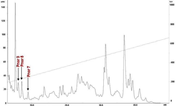
The lyophilized SPE-Phyllomedusa nordestina crude skin secretion was fractionated
based only on peak shape and distribution, according to the analytical RP-HPLC
profile presented in Figure 1. All pooled peptides were tested for vascular effects and
those pools that were active had their molecular mass distribution profiled by mass
spectrometry (Figure 2) (MALDI/TOF and/or ESI-Q-TOF). This work has focused
solely on bioactive peptides ranging from 700 to 1100 Da; therefore, the arrowed
peaks in Figure 1 were the only ones falling into this profile: small vasoactive
peptides. These pools were subsequently submitted to de novo MS/MS sequencing.
Figure 1. RP-HPLC profile of P. nordestina pooled crude skin secretions after solid
phase extraction, performed according to material and methods section. The arrows
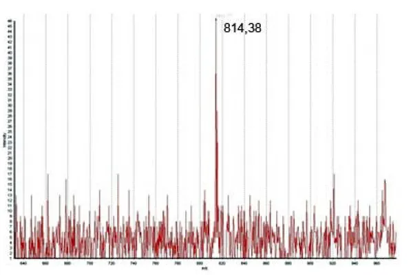
Figure 2. Mass spectrum (MALDI-ToF) of the novel peptides called Pnor 3, Pnor 5
and Pnor 7 (m/z values 811.41, 996.59 and 814.38, respectively, are typed besides
each ion for clearer reading).
De Novo Peptide Sequencing
Those pools selected for de novo (or collision-induced fragmentation analysis by
Q-TOF-MS/MS) sequencing were processed accordingly. Briefly, after cystein bridge
reduction and alkylation by iodoacetamide, the obtained peptides were individually
selected for MS/MS analysis and fragmented by collision with argon (CIF), yielding
daughter ion spectra as presented in Figure 3. This routine was repeated until
sufficient information regarding the peptide sequence was gathered. The MS/MS
spectra were analyzed by the BioLynx software module of MassLynx 4.0 and
manually verified for accuracy in the amino acid sequence interpretation. The active
peptides that had their amino acid sequences deduced were: KPLWRL-NH2;
RPLSWLPK and VPPKGVSM, denominated Pnor 3, 5 and 7 respectively. Table 1
shows that Pnor peptides align fairly well with bradykinin and other kinins from other
A
C
Figure 3. Representative CIF spectra of (A) m/z = 406.28, (Pnor 3; [M+2H]2+), (B)
m/z=498.86, (Pnor 5; [M+2H]2+), and (C) m/z=814.37, (Pnor 7; [M+H]+) and
Table 1. Multialignment of vasoactive peptides. Mainly amphibian peptides are
presented in addition the classical vasoactive peptides
Peptide Sequence
Pnor3 (C-ter NH2) KPLWRL
Pnor5 RPLSWLPK
Pnor7 VPPKGVSM
Bradykinin RPPGFSPFR
Kallidin KRPPGFSPFR
Hyp3-Bradykinin a RPPGFSPFR
Phyllokinin a RPPGFSPFRIY
Hyp3-Phyllokinin a RPPGFSPFRIY T6-Phyllokinin a RPPGFTPFRIY T6V10-Phyllokinin b RPPGFTPFRIY T6V10-Phyllokinin-VY b RPPGFTPFRVY Hyp3_T6-Bradykinin a RPPGFTPFR EPV1T6-Bradykinin-LT b EPVPPGFTPFRLT V1 T6-Bradykinin-LT b VPPGFTPFRLT V1Hyp2_T6-Bradykinin-QT b VPPGFTPFRQT V1Hyp2_T6-Bradykinin-QS b VPPGFTPFRQS V1 T6-Bradykinin-VD b VPPGFTPFRVD V1Hyp2T6-Bradykinin b VPPGFTPFR V1T6-Bradykinin b VPPGFTPFR des-R-T6-Bradykinin b RPPGFTPF des-R-V1T6-Bradykinin a VPPGFTPF
Orpotrin c HGGYKPTDK
Angiotensin DRVYIHPFHL
Hemopressin d PVNFKFLSH
LVV-Hemorphin-7 e LVVYPWTQRF
Vasopresin CYFQNCPRG
Uroteonsin II ACFWKYCV
Consensus* ..ppgf.pf...
Hyp stands for hydroxyl-proline. *Calculated by Multalin (20).
aBrand
et al. (7).
bThompson
et al. (8).
cConceição
et al. (21).
dRioli
et al. (22).
ePiot
Bioassay
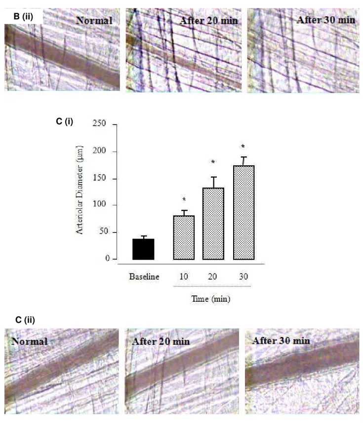
Figure 4 presents the changes in diameter in groups of arterioles in response to local
application of the peptides, over time. The topical administration of 10 nmol of Pnor 3
(Figure 4A) induced vasodilatation. An increase in the diameter of large arterioles was observed after 10 minutes, compared to the initial diameter (75 ± 10 μm and 37 ± 8 μm, respectively). The relative magnitude of arteriolar vasodilatation in response to the peptide was augmented after 20 and 30 minutes (130 ± 12 μm and 175 ± 10 μm, respectively).
Also, for the peptide Pnor 7 (Figure 4C), a significant vasodilatation was observed.
Ten minutes after the topical administration of 10nmol Pnor 7, the arteriolar diameter doubled (Figure 4C; i) relative to the initial diameter (42 ± 5 μm and 87.4 ± 11 μm, respectively). The vasodilatation became more pronounced after 20 minutes (125 ± 18 μm) and remained so until 30 minutes of observation.
Interestingly, Figure 4B displays the changes in diameter of the arteriole groups in
response to the local application of Pnor 5. As observed, the topical administration of
10nmol of Pnor 5 provoked an accentuated arteriolar constriction. This
vasoconstriction activity was evident 10 minutes after the application, when compared to the initial diameter (26 ± 7 μm and 43 ± 9 μm, respectively). The relative
magnitude of arteriolar constriction in response to the peptide had not been restored
by the end of 30 minutes of observation (Figure 4B, ii).
No change in rolling leukocyte velocity or venule diameter was seen over time in
A (i)
A (ii)
B (ii)
C (i)
C (ii)
Figure 4. Intravital micrograph of cremaster muscle (n = 5) after topical application of
10 nmol of the peptides Pnor 3 (A), Pnor 5 (B) and Pnor 7 (C). (i) Arteriolar diameter
variation over time and (ii) time-course evaluation of the vascular effects.
Photographs were obtained from digitized images on the computer monitor (*p <
DISCUSSION
This work reports the purification and characterization of three novel peptides
denominated Pnor 3, Pnor 5 and Pnor 7 (after Phyllomedusa nordestina). Like
bradykinin, Pnor 3 provokes a strong vasodilatation as observed by the
microcirculatory network assay (Figure 4A). This same effect is also induced by the
administration of Pnor 7 (Figure 4C). Interestingly, Pnor 5 was the opposite effect: it
is a strong arteriolar constrictor, as found in the same assay (Figure 4B).
Vasoactive peptides and inflammatory molecules tend to coexist in venoms and
secretions, whereas increased leukocyte adhesion can be an indicator of an inflamed
and dysfunctional endothelium (24). Previous studies by our group reported that the
skin secretion of P. hypochondrialis (although we now know that we were working
with P. nordestina at the time) induces increased leukocyte rolling flux followed by a
gradual increase in cells firmly adherent to the endothelium in intravital experiments
(25). Taking our previous results together with the present findings (characterization
of BRP), one can conclude that they corroborate the previously described local
inflammatory effect of P. nordestina skin administration in mice. Moreover, these skin
secretion components could play a role in the initial rolling and slowing of recruited
leukocytes as well as the transition from slow rolling to firm adhesion. At present, the
mechanisms involved in the local inflammatory process induced by P. nordestina skin
secretion is one of the relevant questions related to the complex pathophysiology
induced by toxins (peptides). Kinins are rapidly produced after tissue injury and play
a role in many of the components of the inflammatory response such as
vasodilatation, plasma extravasation, cell migration, pain and hyperalgesia (26-31).
Peptides described in this report were initially screened for their biological activity
and then for their molecular mass directly from the SPE-processed crude skin
secretion. As presented in Table 1, each of these peptides possesses at least one
Pro residue calculated as consensus by Multalin (20). Not surprisingly, the calculated
consensus nonapeptide based on all the inputted sequences possesses six
bradykinin residues at the correct positions.
Although BK and BRP are not rarely found in amphibian skin secretions, we would
like to call the attention to the point that not only Val1/Thr6-BK variations can be found
on these secretions. However, we were able to identify Pnor7 (VPPKGVSM),
Moreover, Pnor7 presents a Lys residue at position 4. This insertion per se is
sufficient to speculate about a whole new family of peptides, since positions 1, 2, 3, 5
and 7 are conserved (Table 1) and the presence of such a reactive amino acid
should not be considered as a random event.
Also noteworthy are the opposite effects observed for Pnor3 and Pnor5. Apparently
using the same amino acids in a different order, these peptides vary from
vasodilatation activity (Pnor3) to vasoconstrictor activity (Pnor5).
As Harris et al. (32) described, the presence of inflammatory cells in venules may
have a major influence on arteriolar constriction. So, the arteriolar constriction
observed, in which venular adherent leukocytes contributed to the constriction of
paired arterioles, could be attenuated by the injection of a monoclonal antibody
against the adhesion molecules (33). Therefore, as seen in the work of Conceição et
al. (21) on Orpotrin, in our model no change in rolling leukocyte velocity or in
diameter of venules was seen over time in Pnor5-treated animals, suggesting that
Pnor5 exerts a selective and direct action on arterioles.
Some authors speculate that BK and BRP are present in the skin of these tree frogs
as they lack the kallikrein-kinin system (34, 35), and/or that these peptides may act
on and/or potentiate the endogenous BK action on the cardiovascular and/or
gastrointestinal system of their predators, thus acting as defensive peptides (13, 11).
If the latter hypothesis is correct, then the best defensive peptides would not be BK
or BRP by themselves, but rather, a combination of kinins and a potentiator factor. In
a previous study, our group have indeed found a BPP in the skin secretion of
Phyllomedusa hypochondrialis, and it was the first BPP (bearing the canonical
structural motif) to be discovered not only on the frog skin but also in any natural
sources other than snake venoms (9).
Investigating nature’s selected molecules is a fruitful source for identifying
compounds aimed at very specific targets, depending mostly on the source of the
material and the biological system chosen. The present approach, i.e. mass
spectrometry amino acid sequence deduction after a single-step liquid
chromatography, not only requires very little sample manipulation but also has
proven itself to be fast, reliable and effective in identifying bioactive peptides. It is our
allows a rapid and reliable method for screening a large number of
low-molecular-mass peptides present in skin secretions.
ACKNOWLEDGEMENTS
D. C. Pimenta is a CNPq fellow researcher (grant n. 302405/2008-9). Parts of this
work were developed by D. C. Pimenta when working at CAT/CEPID, a FAPESP
grant.
REFERENCES
1. Lazarus LH, Attila M. The toad, ugly and venomous, wears yet a precious jewel in
his skin. Prog Neurobiol. 1993;41(4):473-507.
2. Daly JW. The chemistry of poisons in amphibian skin. Proc Natl Acad Sci USA.
1995;92(1):9-13.
3. Erspamer V. Bioactive secretions of the integument. In: Heatwole H, Barthalmus
GT, editors. The integument, amphibian biology. vol. 1. Chipping Norton: Surrey
Beatty & Sons; 1994. 179-350 p.
4. Zasloff M. Antimicrobial peptides of multicellular organisms. Nature.
2002;415:389-95.
5. Chen T, Zhou M, Gagliardo R, Walker B, Shaw C. Elements of the granular gland
peptidome and transcriptome persist in air-dried skin of the South American
orange-legged leaf frog, Phyllomedusa hypocondrialis. Peptides. 2006;27(9):2129-2136.
6. Conceição K, Konno, K, Richardson M, Antoniazzi MM, Jared C, Daffre S,
Camargo ACM, Pimenta DC. Isolation and biochemical characterization of peptides
presenting antimicrobial activity from the skin of Phyllomedusa hypochondrialis.
Peptides 2006;27(12):3092-9.
7. Brand GD, Krause FC, Silva LP, Leite JRSA, Melo JAT, Prates MV, Pesquero JB,
Santos EL, Nakaie CR, Costa-Neto CM, Bloch C. Bradykinin-related peptides from
Phyllomedusa hypochondrialis. Peptides. 2006;27(9):2137-46.
8. Thompson AH, Bjourson AJ, Shaw C, McClean S. Bradykinin-related peptides
from Phyllomedusa hypochondrialis azurea: Mass spectrometric structural
characterization and cloning of precursor cDNAs. Rapid Commun Mass Spectrom.
9. Conceição K, Konno K, Lopes de Melo R, Antoniazzi MM, Jared C, Sciani JM,
Conceição IM, Prezoto BC, de Camargo ACM, Pimenta DC. Isolation and
characterization of a novel bradykinin potentiating peptide (BPP) from the skin
secretion of Phyllomedusa hypochondrialis. Peptides. 2007; 28(3):515-23.
10. Conlon JM, Aronsson U. Multiple bradykinin-related peptides from the skin of the
frog, Rana temporaria. Peptides. 1997;18(3):361-5.
11. Li L, Bjourson AJ, He J, Cai G, Rao P, Shaw C. Bradykinins and their cDNA from
piebald odorous frog, Odorrana schmackeri, skin. Peptides. 2003; 24(6):863-72.
12. Suzuki H, Iwamuro S, Ohnuma A, Coquet L, Leprince J, Jouenne T, Vaudry H,
Taylor CK, Abel PW, Conlon JM. Expression of genes encoding antimicrobial and
bradykinin-related peptides in skin of the stream brown frog Rana sakuraii. Peptides.
2007;28(3):505-14.
13. Conlon JM. Bradykinin and its receptors in non-mammalian vertebrates. Regul
Pept. 1999;79(2-3):71-81.
14. da Cruz CAG. Sobre as relações intergenéricas de Phyllomedusinae da floresta
atlantica (Amphibia, Anura, Hylidae). Rev Brasil Biol. 1990;50(3):709-26.
15. Frost DR. Amphibian species of the world: an online reference, version 5.3.
Available in: http://research.amnh.org/herpetology/amphibian/index.html. 2009.
16. Caramaschi U. Redefinição do grupo de Phyllomedusa hypochondrialis, com
redescrição de P. megacephala (Miranda-Ribeiro, 1926), revalidação de P. azurea
Cope, 1862 e descrição de uma nova espécie (Amphibia, Anura, Hylidae). Arq Mus
Nac. 2006;64(2):159-79.
17. Daudin FM. Histoire naturelle des rainettes, des grenouilles et des crapauds:
dédiée à B.G.E.L. Lacépèdè. Paris: D I’lmprimerie de Bertrandet: Chez Leurault
Librarie; 1802. 108 p.
18. Atherton E, Sheppard RC. Solid phase peptide synthesis: a practical approach.
Oxford: IRL Press; 1989. 75-160 p.
19. Lomonte B, Lundgren L, Johansson B, Bagge U. The dynamics of local tissue
damage induced by Bothrops asper snake venom and myotoxin II on the mouse
cremaster muscle: an intravital and electron microscopic study. Toxicon.
1994;32(1):41-55.
20. Corpet F. Multiple sequence alignment with hierarchical clustering. Nucleic Acids
21. Conceição K, Konno K, Melo RL, Marques EE, Hiruma-Lima CA, Lima C,
Richardson M, Pimenta DC, Lopes-Ferreira M. Orpotrin: a novel vasoconstrictor
peptide from the venom of the Brazilian stingray Potamotrygon gr. orbignyi. Peptides.
2006;27(12):3039-46.
22. Rioli V, Gozzo FC, Heimann AS, Linardi A, Krieger JE, Shida CS, Almeida PC,
Hyslop S, Eberlin MN, Ferro ES. Novel natural peptide substrates for endopeptidase
24.15, neurolysin, and angiotensin-converting enzyme. J Biol Chem.
2003;278(10):8547-55.
23. Piot JM, Zhao Q, Guillochon D, Ricart G, Thomas D. Isolation and
characterization of two opioid peptides from a bovine hemoglobin peptic hydrolysate.
Biochem Biophys Res Commun. 1992;189(1):101-10.
24. Ulbrich H, Eriksson EE, Lindbom L. Leukocyte and endothelial cell adhesion
molecules as targets for therapeutic interventions in inflammatory disease. Trends
Pharmacol Sci. 2003;24(12):640-7.
25. Conceição K, Miriane Bruni F, Pareja-Santos A, Antoniazzi MM, Jared C,
Lopes-Ferreira M, Lima C, Pimenta DC. Unusual profile of leukocyte recruitment in mice
induced by a skin secretion of the tree frog Phyllomedusa hypochondrialis. Toxicon.
2007;49(5):625-33.
26. Ahluwalia A, Perretti M. B1 receptors as a new inflammatory target. Could this B
the 1? Trends Pharmacol Sci. 1999;20(3):100-4.
27. Calixto JB, Cabrini DA, Ferreira J, Campos MM. Kinins in pain and inflammation.
Pain. 2000;87(1):1-5.
28. Couture R, Harrisson M, Vianna RM, Cloutier F. Kinin receptors in pain and
inflammation. Eur J Pharmacol. 2001;429(1-3):161-76.
29. Dray A, Perkins M. Bradykinin and inflammatory pain. Trends Neurosci.
1993;16(3):99-104.
30. Dray A. Kinins and their receptors in hyperalgesia. Can J Physiol Pharmacol.
1997;75(6):704-12.
31. Perkins MN, Campbell E, Dray A. Antinociceptive activity of the bradykinin B1
and B2 receptor antagonists, des-Arg9, [Leu8]-BK and HOE 140, in two models of
32. Harris NR, Whatley JR, Carter PR, Specian RD. Venular constriction of
submucosal arterioles induced by dextran sodium sulfate. Inflamm Bowel Dis.
2005;11(9):806-13.
33. Zamboni WA, Roth AC, Russel RC, Graham B, Suchy H, Kucan JO. Morphologic
analysis of the microcirculation during reperfusion of ischemic skeletal muscle and
the effect of hyperbaric oxygen. Plast Reconstr Surg. 1993;91(6):1110-23.
34. Rabito SF, Binia A, Segovia R. Plasma kininogen content of toads, fowl and
reptiles. Comp Biochem Physiol A Comp Physiol. 1972;41(2):281-4.
35. Seki T, Miwa I, Nakajima T, Erdos EG. Kallikrein–kinin system in nonmammalian
![Figure 3. Representative CIF spectra of (A) m/z = 406.28, (Pnor 3; [M+2H] 2+ ), (B) m/z=498.86, (Pnor 5; [M+2H] 2+ ), and (C) m/z=814.37, (Pnor 7; [M+H] + ) and respective annotations and sequence deduction](https://thumb-eu.123doks.com/thumbv2/123dok_br/15912544.673728/10.892.128.807.160.630/figure-representative-spectra-pnor-respective-annotations-sequence-deduction.webp)