UNIVERSIDADE DA BEIRA INTERIOR
Ciências
Biosynthesis scale-up of human soluble catechol-O
-methyltransferase
Guilherme Manuel Palha Ruivo do Espírito Santo
Dissertação para obtenção do Grau de Mestre em
Biotecnologia
(2º ciclo de estudos)
Orientador: Prof. Doutor Luís Passarinha
Co-Orientadora: Profª. Doutora Filomena Silva
iii
Acknowledgments
First of all, I would like to express my sincere gratitude to Professor Doctor Luís Passarinha and Professor Doctor Filomena Silva for all their guidance, expertise, and trust. I really appreciate their help, which was crucial for the development of this work, and their vast knowledge, criticism and suggestions.
I would also like to express my gratitude to Augusto, whose expertise, companionship, advice and encouragement made my work much more pleasant and easy.
Furthermore, I would like to thank to all people involved in the Health Sciences Research Centre of the University of Beira Interior, particularly to the Biotechnology and Biomolecular Sciences group.
Finally, I am extremely thankful to all my friends and family for their unconditional support and encouragement through all this time.
v
Resumo
A proteína catecol-O-metiltransferase (COMT, CE 2.1.1.6) foi descrita pela primeira vez em 1958. É uma enzima metiltransferase dependente da S-adenosil-L-metionina (SAM) que catalisa a metilação de substratos catecólicos (catecolaminas, catecolestrogénios). Esta enzima desempenha várias funções importantes no organismo, funcionando como uma barreira entre o sangue e diversos orgãos, através da eliminação de catecóis biológicamente activos ou tóxicos. A enzima COMT desempenha ainda uma função particularmente importante no cérebro, onde participa no metabolismo do neurotransmissor dopamina, pelo que o seu papel nas doenças neurodegenerativas é extremamente relevante, nomeadamente na doença de Parkinson. Esta deonça é caracterizada pela perda progressive de neurónios libertadores de dopamina, levando a uma diminuição acentuada dos níveis deste neurotransmissor no cérebro. Nesta patologia, um dos principais alvos terapêuticos é a enzima COMT, já que a sua ação é indesejável nos pacientes portadores desta doença. Nos últimos anos têm vindo a ser desenvolvidos vários inibidores desta enzima para o tratamento da doença de Parkinson, e há um grande interesse em desenvolver novos inibidores mais eficazes. Para tal, são necessárias grandes quantidades de COMT numa forma activa, e a melhor maneira de o conseguir é a sua produção em larga escala através de processos biotecnológicos. Neste trabalho é proposto um processo de produção em larga escala da isoforma solúvel da COMT, utilizando o sistema de expressão Escherichia coli para a biosíntese da isoforma solúvel desta proteína e testando várias condições de fermentação, com o objectivo de estabelecer um processo de fermentação descontínuo com posterior fornecimento de substrato, controlando vários parâmetros durante esta fase. As condições de fermentação escolhidas, após terem sido testadas em processos de fermentação contínuos, foram uma percentagem de oxigénio dissolvido de 20%, uma concentração de 20 g/L das fontes de carbono e azoto (glicerol e triptona, respectivamente), e por fim, um perfil de adição de substrato no processo de alimentação de 1 g glicerol/L/h. Os resultados finais foram uma densidade óptica máxima de aproximadamente 50 e uma actividade enzimática especifica de 442.34 nmol/h/mg.
Palavras-chave
vii
Resumo alargado
A catecol-O-metiltransferase (COMT, CE 2.1.1.6) foi descrita e purificada pela primeira vez em 1958. É uma enzima metiltransferase que cataliza a transferência do grupo metil da S-adenosyl-L-metionina (SAM) para um dos grupos hidroxilo do substrato catecólico (incluindo catecolaminas e catecolestrogéneos) na presença do ião Mg2+.
Esta enzima existe sob a forma de duas isoformas, uma solúvel (SCOMT) localizada no citosol e outra associada a membranas plasmáticas (MBCOMT), ambas codificadas pelo mesmo gene (localizado no cromossoma 22) a partir de dois promotores. Ambas são expressas na maioria dos tecidos humanos, excepto no cérebro, onde a MBCOMT é a isoforma dominate.
A função geral da COMT é a eliminação de catecóis biologicamente activos ou tóxicos, funcionando como uma barreira desintoxicante entre o sangue e vários tecidos. Além disso, esta enzima desempenha um papel fundamental no metabolismo do neurotransmissor dopamina, associado a várias doenças neurodegenerativas.
Uma destas doenças é a doença de Parkinson, uma das doenças neurodegenerativas mais comuns, afectando cerca de 6 milhões de pessoas a nível mundial, e dados apontam para que esse número duplique nos próximos 20 anos. Esta doença foi descrita pela primeira vez em 1817 por James Parkinson, e caracteriza-se pela perda progressiva e profunda de neurónios libertadores de dopamina, levando a uma diminuição dos níveis deste transmissor no cérebro. Devido a esta diminuição nos níveis de dopamina, as estratégias farmacológicas mais comuns para esta patologia têm como objectivo repor esses níveis, pela administração de levodopa, uma molécula que depois de metabolizada no cérebro dá origem à dopamina, ou de moléculas agonistas da dopamina.
Estes tratamentos, no entanto, são muito limitados, e posteriormente surgiram novas terapias que os complementam, como por exemplo a administração de inibidores da COMT. Tendo em conta a ligação entre a COMT e o metabolismo da dopamina, os inibidores da COMT assumem extrema importância para atenuar os efeitos indesejáveis desta proteína na patologia.
Os primeiros inibidores desenvolvidos, são tóxicos, pouco efectivos in vivo e/ou pouco selectivos. Há portanto um grande interesse científico no desenvolvimento de novos e melhorados inibidores, e para tal são necessárias elevadas quantidades de COMT na sua forma activa.
Assim sendo, o principal objectivo deste trabalho é a produção a larga escala da isoforma solúvel desta enzima, usando a estirpe E. coli BL21 (DE3) como sistema de expressão para a
biossíntese da COMT. Foi portanto desenvolvido um processo de fermentação descontínuo com fornecimento de substrato (“Fed-batch”) em bioreactores de bancada.
Partindo de duas condições de fermentação anteriormente optimizadas pelo nosso grupo de investigação (Temperatura, 40°C e ph 6.5), várias outras condições foram testadas, nomeadamente a percentagem de oxigénio dissolvido, a concentração das fontes de carbono e azoto e os perfis de adição de substrato; e três parâmetros da cultura celular foram analisados durante o processo: concentração de fonte de carbono, viabilidade celular e actividade enzimática.
As condições de fermentação escolhidas, após terem sido testadas em processos de fermentação contínuos, foram uma percentagem de oxigénio dissolvido de 20%, uma concentração de 20 g/L das fontes de carbono e azoto (glicerol e triptona, respectivamente), e por fim, um perfil de adição de substrato no processo de alimentação de 1 g glicerol/L/h. Os resultados finais foram uma densidade óptica máxima de aproximadamente 50 e uma actividade enzimática especifica de 442.34 nmol/h/mg.
ix
Abstract
Catechol-O-methyltransferase (COMT, EC 2.1.1.6) was first described in 1958. It is a S- adenosyl-L-methionine (SAM) dependent methyltransferase which catalyses the methylation of
catechol substrates (catecholamines, catecholestrogens). This enzyme has several important functions in the body, acting as a barrier between the blood and various organs, through the elimination of toxic or biologically active catechols. COMT plays a particularly important role in the brain, where it participates in the metabolism of the neurotransmitter dopamine, and so its role in neurodegenerative diseases is extremely relevant, especially in Parkinson's disease. In recent years, several COMT inhibitors have been developed for Parkinson's disease treatment, and there is considerable interest in developing new and more effective inhibitors. To do so, large amounts of COMT in an active form are necessary, and the best way to achieve this is by up-scaling its production through biotechnological processes. In this work, we propose a fed-batch process for biosynthesis scale-up of the soluble isoform of COMT, using an Escherichia coli expression system for the production of the soluble isoform of this protein, testing various fermentation conditions and controlling various parameters during the process. After several different batch and fed-batch experiments, the selected fermentation conditions were 20% dissolved oxygen, an initial batch concentration of 20 g/L of glycerol and tryptone, and a constant feed of 1 g glycerol/L/h, achieving a final specific COMT activity of 442.34 nmol/h/mg. Cell viability was monitored by flow cytometry and glycerol concentration by an HPLC system.
Keywords
xi
Table of Contents
Chapter 1 - Introduction ... 1
1.1 – General overlook of catechol-O-methyltransferase ... 1
1.1.1 – The V108M polymorphism ... 2
1.1.2 – Crystal structure of human SCOMT ... 2
1.2 –COMT in catecholamine metabolism ... 3
1.3 –COMT and neurodegenerative diseases ... 6
1.3.1 – COMT and Alzheimer’s disease ... 6
1.3.2 – COMT and Parkinson’s disease ... 6
1.3.2.1 – COMT inhibitors in Parkinson’s disease ... 8
1.4 –Recombinant hSCOMT biosynthesis ... 10
1.4.1 – Host strain and promoter selection ... 11
1.4.2 – Fermentation strategies and conditions ... 14
Chapter 2 - Materials and Methods ... 19
2.1 - Materials ... 19
2.2 – Methods ... 19
2.2.1 – Expression vector and bacterial strain ... 19
2.2.2 - Escherichia coli pre-cultivation, batch and fed-batch conditions ... 19
2.2.3 – Flow cytometry assays ... 20
2.2.4 – Glycerol assessment ... 21
2.2.5 – Cell lysis ... 21
2.2.6 – Protein quantitation assay ... 21
2.2.7 – Enzymatic activity assay ... 23
Chapter 3 - Results and Discussion... 25
3.1 – Batch fermentations ... 25
3.1.1 – Definition of batch phase composition ... 29
3.2 – Growth rate and time of fed-batch initiation ... 31
3.3 – Fed-batch fermentations ... 32
3.3.1 – Constant feeding profiles ... 33
3.3.1.1 – Cytometry assays ... 37
3.3.2 – Exponential feeding profiles ... 39
3.3.2.1 – Cytometry assays ... 43
3.4 – COMT production in fed-batch fermentations ... 45
3.4.1– Cytometry assay ... 47
3.4.2 – Enzymatic assay ... 49
Chapter 4 - Conclusions ... 51
xiii
List of Figures
Figure 1 - Crystal structure of human 108V SCOMT. ... 3
Figure 2- Synthesis and metabolism of dopamine into homovanilic acid (HVA) and noradrenaline into vanillylmandelic acid (VMA). ... 4
Figure 3- Dopaminergic transmission in PFC and striatum. ... 5
Figure 4– Representative base structures of some first generation COMT inhibitors. ... 8
Figure 5– Chemical structures of nitecapone, tolcapone, entacapone and nebicapone. ... 9
Figure 6 – Comparative studies of different carbon sources and concentrations in terms of hSCOMT specific activity and biomass yields. ... 15
Figure 7 – Behavior of cultivation parameters during fed-batch procedure.. ... 16
Figure 8 – Calibration curve for BSA solutions absorbance at 570 nm. ... 22
Figure 9 – Calibration curve for BSA solutions absorbance at 595 nm. ... 23
Figure 10 - Growth curve of E. coli in a batch process: 40°C, 30% dissolved oxygen, pH 6.5.. 26
Figure 11 - Growth curve of E. coli in a batch process with 20% dissolved oxygen.. ... 27
Figure 12- Growth curve of E. coli in a batch process with 30% dissolved oxygen.. ... 28
Figure 13 - Growth curve of E. coli in a batch process with 40% dissolved oxygen.. ... 28
Figure 14 - Growth curves of the assays corresponding to the three formulations. ... 30
Figure 15 - Growth curves and glycerol concentration profiles of the assays corresponding to 1 g glycerol/L/h.. ... 35
Figure 16 - Growth curves and glycerol concentration profiles of the assays corresponding to 3 g glycerol/L/h.. ... 35
Figure 17 - Growth curves and glycerol concentration profiles of the assays corresponding to 6 g glycerol/L/h.. ... 36
Figure 18 - Growth curves and glycerol concentration profiles of the assays corresponding to 0.1 h-1.. ... 40
Figure 19 - Growth curves and glycerol concentration profiles of the assays corresponding to 0.2 h-1.. ... 40
Figure 20 - Growth curves and glycerol concentration profiles of the assays corresponding to 0.3 h-1.. ... 41
Figure 21 - Growth curve of the 1 g/L/h fermentation, with 50g/L of initial tryptone concentration.. ... 45
Figure 22 - Growth curve for the final fermentation, with IPTG induction.. ... 46
Figure 23 – HPLC system chromatogram corresponding to the highest glycerol concentration. ... 46
Figure 24 – Cell samples taken at (A) 0h immediately after induction and (B) 4h after induction, for the first replicate.. ... 48 Figure 25 – HPLC system chromatogram for the highest specific activity sample. ... 50
xv
List of Tables
Table 1- Some E. coli strains most frequently used for heterologous protein production and
their key features. ... 12
Table 2- Some Escherichia coli promoter systems that are in use for heterologous protein production and their characteristics. ... 13
Table 3 – Semidefined medium composition. ... 14
Table 4 - Glycerol and tryptone concentrations for the three formulations. ... 29
Table 5 - Growth rates for the three formulations. ... 31
Table 6 – Glycerol monitoring over time for the three formulations tested in subsection 3.1.1. ... 31
Table 7 - Glycerol and tryptone concentrations for the three constant feeds. ... 33
Table 8 - Glycerol concentration in the medium after starting of the feeding process for the first replicates. ... 34
Table 9- Glycerol concentration in the medium after starting of the feeding process for the second replicates. ... 34
Table 10 – BOX/PI dual staining results for the first replicates. ... 38
Table 11 - BOX/PI dual staining results for the second replicates. ... 38
Table 12 - Glycerol concentration in the medium after starting of the exponential feeding process for the first fermentations. ... 41
Table 13 - Glycerol concentration in the medium after starting of the exponential feeding process for the second fermentations... 42
Table 14 - Cytometry results after starting of the exponential feeding process for the first replicates. ... 43
Table 15 - Cytometry results after starting of the exponential feeding process for the second replicates. ... 44
Table 16 - Glycerol concentration after feeding initiation, for both replicates. ... 47
Table 17 - Cytometry results after starting of the exponential feeding process for both replicates. ... 49
xvii
List of Acronyms
3-MT 3-methoxytyramine
AADC aromatic amino acid decarboxylase
AD Alzheimer's disease
BOX Bis-(1,3-dibutylbarbituric acid)trimethine oxonol
BSA Bovine serum albumin
COMT Catechol-O-methyltransferase
DAT dopamine transporter
DCW Dry cell weight
DNC 3,5-dinitrocatechol
DTT Dithiotreitol
EDTA ethylenediamine tetraacetic acid
EGTA ethylene glicol tetraacetic acid
HPLC High performance liquid chromatography
hSCOMT human SCOMT
HVA homovanillic acid
IPTG Isopropyl-b-D-thiogalactopyranoside
MAO-B Monoamine oxidase B
MBCOMT Membrane-bound COMT
OD Optical density
OSA Sodium octyl sulphate
PD Parkinson's disease
PFC Prefrontal cortex
PI Propidium iodide
PVA Potato virus A
SAM S-adenosyl-L-methionine
xix
Justification and Objectives
Cathecol-O-methyltransferase (COMT) is a methyltrasnferase enzyme that catalyzes the transfer of the methyl group from S-adenosyl-L-methionine (SAM) to one of the methyl groups
of a catechol substrate. One of these substrates is the neurotransmitter dopamine, which is associated to several neurodegenerative diseases, such as Parkinson’s disease.
Given the important role played by COMT in the metabolism of dopamine, this enzyme is directly related with Parkinson’s disease, being one of the therapeutical targets for the treatment of this disease.
For this reason, there is a great interest in the development of COMT inhibitors for administration in patients with Parkinson’s disease, and for that, high quantities of the active form of COMT are necessary.
Therefore, the goal of this work was the optimization of soluble COMT biosynthesis, via a fed-batch process applied to bench-top bioreactors, optimizing several culture conditions while monitoring substrate concentration, cell viability and enzymatic activity.
1
Chapter 1 - Introduction
1.1 – General overlook of catechol-O-methyltransferase
Catechol-O-methyltransferase (COMT, EC 2.1.1.6) was first described and partly purified in 1958 by Axelrod and coworkers [1, 2]. COMT is a methyltransferase enzyme that catalyzes the transfer of the methyl group from S-adenosyl-L-methionine (SAM) to one of the hydroxyl
groups of the catechol substrate (including catecholamine neurotransmitters and catechol estrogens) in the presence of Mg2+ [3]. This methylation reaction is a sequentially ordered mechanism, with SAM being the first to bind to the enzyme, followed by the Mg2+ ion and finally the substrate [3].
The enzyme exists as two isoforms: a soluble, cytosolic protein (SCOMT) and a membrane-bound protein (MBCOMT) [4], both coded by the same gene (located in chromosome 22), from two promoters. Most of the human tissues express both transcripts, except for the brain, where MBCOMT is the dominant allozyme, while SCOMT is only present in small amounts [5]. In fact, western blot analysis showed that in human brain 70% of the COMT polypeptides were MBCOMT, and only 30% were SCOMT [3].
SCOMT contains 221 amino acids, and has a molecular mass of 24.4 kDa [3]. The membrane-bound isoform has an additional 50 hydrophobic residues at the N-terminus, 20 of them acting as the membrane-spanning region, and a molecular mass of 30 kDa [3, 4, 5].
The general function of COMT is the elimination of biologically active or toxic catechols and other metabolites, acting as a detoxifying barrier between the blood and other tissues, and it is particularly active in the liver, kidneys and gastrointestinal tract. It is also present in the placenta during the first trimester of pregnancy for embryo protection [3].
This enzyme plays a particularly important role in the metabolism of catecholamines, especially in the breakdown of the neurotransmitter dopamine [5], thus being extremely important in the prefrontal cortex (PFC) because of the relative lack of dopamine transporters in this region [6], as later explained.
1.1.1 – The V108M polymorphism
Human COMT contains a common polymorphism at residue 108, which can be either a valine (V) or a methionine (M), the second having decreased protein levels and lower structural stability, and being more susceptible to oxidation and unfolding [3, 4, 6, 7, 8]. Since the alleles are codominant, there is a trimodal distribution of COMT activity in human populations: 108V homozygotes (higher COMT activity), 108M homozygotes (lower COMT activity) and heterozygotes (intermediate activity) [3, 5]. Approximately 25% of United States and Northern European Caucasians are homozygous for the 108M allele, and it is much less common in African and Asian populations. It has been linked to increased risk for breast cancer [9], obsessive-compulsive disorder [10], schizophrenia [11, 12] and increased sensitivity to pain [13], but also with improved prefrontal cognitive function [14].
This polymorphism hasn’t been found in any other species, therefore the 108M variant may be specific to humans. It appears that COMT activity has decreased during evolution, which may reflect the beneficial effect of lowered COMT activity on prefrontal function. All this complex associations and the referred problems associated with this variant possibly underlie the persistence of both alleles in human race [5]. Because there is only one COMT gene without any known tissue-specific variants, it is likely that the V108M polymorphism leads to functional alterations of COMT in all tissues [3], hence its importance when studying this enzyme.
1.1.2 – Crystal structure of human SCOMT
The structure of human SCOMT (hSCOMT) has been determined with the enzyme complexed with Mg2+, SAM and the inhibitor 3,5-dinitrocatechol (DNC) [4]. The 108V SCOMT structure was obtained via X-Ray diffraction data (based on rat COMT structure [15]) to a 1.98 Å resolution, and this structure was then used as the initial phasing model for the isomorphous 108M SCOMT crystal, set to a 1.30 Å resolution [4].
hSCOMT is composed of a seven-stranded β-sheet core sandwiched between two sets of α-helices (Fig. 1). This structure is characteristic of the SAM-dependent methyltransferase fold family [16], differing only in the presence of an extended loop between β-strands 6 and 7 which forms part of the catechol-binding site [4]. The cofactor SAM, Mg2+ and DNC all bind in shallow clefts on the protein surface (Fig. 1). The molecule SAM interacts with conserved motifs along the first half of the core β-sheet (Fig. 1) [4].
3
Figure 1 - Crystal structure of human 108V SCOMT (adapted from [4]). Ribbon diagrams colored from blue (N-terminus) to red (C-terminus). SAM (magenta), DNC (dark blue) are shown in stick representation. K+ (cyan), Mg2+ (green) and the side chain of residue V108 (gray) are shown in space-filling representation.
The catechol-binding site is a shallow pocket defined by five residues, including “gatekeeper” residues W38 and P174, which hold the substrate in the correct orientation for methylation. Mg2+ is coordinated in the active site by the side chains of three residues, the two hydroxyl groups of the catechol substrate, and a water molecule [4].
Human SCOMT contains seven cysteine residues, three of which (C95, C173, C188) are not conserved in the rat enzyme. C95 is located in the catechol binding site, and C173 is located next to the P174 “gatekeeper” residue. The crystal structure reveals that four of the seven residues are located in surface loops with their sulfur atoms exposed to solvent, making them potential sites for intermolecular disulfide bond formation [4].
There are very few structural differences between the two human holoproteins, and the orientations of SAM, Mg2+ and DNC, as well as their interaction with residues within the SAM and catechol binding sites are virtually identical. However, intramolecular packing deviations are present in the vicinity of the polymorphic residue, due to its location in a surface loop between helix α5 and strand β3, both of which have SAM binding residues at their distal ends. These conformational changes may be restricted by SAM’s ability to stabilize both secondary and tertiary structures of 108M COMT [4].
1.2 –COMT in catecholamine metabolism
As mentioned above, COMT plays a particularly important role in the breakdown of catecholamines, despite historically being considered of minor importance compared with that of monoamine oxidase [5]. In fact, mounting evidence suggests that COMT is extremely important for the breakdown of dopamine, particularly in the PFC (Fig. 2) [5]
Figure 2- Synthesis and metabolism of dopamine into homovanilic acid (HVA) and noradrenaline into vanillylmandelic acid (VMA) (adapted from [5]).
As seen in Fig. 2, COMT takes part in several steps of this process. First, the O-methylation of dopamine by COMT forms 3-methoxytyramine (3-MT), which is the major metabolite of released dopamine in rat PFC, suggesting that this reaction is a prominent pathway in this region [5]. Also, studies using COMT knockout mice revealed an almost 3-fold increase of dopamine levels in the frontal cortex of males, with no changes in other brain regions or in noradrenaline levels [3, 5, 17]. In support of these findings, Tunbridge and coworkers showed that the administration of the specific and brain-penetrant COMT inhibitor tolcapone greatly increases extracellular dopamine, but not noradrenaline, in rat PFC [18].
Thus, it seems that COMT is important for regulating dopamine but not noradrenaline levels in the PFC, although the underlying mechanism remains unclear. Also, despite the complete lack of COMT gene and protein, residual homovanillic acid (HVA) levels were detectable in several brain areas, pointing to the possibility that there is still an unidentified methylation pathway in the brain [5, 18].
Besides PFC, the only other brain region in which the importance of COMT for modulating catecholamine function has been extensively studied is the striatum, where this enzyme plays a minor role in the removal of dopamine (Fig. 2).
5
Figure 3- Dopaminergic transmission in PFC and striatum (adapted from [5]).
The critical relevance of COMT in the PFC is due to the relative lack of dopamine transporter (DAT) in this region, compared to the striatum. Since this transporter is responsible for removing dopamine from the synaptic cleft, it is likely that PFC dopamine is able to diffuse to extra synaptic sites, increasing the likelihood to be degraded by COMT during this process [5]. Since the DAT in the striatum is more abundant than in PFC, dopamine is more effectively removed from the synaptic cleft, with COMT playing at most a minor role (Fig. 2).
As mentioned above, the polymorphism in residue 108 leads to two different results: when this residue is a valine, an increase in COMT activity is observed, which results in reduced prefrontal dopamine levels, subsequently leading to an increased regulation of striatal dopamine activity; however, when the residue is a methionine, a significant reduction in COMT’s enzymatic activity is observed [3, 5, 6, 19]. Therefore, the valine/methionine polymorphism has been reported as a risk factor for several psychiatric disorders.
1.3 –COMT and neurodegenerative diseases
Neurodegenerative diseases result from the gradual and progressive loss of neural cells, leading to nervous system dysfunction [20]. Known risk factors for neurodegenerative diseases include certain genetic polymorphysms [19, 21, 22], increasing age [20], among others. These are incurable and debilitating diseases that cause problems with movement (ataxias) or mental functioning (dementias), with dementias being responsible for the greatest burden of disease [8, 20, 23].
1.3.1 – COMT and Alzheimer’s disease
Alzheimer’s disease (AD) is the most common type of dementia, and is ultimately fatal. It was first identified more than 100 years ago, and although research has revealed a great deal of information about AD, the precise brain changes that trigger the development of the disease, and the order in which they occur, remain largely unknown.
AD is characterized by a variety of behavioral and psychological symptoms, which are the most distressing manifestations of dementia and result in considerable social and economic costs [8]. These psychotic symptoms denote a severe phenotype, characterized by rapid cognitive decline, hostility and aggressive behavior [6].
In recent years, it has been suggested that an underlying genetically distinct condition is present in AD’s psychotic symptoms, with the genes involved in dopamine metabolism being considered as potential candidates [19] for this genetic condition, which could imply the involvement of COMT, as this enzyme plays a crucial role in this process. Thus, the presence of valine (H allele = high activity) in the coding sequence of this protein corresponds, in a dose dependent manner, to reduced prefrontal dopamine levels [6], and, therefore, the COMT gene could be a susceptibility gene for AD related psychosis, with the valine/methionine genetic polymorphism being reported as a risk factor psychosis susceptibility in AD [19, 24].
7 population over 60 years old (about 6 million people worldwide) and data indicate that this number is expected to double within the next two decades [23]. This disease was first described by James Parkinson in 1817, and it is characterized by progressive and profound loss of dopaminergic neurons, with the presence of Lewy bodies (protein aggregates) in surviving neurons [25]. Symptoms of PD include motor impairment (resting tremor, rigidity) and non-motoric symptoms (cognitive and psychiatric problems), and, despite extensive in the field, neither the cause nor the mechanisms underlying the condition have been firmly established [23, 25].
Despite the above mentioned lack of certainties regarding the causes of PD, it is known that the lack of dopamine may lead to PD (due to a loss of dopamine-producing neurons) [25, 26], and so, levodopa (a dopamine precursor, its administration leads to an increment in dopamine levels) and dopamine agonists (chemicals that mimic dopamine action) are currently the drugs of choice for PD when a significant symptomatic effect needs to be achieved [27]. Unlike dopamine, levodopa crosses the blood-brain barrier and is then decarboxylated to dopamine by aromatic amino acid decarboxylase (AADC) and released by presynaptic terminals in the stratium, replenishing the dopaminergic deficiency [28].
The administration of levodopa is the most common dopaminergic therapy used [27], but about 40% of patients subjected to this treatment may suffer from a “wearing-off” effect, when previously controlled symptoms re-emerge towards the end of the dosing interval, or “on-off” effects, characterized by unpredictable, abrupt fluctuations in the motor system from controlled to uncontrolled states [29, 30]. It was thought that if levodopa was used for a long time, dopamine metabolism produced reactive oxygen species and toxic quinine species [31], but this toxicity concerns have now been mostly discarded [27]. However, the bioavailability of this drug is low, since, when taken orally, levodopa is quickly decarboxylated before reaching the brain, meaning that only a small proportion (about 1% of the administrated dose [28]) of this drug is able to reach the central nervous system [27]. The combination of levodopa with a peripheral AADC inhibitor (unable to enter the central nervous system) remains the most effective treatment for PD, however, even with this combination, only 5 to 10% of administrated levodopa reaches the brain [28]
In order to overcome these limitations, levodopa should be administrated as a formulation that effectively increases its bioavailability [27], and an adjuvant therapy, such as dopamine agonists, is often initiated [29].
Dopamine agonists have also been widely used as an initial therapy for PD because of their effectiveness and their increased capacity to reduce the risk of motor complications when compared with levodopa. However, 50% of the patients initiated on this treatment required
the administration of levodopa to maintain symptom control, and dopamine agonists can also cause behavioral side effects [27].
The use of COMT or monoamine oxidase B (MAO-B, another enzyme involved in the degradation of dopamine and noradrenaline) inhibitors plus levodopa is superior at reducing PD symptoms, compared to using levodopa alone, in patients with advanced PD. They are especially useful amongst patients experiencing fluctuations from levodopa therapy that may already be on dopamine agonists [29].
Another option for complementary levodopa therapy is the administration of MAO-B or COMT inhibitors [27, 29, 31]. Patients receiving these treatments exhibit significant improvement in their PD symptoms, decreasing the necessary levodopa dose [29]. However, studies evaluating MAO-B inhibitors are still scarce, and these molecules carry the risk of many different interactions compared to COMT inhibitors [29].
1.3.2.1 – COMT inhibitors in Parkinson’s disease
First generation COMT inhibitors (gallates, tropolone, and others) have been used for several years. Those compounds contain a catechol structure, or some related bioisosteric moiety, and are typically competitive substrates of COMT [28] (Fig. 4).
Figure 4– Representative base structures of some first generation COMT inhibitors (adapted from [28]).
Some of this early COMT inhibitors have been subjected to some limited clinical tests, but they were found to be short acting, with low in vivo efficacy, low selectivity and were rather toxic [3, 28, 32], thus showing little value as pharmacological agents [28].
9 group of the catechol moiety. The best results were obtained with the nitro group, hence giving rise to a new class of nitrocatecholic COMT inhibitors [28], which are very potent, highly selective and orally active [3].
The concentration of the most potent nitrocatechols necessary to inhibit 50% of in vitro COMT activity was typically in the low nanomolar range, which indicates a potency three orders of magnitude higher than that of typical first generation COMT inhibitors. Also, they are kinetically characterized as reversible tight-binding inhibitors of COMT, behaving competitively with respect to the catechol substrates [28].
Second generation COMT inhibitors can be divided into three groups [3]: mainly peripherally acting compounds, broad-spectrum compounds (working in both the periphery and the brain) and atypical compounds (acting preferably in the brain).
Peripherally acting COMT inhibitors are perhaps the most interesting compounds, since they can be used as adjuncts to levodopa in PD, in order to inhibit the degradation of this compound before reaching the brain [3].
Second generation inhibitors are exemplified by nitecapone, tolcapone, entacapone and nebicapone [3, 28] (Fig. 5).
Figure 5– Chemical structures of nitecapone, tolcapone, entacapone and nebicapone (adapted from [28]).
Despite the subtlety of the differences in the mode of COMT inhibition by these inhibitors, they have very different efficacies. Entacapone is short acting and peripherally selective, and it is taken concomitantly with every dose of levodopa. It is the most widely marketed COMT inhibitor [32]. Tolcapone is more potent and longer acting than entacapone, but it is nonselective, inhibiting both cerebral and peripheral COMT. The clinical use of tolcapone is severely restricted due to its elevated hepatotoxicity risk [33]. Nebicapone has a longer duration of peripheral COMT inhibition than entacapone and more limited access to the brain than tolcapone, but due to limited clinical experience, firm conclusions concerning its safety have not yet been established [32]. Finally, nitecapone is a strictly peripheral COMT inhibitor that increases striatal levels of HVA, which reflects an increased amount of levodopa reaching and being utilized in the brain, due to inhibition of COMT in the peripheral tissues, but not in the brain [28].
Given all the problems related with first and second generation COMT inhibitors, there is a requirement for improved inhibitors to address the unmet medical needs of many PD patients [28, 32]. In an attempt to perhaps develop a third generation of COMT inhibitors, different structures have been studied, such as heterocyclic COMT inhibitors, that were found to have reduced toxicity risk and guarantee a longer duration of inhibition [32].
In the present, only two compounds are currently available, namely tolcapone, the use of which is restricted; and entacapone, a safer but less efficient compound. Nebicapone, with an efficiency between the two commercially available inhibitors and apparently safer than tolcapone, has been studied in humans. So this field still has the potential for the development of newer and different COMT inhibitors with a good and safe therapeutic profile [28].
In order to develop new COMT inhibitors, a relatively high quantity of enzymatically active COMT is needed, whether for crystallization studies needed to develop structure-based inhibitors [34], or for the in vitro studies needed in the first stage of drug development.
1.4 –Recombinant hSCOMT biosynthesis
The best way to obtain considerable amounts of human proteins is by recombinant technology. In the case of recombinant human SCOMT (hSCOMT), it has been produced via different expression systems, such as transfected mammalian cells [35], insect cells (via mammalian and baculovirus vectors) [36], plant cells (via a potyvirus) [37] and prokaryotic cells, such as E. coli [38 - 40].
11 determine their subcellular localization. The main disadvantage of this expression system for large scale recombinant protein production is the high costs of media components necessary for cell growth [41]. Also, overexpression of recombinant proteins may exceed the cells’ capacity to process synthesized polypeptides properly, leading to accumulation of proteins in anomalous localizations [35]
In 1992, Tilgmann and coworkers produced active human recombinant soluble and MBCOMT, using expression systems in transfected mammalian and recombinant baculovirus infected insect cells, obtaining a much higher amount of recombinant COMT using the baculovirus expression system [36].
More recently, plant cells infected with potato virus A (PVA) based vectors have also been used for recombinant SCOMT production, achieving high amounts of active SCOMT in plant tissues [37]. However, recombinant SCOMT obtained with this expression system had a methylation activity of only 5 to 30% of those measured from E. coli lysates [37].
The main sources for recombinant hSCOMT production throughout the years, however, have been E. coli host systems [38 - 40]. Since this expression system has an ability to grow rapidly and at high cell density on inexpensive substrates [42, 43], E. coli was the selected microorganism for the development of our work.
1.4.1 – Host strain and promoter selection
Escherichia coli is a gram-negative bacterium and is the most commonly used organism for
heterologous protein production [41 - 45], allowing the establishment of large scale production systems due to its ability to quickly reach high cell densities in inexpensive media [42, 43, 45, 46]. Additionally, its well-known genetics with publicly available genome sequences and also the availability of a large number of cloning vectors and mutant strains [43] makes E. coli the microorganism of choice for recombinant protein production.
Despite all this advantages, E. coli presents some limitations regarding protein production issues, such as the limited ability to build disulfide bonds, the lack of an efficient secretion system (due to the complexity of its two membranes), and the inability to perform posttranslational modifications [42, 44, 47]. However, as most proteins retain their full biological activity in a non-glycosylated form, they can be effectively produced in E. coli, overcoming the drawbacks associated with this prokaryotic protein expression [43].
For routine protein expression, E. coli B and K-12 strains, along with their derivatives, are the most frequently used (Table 1), and BL21 is more suitable for protein production due to the lack of two proteases (lon and ompT) present in K12 strains [44], thus avoiding protein degradation.
One particular BL21 derivative, E. coli BL21 (DE3) was especially designed for the overexpression of membrane proteins, but thousands of homologous and heterologous soluble proteins were successfully expressed to high levels in E. coli BL21 (DE3) [44, 47]. This strain has been used for all three referred studies of COMT production in this microorganism [38], [39], [40], and so E. coli BL21 (DE3) was the selected strain for the development of this work.
Table 1- Some E. coli strains most frequently used for heterologous protein production and their key features (adapted from [44]).
Another important step in recombinant protein production is the selection of an appropriate promoter. In order to obtain a high-level production in E. coli, the promoter must be strong and inducible, to enable high-level gene expression [48], resulting in the accumulation of desired protein up to 10 to 30% or more of total cell protein [47]; have a minimal level of basal transcriptional activity [47], as large-scale gene expression preferably employs cell growth to high density and minimal promoter activity, followed by induction of the promoter [43, 47]; and be able to provide a simple and cost-efficient induction [44].
13 T7 RNA polymerase is one of the most widely used expression systems. It elongates chains faster than E. coli RNA polymerase and it is present in the selected E. coli BL21 (DE3) strain chromosome, under control of a lac promoter derivative, meaning that it can be strongly induced by isopropyl-b-D-thiogalactopyranoside (IPTG) [44]. Typically, pET vectors are extremely popular for recombinant protein production. In this system, target genes are positioned downstream of the T7 promoter [42], and so the characteristics of T7 RNA polymerase expression system are maintained [49].
Table 2- Some Escherichia coli promoter systems and their specific characteristics that can be applied for heterologous protein production (adapted from [44]).
As mentioned above, IPTG is the inducer of choice for the selected expression system. Although IPTG’s cost is often argued to limit the usefulness of IPTG-inducible promoters [42], they are powerful and widely used for basic research [47], and cost-related problems are rarely applicable for high added value products [42]. Another problem associated with IPTG is its toxicity, and so, the minimum amounts needed to achieve full induction should be used [42].
1.4.2 – Fermentation strategies, conditions and monitoring
It is known that E. coli is able to grow in salt-based chemically defined media, as long as an organic carbon source and a nitrogen source are provided, as well as in complex organic media [40]. However, it has been proved that it’s not enough to meet bacterial nutritional requirements to ensure that they continue to grow [46], in fact, several aspects affect bacterial growth, such as temperature, pH, agitation rate (directly related to oxygen transfer) and the presence of toxic or inhibitory compounds in the medium [45, 46]. For this particular strain, a semi defined medium has already been described, and in our research group, hSCOMT has already been produced via a fed-batch process [40].
Passarinha and coworkers [40] studied the best growth conditions for the production of hSCOMT in this E. coli expression system, firstly in shake flask experiments. The semidefined medium (Table 3) was tested with different carbon sources (glucose and glycerol) concentrations (Fig. 6).
15
Figure 6 – Comparative studies of different carbon sources (gly, glycerol; glu, glucose) and
concentrations in terms of hSCOMT specific activity (nmol/h/mg) and biomass (g/L) yields (adapted from [40]).
Their results showed that higher glucose concentrations inhibit cell growth, leading to lower hSCOMT specific activity. The best results in terms of cell growth and enzymatic activity were achieved with a low (15 g/L) concentration of glucose, but this concentration can promote several adverse effects during scale-up. Glycerol concentration did not show this effect, and the best performance at this scale was obtained with 10 g/L of glycerol in the culture medium [40].
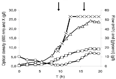
Subsequently, fed-batch fermentations were performed, and a feeding strategy of exponential followed by constant feeding was found to provide the best results in terms of biomass (10 g dry cell weight (dcw)/L in 19h), with a growth rate of 0.3 h-1 (Fig. 7). These results were then validated, achieving a final volumetric accumulation after 3 h of induction around 580 U/L.
Figure 7 – Behaviour of cultivation parameters during fed-batch procedure. The starting of the feeding and induction are indicated by the left and right arrows, respectively. OD, optical density [dcw (g/L) = 0,3863 s OD]; X, biomass concentration (g/L) (adapted from [40]).
In a different study, Silva and coworkers [39] optimized the production of hSCOMT using the same E. coli expression system but with a different approach based on artificial neural networks, in shake flask experiments. A method combining central composite design and artificial neural networks was applied to the above mentioned semidefined medium and a complex medium (SOB medium), to optimize the fermentation conditions for the production of hSCOMT. Temperature, pH and stirring rate were selected as input variables, with total hSCOMT activity being the response variable, and also monitoring biomass concentration levels. The optimization procedure adopted in this work lead to a maximum hSCOMT activity of 183.73 nmol/h onto a semi-defined medium at 40°C, pH 6.5 and stirring rate of 351 rpm [39]. The experimental strategy and validation presented left new perspectives for further trials.
Apart from the optimization of growth conditions mentioned above, to achieve high quantities of recombinant protein production, large-scale culture processes have to be applied, mostly based on fed-batch mode cultures [45, 46, 50].
A fed-batch culture is generally started with an inoculum growing at the maximum specific growth rate that can be sustained using the nutrients initially present in a bioreactor, followed by the imposition of a specific regime of nutrient feed until fermentation is complete [45]. These methods are based on mathematical models that describe growth patterns and the expected demand for nutrients [46].
17 The method of nutrient feeding is critical to the success of a fermentation process, as it affects both the maximum attainable cell concentration and productivity. Regarding the pattern of nutrient addition, three main types of pre-determined feeding profiles can be considered: constant, exponential and stepwise feeds [45].
In constant feeding, nutrients are fed into the bioreactor at a predetermined rate that does not change during the process. Due to the increase in culture volume and cell concentration, the specific growth rate continuously decreases [45]. In stepwise (or gradual) increasing feeding, nutrients are fed at ever increasing rates. It can enhance growth by supplying more nutrients at higher cell concentrations. Cell growth can be exponential during the entire culture period if the feed rate is increased in proportion to cell growth [45].
Exponential feeding allows cells to grow at predetermined specific growth rates, usually between 0.1 and 0.3 h-1, that can be set to fall below the maximum specific growth rate and also avoid acetate formation (a growth limiting by-product for E. coli) [45, 51].
Typically, the feeding rate can be calculated using a standard equation [45, 46] (1).
(1)
M – mass-flow rate of the carbon source (g/h) F – feed flow-rate (h-1)
Sf – substrate concentration in the feeding solution (g/L)
µ - specific growth rate (h-1)
YX/S – cell yield on carbon substrate, specific for each strain (g dcw/g substrate)
m – specific maintenance coefficient (g/(g dcw h) X – cell concentration (g/L dcw)
V – culture volume (L) t0 – time of feeding start (h)
t – process time (h)
Another feeding approach that can be used is the direct and indirect feedback control systems for the controlled addition of nutrients. Indirect control is based on online monitoring of parameters such as pH, dissolved oxygen, CO2 evolution rate and cell concentration. Direct
Besides all the fermentation conditions and strategies mentioned above, there are important additional factors that can interfere with cell growth and, consequently, with protein production.
One of the most important factors to monitor is cell physiology, a critical parameter in recombinant fermentations, since alterations in cell physiological state may reflect alterations in the host cell metabolism [52].
Bacteria are relatively difficult to analyze by flow cytometry, the best tool to assess cell viability, mostly due to their reduced size, but technical advances in instrumentation and methodology are leading to an increased popularity of flow cytometry and its range of applications in biotechnological processes [53].
One of the most used parameters to determine cell viability is membrane potential, which is considered a measure of the health of microorganisms, because only live cells are able to maintain its membrane potential. Membrane depolarization means a decrease in cell activity, but it does not imply cell death, it means that the membrane is structurally damaged and ions can go across it freely [54].
Membrane potential measurements are carried out by means of lipophilic dyes which go through the cell membrane and accumulate inside the cell when the membrane is depolarized according to its charge. Oxonols, such as BOX (Bis-(1,3-dibutylbarbituric acid)trimethine oxonol) are anionic and lipophilic, accumulating inside non-viable cells [54].
The evaluation of membrane integrity is the most definitive proof of cell viability. Cells showing intact membranes are impermeable to multiple charged dyes, the most common being propidium iodide (PI), but if cells loose membrane integrity, these dyes enter into the cells and emit fluorescence upon binding to nucleic acids [54]. Cells with damaged or compromised membranes cannot maintain or generate the electrochemical gradient and hence the membrane potential, and if a PI/BOX dual staining is used, these cells will emit fluorescence corresponding to both of these dies [53, 54].
19
Chapter 2 - Materials and Methods
2.1 - Materials
Ultrapure reagent-grade water was obtained from a Mili-Q system (MIlipore/Waters). Carbenicillin disodium salt, calcium chloride dihydrate, magnesium sulfate heptahydrate, lysozyme, cobalt(II) chloride hexahydrate, dithiotreitol (DTT), SAM chloride salt, DNase, epinephrine (bitartrate salt), disodium ethylenediamine tetraacetic acid (EDTA), sodium octyl sulphate (OSA), bovine serum albumin (BSA), LB-Agar, IPTG, tryptone, glycerol and PI were obtained from Sigma Chemical Co. (St Louis, MO, USA). Potassium chloride, sodium chloride, boric acid were supplied by Fluka (Buchs, Switzerland). Sodium phosphate dibasic and potassium dihydrogen phosphate monobasic were obtained from Panreac (Barcelona, Spain). Bis-(1,3-dibutylbarbituric acid)trimethine oxonol (BOX) was obtained from Invitrogen Corporation (Carlsbad, CA, USA). All other chemicals were of analytical grade and used without further purification.
2.2 – Methods
2.2.1 – Expression vector and bacterial strain
The Champion pET101 Directional TOPO expression kit (Invitrogen Corporation, Carlsbad, CA, USA) was used for the expression of hSCOMT on E. coli BL21(DE3) strain gently provided by Bial (São Mamede do Coronado, Portugal).
2.2.2 - Escherichia coli pre-cultivation, batch and fed-batch conditions
In this study, except for tryptone and glycerol levels, all media components for the semi-defined medium were kept constant (5.5 g/L Na2HPO4, 0.5 g/L NaCl, 1.64 g/L citric acidmonohydrate, 2 g/L potassium citrate, 1.21 g/L MgSO2·7H2O, 50 µg/mL carbenicillin and 1.5
mL/L trace elements solution) for the pre-cultivations batch and batch phase of fed-batch experiments. The trace elements solution consisted of 27 g/L FeCl3·6H2O, 2 g/L ZnCl2, 2 g/L
CoCl2·6H2O, 2 g/L Na2MoO4·2H2O, 1 g/L CaCl2·2H2O, 1.2 g/L CuSO4 and 0.5 g/L H3BO4,
prepared in 1.2 M HCl. The LB Agar plates with 50 µg/mL carbenicillin were inoculated from a cell bank aliquot and grown overnight at 37°C. From the plate, a single colony was inoculated in a 500 mL shake flask containing 125 mL of semi-defined medium, and grown at 37°C and
250 rpm until an OD600 (optical density at 600 nm)of approximately 2.6 was reached. Batch
and fed-batch processes were carried out in 750 mL bench-top parallel mini-bioreactors (Infors HT, Switzerland) with 250 mL of the semi-defined medium. The bioreactors were inoculated from the pre-cultivation to obtain a starting OD600 of approximately 0.2. The
temperature and pH were kept constant throughout the batch and fed-batch phases at 40°C and 6.5 (previously optimized by our research group [39]) respectively, with the pH value controlled by the automatic addition of 0.75 M H2SO4 and 0.75 M NaOH through two peristaltic
pumps. The pO2 (dissolved oxygen percentage) was controlled by a two-level cascade of
stirring (between 250 and 900 rpm) and air flow (between 0.2 and 2).
The feeds consisted of different concentrations of tryptone and glycerol dissolved in deionized water and the different feeding profiles tested were maintained by the automated peristaltic pumps controlled by IRIS software (Infors HT, Switzerland).
2.2.3 – Flow cytometry assays
In order to assess cells’ viability during the fermentation experiments, samples were retrieved at specific times and treated for the flow cytometry assays. The OD600 of the samples was measured and a dilution with PBS (8 g/L NaCl, 0.2 g/L KH2PO4, 1.5 g/L Na2HPO4 and 0.2 g/L KCl) was prepared to obtain a final OD600 of 0.2 (approximately 1x10⁸ cells/mL and further diluted in PBS with 4mM NaEDTA to a cell concentration of about 1x10⁶ cells/mL). To this cell suspension, the appropriate volumes of PI and BOX were added in order to attain final concentrations of 10 and 2.5 µg/mL, respectively. The samples were incubated for 15 min at room temperature in the dark, centrifuged for 5 min at 5000 rpm and resuspended in PBS prior to analysis in the CyAn ADP flow cytometer (Beckman Coulter Inc., California, United States). Acquisition was performed with Summit Software (Beckman Coulter Inc., Brea, CA). The acquisition was based on light scatter and fluorescence signals resulting from 25 mW solid state laser illumination at 488 nm. Fluorescence signals were collected by FL1 (530/40 nm, BOX) and FL4 (680/30 nm, PI) bandpass filters. Light scattering, BOX and PI fluorescence measurements were acquired logarithmically. Threshold was set on SSC to exclude noise, other particles and debris. Cells were gated according to light scatter parameters. Sample acquisition was operated at flow rate of no more than 300 events per second and a total of 5000 events were acquired for each sample.
21
2.2.4 – Glycerol assess
For glycerol assessment, samples were also retrieved at specific times and centrifuged at 4°C and 16000g for 5 minutes. The resulting supernatant was then filtered through a 0.22 µm filter for HPLC (high performance liquid chromatography) injection.
Quantification was carried out using an Agilent 1290 Infinity LC HPLC system (Waldbronn, Germany) coupled with a Refractive Index Detector (RID) (Agilent 1260 Infinity). Compound separation was done using a Hi-Plex H ion-exchange analytical column (Agilent, Santa Clara, CA, USA) with a 7.7 x 300 mm and 8 μm pore size. The mobile phase consisted of a 2.5 nM H2SO4 solution prepared with ultrapure water, filtered through a 0.2 μm pore membrane and
degassed for 15 min before use. Flow rate was set to 0.6 mL/min and column temperature was set to 65°C.
2.2.5 – Cell lysis
Intracellular SCOMT was obtained via a combined lysis process. Typically, 2 mL of samples from fermentations were centrifuged at 4°C and 16000g for 5 minutes, ressuspended with 500 µL of a standard buffer buffer (150 mM NaCl, 10 mM DTT, 50 mM Tris, 5 µg/mL leupeptin and 0.7 µg/mL pepstatin), transferred to lysis tubes and kept on ice. Subsequently, in order to perform the enzymatic lysis, 100 µL of 10 mg/mL lysozyme solution were then added to each tube and incubated at room temperature for 15 minutes. The disruption step continued by a freeze-thaw method, with the tubes submerged in liquid nitrogen until frozen and transferred to a 42°C bath for thawing. This process was repeated for 6 times to ensure maximum lysis efficiency. After this procedure, 50 µL of 1 mg/mL DNase solution were then added to each tube and the samples were centrifuged for 20 minutes at 4°C and 16000g. The resulting supernatant, containing the solubilized SCOMT, was then used as sample for the activity and protein quantitation assays.
2.2.6 – Protein assessment assay
The protein quantitation assay was carried out using a Pierce BCA Protein Assay kit (Thermo Scientific, USA) on a 96 well plate, and a specific volume of working reagent (WR) was prepared from solutions A and B from the kit according to the number of existing samples and blanks as follows:
Given this volume, the proportion of both solutions was calculated based on equations 3 and 4:
(3)
(4)
Then, 25 µL of each sample or blank (in triplicates) and 200 µL of WR were added to each well and homogenized. The plate was then incubated at 37°C for 30 min, after which the absorbance at 570 nm was measured and the values applied to a previously calculated calibration curve.
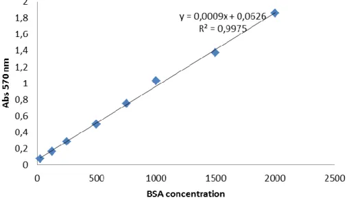
For the calibration curves, several solutions of different BSA concentrations (25, 125, 250, 500, 750, 1000, 1500 and 2000 µg/mL) were prepared in triplicates using a standard buffer (150 mM NaCl, 10 mM DTT, 50 mM Tris, 5 µg/mL leupeptin and 0.7 µg/mL pepstatin), 1/100 diluted. BSA solutions and 1/100 diluted standard buffer (working as blank) were subjected to the quantitation assay as mentioned above, and their absorbance measured at 570 and 595 nm. The results of the three replicates for each BSA concentration and the blank were averaged and two calibration curves were plotted, on for each wave length (Fig. 8 and 9).
23
Figure 9 – Calibration curve for BSA solutions absorbance at 595 nm.
Given the plots, the chosen wave-length for the assay was 570 nm, as it presented the best results (a R2 of 0.9975) compared to the 595 nm curve, and its equation was used to assess
the quantity of total protein in the samples.
2.2.7 – Enzymatic activity assay
The enzyme activity was measured via the quantity of metanephrine produced as a result of the reaction between hSCOMT and the substrate epinephrine.
The samples for this assay were prepared to a final volume of 250 µL by diluting a certain amount of the resulting supernatant of the lysis process in standard buffer (described above) in order to obtain the same amount of protein on each sample (150 µg/mL), based on the quantitation assays results (5).
(5)
The experiments were carried out in refrigerated, foil covered test tubes, adding 250 µL of the diluted sample and 200 µL of incubation solution (0.272 mg/mL SAM, 1 µL/ML MgCl2 (0.2
M), 40 µL/mL ethylene glycol tetraacetic acid (EGTA) (50 mM) and 959 µL/mL phosphate buffer) and incubating them for 5 min in a water bath at 37°C and 83 RPM. After 5 min, 50 µL of epinephrine solution (3.331 µg/mL, dissolved in incubation solution) were added, in order to let the enzymatic reaction to occur for 10 minutes. After 10 minutes, the reaction was stopped by transferring the tubes to ice and adding 100 µL of perchloric acid (2 M). The tubes were then kept at 4°C for 1 hour, after which the resulting samples were transferred to
eppendorfs and centrifuged for 10 min at 4°C and 6000 RPM. The samples were the filtered through a 0.22 µm pore size filter to remove precipitated biomaterial and injected to an HPLC system.
The HPLC injections were performed using a HPLC model Agilent 1260 system (Agilent, Santa Clara, CA, USA) equipped with an autosampler and quaternary pump coupled to an ESA Coulochem III (Milford, MA, USA) coulometric detector. Chromatographic separation was achieved on an analytical column Zorbax 300SB C18 reverse phase analytical column (250 x 4.6
mm i.d. 5 µm) (Agilent, Santa Clara, CA, USA). The mobile phase (0.1 M sodium dihydrogen phosphate, 0.024M citric acid monohydrate, 0.5 mM OSA and 9% acetonitrile, v/v), pH 2.9, was filtered under vacuum (0.2 μm hydrophilic polypropylene filter), degassed in ultrasonic bath before use. Column effluent was monitored with an electrochemical detector by a coulometric mode, which was equipped with a 5011 high sensitivity dual electrode analytical cell (electrodes I and II) using a procedure of oxidation/reduction (analytical cell #1: +410 mV; analytical cell #2: -350 mV). The flow rate applied was 1 mL/min. Column temperature
was optimized to 30ºC. The chromatograms were obtained by monitoring the reduction signal
25
Chapter 3 - Results and Discussion
In recent years, several attempts have been performed to obtain a large quantity of enzymatically active and pure hSCOMT. The first step to achieve this aim is to develop an adequate up-stream strategy, which means high levels of recombinant enzymatic production in an active form, in order to facilitate the subsequent purification steps. One of the most effective ways of enhancing recombinant protein production is the application of a fed-batch process, which highly increases cell density and, subsequently, protein production. However, a fed-batch process has its particularities, and it must be finely tuned to obtain the desired results.
In this work, a fed-batch bioprocess based on previously optimized culture conditions [39] was developed, via an up-scaling of hSCOMT production. Initially, several batch fermentations were carried out, in order to establish and optimize culture conditions, batch phase and bioreactor operation for the fed-batch fermentations. After this stage, a series of fed-batch fermentations with different feeding strategies were tested in order to obtain the maximum biomass production.
3.1 – Batch fermentations
As mentioned above, Silva and coworkers [39] optimized three physical culture conditions for the production of hSCOMT in shake flasks, namely temperature (40° C), pH (6.5) and stirring rate (351 rpm), and this was the starting point for the strategy described in the present work. Since the optimization was performed in bench-top parallel mini-bioreactors, and at this scale the stirring rate was not kept constant throughout the fermentations, the optimized 351 rpm stirring rate wasn’t applicable. As the agitation speed is associated with the dissolved oxygen concentration, the dissolved oxygen concentration was the parameter to optimize in this first step of our work, in order to assess the best value to be used in subsequent experiments, while temperature and pH values were kept constant in all of the experiments carried out through the whole work. Dissolved oxygen concentration in culture media, maintained by a cascade of agitation and airflow, in which agitation is constantly increased from 250 up to 900 rpm, followed by a constant increase of air flow from 0.2 to 2 (starting when maximum agitation is reached).
Dissolved oxygen concentration is one of the most difficult variables to reproduce, due to the combination of low solubility in water (even lower at higher temperatures) and, when cell density increases, the high nutritional demand [55] demands for pure oxygen supplementation to the culture media.
Since our studies were carried out at relatively high temperature (40°C), oxygen solubility is consequently lower, and so this is in fact an important parameter to establish in the first stages of a bioprocess.
Firstly, two batches were performed at 30% dissolved oxygen [40] to determine the typical growth curve under these conditions (Fig. 10). The medium composition was the previously described in section 2.2.2 of the Materials and Methods section, with a concentration of glycerol and tryptone of 30 and 20 g/L, respectively.
Figure 10 - Growth curve of E. coli in a batch process: 40°C, 30% dissolved oxygen, pH 6.5.
The growth curve presented in Fig. 10 showed that the stationary phase is reached after approximately 8 hours of fermentation. Under these conditions the maximum OD attained is of about 28.
The maximum obtained OD is surprisingly higher than the values previously obtained in our research group [40] for fed-batch fermentations applied to the same strain, medium and dissolved oxygen concentration.
In fact, just by applying the physical parameters optimized by Silva and coworkers [39] to a bioreactor scale, maximum OD values reached were promising, since no further optimization
27 Subsequently, the next step was to test three standard set-points for dissolved oxygen concentrations (20 and 40%, as well as the already used 30%), accomplishing this strategy by recombinant COMT induction
When using recombinant E. coli, growth and production phases can be separated, taking advantage of regulated promoters to achieve high cell densities in the first phase (while the promoter is “off”), and then high rates of heterologous protein production in the second phase (following induction) [56]. Given this information, cell culture should be grown until the beginning of the stationary phase, so that high cell densities are achieved prior to induction.
So, the three different dissolved oxygen set-points (20%, 30% and 40%) were tested in duplicates (Fig.11, 12 and 13). Fermentations were stopped four hours after induction. For activity assays, samples were retrieved at the end of the fermentation.
Figure 11 - Growth curve of E. coli in a batch process with 20% dissolved oxygen. Arrow indicates the induction time.
time (h)
OD
Figure 12- Growth curve of E. coli in a batch process with 30% dissolved oxygen. Arrow indicates the induction time.
Figure 13 - Growth curve of E. coli in a batch process with 40% dissolved oxygen. Arrow indicates the induction time.
Based on the maximum OD reached, these results showed that the batch at 20% oxygen gives better results than 30% and 40%. This may not correspond to the expected results as higher percentages of dissolved oxygen should allow increased cell growth. However, the maintenance of the set value of dissolved oxygen is not possible throughout the whole batch process using agitation and airflow cascade, indicating that oxygen supplementation might be required to conserve such dissolved oxygen levels. In fact, at advanced stages of fermentation, even the highest values of agitation and aeration cannot compensate oxygen
OD 600 time (h) time (h) OD 600
![Figure 2- Synthesis and metabolism of dopamine into homovanilic acid (HVA) and noradrenaline into vanillylmandelic acid (VMA) (adapted from [5])](https://thumb-eu.123doks.com/thumbv2/123dok_br/18068160.864263/24.893.159.742.150.420/figure-synthesis-metabolism-dopamine-homovanilic-noradrenaline-vanillylmandelic-adapted.webp)
![Figure 5– Chemical structures of nitecapone, tolcapone, entacapone and nebicapone (adapted from [28])](https://thumb-eu.123doks.com/thumbv2/123dok_br/18068160.864263/29.893.247.639.700.1057/figure-chemical-structures-nitecapone-tolcapone-entacapone-nebicapone-adapted.webp)
![Table 1- Some E. coli strains most frequently used for heterologous protein production and their key features (adapted from [44])](https://thumb-eu.123doks.com/thumbv2/123dok_br/18068160.864263/32.893.145.745.450.828/table-strains-frequently-heterologous-protein-production-features-adapted.webp)
![Table 2- Some Escherichia coli promoter systems and their specific characteristics that can be applied for heterologous protein production (adapted from [44])](https://thumb-eu.123doks.com/thumbv2/123dok_br/18068160.864263/33.893.130.688.393.878/escherichia-promoter-systems-specific-characteristics-applied-heterologous-production.webp)
![Table 3 – Semidefined medium composition (adapted from [40]).](https://thumb-eu.123doks.com/thumbv2/123dok_br/18068160.864263/34.893.307.600.552.843/table-semidefined-medium-composition-adapted-from.webp)