Analysis of failures in total hip arthroplasties
77
0
0
Texto
(2) Analysis of Failures in Total Hip Arthroplasties. II. Carla Bibiana Santos.
(3) Analysis of Failures in Total Hip Arthroplasties. MESTRADO INTEGRADO EM BIOENGENHARIA 2010/2011. Editado por FACULDADE DE ENGENHARIA DA UNIVERSIDADE DO PORTO Rua Dr. Roberto Frias 4200-465 PORTO Portugal. Tel. +351-22-508 1400 Fax +351-22-508 1440 Correio electrónico: [email protected] Endereço electrónico: http://www.fe.up.pt. Reproduções parciais deste documento serão autorizadas na condição que seja mencionado o Autor e feita referência a Mestrado Integrado em Bioengenharia – 2010/2011 – Faculdade de Engenharia da Universidade do Porto, Porto, Portugal, 2011.. As opiniões e informações incluídas neste documento representam unicamente o ponto de vista do respectivo Autor, não podendo o Editor aceitar qualquer responsabilidade legal ou outra em relação a erros ou omissões que possam existir.. III. Carla Bibiana Santos.
(4) Analysis of Failures in Total Hip Arthroplasties. IV. Carla Bibiana Santos.
(5) Analysis of Failures in Total Hip Arthroplasties. 'Pedras no caminho? Guardo todas. Um dia vou fazer um castelo.' Fernando Pessoa. V. Carla Bibiana Santos.
(6) Analysis of Failures in Total Hip Arthroplasties. VI. Carla Bibiana Santos.
(7) Analysis of Failures in Total Hip Arthroplasties. Resumo O objectivo deste trabalho é analisar as razões das falhas das artroplastias totais da anca numa amostra de doentes que realizaram uma cirurgia de revisão nos dois principais hospitais do Porto entre 21 de Novembro de 2005 e 23 de Novembro de 2010. Todos os dados recolhidos foram armazenados numa base de dados. Os tecidos foram analisados histologicamente. A análise dos componentes das próteses foi efectuada através de microscopia e técnicas de análise de imagem para quantificação. do. desgaste.. Todos. estes. resultados. foram. analisados. estatisticamente. As próteses foram explantadas de 39 doentes, sendo que 22 (56,4%) eram mulheres. A idade média de homens e mulheres era, respectivamente 51,9 e 56,4, não sendo estas diferenças estatisticamente significativas (p=0,338). O tempo médio de vida das próteses foi 13,9 anos nos homens e 11,1 anos nas mulheres, não sendo estas diferenças estatisticamente significativas (p=0,275). Artrose foi a principal causa para realizar uma artroplastia da anca (54,8%), e a principal razão de revisão da prótese foi a osteólise (70%). As próteses cimentadas apresentam em média um tempo de vida de 14,2 anos e as não cimentadas de 10,6 anos, no entanto este resultado não é estatisticamente significativo (p=0,252). Não foram observadas diferenças no tempo médio de vida das próteses entre os grupos de idade entre homens e mulheres (p=0,247). O nível de desgaste dos componentes apresenta uma diferença estatisticamente significativa entre as diferentes constituições do material (p<0,001). Os principais factores que influenciaram o desgaste das próteses foram o índice de massa corporal e o material constituinte das próteses. O material que constitui as próteses deve ser biocompatível e resistente de modo a evitar o desgaste e consequentemente a falha. Se o número de falhas for reduzido é possível aumentar o tempo de vida da prótese e por isso a qualidade de vida dos pacientes.. VII. Carla Bibiana Santos.
(8) Analysis of Failures in Total Hip Arthroplasties. VIII. Carla Bibiana Santos.
(9) Analysis of Failures in Total Hip Arthroplasties. Abstract The purpose of this study is to analyze the reason for failures of the total hip arthroplasties in a sample of patients that went to a revision surgery in the two main hospitals of Porto city, Portugal, from November 21th, 2005 to November 23th, 2010. In order to organize all data collected, a database was created. The tissues were histological analyzed. Device analysis was performed by microscopic analysis and a method of image analysis for wear quantification. All this results were statistically analyzed. Explanted devices were collected from 39 patients, being 22 female (56.4%). The mean of male age is 51.92 and the female have a mean of age of 56.41, but this difference was not statistically significant (p-value = 0.338). The average lifetime of the prostheses was 13.9 years for male and 11.1 years for female, these differences were not statistically significant (p-value = 0.275). Osteoarthrosis was the main reason to perform hip arthroplasty (54.8%), and the main reason for the revision of the prosthesis was osteolysis (70%). The average lifetime of cemented prostheses was 14.2 years and 10.6 years for uncemented prostheses, but this result was not statistically significant (p-value = 0.252). There was not a significant difference in the effect of age on lifetime prosthesis for males and females (p-value=0.247). Comparing wear level and device material it was verified a statistically significant difference between materials (p-value<0.001). The main factors influencing the wear of the prosthesis were the body mass index and the composition of the devices. The constitution of the materials of the devices must be biocompatible and hard to avoid wear and failure consecutively. If the number of failures is reduced, it is possible to increase the lifetime of the prosthesis and therefore improving the quality of life of patients.. IX. Carla Bibiana Santos.
(10) Analysis of Failures in Total Hip Arthroplasties. X. Carla Bibiana Santos.
(11) Analysis of Failures in Total Hip Arthroplasties. Agradecimentos Quero começar por agradecer à minha orientadora Prof. Doutora Maria de Fátima de Pina por ter orientado o trabalho de uma forma dedicada e ter estado sempre disponível para esclarecer as dúvidas que iam surgindo. Além disso queria também agradecer por todas as palavras de alento nas horas de maior aperto, por todo o empenho, rigor científico, ideias, sugestões, informações e opiniões durante todo o trabalho. De seguida queria agradecer à Prof. Doutora Meriem Lamghari por ter coorientado o trabalho e ter sempre presente a preocupação relativamente à evolução do trabalho. Bem como por todas as sugestões e ter estado sempre disponível para esclarecer as minhas dúvidas. À Dra. Inês Estevâo por todo o apoio e esclarecimento de dúvidas relativamente ao trabalho laboratorial e procedimentos tanto ao nível da histologia como da análise dos componentes. Quero também deixar uma palavra de agradecimento para a Dra. Daniela Silva do Laboratório de Microscopia Electrónica de Varrimento do CEMUP por toda a paciência e disponibilidade nas inúmeras horas de análise dos componentes. Aos compinchas de Bioengenharia por todos os momentos de desespero partilhados, as palavras de apoio, os desabafos, os silêncios e os momentos de alegria, brincadeira e animação que muito contribuíram para todo o processo. Não posso também esquecer a importância que tiveram os amigos do peito para dar apoio, ideias, sugestões, incentivos, ouvir desabafos, telefonemas a meio da noite ou até tirar dúvidas. Obrigada por toda a vossa paciência: Adriana, Miguel, João, Pedro, Mota, Sylvie, Carolina, Gustavo, Luís, Diana e Gonçalo. Por fim mas não menos importante quero agradecer à minha família: Mãe, Pai, Irmão, Avós e Avôs, Tias e Tios, Primas e Primos por toda a preocupação demonstrada, toda a ajuda, toda a paciência e todo o carinho que me transmitiram durante esta jornada.. XI. Carla Bibiana Santos.
(12) Analysis of Failures in Total Hip Arthroplasties. XII. Carla Bibiana Santos.
(13) Analysis of Failures in Total Hip Arthroplasties. Table of Contents. 1.. Introduction .......................................................................................................................... 1 1.1.. Motivation ..................................................................................................................... 1. 1.2.. Aim ................................................................................................................................ 2. 1.3.. Overview ....................................................................................................................... 2. 2.. State-of-the-art ..................................................................................................................... 5. 3.. Total Hip Arthroplasties ........................................................................................................ 9 3.1.. Anatomy and physiology of the hip .............................................................................. 9. 3.2.. Diseases that reach the hip joint ................................................................................. 13. 3.3.. Hip prostheses ............................................................................................................. 14. 3.3.1.. Cemented total hip prostheses ........................................................................... 15. 3.3.2.. Uncemented total hip prostheses ....................................................................... 17. 3.4.. 4.. 5.. Evolution of surgical techniques ................................................................................. 18. 3.4.1.. Technique ............................................................................................................ 18. 3.4.2.. New technique .................................................................................................... 21. 3.5.. Failure .......................................................................................................................... 23. 3.6.. Analysis of the failures ................................................................................................ 24. Materials and Methods ....................................................................................................... 27 4.1.. Histological Analysis .................................................................................................... 27. 4.2.. Component Analysis .................................................................................................... 28. Results ................................................................................................................................. 31 5.1.. Sample Description ..................................................................................................... 31. 5.2.. Surgeries and Analyzed prostheses ............................................................................. 31. 5.3.. Histologycal analysis.................................................................................................... 35. 5.4.. Devices analysis ........................................................................................................... 38. 5.5.. Statistical analysis........................................................................................................ 41. 6.. Discussion ............................................................................................................................ 47. 7.. Conclusions ......................................................................................................................... 53. References ................................................................................................................................... 55. XIII. Carla Bibiana Santos.
(14) Analysis of Failures in Total Hip Arthroplasties. XIV. Carla Bibiana Santos.
(15) Analysis of Failures in Total Hip Arthroplasties. List of figures. Figure 3.1- Anatomy of hip joint (Mattei, et al. 2010). ....................................... 9 Figure 3.2 - Schematic diagram showing parts of a standard femoral component for THA (Mirza, et al. 2010). ............................................................................. 11 Figure 3.3 - (A) Normal hip joint; (B) coxa vara; (C) coxa valga (Dorland 2007). 11 Figure 3.4 - Main components of an artificial hip joint (Mattei, et al. 2010). ........ 15 Figure 3.5 – A total hip arthroplasty with the use of methylmethacrylate cement to fix the prosthetic femoral and acetabular components to the bony structures (Siopack e Jergesen 1995)........................................................................... 16 Figure 3.6 – Antero external hip incision (Vail e Callaghan 2007). ...................... 19 Figure 3.7 - (A) initially well placed prosthesis, (B) after a few years reveals the cement fragmentation and osteolysis (Serra 2001).......................................... 20 Figure 3.8 - Technique for application of cement of second generation. (A) Scraping in pre-calculated caliber. (B) Introduction of plug channel. (C) Application of cement in the liquid phase. (D) Retrograde filling of the channel. (E) Prosthesis in the cement mantle thick homogeneous (Serra 2001). ........................................... 22 Figure 5.1 – Prosthesis (P) during a surgery. .................................................. 32 Figure 5.2 – X-ray prosthesis. ...................................................................... 32 Figure 5.3 – Total Hip Prosthesis composed by a stem, a metal head, a polyethylene acetabulum and a metallic acetabulum. ....................................... 33 Figure 5.4 – (a) Monoblock Titanium stem prosthesis with the polyethylene acetabulum, (b) Polyethylene acetabulum with severe wear. ............................ 33 Figure 5.5 – Two Titanium Acetabular cup with moderate wear. ........................ 34 Figure 5.6 – (a) Monoblock Titanium Stem, (b) Modular Cobalt Alloy stem. ......... 34 Figure 5.7 – Microscopy image with light transmission: Inflammatory process characterized by the presence of Macrophages (M) and Lymphocytes (L), HE Staining, (40x). ......................................................................................... 35 Figure 5.8 - Microscopy image with light transmission: Some metallic debris(MD), HE Staining, (40x). ..................................................................................... 36 Figure 5.9 – Microscopy image with light transmission: metallic debris (MD) of prosthesis wear, HE Staining, (40x). ............................................................. 36 Figure 5.10 - Microscopy image with polarized light: Some polyethylene debris (PD), HE Staining, (40x).............................................................................. 37 Figure 5.11 – SEM Images of a metallic stem surface: a) SE electrons e b) BE electrons. .................................................................................................. 38. XV. Carla Bibiana Santos.
(16) Analysis of Failures in Total Hip Arthroplasties Figure 5.12 - EDS Spectrum of the metallic stem observed in Figure 5.11. This spectrum show that the metallic stem is composed by Co-Cr. ........................... 38 Figure 5.13 - SEM Images of a polyethylene component with a metallic debris.: a) SE electrons e b) BE electrons . .................................................................... 39 Figure 5.14 - EDS Spectrum of the polyethylene cup with metallic debris presented in figure 5.13. This spectrum show that the metallic debris is composed by Titanium. .................................................................................................. 39 Figure 5.15 – Example of the method of image analyses used to quantify polyethylene wear. ..................................................................................... 40 Figure 5.16 – Results obtained for the method explained before for the %wear in the images analyzed in function of lifetime of prostheses. ................................ 41. XVI. Carla Bibiana Santos.
(17) Analysis of Failures in Total Hip Arthroplasties. List of tables Table 5.1 – Normality Tests. ........................................................................ 41 Table 5.2 – Comparison of lifetime prostheses in sex, age group, wear level, type of surgical technique, BMI, implant reason and material composition. .................... 42 Table 5.3 – Sex, age group, type of surgical technique, BMI, implant reason and material composition in Prostheses wear level. ............................................... 43 Table 5.4 –Reason of explants in cemented and non-cemented prostheses. ........ 44 Table 5.5 – Implant reason in failure, osteolysis, dislocation, infection, pain and fracture. ................................................................................................... 44 Table 5.6 – Mean lifetime prostheses by age group in male and female. ............. 45. XVII. Carla Bibiana Santos.
(18) Analysis of Failures in Total Hip Arthroplasties. XVIII. Carla Bibiana Santos.
(19) Analysis of Failures in Total Hip Arthroplasties. List of abbreviations and symbols. THA – Total Hip Arthroplasty BMI – Body Mass Index Sig. – Significance value µm - micrometers SPSS - Statistical Package for Social Sciences MRI - Magnetic Resonance Image INEB – Instituto de Engenharia Biomédica EDS - Dispersive Spectroscopy X-ray HE - Hematoxylin-eosin BE - Back-scattered electrons SE –Secondary electrons M/M - Metal-on-metal. XIX. Carla Bibiana Santos.
(20) Analysis of Failures in Total Hip Arthroplasties. XX. Carla Bibiana Santos.
(21) Analysis of Failures in Total Hip Arthroplasties. 1. Introduction. 1.1.. The. Motivation. main. critical. issues. for. implant. success. are:. the. implant. fixation/loosening related to the implant/bone interaction and the wear of the articulating surfaces (femoral head and cup surfaces). The adverse tissue reactions to wear debris cause loosening and implants failure, when it happens it is necessary to do a revision surgery. Total Hip Arthroplasty (THA) is one of the most successful operations of the 20th century because it gives to the patients, with some diseases that cause pain in the hip, a better quality of life (Skinner e Kay 2011). In 2007, in Germany, 152,338 primary hip prosthesis implantations and 21,782 hip prosthesis revisions and component revisions were performed (Efe e Schmitt 2010) while in France, the number of such surgeries was 117,400 and 22,427 respectively (Klouchea, Sariali b e Mamoudya 2010). Total Hip Arthroplasty is currently performed in 70,000 patients per year in the UK, 250,000 a year in the United States, and one million worldwide (Skinner e Kay 2011). In Portugal it is estimated that between June 1st, 2009 and May 31th, 2010 about 8000 primary THA and 1200 total hip revisions (THR) have been performed (R.P.A. 2010). Moreover there are costs associated with these surgeries, according to a research group that performed a cost analysis of aseptic revision surgery of total hip prostheses, those can cost 1.4 times more than the cost of the first THA (Klouchea, Sariali b e Mamoudya 2010). The achievement of a revision surgery to replace the device due to a failure is a complex procedure and has more risks for the patient than the first replacement surgery, also involves larger time of recuperation and major incapacitation to the patient (Klouchea, Sariali b e Mamoudya 2010), furthermore the economical costs are also a problem for the national health service. The state budgets assigned to the health systems in all countries is unable to bear all the expenses carried out by the high number of revisions that occur each year. 1. Carla Bibiana Santos.
(22) Analysis of Failures in Total Hip Arthroplasties worldwide, so it is pertinent that there is a unit responsible for reviewing the cases of failures especially those that occur in a short time after deployment because may occur any fault in the process, since the manufacture of the device until the surgery or a physiological fault, that will lead to an increased number of revision surgeries and therefore increase costs. For these reasons, understanding the causes of failures is crucial for patients and physicians and may allow development of more durable materials and effective ways to reduce the state costs in this kind of services.. 1.2.. Aim. The purpose of this study is to analyze the reason for failures of the total hip arthroplasties in a sample of patients that went to a revision surgery in the two main hospitals of Porto city, Portugal, from November 21th, 2005 to November 23th, 2010.. 1.3.. Overview. The dissertation will focus on the failure of total hip arthroplasties. Initially, it was necessary to have contact with the techniques that were used for the histological analysis of tissues explanted in revision surgery of hip prosthesis, get insight into techniques for analyzing material composition of the prostheses that were rejected, and it was necessary to organize all the data collected in a database system for later analysis. The data were analyzed in order to draw conclusions on which materials are more associated with failures, and the main reasons for revision surgeries, understanding the cause of these failures. These topics were addressed throughout this work, which can contribute to improve the quality of life of people in need of these prostheses. The first part of the dissertation, after the aims, is the state-of-the-art with the story of the beginning of the prosthesis since Charnley until now. Then there is the theoretical chapter with the following subjects: anatomy of the hip, the diseases that reach the hip joint, the several types of hip prostheses, the evolution in the surgical techniques, the causes of failure in the arhtroplasties and the analyzes performed for the failures.. 2. Carla Bibiana Santos.
(23) Analysis of Failures in Total Hip Arthroplasties After this chapter is the description of the methods used in all the work, and finally the results and discussion of them.. 3. Carla Bibiana Santos.
(24) Analysis of Failures in Total Hip Arthroplasties. 4. Carla Bibiana Santos.
(25) Analysis of Failures in Total Hip Arthroplasties. 2. State-of-the-art. Total Hip arthroplasty has been quite performed since its development by Charnley. It is a very important intervention, because it can produce dramatic improvements in functional status and health-related quality of life (Learmonth, Young e Rorabeck 2007). The use of this procedure is still increasing in developed countries, but surgical rates vary worldwide. The differences can be explained by differences in the prevalence of hip diseases and in clinical decision (Quintana, et al. 2000). To produce implants with a greater durability two primary requirements are needed: to achieve fixation to the host (with or without cement and in a variety of shapes, surfaces, and materials) which is practically solved; and the other one, the major limitation in this procedure, is to provide a durable bearing surface because if that not happens it leads to production of wear debris, with subsequent osteolysis and component loosening (Gard, Iorio e Healy 2000). Components must, therefore, provide durable fixation in the face of high stresses, whereas bearing surfaces need to be resilient and show low wear (Learmonth, Young e Rorabeck 2007). The principal failures that occur in THA include fracture of the implant, aseptic loosening as a result of mechanical failure of the fixation interface, infection, polyethylene wear, and dislocation. Developments in THA have been directed to the reduction of the failure rate and increased longevity of the patient, since higher failure rates were reported in younger patients, mostly because of a more active life (Learmonth, Young e Rorabeck 2007). Since 1976 (approval of the “Medical Device Amendments” law) medical devices in the United States have been placed under the regulatory umbrella of the Food and Drug Administration (FDA). Total hip prostheses fall under this agency's jurisdiction and have been generally classified as type II devices, which means that this device have a moderate risk for the patient (Heck, et al. 1995). Nowadays, implants may be grouped according to a basic hierarchy based on design type. Other features vary within the groups: metal component, collars, flanges, modularity, stiffness, neck geometry, head options, supplemental coatings, size, shape, proximal/distal matching, left and right sided (Gard, Iorio e Healy 2000). Osteoarthritis of the hip has been diagnosed in ancient skeletons and its prevalence and distribution seems no different from today. More than 100 years ago, the first attempt to treat hip arthritis surgically was made. This procedure, offered in the late 19th and early 20th centuries, consisted of the replacing of various tissues between the articulating surfaces of the hip. However a new era of. 5. Carla Bibiana Santos.
(26) Analysis of Failures in Total Hip Arthroplasties arthroplasty was introduced by Smith-Peterson in 1938 with an interposition of a vitallium cup, which covered the reshaped femoral head. In 1938, Wiles developed the first prosthetic total hip replacement (precedent of the modern ones). Other attempts were made but without good results due to poor design, inferior materials, and mechanical failure. In the early 1960s Charnley revolutionized management of the arthritic hip with the introduction of new ideas: the idea of low friction torque arthroplasty; use of acrylic cement to fix components to living bone; and introduction of high-density polyethylene as a bearing material (Learmonth, Young e Rorabeck 2007). Theoretical models have the effort of increasing service life of hip implants and they are a cheaper way to predict long-term behavior. Models of wear in hip implants have been developed only recently and are typically oversimplified with respect to the real case; for instance the wear caused by adhesion and abrasion is simulated as a whole, without distinguishing the separate contributions (Mattei, et al. 2010). Metal-on-metal (M/M) total hip replacements were used frequently in the 1960s. However, by 1975 the M/M combination was phased out and replaced by metal-on-polyethylene bearings because of the higher loosening rates with M/M hip prosthesis and concerns over biological reaction to the alloy constituents. But, in the late 1980s, concerns over osteolysis attributed to polyethylene wear debris led to the reintroduction of M/M bearings (Dumbleton e Manley 2005). These new devices have improved polished surfaces to reduce friction and increase longevity. Likewise, the ceramic implants are becoming popular with newer material of increased strength and smooth surfaces. But these alternatives are also challenges. There are reports of high levels of metal ions and a report of malignant change secondary to deterioration of the metal. Although the concern about the use of metal and other materials in the manufacture of implants still exists, regarding to malignancy may be lower than previously thought (McDermott 2004). Improvements in prosthetic materials, prosthesis design, and surgical techniques have increased the attractiveness of total hip resurfacing as an alternative to total hip arthoplasty for the treatment of younger patients. For the reason that hip resurfacing arthroplasty have many advantages over the traditional total replacement, including the preservation of bone stock, less stress shielding and thigh pain, fewer dislocations, reduced osteolysis, improved biomechanics and ease of revision (Jiang, et al. 2010). Research is currently focused on creation of an osteogenic stimulus to enhance bone on growth or heal bony defects. Work in nanotechnology to investigate the effectiveness of incorporating biologically active proteins onto. 6. Carla Bibiana Santos.
(27) Analysis of Failures in Total Hip Arthroplasties implants to enhance fixation it is still early, but, if successful, it could provide the implant coating of the future (Learmonth, Young e Rorabeck 2007). The large number of studies that report the THA failures leads to a demand for new alternatives for this procedure. A study of long-term success of cementless acetabular revision found a survival rate of 95% in 12 years. In primary total hip arthroplasty, however, the use of methylmethacrylate glue-type can still have excellent results. In a study of 30 years follow-up, 88% of implants were functional, with a revision rate of 7.3% at the time of death or end of study. Analysis of faults and their associated problems led to modifications of surgical techniques to preserve the attachment muscle and changes in the implants. Protection of bone attachment and bone interface surface led to a more judicious use of cement, the preservation of bone stock, and efforts to improve the fixation of the bone surface of the implant. There is evidence that particle debris from implants increases wear levels and provides more debris, and ultimately results in pathological failure of the implants. The use of Magnetic Resonance Image (MRI) to assess the pathology of the pelvis, colon, uterus, rectum and prostate, often leads to early detection of pathology of the hip. This interest was created in revascularization of the femoral head and other preventive efforts, in particular for avascular necrosis of the femoral head, a common cause of destructive arthritis of the hip. For over 30 years, THA has been a common procedure. Now, a growing number of arthroplasty revision procedures are being done (McDermott 2004).. 7. Carla Bibiana Santos.
(28) Analysis of Failures in Total Hip Arthroplasties. 8. Carla Bibiana Santos.
(29) Analysis of Failures in Total Hip Arthroplasties. 3. Total Hip Arthroplasties. Although hip arthroplasty is considered one of the greatest achievements in orthopedic surgery in the last decades, from an engineering point-of-view hip implants are not a complete success and still need further developments. In particular, because they tend to have a limited lifetime of about 15 years, not satisfactory for patients, especially the youngest ones. For those patients, recently emerged an alternative resurfacing technique, which is a surgery less invasive. (Mattei, et al. 2010).. 3.1.. Anatomy and physiology of the hip. A full knowledge of the functions and the behavior of the joint to be replaced is of enormous importance for the construction and the choice of the replacement part. The hip region is located lateral to the gluteal region, inferior to the iliac crest, and overlying the greater trochanter of the femur, or "thigh bone"; this joint is spherical and it is positioned in the pelvis between the femoral head and the acetabulum (Figure 3.1); it is a diarthrosis or synovial joint, because it has a capsule that contains the synovial fluid, a biological lubricant that acts also like a shock- absorber (Mattei, et al. 2010).. Figure 3.1- Anatomy of hip joint (Mattei, et al. 2010).. 9. Carla Bibiana Santos.
(30) Analysis of Failures in Total Hip Arthroplasties The basic structure around the hip, is the bony surface of superficial and deep muscular and neurovascular anatomy. This complex system of muscles and ligaments around the joint and bone setting the ball and socket provides stability to the joint (Mirza, et al. 2010). The clinically relevant surface anatomy of the hip is composed by several superficial palpable bony prominences. The reference points highlighted earlier are the anterior superior iliac spine and anterior inferior iliac spine, which serves as insertion point for the sartorius and direct head of the rectus femoris, respectively. The greater trochanter and posterior superior iliac spine are also easily identifiable in the posterior lateral aspect of the hip. Accurate identification, palpation, and understanding the relationship of these structures with the rest of the anatomy is vital for any hip surgery planning. As the detailed understanding of the accessories and muscle innervations are essential for the safety of open surgical procedures (Ranawat e Kelly 2005). The hip is a diarthrodial joint and is defined by restricted joint bone of the proximal femur and acetabulum. The acetabulum, which consists of three bones of the pelvis, the ischium, illium and pubis, form the Y-shaped triradiate cartilage, which usually fuses from 15 to 16 years old. It is oriented approximately 45° caudally (abduction) and 15° anteriorly (anteversion). Its hemispherical shape covers 170 degrees of the femoral head. Moreover, the proximal femur has only two primary growth centers of the femoral epiphysis and the trochanteric apophysis, which both ossify by age 16-18 depending on the sex of the individual. The muscle attachments that crossing the hip joint are extensive and they can be divided by function: primary flexors, extensors, the abductors, the adductors and finally, the external rotators. The vascular anatomy of proximal femur and acetabulum are extensions of internal and external iliac vessels (Ranawat e Kelly 2005). The femoral head diameter averages about 46mm. Two critical angular relationships of the femoral neck with the stem include the neck stem angle which averages 130 degrees and the femoral anteversion angle which averages 12 degrees. Femoral neck version is the angle of the femoral neck with the intercondylar plane. The hip joint contribution to lower limb length is the vertical distance from the femoral head centre to the lesser trochanter. Femoral offset is the horizontal distance from the midline of the longitudinal axis of the femur and the centre of rotation of the femoral head (Figure. 3.2).. 10. Carla Bibiana Santos.
(31) Analysis of Failures in Total Hip Arthroplasties. Figure 3.2 - Schematic diagram showing parts of a standard femoral component for THA (Mirza, et al. 2010).. Individual variations and conditions that affect head neck angle and femoral anteversion lead to changes in femoral offset and hip joint contribution to limb length. For example patients with hip dyplasia may have coxa valga and increased femoral anteversion, with resultant decrease in offset and increase in limb length while in coxa vara, the femoral neck angle is reduced, leading to greater offset and tendency to shortening (Figure 3.3). These conditions also pose a challenge in THA and need careful preoperative consideration (Mirza, et al. 2010).. Figure 3.3 - (A) Normal hip joint; (B) coxa vara; (C) coxa valga (Dorland 2007).. The hip joint is the articulation between the head of the femur and the acetabulum of the innominate bone, both joint surfaces are covered with a strong but lubricated layer called articular hyaline cartilage. It is a synovial ball and socket joint with the synovial fluid, which permits a wide range of movements to carry out the locomotors activities and transmit high dynamic loads (Mattei, et al. 2010). The movements of the hip joint are flexion, extension, abduction, adduction and medial and lateral rotation. The hip joint connects the lower limb to the trunk, and therefore is involved in the transmission of weight. This joint have severe mechanical requirements because it must be capable not merely of supporting the. 11. Carla Bibiana Santos.
(32) Analysis of Failures in Total Hip Arthroplasties entire weight of the body, but of stable transference of the weight, particularly during movement of the trunk on the femur, as occurs during walking and running. The joint must therefore possess great strength and stability, even at the expense of limitation of range of movement. The stability of the joint is determined by the shape of the articular surfaces (a deep socket securely holding the femoral head), the strength of the joint capsule and associated ligaments, and the insertion of muscles crossing the joint, which tend to be at some distance from the centre of movement (Palastanga, Field e Soames 2006). The ligament of the head of the femur, which is covered by a synovial membrane, extends from the acetabular fossa, where there is a fatty cushion, to the head of the femur. This ligament contains the artery to the head of the femur, which comes from the acetabular branch of the obturator artery. The head of the femur is also supplied with blood branches of the medial and lateral circumflex femoral arteries. The middle part of the upper rim of the acetabulum appears thickened in radiographs and may be called the roof of the socket (Platzer 2008). An angle of 30° that can be easily measured on radiographs is formed between a vertical line through the centre of the femoral head and a line from this centre to the bony margin of the acetabulum (angle of Wiberg). Decreases in this angle have implications for stability of the joint. Another measure which can be used to identify fractures and dislocations of the femur is the Shenton’s line, this is drawn along the upper margin of the obturator foramen and the inferior margin of the femoral neck to the medial side of the shaft. The head of the femur projects into the orbicularis zone like a button in a buttonhole. Together with the acetabular lip and atmospheric pressure, the zone orbicularis serves as an additional arrangement to maintain contact between the head and the socket. The ligament of the head of the femur runs within the capsule. Those regions of the capsule which are not strengthened by ligaments represent areas of weakness (Palastanga, Field e Soames 2006). Despite its remarkable characteristics, the hip joint can be affected, more often in aged people, by chronic pain and diseases such as osteoarthritis, rheumatoid arthritis, bone tumors or traumas. In these cases, the best clinical solution is the THA to replace the unhealthy hip joint with an implant, preserving the synovial capsule (Mattei, et al. 2010).. 12. Carla Bibiana Santos.
(33) Analysis of Failures in Total Hip Arthroplasties. 3.2.. Diseases that reach the hip joint. The hip diseases are commonly associated with people with more than 50 years old, but they may also appear in younger people. These diseases are characterized by a progressive destruction of joint components leading to a decreased function accompanied by pain that may be more or less intense, depending on the wear suffered by the joint. In some cases may require surgery to remove bone, cartilage or damaged joint, realign or alter the articular surfaces that are loading; or remodel the joint, replacing the same by means of another made with synthetic material, whose morphology is close to the human anatomy. There. are. many. reasons. to. perform. a. hip. replacement. however. osteoarthrosis (coxarthrosis) is the most common cause to do a hip arthroplasty. In the United Kingdom 93% of hip replacements are performed for osteoarthrosis (Murnaghan e Hamer 2010). Osteoarthrosis is a major cause of pain and disability, particularly in the elderly (CROFT, et al. 1990), because affects the articular surfaces of the joint and subchondral bone (Learmonth, Young e Rorabeck 2007). Coxarthrosis is a degenerative disease of the nature of the hip joint. Thus, there is a limitation of movement of the hip and is quite marked limitation of motion in abduction, adduction and rotation, but keeping up the movement of flexion (Bockstahler, Müller e Mlacnik 2004). This disease is often accompanied by deformations of the acetabulum, the femoral head and also pain in the affected area. Although the factors that lead to a primary osteoarthrosis are not entirely understood, the main causes known for the manifestation of this disease are: genetic factors and race, mechanical abnormalities, certain occupations, high body mass index and poor nutrition. Pain is the main symptom presented in hip arthritis. In the first phase of the disease symptoms include pain due to activity and loss of internal rotation, particularly in flexion, but with disease progression flexion and adduction contractures occur also, the pain becomes constant also occurring at rest and at night. Patients often limp and exhibit a decrease in the support phase of the affected limb in order to limit pain (Murnaghan e Hamer 2010). Thus, when traditional procedures (eg, analgesics, anti-inflammatory drugs, physiotherapy, etc.) do not produce pain relief, there is a prevailing need for joint replacement with an implant (Matos 2006). There are other reasons for performing hip replacement in cases such as bone fracture, for instance of the femoral neck. These fractures can occur through. 13. Carla Bibiana Santos.
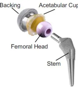
(34) Analysis of Failures in Total Hip Arthroplasties direct trauma, such as in accidents and falls, or due to osteoporosis. The patient has pain in the hip, with a fractured limb in external rotation and shortening. However it is mostly for orthopedic reasons that run an implantation of hip prosthesis, some other diseases that require deployment of a hip prosthesis are: rheumatoid arthritis, ankylosing spondylitis, Legg-Calvé-Perthes syndrome and congenital hip dislocation. Other less common diseases that lead to hip replacement are: Paget’s disease of bone and tumors of the neck and the head of the femur. There are cases that the surgery to hip arthroplasty is not recommended, like morbid obesity, history of infections and severe mental disorders (Muirhead-Allwood, Sandiford e Kabir 2008).. 3.3.. Hip prostheses. Hip joint replacement is more frequently done in individuals age 60 and older. Younger people who have a hip replaced may put extra stress on the artificial hip because of their life style; this extra stress can cause it to wear out and if that happens, part or all of the joint may need to be replaced; so hip replacement are performed in younger people only if there is no other alternative. The goals of hip replacement are to provide sustained pain relief and physiologic hip function. To achieve these objectives, there are some important steps to perform: the selection and preparation of the patient, the selection of a quality implant, and the execution of the operation. The accurate accomplishment of these criteria contributes to decreasing the probability of failure, which leads to the successful of arthroplasty (Gard, Iorio e Healy 2000). The success of hip arthroplasty as a treatment method of the joint resulted in a better quality of life for older people and increased the indications for this procedure. This growing consumer demand for new products stimulated the introduction of new materials and methods in the arthroplasties (Wroblewski, Siney e Fleming 2008). Hip prosthesis can be of two types, partial or total; partial hip prostheses have only one component, the femoral component (Moore partial prosthesis or Thompson prosthesis). The total hip prosthesis, which will be focused on this work, has two main parts (acetabulum and stem) that may consist in four components (Figure 3.4) (Mattei, et al. 2010):. 14. Carla Bibiana Santos.
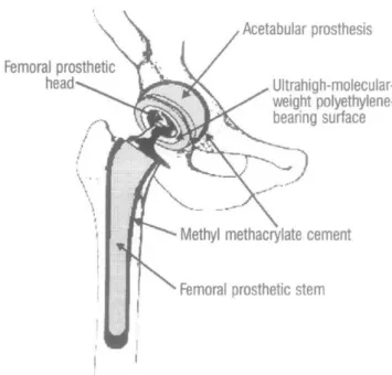
(35) Analysis of Failures in Total Hip Arthroplasties • The acetabular component that replaces the hip socket and consists of a metal shell coated on the inside of a hard plastic and slippery, that can be composed by one or two components; • The femoral component replaces the femoral head and sunk into the medullary canal of the femur, that can be composed by two pieces (head and stem separate) or only one piece with the head coupled to the stem.. Figure 3.4 - Main components of an artificial hip joint (Mattei, et al. 2010).. A very important step before an arthroplasty is the choice of the prosthesis according to materials, and this will depend on several factors, of which the most important one is to choose a prosthesis that, in principle, will have a greater longevity. Total hip prosthesis may be constituted by various materials and can be cemented or uncemented as explained below.. 3.3.1.. Cemented total hip prostheses. In the 1960s Charnley introduced and popularized the use of polymethyl methacrylate bone cement for fixation of total hip prostheses. Cemented fixation includes: bone-cement and cement-implant interfaces (Figure 3.5). The bone cement surface is the one that provides the foundation for durable fixation. Cemented total hip replacement is highly technique-dependent because the surgeon manufactures the bone-cement-implant composite at the time of surgery.. 15. Carla Bibiana Santos.
(36) Analysis of Failures in Total Hip Arthroplasties. Figure 3.5 – A total hip arthroplasty with the use of methylmethacrylate cement to fix the prosthetic femoral and acetabular components to the bony structures (Siopack e Jergesen 1995).. Cement is a grout not glue: fixation is achieved by mechanical inter lock rather than adhesion. To increase cement intrusion into bone and enhanced the interface shear strength there are some techniques like: cleaning of the endosteal bone with pulsed lavage, increased pressurization of cement enhanced penetration. In fact, the "cement" has no adhesive properties. Its role is to expander surface of the prosthetic components to the contour of the bone that houses the second smallest crevices, making them so congruent with the bone that there are fixed and, above all, thus creating a comprehensive interface for transmission of forces between the bone and the elements of the prosthesis (Serra 2001). The main concepts to consider in the design of the cemented stem are a taper-slip and a composite-beam. The first is a highly polished tapered stem designed to settle within the cement mantle and re-engage the taper, on the other hand, fixation of the composite beam relies on the shape of the implant and the composite fixation of stem to cement and cement to bone. To an improvement in cemented fixation of the acetabular component it is essential. cleaning. and. drying. of. the. reamed. acetabulum. and. sustained. pressurization of cement. For the medium term improvement is necessary to amend the stem design, changing the visco-elastic properties of cement and enhanced cementation techniques. The design of the polyethylene cemented cup has changed little over the decades (Learmonth, Young e Rorabeck 2007). The main characteristics of cemented stems are: smooth, rough, tapered shape and the geographic surface; and the cemented sockets are of two types: all polyethylene or. 16. Carla Bibiana Santos.
(37) Analysis of Failures in Total Hip Arthroplasties metal-backed polyethylene (Gard, Iorio e Healy 2000). The main advantages of cemented prostheses are their reduced cost (Gard, Iorio e Healy 2000), and the ability to add antibiotics in cement to keep the infection under control (Mokete e Naudie 2006).. 3.3.2.. Uncemented total hip prostheses. Implant stability and fixation are crucial for durability, therefore the conception of cementless femoral and acetabular components must be provide a great stability and to encourage bone to osteo integrate. Most published studies show that anatomically shaped stems indicate a higher frequency of thigh pain than with other traditional designs (tapered or cylindrical). Stems should have a rough surface (porous coating) to aid bony apposition to anchor the implant, thus the biomechanical forces can be transmitted to the femoral component through the joint. Tapered stems are fixed in threepoints of proximal cancellous bony to obtain immediate stability. The survival results obtained in this type of prosthesis have been good and only occasionally occurred thigh pain. Most of conical stems are made of chromium-cobalt, however there are no records of different survivals times in titanium stems. Titanium has a lower modulus of elasticity, similar to the host bone and is more biocompatible than cobalt chrome. Moreover, titanium is notch sensitive, which predisposes to cracking if the stem is not well supported. In the cylindrical stems is performed a distal cortical fixation is thus create greater leverage to resist torsion forces, compared with proximally coated stems, and also benefits from an immediate stabilization. To execute this procedure is necessary to have a large implant diameter and a canal-filling, the harshness of the stem depends on the elastic modulus of the material and is proportional to the fourth power of the diameter, this increase in diameter has been associated with pain in the distal thigh and proximal stress shielding. To lessen this pain several tests have been performed that attempt to decrease the stiffness of the stem (Learmonth, Young e Rorabeck 2007). Cementless acetabular cups are hemispherical in shape and most are totally porous-coated for bone ingrowth. Initial stability and fixation can be obtained by press-fit of the component; additional accessory can be provided by pegs, spikes, screws, or a threaded-cup design. This component was introduced to alleviate the difficulty fixation failure of cemented sockets made of polyethylene. Many uncemented components have predominantly fibrous tissue at the fixation interface instead of bony ingrowth. A substance used to enhance bone ingrowth and. 17. Carla Bibiana Santos.
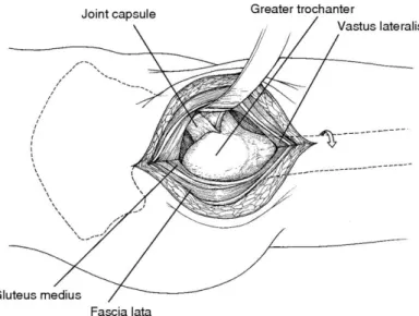
(38) Analysis of Failures in Total Hip Arthroplasties stimulate bony gap closure is Hydroxyapatite. Long-term results for uncemented total hip arthroplasty are poor compared with its cemented counterpart; however uncemented femoral components are the best option for younger patients (Learmonth, Young e Rorabeck 2007), (Gard, Iorio e Healy 2000).. 3.4.. Evolution of surgical techniques. The prosthetic total hip replacement is now the most widely used operation for the surgical treatment of coxarthrosis. The short and medium term results are good or excellent in 90% of patients. The improvement is mainly in pain and, to a lesser extent, mobility. The effect of such positive results, the operation has revolutionized the concepts of hip surgery, passed over other surgical solutions and making possible the treatment of situations, even outside of osteoarthrosis, so far no satisfactory resolution. Over the past twenty years, there have been made in Portugal a few thousand total hip prostheses (Serra 2001).. 3.4.1.. Technique. The operation consists of replacing the articular surface of the acetabulum and the femur by artificial materials. The intervention can be implemented in various ways and by different approaches. The most used incisions are the anteroexternal (Figure 3.6), between the fascia lata and the medium-and small gluteus, and posterior longiditunally separating the fibers of the large gluteal muscles and focusing the posterior capsule.. 18. Carla Bibiana Santos.
(39) Analysis of Failures in Total Hip Arthroplasties. Figure 3.6 – Antero external hip incision (Vail e Callaghan 2007).. The remains of the cartilage and subchondral bone acetabulum are scraped according acetabular dimensions for place the artificial cup in that location. The femoral head is removed and replaced by a metal head. There are many combinations of materials in various types of total hip prostheses currently in use but in the more usual pattern, the acetabulum is made of polyethylene chains of high density and the femoral stem is made of cobalt-chromium-molybdenum alloy. This pattern remains identical to the first successful implant, developed in the 1960s by John Charnley, English surgeon, still considered the most reliable in assessing long-term results (more than twenty years). The Poly (methyl methacrylate) (used in cemented prostheses) get a paste phase, by mixing a powder (polymer) and liquid (monomer) and then in exothermic (releases temperatures between 40º and 60º degrees), is as rigid as glass after a few minutes. Before placing the cup, is interposed a layer of cement in pasty phase in the acetabulum and then presses the cup to the hardening of the substance. To fix the femoral component, fill the medullar cavity of proximal femur with cement and put the stem inside it. The results obtained with these process seems to be excellent, this solution developed by Charnley seemed to have the mechanical attributes to withstand years of use. In the mid 1980s many of the prostheses placed by this method began to fail. Between cement and bone, previously in close contact, a type of synovial membrane with areas of bone destruction appeared (osteolysis) and leads to the loosening of prosthetic components (Figure 3.7). Then the movement between components and the bone leads to such pain that it was necessary to replace the prostheses with new ones, filling the cavities of bone destruction with greater. 19. Carla Bibiana Santos.
(40) Analysis of Failures in Total Hip Arthroplasties amounts of cement. After a year or two, the second prosthesis was again released by accretion of destruction and there was no bone to implant the new device. This peri-prosthetic osteolysis was interpreted as a foreign body reaction to the cement; later it was realized that this interpretation was wrong but the construction of prostheses without cement had already started.. Figure 3.7 - (A) initially well placed prosthesis, (B) after a few years reveals the cement fragmentation and osteolysis (Serra 2001).. It was not easy to dispense that expander surface tension that eliminated the tension crisis in the bone-prosthesis interface, decreasing the enormous distance between the modulus of elasticity of bone and the metal that, in preCharnley, did fail the first models. The solutions immediately available were selffixing acetabulum with varied relief for the surface of the metal-bone contact on femoral stems to encourage bone inlay. In the next generation, titanium arose between the metals, as the closest of the elastic modulus of bone. In the next phase, acetabula were themselves coated porous metal and with encrustations of hidroxyapatite to it and the femoral stem, used as a "generator of bone" to the interface, so for the adherence of bone to prosthesis.. 20. Carla Bibiana Santos.
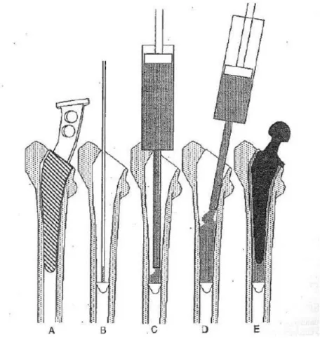
(41) Analysis of Failures in Total Hip Arthroplasties All this technology was more expensive than the models based on the "Charnley" to the point of the new prosthesis triple, and even in some, fivefold prices. The economic effort, that dignified the generation of non-cemented prostheses, it became questionable as soon as it was found that peri-prosthetic osteolysis also appeared with it, and even, in some cases, much faster and seriously. During all the structural changes attempted in the total hip prosthesis there was a common point: with the materials available, the lowest friction coefficient of articular technologically possible to build a lasting artificial joint in mechanical terms, was the metal with ultra density polyethylene. In the mid 1990s, with the persistent presence of peri-prosthetic osteolysis in cementless prosthesis, it became clear that this was not derived from the cement but the inflammatory reaction to polyethylene particles ultramicroscopic that absorbed by macrophages, generate a typical foreign body reaction. Under the effect of gravity or the pressure of joint fluid, these particles of less than 1 µm migrate distally into the femoral stem or proximally to the region of the cup. Then, finding passage between cement and bone or between the prosthesis cementless and bone, are phagocytized by cells macrophytes local, triggering cellular irritation that produces a range of inflammatory mediators. This reaction starts the periprosthetic. osteolysis,. generates. polynucleated. cells. similar. to. osteoclasts,. destroying bone and causes the dislocation of prosthetic components.. 3.4.2.. New technique. After the use of metallic acetabulum (alternative of the acetabular polyethylene) have indicated that it is not a good option too, it is necessary reshape the method. The cement is a good way to join the bone with the prosthesis. Applying the improved techniques (named third generation), prevents the existence of cracks between this and the bone. In the first prosthesis, the cement was introduced into the medullar canal of the femur with the aid of finger pressure to the filling completely. Then, inside that bulk, the femoral stem was placed. For the cement does not adhere to gloves as a glue, it was necessary to wait until passing the sticky state, and only uses it in the plastic phase. At this stage, the various layers of cement that were progressing into the spinal canal have not coalesced, remaining as a "puff pastry" streaky blood and. 21. Carla Bibiana Santos.
(42) Analysis of Failures in Total Hip Arthroplasties air vacuoles. The paste was introduced in this phase of polymerization and was also too strong to penetrate the recesses of the bone, leaving very little congruent bone-cement interface, and its thickness was not uniform around the prosthesis, causing tension points. Laboratory experiments have found that all these facts caused the mechanical failure that led to the fragmentation of the cement. New technique were created, called "second generation" (use of a plug cord to allow pouring cement in the liquid phase with a syringe in retrograde motion see Figure 3.8) and "third generation" (pressurization, reduction of bubbles air mixture by centrifugation or vacuum, pre-coating, centering rod, etc..), which began to promote a uniform cement mantle in texture and thickness, finally acceding to the crevices of the femoral channel. In support to this concept appear the reports of the surprising longevity of many cemented prostheses of Charnley, 25 and 30 years, revealing that no defect was found in cement, which is well able to avoid osteolysis, when applied with proper technique (Serra 2001).. Figure 3.8 - Technique for application of cement of second generation. (A) Scraping in precalculated caliber. (B) Introduction of plug channel. (C) Application of cement in the liquid phase. (D) Retrograde filling of the channel. (E) Prosthesis in the cement mantle thick homogeneous (Serra 2001).. 22. Carla Bibiana Santos.
(43) Analysis of Failures in Total Hip Arthroplasties If it is important to prevent the penetration of microscopic particles of polyethylene between the bone and the prosthesis, then it is important to promote rapid bone-implant integration in the margins of the acetabulum and the femoral neck, the areas of access to prosthetic interface. In this way, important changes have arisen in recent generations of these prostheses, creating a porous ring that stimulates bone growth by integrating either the acetabulum or the proximal femoral stem and prevent migration of particles (Serra 2001).. 3.5.. Failure. Not all surgeries are successful. An early or late infection may occur in 1-5% of cases, leading to devices dislocation of peri-prosthetic osteolysis. In such cases, it is necessary to remove the prosthesis and the decision to relocate or not is a major challenge to experience and sense of an orthopedic surgeon (Serra 2001). A hip revision surgery can have many reasons; in several years or just few months a failure can occur and there are several factors that can influence the onset of these failures. Major complications following hip arthroplasty are osteolysis, dislocation and infection and the causes leading to these states may be material defects, excessive wear of polyethylene elements, loosening of the artificial acetabulum, or wrong surgical techniques (Michalski, Plominski e Watral 2005). Osteolysis and/or aseptic loosening are the major cause for the revision of the hip surgery. Osteolysis is a process of active resorption of bone matrix by osteoclasts. There are many causes that can trigger this biological mechanism, referring to the main explanation to the formation of particles from the friction between the materials and the consequent immune response, leading to an active resorption of bone (Harris 1995). Symptoms can be minimal (pain on weight bearing at the beginning of the walk), particularly when there is lysis but limited loosening. Patients with large areas of osteolysis are at significant risk of periprosthetic fracture which requires frequent control. Radiographic evidence of loosening includes cracks in the cement mantle, subsidence or movement of the implant, and areas of lysis between the bone cement, bone-implant or implant cement interface. Treatment of osteolysis normally involves a revision surgery (Murnaghan e Hamer 2010). In total hip prosthesis, the dislocation tends to occur in the first three months after surgery if patients place the leg in inappropriate positions; or many. 23. Carla Bibiana Santos.
(44) Analysis of Failures in Total Hip Arthroplasties years after surgery due to implant wear and in these cases will, most likely, be held a revision surgery. If the hip move several times it is necessary to do a revision surgery. That instability could be related to the patient, disease, and implant technique or other factors. The risk of infection after a hip joint arthroplasty is common. Staphylococcus aureus and Staphylococcus epidermidis are the two most commonly identified pathogens and these originate from the patient’s skin, operating room environment or blood. Infection can be characterized as either early postoperative, chronic or haematogenous (the less common). There are some important points to try to prevent infections such as a coordinated approach to organism identification. Appropriate antibiotic management between surgeons and microbiologists is also an essential prerequisite to success with this type of surgery. Treatment of infection can involve thorough washout, debridement, exchange of modular components and antibiotics (acute and haematogenous infections); or radical debridement, removal of all foreign material and implantation of antibiotic loaded cement for chronic infection (Murnaghan e Hamer 2010). In cemented stems implanted the early failure was frequent and these were associated with localized areas of bone destruction and resorption (osteolysis). Their cause was initially believed to be infection but subsequently it was attributed to a local inflammatory response initiated by cement particles (Learmonth, Young e Rorabeck 2007). Failures. of. cementless. cups. include. accelerated. polyethylene. wear,. malfunction of the locking mechanism of the polyethylene liner in the metal-backed shell, and extensive periacetabular osteolysis. Screw holes in the shell enable debris to access the periacetabular cancellous bone, a further extension of the effective joint space. Modifications to acetabular shells with polished internal surfaces and better locking mechanisms should reduce these complications. Acetabular component survival is poor: a high proportion of failures is due to polyethylene wear and osteolysis (Learmonth, Young e Rorabeck 2007).. 3.6.. Analysis of the failures. The causes of failure mentioned above lead to a revision surgery, in which is often performed a replacement of the prosthesis. After such substitution it is necessary to analyze the components of the prosthesis removed and the tissues placed at the interface of the prosthesis to the human body.. 24. Carla Bibiana Santos.
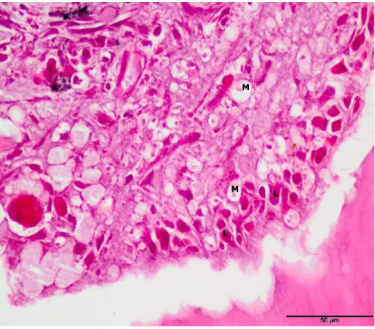
(45) Analysis of Failures in Total Hip Arthroplasties Tissue samples taken from the interface are prepared to be viewed in a microscope; it is with a microtome that extremely thin slices of material are cut. In some cases markers can be placed in these samples to detect the presence of certain substances in the tissue (immunohistochemical analysis). Particles extracted from interface tissues are obtained during revision surgery of total hip replacements according to the method of Yamac (sterilization, washing, resuspention and sonication) (SAEED e REVELL 2001). The tissue sections are fixed in buffered formalin and after stained with hematoxylin and eosin. All the sections will be studied microscopically, both with bright field and polarized light. The histological parameters analyzed are: inflammation and particulate debris (CHAROSKY, BULLOUGH e WILSON JR. 2007). Together with the histological analysis of the tissues, the explanted materials will be also analyzed, to further understand the reason of hip failures. The taper stem and the sleeve are sectioned for microscopic analysis using a water-cooled diamond. The implant is inspected using polarized light microscope and scanning electron microscope. In addition, a high resolution inductively coupled plasma mass spectrometry is performed on the blood serum sample (collected from the patient at the time of revision surgery) to measure the titanium ion levels (Paliwal, Allan e Filip 2007). The techniques mentioned above are the most commonly used for the analysis of the failed prosthesis, however there are other tests that can be performed in the devices of the explanted prostheses, such as are gel permeation chromatography for polymer components. A characterization of the materials from the point of view of their viscoelastic properties using a technique of dynamic mechanical analysis (DMA) can also be done. The finite element modeling, despite not allowing understand the failures of the prostheses, can be useful because it allows to recreate the model of the prosthesis placed on the patient and perform a more rigorous planning of surgery which can surely lead to less failures. Another analysis that is important to do is the quantification of the wear of the prosthesis material, such as polyethylene, since this has usually a higher wear. This quantification can be accomplished in several ways: quantification of the polyethylene that stay in the tissues (Slouf 2007) or with techniques of digital image analysis.. 25. Carla Bibiana Santos.
(46) Analysis of Failures in Total Hip Arthroplasties. 26. Carla Bibiana Santos.
(47) Analysis of Failures in Total Hip Arthroplasties. 4. Materials and Methods. In order to organize all data collected between 2005 and 2010, a database was created to register data about the patient, the revision surgery, the devices and the level of degradation. In the various fields of the different tables all relevant data were registered in order to have it organized and available for statistical analysis carried out later. This database was created based on ISO standards for retrieved and analysis of polyethylene and metal devices, ISO 12891-1, ISO 128912 and ISO 12891- 3 respectively. The participants of this study are patients who went to a revision surgery of a THA in São João or Santo António hospitals, in Porto city. The diagnosis of the prosthesis failure was confirmed by the surgeons who carried out the operation. The database was used to register the important data for the analysis performed later, for example, patient age, profession (it is important to know the type of activity performed daily), date of implantation of prosthesis, date of revision surgery, the materials that compose the prosthesis devices, tissues removed for analysis, and other information. The database consists of several tables, with data acquired from different formularies: one for information about the patient, another for the information about surgery and other one for materials and tissues explanted as well as results from their analyzes. In the database it is also possible to introduce graphic data such as X-rays and pictures of the prostheses in place, before the removal. All these information can help understanding and quantifying the bone degradation, associated to each patient.. 4.1.. Histological Analysis. The samples were received and identified with an internal code that was used throughout all subsequent procedures. These tissues were collected in zones: acetabular, femoral and articular. The first step in the processing of tissues was the total demineralization of the sample that allows the removal of calcium salts that were deposited in tissues. After this process it was necessary to prepare the samples for cutting. The samples were placed in paraffin that solidified in contact with a cold plate. The cuts were made on a rotation microtome with 3-4 µm.. 27. Carla Bibiana Santos.
(48) Analysis of Failures in Total Hip Arthroplasties The staining made in most samples was hematoxylin-eosin (HE). Before the use of optical microscopy with transmitted light to view the samples it was necessary to mount them with a cover slip. The details of the protocol followed in this analysis cannot be revealed because it is a confidential protocol of Instituto de Engenharia Biomédica (INEB).. 4.2.. Component Analysis. The explanted prostheses were received and identified with the internal code, and was previously sterilized according to manufacturer's recommendation and hospital procedures (autoclave). After a quick check of the condition of the received material, one may need to perform a cleaning process of the explants. If the device were not sterilized, should be placed in hydrogen peroxide 3%, to remove adhering blood. The devices analyses consist of a macroscopic and microscopic analysis. The first, aims at assess the existence and degree of wear, scratches caused in extraction or other relevant aspects, and these are done by evaluating each prosthesis and by acquiring multiple images of the prostheses received. Microscopic analysis was performed in a scanning electron microscope (SEM) to identify defects and impurities. The technique is usually associated with Energy Dispersive X-ray Spetroscopy (EDS), which allows a semi-quantitative analysis of the chemical elements on the surface of materials. This technique involves a preparation for mounting samples in an aluminum holder using carbon tape to the sample surfaces flat or araldite if the surface is uneven. The operating principle is based on the incidence of an electron beam at a point on the surface of the target sample, and subsequent collection of electronic signals emitted by the target material. The samples are scanned by a beam of electrons accelerated by a voltage that varies between 0 and 40 KV. This interaction results in the emission of various types of radiation and electrons, including secondary electrons (SE), used in the formulation of sample image and the back-scattered electrons (BE). The SE are originated in the process of no-elastic interaction of primary electrons, which suffer excitement. The electron back-scattered allows the distinction in the tested sample, the regions of light and heavy atoms, with higher binding energy.. 28. Carla Bibiana Santos.
(49) Analysis of Failures in Total Hip Arthroplasties In order to quantify polyethylene wear in each of the acetabular cup a method of image analysis was used. Based on the outer and inner diameter of the acetabulum, total area was calculated between these two circles by counting the pixels of the image in this region using the software MATLAB®. The region of wear was selected and its area also calculated. Based on the following formula, the percentage of wear in each of the acetabulum border could be calculated.. The acetabula that not entered in these analyses were those that showed no wear, those who were so damaged that it was impossible to identify their internal and external diameter and those on which we had no information regarding the lifetime of the prosthesis. For the statistical analysis, Statistical Package for Social Sciences (SPSS) software and statistical analysis to compare between different groups was used.. 29. Carla Bibiana Santos.
(50) Analysis of Failures in Total Hip Arthroplasties. 30. Carla Bibiana Santos.
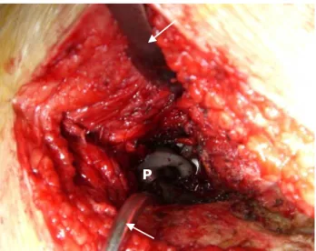
(51) Analysis of Failures in Total Hip Arthroplasties. 5. Results. In this section the description of the data collected and the results obtained in the analyses are presented. The images shown are just some representative examples of the results obtained.. 5.1.. Sample Description. Explanted devices were collected from 39 patients, being 17 men (43.6%) and 22 women (56.4%), corresponding to 40 revision surgeries (one patient went to 2 revisions surgeries in the study period). With the height and weight of the patients, the Body Mass Index (BMI) was calculated and a value above 25 was considered as overweight and below it was considered normal. From the total of patients, 19 had overweigh (48.7%) and 9 (23.1%) were normal. For some patients the BMI could not be calculated because of missing information either about their heights or weights. Missing data was observed in other variables. Concerning the age of the patient when the prosthesis was implanted, information about 30 patients were available. Ages ranged from 31 to 78 years old, with a mean and standard deviation (SD) of 54.5 (12.5). Lifetime of the prostheses, varies from zero to 30 years, with a mean (SD) of 12.3 (7.10). Regarding the type of prostheses, 21 (53.8%) were cemented and 13 (33.3%) non-cemented. Analyzing their composition, 24 (43.6%) devices were made of polyethylene 16 (29.1%) components by Titanium and 14 (25.5%) were made with a cobalt alloy.. 5.2.. Surgeries and Analyzed prostheses. All the analyzed prostheses were removed in a surgery made for the replacement of the device. The figure 5.1 is obtained during the surgery, and it is possible see the prosthesis and the lots of tissue that is necessary to separate with the retractors (marked whit a white arrow) for remove the prosthesis.. 31. Carla Bibiana Santos.
(52) Analysis of Failures in Total Hip Arthroplasties. P. Figure 5.1 – Prosthesis (P) during a surgery.. In the next figure (5.2) it is possible to see an x-ray of the prosthesis placed in the person. The database contains twelve x-ray images.. Figure 5.2 – X-ray prosthesis.. In the next figures, several types of prostheses analyzed in the context of this work are showed. Moreover it is possible to observe the different devices, shapes and the wear in some prostheses. In figure 5.3 a prosthesis with all the components: stem, head and acetabular cup. Acetabular component is constituted. 32. Carla Bibiana Santos.
(53) Analysis of Failures in Total Hip Arthroplasties for 2 parts, one of metal and another of polyethylene is presented. This prosthesis was placed with cement because it is possible to observe some residual of the cement in the metal cup (marked with a black arrow). The material composition of this device is cobalt-chromium-molybdenum alloy.. Figure 5.3 – Total Hip Prosthesis composed by a stem, a metal head, a polyethylene acetabulum and a metallic acetabulum.. In figure 5.4 a) a monoblock stem made by titanium and a polyethylene acetabulum of a 30 years-old prosthesis can be observed. The acetabulum is zoomed in figure 5.4 b) and a severe wear can be observed.. (b). (a). Figure 5.4 – (a) Monoblock Titanium stem prosthesis with the polyethylene acetabulum, (b) Polyethylene acetabulum with severe wear.. The figure below (5.5) presents two different acetabular cup shells with screws, and both constituted by titanium. As it is possible to observe, both prostheses have a moderate wear. The prosthesis on the right side had a lifetime of. 33. Carla Bibiana Santos.
(54) Analysis of Failures in Total Hip Arthroplasties 15 years and the other of 18 years. Both patients from whom the devices were explanted are overweight.. Figure 5.5 – Two Titanium Acetabular cup with moderate wear.. The stems rarely present wear, as can be observed in the next figure (5.6), where two different kind of devices are presented. In the right side there is a monoblock stem made by titanium and on the left side there is a modular stem constituted by a cobalt chromium alloy.. (a). (b). Figure 5.6 – (a) Monoblock Titanium Stem, (b) Modular Cobalt Alloy stem.. After the macroscopic analysis of the prostheses other analyses like histological were performed.. 34. Carla Bibiana Santos.
Imagem


+7


Documentos relacionados