Universidade de Aveiro Ano 2018/2019
Departamento de Química
CELESTINA GOMES
LOPES
CARATERIZAÇÃO DO PERFIL LIPÍDICO DE Bacillus
licheniformis I89 E DA SUA ALTERAÇÃO EM
RESPOSTA ÀS CONDIÇÕES DE CRESCIMENTO
CHARACTERIZATION OF THE LIPID PROFILE OF
Bacillus licheniformis I89 AND ITS CHANGES IN
Universidade de Aveiro Ano 2018/2019
Departamento de Química
CELESTINA GOMES
LOPES
CARATERIZAÇÃO DO PERFIL LIPÍDICO DE Bacillus
licheniformis I89 E DA SUA ALTERAÇÃO EM
RESPOSTA ÀS CONDIÇÕES DE CRESCIMENTO
CHARACTERIZATION OF THE LIPID PROFILE OF
Bacillus licheniformis I89 AND ITS CHANGES IN
RESPONSE TO THE GROWTH CONDITIONS
Tese apresentada à Universidade de Aveiro para cumprimento dos requisitos necessários à obtenção do grau de Doutor em Bioquímica, realizada sob a orientação científica da Doutora Maria do Rosário Gonçalves dos Reis Marques Domingues, Professora Associada com Agregação do Departamento de Química da Universidade de Aveiro e da Doutora Sónia Alexandra Leite Velho Mendo Barroso, Professora Auxiliar com Agregação do Departamento de Biologia da Universidade de Aveiro
Apoio financeiro da Fundação Calouste Gulbenkian, através de uma Bolsas de investigação para Pós-graduação e Especialização destinados a Estudantes PALOP
o júri
presidente Prof. Doutor Aníbal Manuel de Oliveira Duarte
professor Catedrático da Universidade de Aveiro
Prof. Doutora Ana Maria Pereira Gomes,
Professora associada da Escola Superior de Biotecnologia da Universidade Católica Portuguesa do Porto
Prof. Doutora Maria Amália da Silva Jurado
professora auxiliar da Universidade de Coimbra
Prof. Doutora Maria Manuel Santos Oliveira
professora auxiliar da Universidade de Trás-os-Montes e Alto Douro
Doutora Tânia Isabel de Sousa Caetano
Investigadora auxiliar da Universidade de Aveiro
Prof. Doutora Maria do Rosário Gonçalves dos Reis Marques Domingues
agradecimentos Escrever a tese não foi uma tarefa fácil, não é minha intenção aborrecer-vos com os motivos subjacentes e, sem o apoio das pessoas que passo a enumerar, provavelmente ainda me encontraria a trabalhar nela. Passo, assim, a expressar os meus sinceros agradecimentos.
À Professora Rosário Domingues e à Professora Sónia Mendo pela orientação deste trabalho, pelos conhecimentos transmitidos, pela disponibilidade e pela motivação. Mas acima de tudo, por me terem acolhido no vosso grupo de investigação e me terem oferecido a possibilidade de trabalhar neste projeto. Sou muito grata por tudo.
Aos restantes coautores dos trabalhos apresentados nesta tese pelos seus contributos.
Aos colegas do grupo de Espectrometria de Massa e do Laboratório de Biotecnologia Molecular do DBio pelo auxílio e pelo agradável ambiente de trabalho, e em particular à Joana Barbosa pela sua disponibilidade, acolhimento e conhecimento transmitidos. Á Dra. Cristina Barros pelo apoio no laboratório. Um especial obrigado à Iara, à Emeline, à Stephanie, à Cesária, à Eveline Brito, à Fernandina e o Malam Sanha que foram o meu porto de abrigo durante estes anos.
Um especial obrigado a todos os amigos que de alguma forma me incentivaram a ser resiliente.
À minha mãe, os meus irmãos e os sobrinhos pelo amor e apoio incondicional. À unidade de investigação de Química Orgânica, Produtos Naturais e Agroalimentares (QOPNA) (UID/QUI/00062/2019) e à Rede Nacional de
Espectrometria de Massa (REDE/1504/REM/2005) da Universidade de
Aveiro. Ao Centro de Estudos do Ambiente e do Mar (CESAM)
(UID/AMB/50017/2019), da Universidade de Aveiro.
À Fundação Calouste Gulbenkian pelo financiamento através de uma Bolsas de investigação para Pós-graduação e Especialização destinados a Estudantes Africanos de Língua Portuguesa e de Timor-Leste sem o qual este trabalho não teria sido possível.
palavras-chave Bacillus licheniformis I89, Acidos gordos, Espectrometria massa, Bacteria de Gram positivo, Fosfolípidos e Glicolipidos.
resumo Bacillus licheniformis I89 é uma bactéria de Gram positivo formadora de
endósporos, que possui a capacidade de produzir vários compostos de interesse biotecnológico incluindo proteases, amilases, surfactantes e antibacterianos. Não existe informação sobre a composição lipídica desta estirpe. Alguns estudos publicados sobre outras espécies B. licheniformis reportam apenas a composição em ácidos gordos. Entre os ácidos gordos identificados, estão descritos ácidos gordos ramificados, também encontrados em outras bactérias de Gram positivo. Estes ácidos gordos ramificados estão descritos como sendo benéficos para a saúde humana e úteis na prevenção de doenças, para além das potenciais aplicações biotecnológicas. Assim, o presente trabalho teve como objetivo alargar o conhecimento sobre o lipidoma de B. licheniformis I89, nomeadamente a) identificar o perfil de ácidos gordos de estirpe B. licheniformis I89 e analisar a adaptação do perfil de ácidos gordos em função da temperatura de crescimento (37 e 50 ºC) e nas diferentes fases do crescimento (lag, exponencial e estacionária), b) caracterizar o lipidoma de B. licheniformis I89 nas diferentes fases de crescimento (lag, exponencial e estacionária) a 37 ºC, c) avaliar o efeito do antibiótico vancomicina na alteração do perfil de lípidos de B. licheniformis I89, nas fases lag e exponencial de crescimento a 37 ºC. Para alcançar os objetivos propostos foram utilizadas as metodologias de cromatografia gasosa acoplada à espectrometria de massa (GC-MS) para identificação e quantificação de ácidos gordos e a cromatografia líquida de fase normal, usando uma coluna de interação hidrofílica, acoplada a espectrometria de massa (HILIC-ESI-MS) e MS/MS, para a identificação do perfil de lípidos polares. As mesmas metodologias foram também aplicadas para analisar o efeito de antibiótico vancomicina na alteração de perfil de lípidos da estirpe. O perfil de ácidos gordos (FAs) de B. licheniformis I89 obtido por GC-MS revelou a predominância de FAs ramificados das séries iso e anteiso (15:0, a15:0, i-16:0, i-17:0 e ai-17:0) e menor quantidade de ácidos gordos saturados (14:0, 16:0 e 18:0) em todas as condições de crescimento. O perfil de FAs variou com a temperatura e também com as fases de crescimento. Da fase lag para a fase estacionária, a 50 ºC, houve uma diminuição dos ácidos gordos ai-17: 0 e i-16: 0, enquanto que o ácido gordo i-15: 0 aumentou. Para a temperatura de 37 ºC, observou-se um aumento dos ácidos gordos i-15: 0 e i-16: 0 e uma diminuição dos ácidos gordos ai-15: 0 e ai-17: 0. A análise do extrato lipídico por HILIC-ESI-MS e HILIC-ESI-MS / HILIC-ESI-MS permitiu identificar o lipidoma de B. licheniformis I89, o qual ainda não tinha sido descrito até a data.
resumo No lipidoma de B. licheniformis I89 foram identificadas quatro classes de fosfolípidos: fosfatidiletanolamina, fosfatidilglicerol, lisil-fosfatidilglicerol e cardiolipina; duas classes de glicolípidos: monoglicosildiacilglicerol e diglicosildiacilglicerol; e duas classes de fosfogliceroglicolípidos: primer de ácido lipoteicóico monoalanilado e primer de ácido lipoteicóico. Todas as classes de lípidos foram identificadas nas três fases de crescimento analisadas, tendo-se observado variações na abundância em algumas espécies moleculares. Entre as fases exponencial e estacionária observou-se um aumento significativo nas espécies lipídicas 30:0 e uma diminuição significativa nas espécies lipídicas 32: 0, quando comparadas com as da fase lag. Para além disso, estudou-se ainda a alteração na composição do perfil lipídico de B. licheniformis I89 na presença de vancomicina nas duas fases de crescimento (lag e exponencial) a 37 ºC. Os resultados obtidos permitiram observar uma redução de algumas espécies moleculares de fosfatidilglicerol (PG), em resposta à presença da vancomicina. Uma vez que B. licheniformis I89 é sensível a este antibiótico, esta alteração pode ser justificada pelo facto da vancomicina atuar ao nível da inibição da síntese da parede celular podendo, por isso, afetar os lípidos que a constituem. Neste contexto, a abordagem lipidómica revelou-se uma ferramenta muito promissora no estudo da composição lipídica bacteriana, uma vez que esta permite analisar com precisão a alterações do perfil lipídico em resposta a diferentes condições de crescimento, nomeadamente, as que se observam na presença de antibióticos. Por outro lado, e dado a sua composição em ácidos gordos iso e anteiso, esta espécie de Bacillus pode vir a ser usada como fonte de ácidos gordos ramificados, uma vez que estes têm sido descritos como podendo ter atividade anti-tumoral. Muito embora essa atividade ainda tenha que ser investigada, os resultados obtidos fazem prever uma possível aplicação biotecnológica destes compostos com atividades terapêuticas.
keywords Bacillus licheniformis I89, Lipidomic, Fatty acids, polar lipids, Mass spectrometry, Gram-positive bacteria, Phospholipids, Glycolipids.
abstract Bacillus licheniformis I89 is a Gram-positive endospore-forming bacterium that
has the ability to produce various compounds with biotechnological application including proteases, amylases, surfactants and antibacterial. There is no information on the lipid composition of this strain. Some published studies on B. licheniformis species report the composition in fatty acids. However, some branched fatty acids, already described in Gram positive bacteria, have also been detected and are predominant constituents of B. licheniformis I89. These branched fatty acids have been described as beneficial to human health and useful in disease prevention, and thus have potential biotechnological applications. In the present work we studied the lipidome of B. licheniformis I89. The main objectives of the study were: a) to identify the fatty acid (FA) profile of B. licheniformis strain I89 and to evaluate the adaptation of the fatty acid profile in response to the growth temperature (37 and 50ºC) and in the different growth phases (lag, exponential and stationary), b) characterize the lipidome of B. licheniformis I89 in the different phases of growth (lag, exponential and stationary) at 37 ºC and c) evaluate the effect of the antibiotic vancomycin on the alteration of the lipid composition profile of B. licheniformis I89 in the lag and exponential growth phases at 37 ° C. In order to reach the proposed objectives, gas chromatography coupled to mass spectrometry (GC-MS) was used to identify and quantify the FAs and normal phase liquid chromatography using a hydrophilic interaction column coupled to mass spectrometry (HILIC-ESI-MS) and MS/MS was used to identify the polar lipid profile. In addition, GC-MS and HILIC-ESI-MS were also used to analyze the effect of the antibiotic vancomycin on the lipid profile change. The FAs profile of B. licheniformis I89 obtained by GC-MS revealed the predominance of branched FAs of the iso and anteiso series (i-15:0, ai-15:0, i-16:0, i-17:0 e ai-17:0) and a low abundance of saturated FAs (14:0, 16:0 and 18:0) in all the growth conditions.
abstract The FA profile showed variation with temperature and also with the growth phases. From lag phase to stationary phase at 50 ° C, there was a decrease of the FAs ai-17: 0 and i-16:0, while the FA i-15: 0 increased, whereas at 37 ° C, there was an increase of FA i-15:0 and i-16: 0 and a decrease of the FA ai-15:0 and ai-17:0. On the other hand, the lipidome of B. licheniformis I89 was identified for the first time by HILIC-ESI-MS and MS/MS. In the lipidome of B. licheniformis I89, four classes of phospholipids were identified: phosphatidylethanolamine, phosphatidylglycerol, lysylphosphatidylglycerol and cardiolipin; two classes of glycolipids: monoglycosyldiglycerol and diglycosyldiglycerol; and two classes of phosphoglyceroglycolipids: mono-alanylated lipoteichoic acid primer and lipoteichoic acid primer. All lipid classes were identified in the three growth phases analyzed, with a significant increase in the lipid species with 30:0 and a significant decrease in the lipid species with 32:0, between the exponential and stationary phases, when compared to the lag phase. In addition, a change in the composition of the lipid profile of B. licheniformis I89 was observed in the presence of vancomycin in the two growth phases (lag and exponential) at 37 ° C with the reduction of the levels of some PG molecular species. Vancomycin acts at the level of inhibition of the synthesis of the cell wall. B. licheniformis I89 is sensitive to this antibiotic and therefore, it seems to affect the membrane lipids. In this particular context the lipidomic approach employed is a very promising tool to study bacterial lipid composition. Since this allows to accurately analyze changes in lipid profile in response to different growth conditions, namely, those observed in the presence of antibiotics. Branched fatty acids have been described as having antitumor activity. Considering that B. licheniformis I89 is rich in branched fatty acids, this bacterium may be used as the source of this type of FA. However, it is still necessary to investigate a possible biotechnological application of these compounds as therapeutic agents.
xix
Contents
List of figures………xxiii
List of tables………. xxvi
Abbreviations………...xxvii
CHAPTER I- INTRODUCTION
General Introduction ……….3I.1 The genus Bacillus………..4
I.2 Bacillus licheniformis, a species with interesting biotechnological applications...4
I.3 The composition and role of lipids in the Gram-positive bacterial cell wall ………6
I.4 Methods used in lipid analysis in Gram-positive bacterium ………9
I.4.1 Analytical approaches in the study of lipid in bacteria ………...9
I.4.1.1 Analysis of lipid classes of Gram-positive bacteria by TLC …….13
I.4.1.2 Analysis of the fatty acid profile of Gram-positive bacteria by GC-FID and GC-MS………14
I.4.1.3- Analysis of the lipid profile of Gram-positive bacteria by mass spectrometry-based approaches ………...15
I.4.1.4 Lipidomic profile typical of Gram-positive bacteria using LC...18
I.5 The aim of the work ………20
I.6 References ………..21
CHAPTER II – DECODING THE FATTY ACID PROFILE OF Bacillus
licheniformis I89 AND ITS ADAPTATION TO DIFFERENT GROWTH
CONDITIONS TO INVESTIGATE POSSIBLE BIOTECHNOLOGICAL
APPLICATIONS.
II.1 Abstract………..32II.2 Introduction………32
II.3 Materials and Methods………...33
xx
II.3.2 Lipid extraction………...34
II.3.3 Quantification of phospholipids by phosphorus assay………34
II.3.4 Fatty acid analysis by gas chromatography-mass spectrometry (GC-MS) ...34
II.3.5 Statistical analysis………...35
II.4 Results………35
II.4.1 FA Profile of B. licheniformis I89………35
II.4.2 FA Profile at Different Growth Temperatures ……….36
II.4.3 FA Profile at Different Growth Phases ………37
II.4.4 Discussion……….37
II.4.5 References……….39
CHAPTER III- LIPIDOMIC SIGNATURE OF Bacillus licheniformis I89
DURING THE DIFFERENT GROWTH PHASES UNRAVELLED BY
HIGH-RESOLUTION
LIPID
CHROMATOGRAPHY-MASS
SPECTROMETRY
III.1 Abstract………43III.2 Introduction……….43
III.3 Materials and Methods………44
III.3.1 Bacteria and growth conditions………..44
III.3.2 Lipid extraction………...44
III.3.3 Quantification of phospholipids by phosphorus assay………44
III.3.4 Hydrophilic interaction liquid chromatography - electrospray ionization - mass spectrometry (HILIC-ESI-MS) ………45
III.3.5 Data and Statistical analysis………45
III.4 Results………..47
III.4.1 Phospholipid profile………47
III.4.2 Glycolipid profile………47
III.4.3 Phosphoglyceroglycolipid profile………..49
III.4.4 Lipid profile is growth phase-dependent………49
III.5 Discussion………49
xxi
III.7 References………52
CHAPTER IV- CHANGES IN Bacillus licheniformis I89 MEMBRANE
LIPID COMPOSITION AFTER EXPOSURE TO VANCOMYCIN
IV .1 Introduction……….57IV .2 Materials and Methods………....58
IV.2.1 Determination of minimum inhibitory concentration for vancomycin (MIC)………58
IV.2.2 Growth of B. licheniformis I89 in the presence of vancomycin………58
IV.2.3 Lipid extraction………..59
IV.2.4 Quantification of phospholipids by phosphorus assay………60
IV.2.5 Fatty acid analysis by gas chromatography-mass spectrometry (GC-MS)………...60
IV.2.6 Hydrophilic interaction liquid chromatography - electrospray ionization - mass spectrometry (HILIC-ESI-MS) ……….61
IV.2.7 Data and Statistical analysis………....62
IV.3 Results………...63
IV.3.1 MIC determination and establishment of vancomycin assay conditions…63 IV.3.2 Adaptation of FA profile from B. licheniformis I89 in the presence of vancomycin ………..63
IV.3.3 Adaptation of polar lipid profile from B. licheniformis I89 in the presence of vancomycin………..65
IV.4 Discussion……….70
IV.5 Concluding Remarks……….72
IV.6 References……….73
xxii
List of figures
CHAPTER I
Fig. I.1 Structure of the lantibiotic lichenicidin which is composed of two peptides Bliα and
Bliβ……….5
Fig. I.2 - General structure of the Gram-positive cell wall………..6
CHAPTER II
Fig. II.1 Main classes of fatty acids identified in B. licheniformis I89: a) Saturated fatty
acids represented here by 15:0; Branched fatty acids represented here by b) i-15:0, c) ai-15:0………...33
Fig. II.2 Principal coordinates analysis of the fatty acid composition of Bacillus
licheniformis I89 in growth phase and growth temperature at 37 ºC and at 50 ºC…………36
Fig. II.3 Comparison of the variation of fatty acid profiles of Bacillus licheniformis I89 in
each growth phase, depending on the growth temperature: lag at 37 °C and at 50 °C (a), exponential (Exp) at 37 °C and at 50 °C (b) and stationary (Sta) 37 °C and at 50 °C (c). Values are means ± standard deviation. * p < 0.05, ** p < 0.01, *** p < 0.001………36
Fig. II.4 Comparison of the fatty acid profiles of Bacillus licheniformis I89 between the lag,
exponential (Exp) and stationary (Sta) growth phases at 37 °C (a) and at 50 °C (b). Values are means ± standard deviation, * p < 0.05, ** p < 0.01………37
CHAPTER III
Fig. III.1. HILIC-ESI-MS chromatograms of total lipid extracts of B. licheniformis I89 in
(a) negative ion mode and (b) positive ion mode and the retention time (RT) of each polar lipid class. PG: Phosphatidylglycerol, RT 2.3 min; CL: Cardiolipin, RT 2.3 min; LTAP: Lipoteichoic acid primer, RT 2.5 min; DGDG: Diglycosyldiacylglycerol, RT 3.0 min; MGDG: Monoglycosyldiacylglycerol, RT 3.0 min; LTAP-Ala: Mono-alanylated
xxiii
lipoteichoic acid primer, RT 4.0 min; PE: Phosphatidylethanolamine, RT 5.1 min; lys-PG: lysyl-phosphatidylglycerol, RT 15.7 min………44
Fig. III.2 Chemical structures of the polar lipids identified in B. licheniformis I89……..45 Fig. III.3 LC-MS spectra of the phospholipid classes identified in B. licheniformis I89
lipidome in the negative ion mode: PG (a), lys-PG (c), CL (e) and in positive ion mode for PE (g). LC-MS/MS spectra and fragmentation pattern of one of the possible isomers of the
[M – H]− ions of PG (17:0/15:0) at m/z 721.5 (b), lys-PG (17:0/15:0) at m/z 849.6 (d), CL
(15:0/15:0/15:0/17:0) at m/z 1323.9 (f) and of the [M + H]+ ion of PE (17:0/15:0) at m/z
692.3 (h)………48
Fig. III.4 LC-MS spectra of glycolipids identified in B. licheniformis I89 lipidome: MGDG
(a) and DGDG (c). LC-MS/MS spectra acquired in positive ion mode and fragmentation pattern of one of the possible isomers of MGDG (17:0/15:0) at m/z 748.6 (b) and DGDG (17:0/15:0) at m/z 910.6 (d)………..50
Fig. III.5 LC-MS spectra of phosphoglyceroglycolipids identified in B. licheniformis I89:
LTAP (a) and LTAP-Ala (c). LC-MS/MS spectra acquired in negative ion mode and
fragmentation pattern of one of the possible isomers of the [M – H]− ions LTAP (15:0/15:0)
at m/z 1017.6 (b) and LTAP-Ala (17:0/15:0) at m/z 1116.6 (d)………51
Fig. III.6 Comparison of the polar lipid species of B. licheniformis I89 between the lag,
exponential (Exp) and stationary (Sta) growth phases at 37 °C: (a) PG, (b) lys-PG, (c) CL (d) PE, (e) DGDG, (f) MGDGD, (g) LTAP and (h) LTAP-Ala. Values are means ± standard deviation, * p < 0.05, *** p < 0.01 and *** p < 0.001………52
Chapter IV
Fig. IV.1 Variation of FA composition of B. licheniformis I89 membrane in the lag (a) and
exponential (b) phases, after exposure to vancomycin at concentrations of 0.008 and 0.125 µg/mL. Values are means ± standard deviation, * p < 0.05………...64
Fig. IV. 2 Variation of B. licheniformis I89 membrane polar lipid composition of the classes
PG, lys-PG, PE and DGDG in the lag phase, after exposure to vancomycin in concentrations of 0.008 and 0.125 µg/mL. Variation was observed for the polar lipid classes PG (a), lys-PG (b) PE (c), and DGDG (d). Values are means ± standard deviation, * p < 0.05, ** p < 0.01 and *** p < 0.001………..67
xxiv
Figure IV.3 Variation of the polar lipid composition of B. licheniformis I89 membrane in
the exponential phase, after exposure to vancomycin in concentrations of 0.008 and 0.125 µg/mL. Variation was observed for the polar lipid classes lys-PG (a) and PE (b), Values are means ± standard deviation, ** p < 0.01………69
APPENDIX SUPPLEMENTARY MATERIAL OF SECTION III
Supplementary fig. III.1 LC-MS spectra of the phospholipid classes (PLs) acquired in
positive ion mode for PG (a), lys-PG (c), and in negative ion mode for PE(e) and CL(g), identified in Bacillus licheniformis I89 lipidome. LC-MS/MS spectra and possible
fragmentation pattern of one of the possible isomers of the [M + NH4]+ ions of PG
(17:0/15:0) at m/z 740.5(b), [M + H]+ ion of Lys-PG (17:0/15:0) at m/z 851.6 (d), [PE – H]−
ion of PE (17:1/15:0) at m/z 690.3(f) and [CL – 2H]2− ion of CL (15:0/17:0) m/z 661.5(h)………80
Supplementary figure III.2 LC-MS spectra of the glycolipid classes (GLs) acquired in
negative ion mode for MGDG (a), DGDG (c) identified in Bacillus licheniformis I89 lipidome. LC-MS/MS spectra and possible fragmentation pattern of one of the possible
isomers of the [M – CH3COO]− ions of MGDG (17:0/16:0) at m/z 775.6 (b) and DGDG
xxv
List of tables
Chapter I
Table I.1 Summary at the results work using MS- approaches for the identification in lipids
composition of Gram-positive bacteria reported in the literature……….10
Table I.2 Typical specific fragmentation pathway (product ions and neutral loss (NL)) of
polar lipid from Gram-positive bacteria identified in the MS/MS spectrum………16
Chapter II
Table II.1 Variation of the fatty acid profiles of Bacillus licheniformis I89 in each growth
phase, depending on the temperature: lag at 37 °C and at 50 °C, exponential (Exp) at 37 °C and at 50 °C and stationary (Sta) 37 °C and at 50 °C and the lag, exponential and stationary growth phases at 37 °C and at 50 °C……….35
Table II.2 Similarity percentage analysis (SIMPER) identifying which FA contributing to
the differences recorded at the growth temperatures of 37 ºC and at 50 ºC in Bacillus licheniformis I89………...36
Chapter III
Table III.1 Molecular species of phospholipids from B. licheniformis I89 identified by
LC-MS in positive and negative ion modes. The iso and anteiso C15 and C17 are the most abundant FAs of B. licheniformis I89. It is not possible to distinguish the linear C15 and C17 from their branched isomers by LC-MS and MS/MS analysis……….46
Table III.2 Molecular species of glycolipids from B. licheniformis I89 identified by LC-MS
in positive and negative ion modes. The iso and anteiso C15 and C17 are the most abundant FAs of B. licheniformis I89. It is not possible to distinguish the linear C15 and C17 from their branched isomers by LC-MS and MS/MS analysis……….49
Table III.3 Molecular species of phosphoglyceroglycolipids from B. licheniformis I89
xxvi
abundant FAs of B. licheniformis I89. It is not possible to distinguish the linear C15 and C17 from their branched isomers by LC-MS and MS/MS analysis……….50
xxvii
List of Abbreviations
B. licheniformis Bacillus licheniformis CL Cardiolipin
ESI-MS Electrospray ionization mass spectrometry GC- FID Gas chromatography - flame ionization detector GC- MS Gas chromatography - mass spectrometry GL Glycolipids LC Lipid chromatography Lys-PG Lysyl-phosphatidylglycerol PE Phosphatidylethanolamine PG Phosphatidylglycerol PGL Phosphoglyceroglycolipids TA Teichoic acid
FID Flame ionization detector MS Mass spectrometry
HPLC High performance liquid chromatography,
HILIC Hydrophilic interaction chromatography
UHPSFC Ultrahigh performance supercritical fluid chromatography LTAP Lipoteichoic acid primer
LTAP-Ala Mono-alanylated lipoteichoic acid primer
HILIC-ESI-MS Hydrophilic interaction liquid chromatography coupled to electrospray ionization mass spectrometry
MS/MS Tandem mass spectrometry MGDG Monoglycosyldiacylglycerols DGDG Diglycosyldiacylglycerols
CHAPTER I. INTRODUCTION
GENERAL INTRODUCTION
I.1 THE GENUS BACILLUS
I.2 Bacillus licheniformis, A SPECIES WITH INTERESTING
BIOTECHNOLOGICAL APPLICATIONS
I.4 THE COMPOSITION AND ROLE OF LIPIDS IN THE
GRAM-POSITIVE BACTERIAL CELL WALL
I.5 METHODS USED IN LIPID ANALYSIS IN GRAM-POSITIVE
BACTERIUM
I.5.1 ANALYTICAL APPROACHES IN THE STUDY OF LIPID IN
BACTERIA
I.5.1.1 ANALYSIS OF LIPID CLASSES OF GRAM-POSITIVE
BACTERIA BY TLC
I.5.1.2 ANALYSIS OF THE FATTY ACID PROFILE OF
GRAM-POSITIVE BACTERIA BY GC-FID AND GC-MS
I.5.1.3 ANALYSIS OF THE LIPID PROFILE OF
GRAM-POSITIVE
BACTERIA
BY
MASS
SPECTROMETRY-BASED
APPROACHES
I.5.1.4 LIPIDOMIC PROFILE TYPICAL OF GRAM-POSITIVE
BACTERIA USING LC-MS
General Introduction
Bacteria comprises a large group of microorganisms with interest in the different fields, not only due to their effect in health, disease prevention, and treatment but also as pathogens. Bacteria can produce many compounds of interest with several biotechnological applications [1]. Bacteria have the ability to adapt to different growth conditions including temperature and pH which make them attractive to be used in industrial processes, since growth conditions can be adjusted and manipulated to improve the production of the biotechnological compound(s) of interest [2]. The bacterial lipid composition varies between species, environmental conditions, and growth phase [3–5]. Lipids seem to play an important role in the adaptation process and in the metabolism of bacteria. Nevertheless, the adaptation of lipid metabolism and the variation of lipid composition in bacteria in response to different growth conditions is far from being completely understood. Lipids are the main components of cell membranes, and different bacteria display different lipid composition [6]. Fatty acid profile is quite specific among different bacteria [7]. Nonetheless, it is recognized that under different growth temperatures, a shift in the fatty acid profile is observed [4]. Nonetheless, fatty acids are usually esterified to other lipids, such as phospholipids and glycolipids that are in fact the main constituents of the cell membranes and the cell wall of bacteria. But the knowledge of the composition and importance of polar lipids is far from being completely understood mainly because the bacterial lipidome is quite complex, and the lipidome of most bacteria remains unknown. Only in the last decade, with the use of modern mass spectrometry-based approaches, new studies have addressed the total lipidome of bacteria at the molecular level. This will contribute with new knowledge and understanding as regards the role of lipids in bacteria, with benefits for the discovery of new antimicrobials, or new biotechnological application of bacteria, their constituents and their products. The present work addresses the lipidome of B. licheniformis I89, a non-pathogenic Gram-positive bacterium producer of the lantibiotic (lanthipeptides with antibacterial activity) lichenicidin and other compounds with biotechnological interest.
I.1 The genus Bacillus
Bacillus is a genus of Gram-positive bacteria. It comprises rod-shaped endospore-forming bacterium with an aerobic or facultative anaerobic metabolism [8]. Members of the genus Bacillus have been isolated from a wide variety of environments such as plant and soil [9–11]. Some Bacillus species are pathogenic, yet many others are of industrial, and commercial interest [12]. Among the pathogenic species are B. anthracis, the causative agent of anthrax in humans and other animals, and B. cereus, which is a common cause of food poisoning [13]. B. thuringiensis and some strains of B.cereus are pathogenic to certain insects. Other Bacillus species have special characteristics that make them good candidates as biological control agents (fungicides, bactericides, and fertilizers) [14,15]. With regard to species with industrial applications [16], B. subtilis, B. licheniformis and B. amyloliquefaciens have a long history of safe commercial application in the food, detergents and pharmaceuticals industries [17]. Due to their applications, Bacillus species are of great economic interest and are of value to the biotechnology industry.
I.2. Bacillus licheniformis, a species with interesting biotechnological applications
Bacillus licheniformis is a non-pathogenic, Gram-positive endospore-forming bacterium frequently found in soil. It is a facultative anaerobe bacterium that belongs to the B. subtilis group II [18,19]. Members of this genus have been described as having several biotechnological applications as produces of valuable compounds such as proteases, amylases, surfactants, lantibiotics, biofuels, and other secondary metabolites [20]. It is usually considered safe, and some strains of this species are considered as probiotic and are used in the food and feed industry [12]. Also, this species produces several compounds of interest. Bacitracin is a peptide antibiotic produced by B. licheniformis with activity against Gram-positive bacteria, a few Gram-negative such as meningococci and gonococci and also against gas gangrene infection in guinea pigs and haemolytic streptococcal infection [21]. Lichenysin A, a cytotoxic substance, is a small cyclic lipopeptide structurally very similar to surfactants, also produced by B. licheniformis, exhibits antibacterial activity. It is not commercialized as antibacterial due of its toxicity [22]. Some B. licheniformis isolates can
mitigate the effects of fungal pathogens on maize, grasses and vegetable crops [19]. Other applications can be found for B. licheniformis. Its great fermentative capacity combined with low toxicity has made B. licheniformis 2336 a popular choice as probiotics [23]. In another recent study, antimicrobial compounds derived from B. licheniformis BFP011 isolated from papaya (Thailand) were shown to inhibit the growth of several important phytopathogens, as well as human pathogenic and food spoilage bacteria, namely, Colletotrichum capsici, Escherichia coli O157: H7 and Salmonella typhi ATCC 5784 [24]. The proteases from B. licheniformis are used in the detergent industry as well as for dehairing and bating of leather [25] and amylases. produced by this species are employed for the hydrolysis of starch, desizing of textiles and sizing of paper [19]. The antagonistic compounds, bacillocin from B. licheniformis is able to inhibit the growth of Gram-positive isolates such as B. anthracis, L. innocua, S. epidermidis [26]. Apart from peptides, polyketides are another important family of secondary metabolites produced from B. licheniformis that have also antimicrobial, immunosuppressive, antitumor or other physiologically relevant bioactivities. Although polyketides are widespread secondary metabolites from bacteria, only a few have been isolated and characterized from Bacillus [27]. In addition, the B. licheniformis I89 strain that will be studied in the present thesis was also reported as a producer of other compounds with biological interest. This strain, isolated from a hot spring environment in São Miguel, Azores, Portugal [28], is producer of lichenicidin, a two-component lantibiotic consisting of two peptides, Bliα and Bliβ (Fig 1) [29] with activity against methicillin-resistant Staphylococcus aureus (MRSA) and L. monocytogenes [30,31].
Fig. I.1 Structure of the lantibiotic lichenicidin which is composed of two peptides Bliα and
Bliβ [32].
Considering the interest and wide applications of B. licheniformis in the industry, in the manufacture of various biotechnological relevant products, it will be interesting to deepen the knowledge of the lipid composition of this bacterium. The information retrieved can be useful in searching for new applications of these molecules in other less explored areas of biotechnology.
I.3 The composition and role of lipids in the Gram-positive bacterial cell
wall
Gram-positive bacteria cell wall is a complex structure (Figure 1). The cell wall comprises a thick peptidoglycan layer of about 40-80 nm that stabilizes the cell membrane [33]. In addition, the cell wall of Gram-positive bacteria contains a unique component, the teichoic acid (TA), that can be covalently attached to the N-acetylmuramic acid unit of peptidoglycan (wall teichoic acids, WTAs) or anchored to a glycolipid to the cytoplasmic membrane (lipoteichoic acids, LTAs) [34,35]. The TA or related glycopolymer play crucial roles in bacterial survival under disadvantageous conditions and in other basic cellular processes such as protection against harmful substances and environmental stresses, control of enzyme activities and cation concentrations in the cell envelope, and binding to receptors and surfaces [34]. Gram-positive bacteria lack the outer membrane as well as phosphate-rich lipid A and lipopolysaccharide found in Gram-negative bacteria.
Fig. I.2 General structure of the Gram-positive cell wall adapted [36].
The bacterial cytoplasmic membrane forms a crucial barrier to the cell. It is composed of a glycerolipid bilayer with associated protein molecules and is very important for cell physiology, homeostasis, and survival. The biophysical properties of the membrane are, to a large extent, determined by the fatty acyl residues of the membrane and also by their composition in phospholipids and glycolipids [3,37].
Lipids are major compounds of bacteria cell membranes and share a large variety of biological functions. In bacteria, most lipids are located in the cell wall, mainly in the cytoplasmic membrane [38]. Phospholipids are the major constituent of the cell membrane and play a fundamental role in the maintenance of the membrane integrity, fluidity and charge that, in turn, modulate interactions with membrane-associated proteins [3,39]. The presence of anionic phospholipids renders bacterial cell surface negatively charged. This feature makes bacterial membrane the easy target of host immune molecules such as cationic antibiotic peptides [40–42]. Some of these phospholipids are also signalling molecules and participate in signal transduction [3]. The bacterial cytoplasmic membrane is a heterogeneous highly dynamic structure, comprising diverse domains differing in their phospholipid and protein composition, giving rise to defined membrane microenvironments and play an important role in compartmentation of specific proteins in the membrane.[43]. Some lipids are also carriers of important nutrients such as fatty acids, important for cell metabolism.
Gram-positive bacteria contain branched fatty acids with the major distribution of anteiso C15:0 and C17:0 chains. The most prevalent FAs in bacterial lipids have chain lengths between C12 and C20 [44]. The FAs profile can be straight-chained, branched-chained, unsaturated and cyclopropane. The branched-chain fatty acids are typical of Gram-positive bacteria and can be used as a marker for the differentiation of negative and Gram-positive bacteria [7].
Lipids in the bacteria cell membrane are quite diverse and bacteria specific and have some phospholipid and glycolipids that are not found in mammals. In bacteria, there are three major classes of phospholipids, namely phosphatidylethanolamine (PE), phosphatidylglycerol (PG) and cardiolipin (CL) [45,47]. Other phospholipids specific of Gram-positive bacteria were described. According to previous reports, Gram-positive species express at least one type of aminoacylated phosphatidylglycerol [37,45]. Unlike
Gram-negative bacteria, Gram-positive bacteria typically have less PE but have an abundance of lysyl-PG or other aminoacylated PGs which are absent in most Gram-negative bacteria [46,47]. The most common aminoacylated phosphatidylglycerol lipids found in bacteria are lysyl-phosphatidylglycerol (lysyl-PG) and also amino acyl esters including alanyl-, ornithyl-, or arg-PG , that have been found in several Firmicutes such as B. subtilis (lysyl-PG) [48,49], S. aureus (lysyl-PG) [50], and L. monocytogenes (lysyl-PG) [51,52], B.subtilis (alanyl-PG), [48,53], C. perfringens (alanyl-PG), B. cereus and Mycobacterium (ornithyl-PG), E. faecium and E. faecalis (arg-PG) [54]. Other serine and glycine-containing lipids are also known to exist in bacteria [55].
The presence of these amino acyl esters in the membrane reduces the susceptibility to antimicrobial peptides produced during host infection and their absence in the membrane of bacteria leads to hypersensitivity to antibiotics [56]. Besides, the phospholipid lysyl-PG is practically absent in Gram-negative bacteria. Phosphatidylinositol (PI) a phospholipid widely present in mammal cells is scarce in bacteria but it is found in a few Gram-positive species including Listeria monocytogenes [51]. Phosphatidic acid (PA) and phosphatidylserine (PS), are widespread in bacteria but they are present in small amounts [57]. Other types of polar lipids bearing phosphates groups found in Gram-positive bacteria are the lipo- and wall-teichoic acids found only in Gram-positive bacteria. Besides phospholipids, other lipids were reported in bacteria such as glycolipids (GL) and phosphoglyceroglycolipids (PGL), of which the last is only present in Gram-positive bacteria. PGL play crucial roles in cell shape determination, regulation of cell division, and other fundamental aspects of Gram-positive bacterial physiology and are important in pathogenesis and play key roles in antibiotic resistance [58]. Also, mannosides derivatives of phosphatidylinositol were found in Actinomycetes and in few other bacteria [6].
Different bacteria can have a different phospholipids composition, and their relative abundance in cell membranes can be bacteria specific. In Gram-positive bacteria belonging to the phylum Firmicutes, such as B. subtilis, S. aureus, and L. monocytogenes, PG is the main membrane component of the total phospholipid pool being PE the second most abundant class, followed by CL and Lys-PG [59].
In bacteria, the composition in phospholipids, either the proportion of the PL classes and their composition in fatty acyl chains can change rapidly in response to environmental conditions, such as extreme temperatures, osmotic stress or low pH [3]. The content in
branched fatty acids in membranes can vary substantially depending on the growth conditions. Membrane lipid composition affects membrane viscosity, which modulates membrane permeability and can influence both solute transport and protein interactions. Membrane lipid homeostasis is thus a crucial process and can compromise bacteria viability [60]. Membrane defines the boundary between a single cell and its environment and is, therefore, the main target for antibacterial agents.
The knowledge on the composition of lipid components of both the cell wall and the bacterial membrane is thus important and have been addressed for many years, using different analytical approaches but, only recently, with the development of new methodologies, such as omics base mass spectrometry approaches, this area is reaching new developments, which will be addressed in the following chapters.
I.4 Methods used in lipid analysis in Gram-positive bacterium
The knowledge on the composition of lipid components of the cell wall and the bacterial membrane has been addressed using low technology methodologies as thin layer chromatography (TLC), and gas chromatography (GC) coupled with flame ionization detector (FID) or mass spectrometry (MS) or high-tech approaches, namely mass spectrometry-based approaches, using liquid chromatography coupled with mass spectrometry (LC-MS). Both have been applied to the study of lipid composition of Gram-positive bacteria and including Bacillus as will be described in this chapter, that will highlight the state of the art of the results obtained in the identification of the lipidome in Gram-positive bacteria.
I.4.1 Analytical approaches in the study of lipid in bacteria
Lipid analysis is a multi-step process that starts with the extraction of lipids, followed by the separation of lipids and analysis. Most of the methods reported in bacterial studies are based on the extraction of lipids using conventional methodologies such as Bligh and Dyer and Folch methods [49,61,70,71,62–69] (Table 1). These are, still nowadays, the most
popular methods used in the studies of lipids in bacteria. The total lipid extract obtained can be fractionated followed by analysis of factions, or alternatively, the total lipid extract can be further analyzed. Fractionation can be done by thin layer chromatography (TLC), by solid phase extraction (SPE) or by liquid chromatography (LC). Total extracts or fractions can be analyzed by TLC, GC-FID, gas chromatography coupled with mass spectrometry (GC-MS) or LC-MS. The first approach, TLC, gives information about lipid class, the other following two approaches give information about the composition in fatty acids, while LC-MS is used to identify the total lipidome profile, with information at the molecular level and at the class level. Screening of bacterial lipids have been carried using these different MS methods (Table 1) and the information about these approaches and results will be detailed in the following subsections.
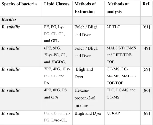
Table I.1 Summary at the results work using MS- approaches for the identification in lipids
composition of Gram-positive bacteria reported in the literature.
Species of bacteria Lipid Classes Methods of Extraction
Methods at analysis
Ref.
Bacillus
B. subtilis PE, PG,
Lys-PG, CL, GL, and GPL Folch / Bligh and Dyer 2D TLC [61] B. subtilis 6PE, 9PG, 2Lys-PG, CL, and 3DGDG, Folch / Bligh and Dyer MALDI-TOF-MS and LIFT-TOF-TOF [49]
B. subtilis 7PE, 4PG,
1Ly-PG, CL, and PA Bligh and Dyer GC-MS, LC-MS/MS, MALDI- TOF/TOF [59] B. subtilis 4PE, 8PG, PS and 6PA Hexane- propan-2-ol mixture TLC, LC-MS and GC-MS [86] B. subtilis PG, CL, alanyl-PG, Lyso-CL,
Bligh and Dyer QTRAP [88]
DGDG, LTAP, LTAP-Ala, and LTAP-(Ala)2
B. cereus PG, PE and CL Bligh and
Dyer TLC and GC-MS [62] B. stearothermopilus PG, PE, CL, and PGL Bligh and Dyer TLC and GC-MS [63] Enterococcus E. faecalis 17PG, 8Lysyl-PG, 23CL, 5DGDG, 3DAG, 4TAG and 3PGL Bligh and Dyer TLC, LC-MS/MS [64] Staphylococcus
S. aureus PG, PE,
Lysyl-PG, and CL Bligh and Dyer TLC [65] S. aureus PG, Lysyl-PG, CL, DGDG, MDGD, and DAG Bligh and Dyer RP-LC–Q-TOF-MS (Q-TOF-RP-LC–Q-TOF-MS) [66] S. warneri PG, CL, PC,
PA, and Lysyl-PG Bligh and Dyer TLC/ESI-MS, HILIC-LC-MS, MS/MS [67] Mycoplasma M. hyorhinis PG, CL, and GL Bligh and Dyer TLC [74] Clostridia
C. perfringens PG, PE, Lysyl-PG, and CL Bligh and Dyer 2D TLC [68 C. novyi PG, PGp, PE, PEp, Lysyl-PG, Lysyl-PG Bligh and Dyer 2D TLC, LC-MS [69] 11
Alanyl-PG, PS, and PT C. botulinum PG, PGp, PE, PEp, CL, CLp, Ala-PG and Lysyl-PG Bligh and Dyer 2D TLC, LC-MS [88] Listeria L. monocytogenes PG, CL, Lysyl-PG, Lysyl-CL and PI TLC [51] L. monocytogenes 8PG, 6CL, 6Lysyl-CL and 6DGDG Chloroform, methanol, and saline LIT MSn [52] Lactobacillus L. plantarum DGD and DGDG Bligh and Dyer ESI-MS/MS, 1D-, 2D-NMR, TLC, GC-MS [70] Streptococcus S. pneumoniae 23MGDG and 2DGDG CHCl3, CH3OH, 0.3% NaCl LIT/ MSn [89] S. pneumoniae PG, CL, DGDG, and MDGD TLC [91]
S. thermophilus DG and TG Folch TLC, GC/FID and
GC/MS.
[75]
I.4.1.1 Analysis of lipid classes of Gram-positive bacteria by TLC
TLC is a technique used to analyze, identify or separate the components of a mixture. It allows rapid and relatively inexpensive separation of the phospholipid classes and can be used prior to mass spectrometry analysis [71]. But this method has several disadvantages, such as low resolution and sensitivity, increased oxidation of lipid compounds, and given limited information in lipidome analysis. TLC is usually used to separate the major classes of lipids such was the case of separation of phospholipid classes [72]. Separation and fractionation of lipid classes are based on the different polarity conferred by the different composition of the polar head groups. The identification of each class is based on the comparison with lipid standards applied to the same TLC plate. After the separation, each phospholipid class can be quantified, by determination of phosphorous in each spot. It requires that each spot corresponding to different classes be scrapped from the plate and used for the quantification of the phosphorus in each class [63]. Analysis of lipids by TLC allowed the identification and separation of phospholipid classes in some Gram-positive bacteria. In TLC analysis, lipid classes were identified and separated in PG, PE, lysyl-PG, CL and DGDG in B. substilis [73], PE, PG, CL and PGL (phosphoglyceroglycolipid) in B. stearothermophilus [63], CL, PA, PG, Ceramide, PE, PC, PS in S. warneri [67], PG, CL, PE and lysyl-PG in S.aureus [65], PE, PG, CL, and lysyl-PG, Ala-PG and lyso-PG in C. perfringens [68], PE, PG, lysyl-PG, alanyl-PG, PS e phosphatidyl threonine (PT) and their plasmalogens in C. novyi ; MDGD, DGDG in L. plantarum ; PG, CL, DGDG and MGDG in S. pneumoniae [63]; in L. monocytogenes PG, CL, lysyl-PG, lysyl-CL [50], M. hyorhinis sphingomyelin (SPM), PG, PC, CL and neutral lipids (NL)[74] . However detailed structural information cannot be obtained. The lipids in each spot can be extracted using organic solvents and the respective extract can be analyzed by GC-FID or GC-MS or by MS to gain more structural information.
I.4.1.2 Analysis of the fatty acid profile of Gram-positive bacteria by FID and GC-MS
The fatty acids are routinely analyzed in the form of respective esters by GC-FID, or GC-MS [7,75]. GC allows an excellent separation of fatty acids prior to analysis, but requires extensive sample preparation, for the chemical derivatization of the fatty acids. Typically, they are esterified into the methyl ester in order to provide enough volatility for the separation by the GC. Most of the published works describe the identification of the fatty acid profile from the total lipid extract, thus allowing to identify the components in the fatty profile of bacteria [62,63]. In fact, these works showed that different Gram-positive bacteria have a characteristic composition in FA that could be used for taxonomic discrimination purposes. GC-MS has been applied for the analysis of the FA profile in the differentiation of bacterial classes [7,76–78]. Analysis of FA profile has been widely employed and it is not the goal of our work to revise all data from Gram-positive bacteria, and thus we will focus only on the main findings related with FA composition of Bacillus.
GC-MS approaches have been widely used to study the FA profile of several Bacillus spp. Overall, the FAs identified included saturated, unsaturated and branched FA, with a chain length between C14 to C19. FA with C12 and C13 have been scarcely reported. For example the FA profile of B.subtilis, B. stearothermophilus, B. simplex, B. vanillea sp Bacillus pumilus, and B. cereus comprised mainly the FAs i-15, ai-15, i-17, ai-17, i-16, 16:0, 18:0 [4,62,63,79–81]. Some other minor FA is sometimes reported, in addition to the previous ones, as is the case of cyclo-16:0 in Bacillus pumilus [81]. Other Gram-positive bacterium such as S.aureus has, the following FA as the most abundant, the FAs 14:0, i-15:, ai-15:0, i-16:0,16:0, i-17:0, ai-17:0, 18:0, i-19:0 ai-19 [82] and other less common FA such cys-18:1, 18:2 1-OH-12:0, tr-18:1, 2-OH-14:0 [83]. In the case of L. monocytogenes FAs i-15:0 ai-15:0 i-17:0 ai-17:0 were reported [84]. However, the percentage of each FA in the total FA profile is dependent on the species and on the growth conditions [36]. Most of the published works describe the FA profile and the composition of the total lipid extracts from bacteria. Other approaches have also been used, such as the TLC combined with GC-MS. In this approach, TLC allows to fractionate the different lipid classes, and after extraction from the silica, each separated class was derivatized and analyzed in terms of fatty acids classes by GC-MS [62,75]. Results of these studies showed that in both S.
thermophilus WT and ST6 strains, the fatty acids 14:0, 16:0, C16:1, 18:1 cyc-19cyc are the most abundant, whereas the seven strains of B. cereus contained i-15:0, ai-15:0, i16:0, 16:0, i17:0 and a17:0 as the main fatty acyl components of the phospholipids.
I.4.1.3- Analysis of the lipid profile of Gram-positive bacteria by mass spectrometry-based approaches
Mass spectrometry (MS) based approaches have been used for the identification and quantification of lipids in bacteria, and to elucidate taxonomic classification and differentiation. MS can be used to analyze the total lipid extract without previous fractionation, but this has the disadvantage of suppression of ions of lipid classes with lower ionization efficiency. To overcome this, several approaches coupled with MS analysis such as offline with TLC-MS or online with LC (LC-MS) can be used. LC-MS allows the largest coverage of lipid identified and reduces cross suppression following a more sensitive and quantitative detection of minor or poorly ionizing lipid species within complex mixtures.
The identification of lipids by MS has achieved at two different levels of analysis. First, the number of lipid species and their molecular weight is achieved by the information gathered in the MS data, by the identification of the type of ions formed in the source of the
mass spectrometry, [M + H]+, [M + NH4]+, [M – H]–, and identified in the MS spectra. Also,
confirmation of the identity of the polar head groups and the composition of fatty acyl chains is achieved by interpretation of MS/MS data. In fact, the MS/MS spectra show specific fragment ions and/ or fragmentation pathways that are specific to the classes of lipids. For example, in the case of, PG, the MS/MS spectra of the [M + NH4] + adduct, showed the typical fragmentation pathways due to neutral loss of the polar head group, seen as a mass
loss of 159 [48] (Table 2). The MS/MS spectra of the [M + H] + ions of Lys-PG showed
typical neutral loss (NL) of 300 Da and PE showed the NL of 141 Da. Table 2 summarizes the typical fragmentation pathways observed for the phospholipids identified in bacteria. The MS/MS spectra of glycolipids MGDG and DGDG showed characteristic NL of sugar units, -162 Da and also –loss of 2x162 Da (for DGDG) (table 2). The lipoteichoic acid
derivatives (LTAP) showed in the MS/MS spectra of the [M – H]– the product ions 79, 153,
and 171, while the LTAP-Ala showed also the product ion at m/z 88.0, 79.0 and the NL of 89.0 Da [52].
Table I.2 Typical specific fragmentation pathway (product ions and neutral loss (NL)) of
polar lipid from Gram-positive bacteria identified in the MS/MS spectrum.
Lipid class Ionization mode Product ion Neutral loss Reference
PG [M- H]- m/z 171 [M + NH4] + m/z 551.5 - 17 Da [49] - 172 da Lys- PG [M- H]- m/z 145.1 [66,67] [M+ H] + m/z 301.1 - 300 Da PE [M- H]- m/z 196.0 [49] [M+ H] + m/z 551.5 - 141 da MGDG [M + NH4] + m/z 551.5 - 197 Da [66] [M + CH3CO2]- [M + HCO2]- [M + Cl]- DGDG [M + NH4] + m/z 551.5 - 359 Da [66] [M + CH3CO2]- [M + HCO2]- [M + Cl]- LTAP [M- H]- m/z 79.0 [52] m/z 153.0 m/z 171.0 LTAP-Ala [M- H]- m/z 88.0 - 89 Da [52] m/z 79.0 m/z 153.0 16
Recently, MS and LC-MS approaches have been successfully used in the analysis of the lipidome of biological samples and, more recently, for the characterization of bacteria [67,69,85]. MS has received attention for bacterial detection and differentiation due to its capability, when used in conjunction with chromatographic separation, to identify compounds in complex mixtures [19,20]. It has the advantage of analyzing, in only one step, the entire organism´s lipidome.
The application of MS to bacterial identification was done initially by direct analysis of the total lipid extract obtained from bacteria using matrix-assisted laser desorption/ionization time-of-flight (MALDI-TOF) mass spectrometry to attempt to analyze phospholipids in the whole cell bacteria [22,23]. The use of MALDI-MS for phospholipids analysis helped to overcome numerous problems related to the complexity and diversity of extracts, as often encountered in bacteria material [24] (Table 1). With this MALDI-TOF approach, 9PE and 1PG were identified in E. coli [49]. Kondakova and co-workers used MALDI-TOF MS imaging coupled to HPTLC to screen of phospholipids classes of P. fluorescens MFAF76a and identified 3PG, 4PE and 2PC[38]. Guan and co-workers 2012 studied the lipid compositions of 11 representative strains of C. botulinum and a strain of C. sporogenes by MS [87]. All strains contained PG, CL, and PE in both the all-acyl and the alk-1’-enyl (plasmalogen) forms and Ala-PG and Lys-PG. C. butyricum, C. beijerinckii and C. acetobutylicum contained lipids characteristic of these saccharolytic species: a glycerol acetal and a PG acetal of the plasmalogen form of PE. C. perfringens was analyzed by LC-MS allowing the identification of PG, PE, lysyl-PG, Ala-PG and CL [68,86].
Electrospray ionization mass spectrometry also has been used for the analysis of the total lipid extract as a tool for bacterial identification [25]. It was used for the study of the Gram-positive bacteria C. botulinum and allowed the identification of the classes PlaGAPlaE, Lys-PG, Ala-PG. L. monocytogenes lipid extract was analyzed by ESI-MS to determinate the fatty acid substituents and their position on the glycerol backbone of the polar lipids, mainly PG, CL, lysyl-CL and DGDG [46]. L. plantarum was analyzed by ESI-MS and allowed identification of four major glycolipids of L. plantarum: b-D-glucopyranosyl-(1-6)-a-D-galactopyranosyl-(1-2)-D-glucopyranosyldiglyceride. The α-D-Glcp-diglyceride, α-D-Galp-(1-2)-α-α-D-Glcp-diglyceride, β-D-Glcp-(1-6)-α-D-Galp-(1-2)-6-O-acyl-a-D-Glcp diglyceride and the b-D-Glcp-(1-6)-α-D-Galp-(1- 2)-α-D-Glcp-diglyceride [82]. In a published work using MS for direct analysis of the total lipid extracts
by MALDI-MS or ESI-MS only few lipid species were identified. More recently the analysis of the total lipid extract by LC-MS has been used with success for the characterization of the lipidome of some bacteria. It allows the identification and quantitation of phospholipid and glycolipids in one single run [21].
I.4.1.4 Lipidomic profile typical of Gram-positive bacteria using LC-MS
Despite the development of the LC-MS-based lipidomics methodologies, there are still few studies on lipid profile analysis of Gram-positive bacteria (Table 1). The few studies published revealed that this approach has the advantage of providing the separation of lipids and analysis with higher sensitivity and more accurate identification. In LC-MS, the identification of each lipid species is achieved by the information of the retention time, MS information and specific identification by tandem mass spectrometry (MS/MS) data analysis and interpretation [26]. The LC-MS analysis of lipids in bacteria was done either using reversed phase as well as normal phase and hydrophilic interaction chromatography (HILIC) columns. Using the reverse phase, the separation of lipid species is based on their hydrophobic components, thus it is dependent on the fatty acid composition. LC-MS using reverse phase have been scarcely used in bacteria and there is only one published work on LC-MS using C18 reverse phase for B. licheniformis lipid species [91]. In LC-MS using the normal phase and HILIC columns, lipid species are separated based on the polar head polarity and allows the separation of lipids class. LC-MS for lipidomic studies is widely using HILIC or normal phase columns. In those cases, the separation is based on polarity and provides lipid class-specific separation. It is quite important for the discrimination of the isobaric species, such as the case of isobaric PE and PC species [30]. In LC-MS the identification of lipid species is achieved by the assignment of the retention time, identification of the ions observed in the MS spectra and also by the analysis of the LC-MS /LC-MS data. Overall, it allows identifying the ions attributed to the molecules within each lipid classes, as well as their composition in fatty acyl chains. Quantification of each lipid species can be obtained by integration of peak area of the RIC chromatogram attributed to each lipid species. Different classes of polar lipid have been identified in bacteria and depending on the chemical structure that can be analyzed in positive or negative ion modes.
Some published work reported the analysis of Bacillus genus and related genera by lipidomic approach. B. subtilis were studied by LC-MS/MS, LIFT-TOF/TOF or DI-MS. These studies allowed the identification of phospholipid classes as 4PG, 7PE, 1lysyl-PG, CL and PA [59], 10PG, 6PE, 2lysyl-PG, and 3DGDG [49] and 8PG, 4PE, 3PS and 6PA [92]. Furthermore, Almasoud and co-workers used MALDI-TOF-MS or LC-MS to study lipid
extracted 33strains from seven bacterial species belonging to the Bacillus and Brevibacillus
genera with the purpose of discrimination of species. Among the species analyzed, the most studied were those of B. licheniformis identified only by Lyso-PI [90]. PLs profile of E. faecalis and E. faecium were characterized by LC-MS and allowed the identification of 17 PGs, 8 LPGs, 23 CL, 3 GPDGDAG, 5 DGDAG, 3 DAGs, and 4 TAGs species [76] and 2 amino-containing phospholipid lysyl-PG, CL, PA and DPDGDAG [93] while in E. faecium 2 amino-containing phospholipid lysyl-PG, CL, PA, and DPDGDAG were identified [93]. As for the genus Staphylococcus, S. aureus, total lipidome was characterized by RP-LC-Q-TOF-MS or HILIC-MS/MS/MS. These studies allowed to identify 7 PG, 7 LPG, 10 CL, 13 DG, 21 DGDG, 8MGDG [77], PG, CL, Lysyl-PG, PC and PA [61]. Conversely, in Gram-positive bacteria and in the genus Clostridium plasmalogens with an alk-1-enyl ether substituent together with a normal fatty acid linked at glycerol were identified [78,79]. Among several polar lipids, in C. novyi NT through and LC-MS approaches the classes PE, PG, lysyl-PG, alanyl-PG, PS e phosphotidylthreonine (PT) and their plasmalogens were identified, [81].
Overall, the use of mass spectrometry techniques, especially ESI-MS and MALDI-MS, and LC-MS have clearly advanced the field of lipidomics in the profiling of diverse lipids or other metabolites in complex biological samples. MS-based approaches have been used for the identification of polar lipids isolated from the membrane, various phospholipid classes PG, PE, CL, lysyl-PG, and plasmalogen and glycolipids classes DGDG and MGDG that are the main predominant component of Gram-positive bacteria. Identification of the bacteria lipidome will be important because, in bacterial pathogens the cell membrane, is the target of several antibiotics and antimicrobial peptides and, thus, the lipid composition of the membrane is essential for the antibiotic-membrane interaction. In the lipidome, the signature is specific per bacteria and can be used for taxonomic and classification purposes. Furthermore, the identification of specific lipids can propose new biotechnological applications for these bacteria.
I.5 The aim of the work
The lipid profile of B. licheniformis I89 is completely unknown. Although several studies report the alteration of the fatty acid profile in the Bacillus spp, membrane, very few have focused on the lipidome characterization. In the present study, the profiling of the polar lipidome of B. licheniformis I89 was studied for the first time. The work aims at contributing to a better understanding of the adaptation of the lipid metabolism of Bacillus under different growth conditions. Herein, gas chromatography and liquid chromatography, mass spectrometry (MS) and tandem mass spectrometry (MS/MS) based approaches were performed to clarify the following aspects:
- identify the fatty acid profile of B. licheniformis I89, and evaluate the adaptation of the fatty acid profile to different growth conditions: temperature (37 and 50ºC) and stages of cell growth (lag, exponential and stationary);
- characterize the lipidome of B. licheniformis I89 at 37 ºC and at different stages of growth (lag, exponential and stationary),
- evaluate the effect of the antibiotic vancomycin on the lipid composition of B. licheniformis I89 at the different growth stages (lag and exponential) at 37 ° C.
The analytical approach employed, and the main results obtained during the development of this study are described in the following chapters:
Chapter II - Decoding the fatty acid profile of Bacillus licheniformis I89 and its adaptation to different growth conditions to investigate possible biotechnological applications
Chapter III - Lipidomic signature of Bacillus licheniformis I89 during the different growth phases unravelled by high-resolution liquid chromatography-mass spectrometry
Chapter IV – Changes in Bacillus licheniformis I89 membrane lipid composition after exposure to Vancomycin.
I.6 References
[1] R. Marchant, I.M. Banat, Microbial biosurfactants: challenges and opportunities for
future exploitation, Cell Press 2012, 30: 1-8.
[2] F. Baruzzi, L. Quintieri, M. Morea, L. Caputo, Antimicrobial compounds produced
by Bacillus spp. and applications in food, Science agianst microbial pathologens: communicating current reaesach and technological advances 2011, 1102–1111.
[3] Y. Zhang, C.O. Rock, Membrane lipid homeostasis in bacteria, Nat. Rev. Microbiol
2008, 6:222–233.
[4] J. Beranová, M. Jemioła-Rzemińska, D. Elhottová, K. Strzałka, I. Konopásek,
Metabolic control of the membrane fluidity in Bacillus subtilis during cold adaptation, Biochim. Biophys. Acta – Biomembr, (2008), 1778:445–453.
[5] T.J. Denich, L.A. Beaudette, H. Lee, J.T. Trevors, Effect of selected environmental
and physico-chemical factors on bacterial cytoplasmic membranes, 2003.
[6] C. Sohlenkamp, O. Geiger, Bacterial membrane lipids: Diversity in structures and
pathways, FEMS Microbiol. Rev 2015, 40:133–159.
[7] Y. Li, S. Wu, L. Wang, Y. Li, F.S. X. Wang, Differentiation of bacteria using fatty
acid profiles from gas chromatography–tandem mass spectrometry, 2010, 380–1383.
[8] K.S. Ko, W.S. Oh, M.Y. Lee, J.H. Lee, H. Lee, K.R. Peck, N.Y. Lee, J.-H. Song,
Bacillus infantis sp. nov. and Bacillus idriensis sp. nov., isolated from a patient with neonatal sepsis, International Journal of Systematic and Evolutionary Microbiology 2006, 56:2541-2544
[9] S.S. Bae, J.H. Lee, S.J. Kim, Bacillus alveayuensis sp. nov., a thermophilic bacterium
isolated from deep-sea sediments of the Ayu Trough, Int. J. Syst. Evol. Microbiol. 55 (2005) 1211–1215.
[10] W. Demharter, R. Hensel, Bacillus thermocloaceae sp. nov., a New Thermophilic Species from Sewage Sludge, Syst. Appl. Microbiol. 11 (1989) 272–276.
[11] S. Mendo, N.A. Faustino, A.C. Sarmento, F. Amado, A.J.G. Moir, Purification and characterization of a new peptide antibiotic produced by a thermotolerant Bacillus licheniformis strain, Biotechnol. Lett. 2004, 26:115–119.
[12] F.M.F. Elshaghabee, R.D.G. Namita Rokana, C. Sharma, H. Panwar, Bacillus as potential probiotics status, concerns, and future perspectives, Front. Microbiol. 2018,
![Fig. I.1 Structure of the lantibiotic lichenicidin which is composed of two peptides Bliα and Bliβ [32]](https://thumb-eu.123doks.com/thumbv2/123dok_br/15914102.1093055/33.892.141.785.847.974/fig-structure-lantibiotic-lichenicidin-composed-peptides-bliα-bliβ.webp)