University of Aveiro 2015
Department of Chemistry
RAQUEL SOFIA
RIBEIRO LOPES
Caraterização de compostos orgânicos solúveis em
água em aerossóis usando cromatografia líquida e
espetroscopia de fluorescência
Characterization of water-soluble organic
compounds in bioaerosols by liquid chromatography
and fluorescence spectroscopy
Dissertação apresentada à Universidade de Aveiro para cumprimento dos requisitos necessários à obtenção do grau de Mestre em Química – Ramo Química Analítica e Qualidade, realizada sob a orientação científica do Doutor Armando da Costa Duarte, Professor Catedrático do Departamento de Química da Universidade de Aveiro e da Doutora Regina Maria Brandão de Oliveira Duarte, Investigadora em Pós-Doutoramento do CICECO – Instituto de Materiais de Aveiro e do Centro de Estudos do Ambiente e do Mar (CESAM) da Universidade de Aveiro.
A Raquel Sofia Ribeiro Lopes usufruiu de uma bolsa ANICT para o desenvolvimento da Dissertação “Characterization of water-soluble organic compounds in bioaerosols by liquid chromatography and fluorescence spectroscopy”.
o júri
presidente Prof. Doutor Artur Manuel Soares de Silva
Professor Catedrático do Departamento de Química da Universidade de Aveiro
Doutor Pedro Emanuel Pato Martins
Responsável de Investigação, Desenvolvimento e Inovação na Controlvet
Prof. Doutor Armando da Costa Duarte
palavras-chave Bioaerossóis, compostos orgânicos solúveis em água, amostragem passiva, cromatografia líquida, separação cromatográfica de aminoácidos.
resumo Nas últimas décadas, os efeitos da poluição atmosférica têm aumentado, essencialmente no caso das doenças nos seres humanos. De modo a ultrapassar este problema, a comunidade científica tem-se dedicado ao estudo dos componentes atmosféricos. Enquanto parte dos compostos orgânicos solúveis em água, os aminoácidos estão presentes na atmosfera como componentes dos organismos vivos, seres responsáveis por dispersarem doenças através do ar. A cromatografia líquida consiste numa das técnicas capazes de separar os diferentes aminoácidos entre si. Neste trabalho, com o intuito de separar os aminoácidos presentes em amostras de aerossóis recolhidas em Aveiro, foi avaliada a capacidade de separação de quatro colunas cromatográficas (Mixed-Mode WAX-1, Mixed-Mode HILIC-1, Luna HILIC e Luna C18) para quatro aminoácidos diferentes (ácido aspártico, lisina, glicina e triptofano) e a forma como a interação da fase estacionária destas colunas com os analitos é influenciada pela percentagem de solvente orgânico e pela presença/concentração do tampão.
Na coluna Mixed-Mode WAX-1, os cromatogramas dos diferentes aminoácidos revelaram que a separação não era eficiente, sendo os tempos de retenção bastante similares. No caso da lisina, na eluição a 80% (V/V) MeOH, os picos apareceram durante o volume morto. No caso da coluna Mixed-Mode HILIC-1, a variação da concentração do solvente orgânico não influenciou a eluição dos quatro aminoácidos em estudo. Considerando a coluna Luna HILIC, os tempos de retenção dos vários aminoácidos eram demasiados próximos para garantir uma separação entre os mesmos. Por fim, a coluna Luna C18 mostrou-se útil na separação de aminoácidos no modo gradiente, no qual a variação da constituição da fase móvel ocorreu em termos de volume do solvente orgânico (ACN), tendo sido esta a coluna utilizada para a separação das amostras reais. A fase móvel era constituída por água acidificada e ACN, e o gradiente consistia no seguinte programa: 0 – 2 min: 5% (V/V) ACN, 2 – 8 min: 5 – 2 % (V/V) ACN, 8 – 16 min: 2% (V/V) ACN, 16 – 20 min: 2 – 20 % (V/V) ACN, 20 – 35 min: 20 – 35 % (V/V) ACN.
As amostras de aerossóis foram obtidas com recurso a três amostradores passivos colocados em dois locais distintos de Aveiro, cada amostrador contendo dois filtros - um virado para cima e outro para baixo. Após a amostragem, a matéria orgânica solúvel em água foi extraída com recurso a dissolução em água ultra-pura, banho de ultrassons e filtração. Os filtrados recolhidos foram diluídos em água acidificada para a separação cromatográfica. Os resultados da cromatografia líquida evidenciaram a presença de aminoácidos, não tendo sido possível efetuar a sua identificação individual. Os cromatogramas e os espetros de fluorescência indicaram a existência de padrões: as amostras correspondentes aos filtros de cima possuíam sinais mais intensos, indicando que os filtros de cima recolheram maior quantidade de matéria orgânica.
keywords Bioaerosols, water-soluble organic carbon, passive sampling, liquid chromatography, amino acids chromatographic separation.
abstract In the last decades, the effects of the air pollution have been increasing, especially in the case of the human health diseases. In order to overcome this problem, scientists have been studying the components of the air. As a part of water-soluble organic compounds, amino acids are present in the atmospheric environment as components of diverse living organisms which can be responsible for spreading diseases through the air. Liquid chromatography is one technique capable of distinguish the different amino acids from each other. In this work, aiming at separating the amino acids found in the aerosols samples collected in Aveiro, the ability of four columns (Mixed-Mode WAX-1, Mixed-Mode HILIC-1, Luna HILIC and Luna C18) to separate four amino acids (aspartic acid, lysine, glycine and tryptophan) and the way the interaction of the stationary phases of the columns with the analytes is influenced by organic solvent concentration and presence/concentration of the buffer, are being assessed. In the Mixed-Mode WAX-1 column, the chromatograms of the distinct amino acids revealed the separation was not efficient, since the retention times were very similar. In the case of lysine, in the elution with 80% (V/V) MeOH, the peaks appeared during the volume void. In the Mixed-Mode HILIC-1 column, the variation of the organic solvent concentration did not affect the elution of the four studied amino acids. Considering the Luna HILIC column, the retention times of the amino acids were too close to each other to ensure a separation among each other. Lastly, the Luna C18 column revealed to be useful to separate amino acids in a gradient mode, being the variation of the mobile phase composition in the organic solvent concentration (ACN). Luna C18 was the column used to separate the amino acids in the real samples and the mobile phase had acidified water and ACN. The gradient consisted in the following program: 0 – 2 min: 5% (V/V) ACN, 2 – 8 min: 5 – 2 % (V/V) ACN, 8 – 16 min: 2% (V/V) ACN, 16 – 20 min: 2 – 20 % (V/V) ACN, 20 – 35 min: 20 – 35 % (V/V) ACN.
The aerosols samples were collected by using three passive samplers placed in two different locations in Aveiro and each sampler had two filters - one faced up and the other faced down. After the sampling, the water-soluble organic compounds was extracted by dissolution in ultra-pure water, sonication bath and filtration. The resulting filtered solutions were diluted in acidified water for the chromatographic separation. The results from liquid chromatography revealed the presence of the amino acids, although it was not possible to identify each one of them individually. The chromatograms and the fluorescence spectra showed the existence of some patterns: the samples that correspond to the up filters had more intense peaks and signals, revealing that the up filters collected more organic matter.
Table of Contents
Chapter I Introduction:basic concepts of aerosols and liquid chromatography
1. Aerosols: general considerations ... 1
1.1 - Bioaerosols: a particular case of aerosols ... 2
1.2 - Bulk deposition of aerosols... 3
1.3 - Strategies for sampling (bio)aerosols ... 6
1.4 - Amino acids as proxies of bioaerosols ... 7
1.5 - Methodologies for the analysis of amino acids in aerosols ... 16
2. Liquid Chromatography: basic concepts ... 22
2.1 - Definition and brief history ... 22
2.2 - The development of the HPLC technique ... 23
2.3 - Separation mechanisms in liquid chromatography ... 25
3. Final considerations and proposal for thesis work ... 28
Chapter II Materials and methods used for the study of amino acids separation 1. Liquid chromatography columns ... 31
1.1 - Acclaim® Mixed-Mode WAX-1 ... 31
1.2 - Acclaim® Mixed-Mode HILIC-1 ... 32
1.3 - Phenomenex® Luna HILIC ... 34
1.4 - Phenomenex® Luna C18 ... 35
2. Chemicals ... 36
3. Preparation of the solutions for the preliminary tests ... 36
4. Passive sampling ... 37
4.1 - Passive sampler ... 37
4.2 - Sampling data ... 44
6. Chromatographic instrumentation ... 50
Chapter III Preliminary tests of standard amino acids solutions
1. Aim of the preliminary tests ... 53 2. Results of the preliminary tests on the Acclaim® Mixed-Mode WAX-1 column ... 53 2.1 Influence of the organic solvent concentration of the mobile phase in the elution of an acidic amino acid: aspartic acid... 54
2.2 Influence of the organic solvent concentration of the mobile phase in the elution of a basic amino acid: lysine ... 55
2.3 Influence of the organic solvent concentration of the mobile phase in the elution of a neutral amino acid: glycine ... 57
2.4 Influence of the organic solvent concentration of the mobile phase in the elution of an aromatic amino acid: tryptophan ... 59
2.5 Influence of the ionic strength of the mobile phase in the elution of an acidic amino acid: aspartic acid ... 60
2.6 Influence of the ionic strength of the mobile phase in the elution of a basic amino acid: lysine ... 62
2.7 Influence of the ionic strength of the mobile phase in the elution of a neutral amino acid: glycine ... 63
2.8 Influence of the ionic strength of the mobile phase in the elution of an aromatic amino acid: tryptophan ... 64 3. Results of the preliminary tests on the Acclaim® Mixed-Mode HILIC-1 ... 65 3.1 Influence of the organic solvent concentration of the mobile phase in the elution behavior of four different amino acids ... 65
3.2 Influence of the preparation of the amino acids solutions in the elution of four different amino acids ... 67
3.3 Influence of the organic solvent concentration of a mobile phase without buffer in the elution behavior of four different amino acids ... 69
3.4 Reproduction of the work of Noga et al. (2013) to study the influence of the organic solvent concentration in the elution of different amino acids in a mobile phase
without buffer ... 71
4. Results of the preliminary tests on the Phenomenex® Luna HILIC ... 74
4.1 Influence of the time – from the sample preparation until the injection – and the sonication bath in the stability of an acidic amino acid: aspartic acid... 75
4.2 Influence of the time – from the sample preparation until the injection – and the sonication bath in the stability of a neutral amino acid: glycine ... 76
4.3 Influence of the time – from the sample preparation until the injection – and the sonication bath in the stability of a basic amino acid: lysine ... 77
4.4 Influence of the time – from the sample preparation until the injection – and the sonication bath in the stability of an aromatic amino acid: tryptophan ... 78
5. Results of the preliminary tests on the Phenomenex® Luna C18 ... 79
5.1 Gradient selection for the chromatographic separation ... 79
Chapter IV Results and discunssion 1. Liquid chromatography separation ... 94
2. Excitation-emission matrix fluorescence spectroscopy analyzes ... 99
Chapter VConclusions and suggesting to future work ... 104
Chapter VIReferences... 108
Index of Figures
Figure 1 Processes of atmosphere-surface exchanges related to aerosols
(adapted from Dämmgen et al., 2005). ... 4
Figure 2 Illustration of the principle of operation of the Tauber pollen sampler
(adapted from Vincent, 2007). ... 7
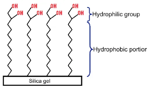
Figure 3 General structure of an amino acid; the R group designates the side
chain. ... 8
Figure 4 Enantiomers of a) glyceraldehyde and b) alanine. In both cases, the
chiral atom is in the centre of the projection, the D- enantiomer is on the left and the L- on the right side. c) Three-dimensional representation of D-alanine and L-alanine, showing the mirror effect between the enantiomers. ... 9
Figure 5 Tswett's experiment that represented the inception of liquid
chromatography (adapted from Arsenault and McDonald, 2014). ... 22
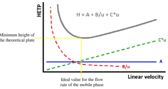
Figure 6 Graphical representation of the contributing terms for the band
broadening (adapted from Yip, 1997). ... 24
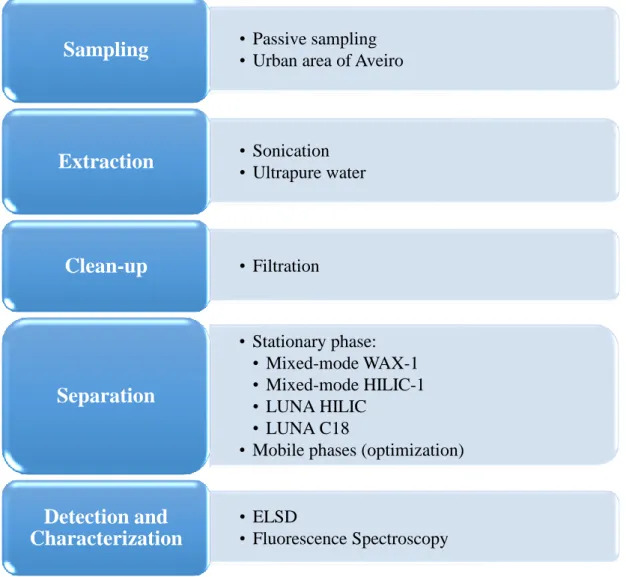
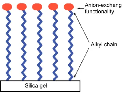
Figure 7 Proposal for the analytical procedure to be employed in this work. . 28 Figure 8 Surface composition of the Acclaim® Mixed-Mode WAX-1 column (adapted from Phenomenex Columns Manual - a))... 31
Figure 9 Surface chemical composition of the Acclaim® Mixed-Mode HILIC-1 column (adapted from Phenomenex Columns Manual - b)). ... 33
Figure 10 Surface composition of the Phenomenex® Luna HILIC (adapted from Phenomenex Columns Manual - c)). ... 34
Figure 11 Surface chemical composition of the Phenomenex® Luna C18 (adapted from Phenomenex Columns Manual - d)). ... 35
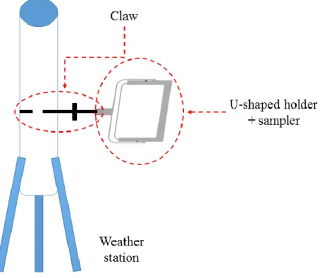
Figure 12 Representation of the sampler and its holder embedded in the weather
station. ... 37
Figure 13 Picture of one model of the samplers. It is possible to distinguish the
three parts that constitute the sampler: the support; the two polyvinyl chloride frames (grey plates); and the Teflon plate where the filters were placed (white plate). ... 38
Figure 14 Front view of the sampler installed near the Glicínias Plaza mall... 39 Figure 15 Lateral view of the sampler installed near the Glicínias Plaza mall.
... 39
Figure 16 Building where the sampler was installed. ... 40 Figure 17 Surrounding of the samplers on the roof of the STIC Department at
the University of Aveiro. ... 40
Figure 18 Surrounding of the samplers on the roof of the STIC Department at
the University of Aveiro. ... 41
Figure 19 Weather station with the two samplers in the roof of the STIC
Department at the University of Aveiro. ... 41
Figure 20 Up filter of the sampler installed near the Glicínias Plaza mall with
biologic matter. ... 42
Figure 21 Procedure implemented for the extraction of the water-soluble organic
compounds from the collected aerosols samples (adapted from Duarte et al., 2007). ... 46
Figure 22 Vials containing the samples residues after the collection from the
round-bottom flasks. The vial number 2 corresponds to the filter sample with biologic material from bird activity. ... 49
Figure 23 Vials of the samples after the filtration via a syringe filter. The vial
number 2 corresponds to the filter with biologic material from bird activity. ... 50
Figure 24 Chromatograms showing the effect of the variation of the organic
solvent concentration at different buffer concentrations - a) 20mM; b) 50mM; c) 100mM;
d) 200mM - in the elution of the aspartic acid. ... 54 Figure 25 Chromatograms showing the effect of the variation of the organic
solvent concentration at different buffer concentrations - a) 20mM; b) 50mM; c) 100mM;
d) 200mM - in the elution of lysine. ... ………56 Figure 26 Chromatograms showing the effect of the variation of the organic
solvent concentration at different buffer concentrations - a) 20mM; b) 50mM; c) 100mM;
Figure 27 Chromatograms showing the effect of the variation of the organic
solvent concentration at different buffer concentrations - a) 20mM; b) 50mM; c) 100mM;
d) 200mM - in the elution of tryptophan………...…59 Figure 28 Chromatograms showing the effect of the variation of the ionic
strength at different organic solvent concentrations - a) 20%; b) 80% - in the elution of the aspartic acid. ... 60
Figure 29 Chromatograms showing the effect of the variation of the ionic
strength at different organic solvent concentrations - a) 20%; b) 80% - in the elution of lysine. ………..62
Figure 30 Chromatograms showing the effect of the variation of the ionic
strength at different organic solvent concentrations - a) 20%; b) 80% - in the elution of glycine……….63
Figure 31 Chromatograms showing the effect of the variation of the ionic
strength at different organic solvent concentrations - a) 20%; b) 80% - in the elution of tryptophan…..………...64
Figure 32 Chromatograms showing the effect of the variation of the organic
solvent concentration at a buffer concentration of 50mM in the retention behaviour of: a) aspartic acid; b) lysine; c) glycine; d) tryptophan. ... 66
Figure 33 Chromatograms of the four tested amino acids eluted under different
organic solvent content conditions: a) aspartic acid; b) lysine; c) glycine; d) tryptophan. ... 68
Figure 34 Chromatograms of the four amino acids eluted under different mobile
phase conditions, in terms of the amount of ACN: a) aspartic acid; b) lysine; c) glycine;
d) tryptophan. ... 70 Figure 35 Results obtained with the isocratic elution of glycine, isoleucine,
leucine, proline, threonine and tryptophan, using 80% (V/V) of organic solvent. ... 72
Figure 36 Results obtained for the isocratic elution of glycine, isoleucine,
leucine, proline, threonine, tryptophan, aspartic acid, lysine and tyrosine, using 20% (V/V) of organic solvent. ... 73
Figure 37 Chromatograms of aspartic acid before (a)) and after (b)) the
sonication bath. ... 75
Figure 38 Chromatograms of glycine before (a)) and after (b)) the sonication
bath. ... 76
Figure 39 Chromatograms of lysine before (a)) and after (b)) the sonication
bath. ... 77
Figure 40 Chromatograms of tryptophan before (a)) and after (b)) the sonication
bath. ... 78
Figure 41 Gradient 1 applied to elute the 20 amino acids mixture. ... 80 Figure 42 Chromatogram obtained with the gradient 1 showing seven peaks in
the separation of the 20 amino acids mixture. ... 80
Figure 43 Gradient 2 applied to elute the 20 amino acids mixture. ... 81 Figure 44 Chromatogram obtained for the gradient 2, showing eight different
peaks in the separation of the 20 amino acids mixture. ... 81
Figure 45 Gradient 3 applied to elute the 20 amino acids mixture. ... 82 Figure 46 Obtained results from the gradient 3, with eight distinct peaks in the
elution of the mixture with 20 amino acids. ... 82
Figure 47 Gradient 4 applied to elute the 20 amino acids mixture. ... 83 Figure 48 Chromatogram obtained for the gradient 4, showing eight different
peaks in the separation of the 20 amino acids mixture. ... 83
Figure 49 Gradient 5 applied to elute the 20 amino acids mixture. ... 84 Figure 50 Chromatogram obtained for the gradient 5, showing four different
peaks in the separation of the 20 amino acids mixture. ... 84
Figure 51 Gradient 6 applied to separate the 20 amino acids in the mixture. . 85 Figure 52 Chromatogram obtained for the gradient 6, showing five different
peaks in the separation of the 20 amino acids mixture. ... 85
Figure 53 Gradient 7 applied to separate the 20 amino acids in the mixture. . 86 Figure 54 Chromatogram obtained for the gradient 7, showing six different
peaks in the separation of the 20 amino acids mixture. ... 86
Figure 56 Chromatogram obtained for the gradient 8, showing seven different
peaks in the separation of the 20 amino acids mixture. ... 87
Figure 57 Gradient 9 applied to separate the 20 amino acids in the mixture. . 88
Figure 58 Chromatogram obtained for the gradient 9, showing eight distinct peaks in the separation of the 20 amino acids mixture. ... 88
Figure 59 Gradient 10 applied to separate the 20 amino acids in the mixture. 89 Figure 60 Chromatogram obtained for the gradient 10, showing ten different peaks in the separation of the 20 amino acids mixture. ... 89
Figure 61 Chromatogram with all the twenty amino acids eluted individually. ... 90
Figure 62 Matching of the results obtained for the mixture and the individual results. ... 91
Figure 63 Chromatogram showing the results of the elution of the six different samples collected in two different sites in the city of Aveiro. The aqueous extracts of the bulk aerosol samples were directly injected after the extraction process. ... 94
Figure 64 Chromatograms related to the sampler location: a) near Glicínias Plaza mall; b) and c) at the University of Aveiro. ... 96
Figure 65 Chromatograms regarding the position of the filters relatively to the samplers: a) bottom filters; b) up filters. Sample 1 was extracted from filter 1; sample 2 was extracted from filter 2; sample 3 was extracted from filter 3; and sample 4 was extracted from filter 4. ... 98
Figure 66 EEM fluorescence spectrum of Sample 1. ... 100
Figure 67 EEM fluorescence spectrum of Sample 2. ... 100
Figure 68 EEM fluorescence spectrum of Sample 3. ... 101
Figure 69 EEM fluorescence spectrum of Sample 4. ... 101
Figure 70 EEM fluorescence spectrum of Sample 5. ... 102
Index of Tables
Table 1 Abbreviations and properties related to the twenty amino acids found in
proteins (adapted from Nelson and Cox, 2004). ... 11
Table 2 Properties of the 20 common amino acids that influence the efficiency
of the liquid chromatography separation process: size, polarity, electric charge at pH 7.0, behaviour at pH 7.0, pI and affinity to water (adapted from Nelson & Cox, 2004; Pommié et al., 2004). ... 15
Table 3 Examples of extraction, clean-up, separation and detection methods
applied in the latest fifteen years for the analysis of amino acids in different matrices. 17
Table 4 Weight values of the filters used during the sampling. The sample
weight was calculated by the difference of the mean value of the after and the before sampling measure. ... 43
Table 5 Atmospheric conditions in Aveiro during the sampling time (data
collected from Current Weather in Aveiro, Portugal). The values shading in light grey refer to the evening moments while the others were registered in the morning. ... 44
Table 6 Volume used during the extraction of the amino acids in each filter. 46 Table 7 Volume used to dissolve the solid extracts obtained after the
lyophilization step. ... 47
Table 8 Weight of the round-bottom flasks taken for the lyophilization step. The
sample weight was calculated by the difference of the mean value of the after and the before lyophilization measure. ... 48
Table 9 Conditions of the preliminary tests for the Acclaim® Mixed-Mode
WAX-1 column. ... 53
Table 10 Conditions of the preliminary tests for the Acclaim® Mixed-Mode
HILIC-1 column in order to study the influence of the organic solvent concentration in the retention of the aspartic acid, lysine, glycine and tryptophan. ... 66
Table 11 Conditions of the preliminary tests for the Acclaim® Mixed-Mode
HILIC-1 column in order to study the influence of the amino acids solution preparation in the retention of the aspartic acid, lysine, glycine and tryptophan. ... 68
Table 12 Conditions of the preliminary tests for the Acclaim® Mixed-Mode
HILIC-1 column to study the effect of the organic solvent content in the elution of the aspartic acid, lysine, glycine and tryptophan. ... 70
Table 13 Conditions of the preliminary tests for the Acclaim® Mixed-Mode
HILIC-1 column in order to study the effect of the organic solvent concentration in the retention behavior of glycine, leucine, isoleucine, proline, threonine and tryptophan. .. 72
Table 14 Conditions of the preliminary tests for the Phenomenex® Luna HILIC
column in order to study the stability of the samples of the aspartic acid, lysine, glycine and tryptophan. ... 75
Table 15 Conditions of the preliminary tests for the Phenomenex® Luna C18
column applied in the selection of the gradient mode which separates more amino acids. ... 79
Table 16 Amino acids and their respective retention times with gradient number
10. ... 90
Table 17 Programs of the 10 gradients used in the improvement of the
chromatographic separation of amino acids. The percentages represent the concentration of the ACN in the mobile phase. ... 92
Index of Abbreviations
ACN – acetonitrile
CE – capillary electrophoresis CSPs – chiral stationary phases EEM – excitation-emission matrix
ELSD – evaporative light-scattering detector FID – flame ionization detector
GABA – gamma-aminobutyric acid GC – gas chromatography
HILIC – hydrophilic interaction liquid chromatography LIF – laser-induced fluorescence
MeOH – methanol MS – mass spectrometry MSD – mass selective detector
PDFOA – pentadecafluorooctanoic acid RP – reversed-phase
SRM – standard reference material TDFHA – tridecafluoroheptanoic acid WAX– weak anionic exchange
I
Introduction: basic concepts of
aerosols and liquid chromatography
Chapter I
2015 1
1. Aerosols: general considerations
Through the years, the interest in developing precise technologies and methods able to detect and identify atmospheric particles has been growing (Després et al., 2012). Atmospheric particles can be emitted into the atmosphere from natural or anthropogenic sources and they can be formed in-situ in the atmosphere through secondary formation processes - aerosols can be primary or secondary, regarding their formation processes. According to Hinds (1999), aerosols consist in colloidal systems of liquid or solid particles suspended in a gas. Primary particles are directly released into the atmosphere. The products of biomass burning, volcanic eruptions, combustion of fossil fuels and wind-driven particles of soil dust, sea salt and biological materials are some examples of primary aerosols (Hallquist et al., 2009). On the other hand, secondary particles are the result of volatile and semi-volatile organic species transformations occurring in the atmosphere and involving gas-to-particle conversion processes, including condensation, nucleation and heterogeneous and multiphase chemical reactions. During these processes, bioaerosols suffer degradation and lose their biologic functions, producing inorganic secondary aerosols particles, such as sulfate (SO42-), nitrate (NO3-) and ammonium
(NH4+), which are products of the conversion of the sulfur dioxide (SO2), nitrogen dioxide
(NO2) and ammonia (NH3) gases into the particulate phase, respectively (Goldstein &
Galbally, 2007; Kroll & Seinfeld, 2008).
When atmospheric particles have a biological origin, such as bacteria, fungi, viruses, debris, dust mites, toxins, spores, cells, pollen, animal or plant organic matter and sub products of biological activities, they are called bioaerosols, and their size can vary from 0.5 to 100 μm (Després et al., 2012; Wathes & Cox, 1995).
Introduction: basic concepts of amino acids and liquid chromatography
2 2015
1.1 - Bioaerosols: a particular case of aerosols
Bioaerosols are present both in outdoor and indoor environments, in a wide range of concentrations, and they may be aerosolized from diverse sources: soil, vegetation, water and living organisms. Commonly, bioaerosols show a considerable diversity in dimension, shape and electric charge. Furthermore, as reviewed by Agranovski (2010), it is often significant to make the distinction between viable and non-viable bioaerosols particles: the first one is referred to microbial cells capable of reproducing or having a metabolic behavior; and the second one is related to organisms unable to reproduce. According to Gao et al. (2009), only less than 10% of the airborne bacteria particles are viable and the higher viabilities have been detected in fungal spores. Considering the importance of the biological activity of bioaerosols due to the possibility of new microorganisms formation, bioaerosols characterization is different from the one traditionally applied on inert aerosols (Agranovski, 2010).
As mentioned by Agranovski (2010), the most common and studied bioaerosols particles are bacteria, pollen, viruses and fungi. Viruses are unique since they have the ability to reproduce but only inside a host cell. However, there is still an absence of information about their shape, size and density. Recently, there has been some evolution in the knowledge regarding the way airborne viral particles travel in the air. It used to be believed these bioaerosols would only survive as airborne if they were attached to a larger particle, in a highly humid environment. Nowadays, due to studies such as the amount of time a laboratory-generated single viruses is able to survive in the air (Lighthart and Shaffer, 1997) and the transmission of the influenza virus (Elbert et al., 2007), it is known that viruses can be diffused through the air without extreme conditions, which can be harmful if the airborne transmission of viral infections increases.
Bacteria are organisms with only one cell which can have various shapes, including spiral, spherical and rod-shaped, and they can be carried through the air by other aerosols like water droplets, fragments of animal skin and plant material (Putaud et al., 2004; Lee et al., 2008). These prokaryote organisms tend to grow in colonies and when aerosolized they frequently aggregate to other materials or to each other in clusters or chains (Conte and Weber, 2002). Both animals and plants can be seriously ill due to airborne bacteria particles. Some bacteria can generate spores related to specific human
Chapter I
2015 3
health diseases such as respiratory allergy and asthma resulting from occupational exposures (Kourtchev et al., 2008).
Fungi are spread in the atmosphere due to the release of spores and they are adapted to several atmospheric environments. According to Lee et al. (2007, 2008), fungi are extremely resistant to high and low temperatures, low humidity and ultraviolet radiation and they can become aerosolized as either individual or agglomerated spores. Fungi airborne particles are associated with allergic reactions, allergic rhinitis and asthma.
Plants produce pollen grains in a significant quantity wherein the characteristics of the airborne pollen are related to the plant that create it. So, particles from different plants diverge in size, shape and surface structure. There is a lack of information related to the aerodynamic diameters of pollen particles, although it is known that the physical size is in the range of 10 to 100 µm. However, numerous types of pollen grains have 25 – 50 µm, values larger than the breathable size fraction (Reponen et al., 2009). Thus, human health effects are associated with smaller and not so usual pollen fragments that may contain allergens (Wathes and Cox, 1995; Miguel et al., 2006).
1.2 - Bulk deposition of aerosols
Every atmospheric particle participates in exchanges with outdoor surfaces, in processes including deposition or removal from the atmosphere and suspension or re--entrainment from underlying surfaces (Wathes and Cox, 1995). Removal methods may occur via either wet or dry deposition or both ways combined, as depicted in Figure 1.
Dry deposition consists in the direct interaction of a particle with a surface, in a continuous process, through diffusion and/or sedimentation. Wet deposition embraces the removal of material from the atmosphere in any falling particle, such as raindrops, snowflakes or hailstones. After deposition, aerosols may experience the process of resuspension or reemission, which can result in the previously deposited material being reemitted into the atmosphere (Després et al., 2012).
Introduction: basic concepts of amino acids and liquid chromatography
4 2015
Dry deposition is the main removal mechanism for particles with a diameter equal or higher than 10 μm. The atmospheric sedimenting particles are the ones that hit a barrier, usually the ground, and stop their movement. They can be deposited via a dry or wet deposition method. On the other hand, there are other atmospheric constituents that do not stop their movement in the atmosphere, not hitting any barrier: the non-sedimenting constituents (Després et al., 2012).
In the non-sedimenting situation, particles can only be removed via wet deposition. When these particles are in the gaseous state, they can be removed by wet deposition after a condensation process, turning in to drops. In the case where the non-
Figure 1 Processes of atmosphere-surface exchanges related to aerosols (adapted from Dämmgen et
Chapter I
2015 5
-sedimenting particles include components in the solid state, they are removed from the atmosphere also via wet deposition, though a wash out process, as shown in Figure 1.
When dry deposition procedures are adopted, particles are studied by their aerodynamic diameter – which is defined as the diameter of a spherical particle with a unit density and with the same terminal velocity in the air as the aerosols’ particle in question (Hinds, 1999). Since biological particles frequently have a complex structure with irregular surfaces, internal pores and asymmetric shape, their aerodynamic and physical sizes may not match perfectly. Therefore, aerodynamic diameter is widely used in bioaerosols analysis (Tong and Lighthart, 2000).
In the case of particles with an aerodynamic diameter within 0.1 – 10 μm, deposition by precipitation is the most efficient removal method, removing a large percentage of particles from the air in many situations, even if it is a single rain event (McDonald, 1962). Dry deposition, on the other hand, is not well understood due to complexities regarding measuring gaseous concentrations and calculating deposition velocities necessary for quantifying dry deposition rates (Elliott et al., 2009).
As shown in Figure 1, bulk deposition is considered as the whole amount of wet and dry deposition collected during sampling. In practical terms, this amount can have some bias due to continuously deposition of gases and fine aerosols to the sampler. Therefore, bulk deposition is frequently known as the total deposition of particles to a constantly opened sampler. During sampling preparation, it is crucial to be aware of the particles that accidentally can enter the sampler. For example, only the material from the surrounding ecosystem must be a part of the sample, which may not always happen due to turbulent deposition. Besides, the sample may also include the contribution from sedimentation particles that have been resuspended by the wind from the adjacent environment. Since these sampling artifacts can easily occur during aerosol collection, it is extremely important to choose well the sampler location and height above ground (Dämmgen et al., 2005).
Introduction: basic concepts of amino acids and liquid chromatography
6 2015
1.3 - Strategies for sampling (bio)aerosols
Relatively to air microorganisms monitoring, there are two main sampling methods: the active and the passive sampling. The theory for passive sampling techniques was introduced by Palmes and Gunnison (1973), who developed a diffusive sampler to determinate the presence of SO2 in the atmosphere. Passive samplers are devices capable
of collecting atmospheric gases or vapors, via a controlled rate by physical processes, such as diffusion or permeation, not involving active movement of the air through the sampler (Brown et al., 1984). Firstly, these samplers were invented in order to be used in indoor studies due to humans’ exposure to high levels of pollutants in working areas. Meanwhile, since the 80’s, the samplers started to be applied to outdoor environmental research, where the pollutants concentrations are extremely lower. Nevertheless, these investigations are challenging, since there is the influence of the meteorological conditions (Palmes et al., 1976; Brown et al., 1984).
Comparatively to active samplers, the passive samplers have been gaining more supporters for gases and vapors sampling since they are simpler, cheaper, smaller, lighter, easier to carry to the sampling site, and do not need electric power or periodic calibrations. Besides, it is not necessary to measure the volume of the sampled air or the every-day presence of a technician in the sampling location, and the sample is more representative of the natural process of deposition (Rose and Perkins, 1982). These samplers are particularly useful in determining the spatial distribution of the atmospheric gases concentrations in large areas, as well as monitoring studies in remote zones and providing data for modeling and comparisons with air quality long term standards. Despite of the aforementioned advantages of these devices, they are not always completely reliable: the passive samplers do not provide instantaneous concentrations but mean values every week or month, thus not allowing obtaining the sampling rate and, consequently, it is not possible to distinguish the transitional moments between highest and lowest concentrations in a certain period (Shooter et al., 1995). Furthermore, they are not as sophisticated as the active samplers (Cruz and Campos, 2002).
In these passive samplers, inertial forces along with the external wind and gravity, provide the transportation of aerosol particles to collectors’ surfaces. Although there was no concept of “passive sampler” at the time, Oren Durham (1946) planned a very simple passive sampling apparatus for atmospheric particles involving essentially the
Chapter I
2015 7
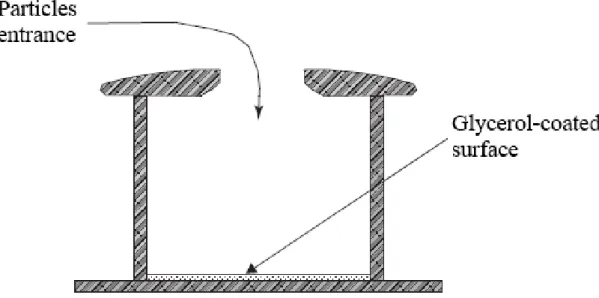
gravitational deposition of those aerosols onto an oil-coated flat slide. However, there was some interference related to the falling of debris and rain. Therefore, the author set a cover in order to direct the collected aerosol into the region of the sampling surface so the sample would experiment the influence of the horizontal wind. Some decades later, in 1974, as mentioned by Vicent (2007), Tauber improved the original idea from Durham and designed a more sophisticated passive sampler, specifically to collect large pollen grains. As shown in Figure 2, aerosols enter the sampler through the circular opening at the top of the cylindrical container by a combination of inertial and gravitational forces. The sample is retained on the glycerol-coated surface on the bottom of the sampler (Vincent, 2007).
1.4 - Amino acids as proxies of bioaerosols
According to some authors, there are several methods with the ability to detect the presence of bacteria, pollen and fungal spores in atmospheric particles, such as the use of chemical biomarkers (Di Filippo et al., 2013), traps (Ong et al., 1995; Ho et al., 2005), molecular biology techniques (Womiloju et al., 2003; Lee et al., 2006) or microorganism sampling, growing and counting (Bauer et al., 2002). The aforementioned biological
Figure 2 Illustration of the principle of operation of the Tauber pollen sampler (adapted from Vincent,
Introduction: basic concepts of amino acids and liquid chromatography
8 2015
methods are difficult to apply and only give partial information since they are limited to the biological component under study. These techniques are applied on amino acids and proteins, important constituents of bioaerosols, allowing the use of these biomolecules as an index of biological occurrence in the atmosphere. Plants, animals, biomass burning and degradation products of peptides and proteins through enzymatic and photo-catalytic reactions in the atmosphere release amino acids directly to the air, which make them responsible for free amino acids contribution to airborne particles (Di Filippo et al., 2014).
Amino acids are composed by a carboxyl and an amino functional groups, an R group and a hydrogen atom, bonded to the same carbon atom (α carbon). All of the 20 common amino acids are α-amino acids and they differ from each other in their R group, also known as side chain. The fact that the α carbon is bonded to four distinct groups makes it a chiral atom, except in the case of glycine since the R group is an hydrogen atom. Having only one chiral center, 19 of the 20 common amino acids are chiral compounds (Nelson and Cox, 2004).
Although amino acids have a common structure, as shown in Figure 3, except in the case of proline, the side chains vary in size, polarity, structure and electric charge, some of the properties capable of influence the molecules solubility in water (Nelson and Cox, 2004).
As a consequence of the tetrahedral arrangement of the bonding orbitals around the carbon atom, the four diverse groups may be organized in two unique spatial adjustments and, thus, amino acids are stereoisomers: the same compound may have divergent spatial arrangements of its’ atoms, although the molecules possess the same atoms and sequence of bonds. It is not possible to convert one molecule’s stereoisomer into the other without breaking the inherent bonds. Since the stereoisomers of amino acids are non-superimposable mirror images of each other, they are called enantiomers. Thus,
Chapter I
2015 9
a special nomenclature has been implemented to describe the absolute configuration of the four substituents of an optical isomer (enantiomer): the D/L system, a convention proposed by Emil Fischer in 1891. According to this system, the enantiomer is named by analogy to glyceraldehyde, shown in Figure 4 – a), consistent with which glyceraldehyde isomer it comes from. Therefore, if an enantiomer comes from D-glyceraldehyde that isomer it also labeled as the D-form (Nelson and Cox, 2004).
Figure 4 Enantiomers of a) glyceraldehyde and b) alanine. In both cases, the chiral atom is in the centre
of the projection, the D- enantiomer is on the left and the L- on the right side. c) Three-dimensional representation of D-alanine and L-alanine, showing the mirror effect between the enantiomers.
Introduction: basic concepts of amino acids and liquid chromatography
10 2015
Besides the relation to the enantiomers of glyceraldehyde, it is possible to name the amino acids according to the atomic number of the atom bonded to the chiral center. The atom with the highest atomic number is the number 1 atom; the atom with the second highest atomic number corresponds to the atom number 2; and so on. If the chiral center is bonded to more than one unit of the same atom, the rule above is applied to the atoms of that group. For example, in the case of alanine (Figure 4 – b) and c)), there are two carbons bonded to the chiral center: a COO- and a CH3 group. Therefore, the atoms that
will be compared are oxygen and hydrogen. The atom that is used for the comparison is always the one with the highest atomic number, as for the atoms S and O in the groups C-HHSH and C-OOH, the atoms S and O are the ones being compared. If the atoms are the same but with different bonds then the type of bond influences the result: a double bond (C=O) has priority over a single bond (C-O). After matching the groups with their priority number, the direction is obtained according to the increasing numerical order: 1 → 2 → 3. If the groups are arranged in the clockwise way, the amino acid is in its D-form (dextrorotary). On the other hand, if the spatial distribution is counterclockwise, the enantiomer is labeled as L- (levorotary). Although this is a simple procedure, it is crucial the hydrogen atom, component of all the amino acids, is away from the viewer in the tri-dimensional conformation, in order to name correctly the enantiomers. If the hydrogen atom is close to the viewer, the result of the nomenclature process is inversed: the D- is related to the counterclockwise direction and the L- becomes the arrangement of the clockwise way (Nelson and Cox, 2004).
In Nature, the fact that amino acids are enantiomers has a significant role in biosynthesis: the amino acids residues present in proteins are exclusively L-enantiomers. When chiral compounds are produced by a conventional chemical reaction, the obtained result is a racemic mixture of both D- and L- isomers. Distinguish and/or separate these two different molecules is a difficult task to achieve. However, if the product is formed by a living organism, D- and L- enantiomers are totally dissimilar. Usually, the formation of stable and consistent structures in proteins requires that only one of the enantiomers is a constituent. In order to keep the organism alive and functional, cells specifically synthetize the L- isomers, since the active sites of enzymes are asymmetric, causing that the reactions they catalyze to be stereospecific. Therefore, and considering that only L-forms of amino acids are used to produce proteins during biosynthesis, it is possible to state that there is a homochirality regarding living organisms. On the other hand, D-amino acids exist in cell walls of bacteria (Barbaro et al., 2014a).
Chapter I
2015 11
Furthermore, enantiomers have the same physical properties but they rotate polarized light in a divergent direction and they interact differently with distinct optical isomers of other compounds. All chiral compounds rotate the plane-polarized light, which means they are optically active (Nelson and Cox, 2004).
In order to indicate the sequence of the amino acids polymerized in proteins, the 20 common amino acids have been assigned three-letter abbreviations and one-letter symbol as shown in Table 1. Regarding human daily basics needs, there are three categories of amino acids: non-essential, conditionally non-essential and essential. In the table below, the non-essential amino acids are marked with green color (▄▄▄) and can be synthesized by the human organism. The conditionally non-essential amino acids, marked with yellow color (▄▄▄), are usually produced in our organism. However, some people are not capable of synthesize them in sufficient quantity, causing a necessity to include those amino acids in their diet. Finally, the essential amino acids, red color (▄▄▄), cannot be produced by the human organism, and consequently, they have to be included in our diet (Nelson and Cox, 2004).
Table 1 Abbreviations and properties related to the twenty amino acids found in proteins (adapted from
Nelson and Cox, 2004).
Amino acid Alanine Cysteine Aspartic acid
(aspartate)
Glutamic acid (glutamate)
Three-letter
code Ala Cys Asp Glu
One-letter code A C D E Molecular formula C3H7NO2 C3H7NO2S C4H7NO4 C5H9NO4 Molecular weight (g/mol) 89.0932 121.160 133.103 147.129 Molecular structure
Introduction: basic concepts of amino acids and liquid chromatography
12 2015
Amino acid Arginine Glycine Asparagine Proline
Three-letter
code Arg Gly Asn Pro
One-letter code R G N P Molecular formula C6H14N4O2 C2H5NO2 C4H8N2O3 C5H9NO2 Molecular weight (g/mol) 174.201 75.0666 132.118 115.130 Molecular structure
Amino acid Glutamine Serine Tyrosine Phenylalanine
Three-letter
code Gln Ser Tyr Phe
One-letter code Q S Y F Molecular formula C5H10N2O3 C3H7NO3 C9H11NO3 C9H11NO2 Molecular weight (g/mol) 146.144 105.093 181.189 165.189 Molecular structure
Chapter I
2015 13
Amino acid Histidine Isoleucine Lysine Leucine
Three-letter
code His Ile Lys Leu
One-letter code H I K L Molecular formula C6H9N3O2 C6H13NO2 C6H14N2O2 C6H13NO2 Molecular weight (g/mol) 155.155 131.173 146.188 131.173 Molecular structure
Amino acid Methionine Threonine Valine Tryptophan
Three-letter
code Met Thr Val Trp
One-letter code M T V W Molecular formula C5H11NO2S C4H9NO3 C5H11NO2 C11H12N2O2 Molecular weight (g/mol) 149.211 119.119 117.146 204.225 Molecular structure
Introduction: basic concepts of amino acids and liquid chromatography
14 2015
The isomerism is not the only property that influences the analysis of the amino acids present in aerosols. Since the aim of this work is to separate amino acids enantiomers through liquid chromatography, there are other fundamental characteristics that play a role in the efficiency of this method, such as size, polarity, charge at pH 7.0 and pI (isoelectric point – pH value at which a certain molecule does not have electric charge), as shown in Table 2.
Although all of the 20 common amino acids are soluble in water, it is necessary to be aware of their affinity to this molecule. Therefore, it is also revealed in Table 2 the hydropathy index - a scale combining hydrophobicity and hydrophilicity of R groups that can be used to measure the tendency of an amino acid to seek an aqueous environment (negative values) or a hydrophobic environment (positive values). These values were studied by Kyte and Doolittle (1982) and obtained by using a computer software that evaluates, progressively, the hydrophilicity and the hydrophobicity of a protein along its amino acids sequence. According to Kyte and Doolittle (1982) results, arginine is the amino acid with the higher tendency to interact with water while isoleucine is the most hydrophobic one. Considering the relation between hydropathy and the polarity of the molecule, it is possible to assert that arginine is the most polar amino acid while isoleucine is the most nonpolar one, in a group of 9 nonpolar and 11 polar amino acids. Although the software assigns numeric values to hydropathy, the results are not completely linear and need to be analyzed. In cases where amino acids are truly large or small, such as tryptophan and glycine, the software is not particularly accurate, and it assigns negative values (hydrophilic properties) to nonpolar molecules (Kyte and Doolittle, 1982).
Arginine is also the amino acid with the highest pH value at which it becomes electrically neutral (highest pI value). On the other hand, aspartic acid is the one with the lowest pI. Among the 20 amino acids, there are three with a positive electric charge at pH 7.0, the ones with a basic behavior: histidine, lysine and arginine. The molecules with a negative electric charge at pH 7.0 are the ones with an acidic behavior: aspartic acid and glutamate. The other 15 amino acids are neutral at pH 7.0. The largest molecule is tryptophan whereas the smallest is glycine (Nelson and Cox, 2004).
Chapter I
2015 15
Table 2 Properties of the 20 common amino acids that influence the efficiency of the liquid
chromatography separation process: size, polarity, electric charge at pH 7.0, behaviour at pH 7.0, pI and affinity to water (adapted from Nelson & Cox, 2004; Pommié et al., 2004).
Ala Cys Asp Glu Gly Asn Pro Gln Arg Ser
P olar it y Polar X X X X X X Nonpolar X X X X Hydropathy index 1.8 2.5 -3.5 -3.5 -0.4 -3.5 1.6 -3.5 -4.5 -0.8 pI 6.01 5.07 2.77 3.22 5.97 5.41 6.48 5.65 10.76 5.68 C ha rge Negative X X Neutral X X X X X X X Positive X Volume Large X Medium X X Small X X X X X X X pH 7.0 Acidic X X Neutral X X X X X X X Basic X
Tyr Phe His Ile Lys Leu Met Thr Val Trp
P olar it y Polar X X X X Nonpolar X X X X X X Hydropathy index -1.3 2.8 -3.2 4.5 -3.9 3.8 1.9 -0.7 4.2 -0.9 pI 5.66 5.48 7.59 6.02 9.74 5.98 5.74 5.87 5.97 5.89 C ha rge Negative Neutral X X X X X X X X Positive X X Volume Large X X X X X X X Medium X X Small X pH 7.0 Acidic Neutral X X X X X X X X Basic X X
Introduction: basic concepts of amino acids and liquid chromatography
16 2015
1.5 - Methodologies for the analysis of amino acids in aerosols
Amino acids, free and combined, can be a component of water-soluble organic
carbon (WSOC), and their presence in the atmosphere has been studied for the last
decades. This study is truly challenging due to their low ambient concentrations. Besides, proteins and peptides may undergo modifications through chemical and physical processes (Haan et al., 2009).
Although the quantification is a difficult task, amino acids extraction can be a simple practice according to the procedure followed by Matsumoto and Uematsu (2005). According to these authors, it is possible to separate 14 of the 20 common amino acids via an easy extraction method: sonication with pure water. Since this research work is focused on the extraction and separation of WSOC, and in amino acids in particular, Matsumoto and Uematsu’s work appears to be a successful study using a simple extraction procedure.
Over the latest fifteen years, there have been studies in a wide range of procedures for extraction and analysis of amino acids in diverse matrices, from mural painting to standard reference material, using liquid chromatography. Examples of some of those studies are summarized in Table 3, considering the following characteristics: matrix, extraction, clean-up methods, separation and detection techniques, composition of the mobile and stationary phases, and the amino acids that were analyzed. As it is possible to observe in Table 3, the extraction method for amino acids can be complex and laborious such as in the works of Johnson and Pregitzer (2007), Zangrando et al. (2010) and Di Filippo et al. (2014), or simple such as in the works of Barbaro et al. (2011), Scalabrin et al. (2012) and Warren (2013). Concerning the clean-up process for the isolation of amino acids from other potential interfering analytes, Barbaro et al. (2011, 2014b), Warren (2008) and Zangrando et al. (2010) used a filtration step in their work. On the other hand, Di Filippo et al. (2014) and Buiarelli et al. (2013) prefered the solid phase extraction as a clean-up methodology to isolate the amino acids in their samples. Although the majority of the listed works (Table 3) has used a mass spectrometry as detector, some authors adopted a two-dimensional liquid chromatography method for the separation of amino acids, coupled to different detectors, namely UV-Vis (Mace et al., 2003), flame ionization (Amelung & Zhang, 2001), fluorescence (Johnson & Pregitzer, 2007) and evaporative light scattering (Chaimbault et al., 1999) detectors.
Chapter I
2015 17
Table 3 Examples of extraction, clean-up, separation and detection methods applied in the latest fifteen years for the analysis of amino acids in different matrices.
Matrix Particulate
Matter Extraction Clean-up
HPLC Detector and
Detected Amino Acids
Reference Mobile Phase Stationary Phase
A
er
oso
ls
Bulk Sonication (MeOH*) Filtration
Eluent A: CH3COONa aqueous buffer - pH
4.00
Eluent B: ACN and HCOOH ZIC-HILIC (+)ESI-MS/MS 20 A, N, D, C, Q, E, G, 3-Hyp, 4-Hyp, I, L, M, MetSO, MetSO2, F, P, S, Y, T, V (Barbaro et al., 2011) PM0.1 PM2.5 PM10 Sonication (H2O/MeOH), evaporation, clean-up, hydrolysis and purification Solid phase extraction Eluent A: H2O and HCOOH (pH 2.3)
Eluent B: MeOH and HCOOH C18 (+)ESI-MS/MS 24 Orn, H, K, R, G, A, S, P, V, T, L, I, N, Q, D, Hyp, E, Cit, C, M, F, Y, W, GABA (Di Filippo et al., 2014) PM10 Sonication (MeOH) Extracts assemblage and filtration Eluent A: CH3COONa aqueous buffer - pH 4.00
Eluent B: ACN and HCOOH ZIC-HILIC (+)ESI-MS/MS 20 F, T, L, I, M, Y, V, 3-Hyp, P, 4-Hyp, A, E, Q, S, N, D, C, G, MetSO2 (Scalabrin et al., 2012)
Bulk Sonication (pure H2O) Filtration Not specified
14 D, S, E, G, A, V, M, I, L, Y, F, H, K, R, (Matsumoto and Uematsu, 2005) PM10 Sonication (ultrapure H2O and ice; addition
of ice); spike and extraction (ultrapure
H2O)
Filtration
Eluent A: ultrapure H2O and HCOOH
Eluent B: MeOH and HCOOH CHIROBIOTIOC TAG 11 A, D, R, E, F, P, Y, T, L, G, V (Barbaro et al., 2014 - a))
Introduction: basic concepts of amino acids and liquid chromatography 18 2015 A er oso ls (f ro m biom ass bu rni ng ) PM10
For the analysis, filtered aerosol extracts were injected onto the column
without further preparation
ACN:MeOH and a pH-neutral potassium phosphate dibasic solution Dionex DX 300 UV-Vis 17 D, E, S, T, G, A, R, P, V, M, I, L, F, C, K, H, Y (Mace et al., 2003) U rban d ust PM10 Accelerated solvent extraction (H2O/MeOH) Solid phase extraction, wash and elution Eluent A: CH3COONH4, adjusted to pH 4.00 (HCOOH) Eluent B: ACN/H2O acidified (HCOOH) C18, LUNA HILIC and Acclaim Trinity MS/MS 14 F, P, L, I, M, V, W, Y, A, T, Q, G, S, N (Buiarelli et al., 2013) Soil s Bulk Dissolution (ultrapure H2O) and centrifugation --- CE* MS/MS Only peptides: G-G, A-A, G-L,G-Q, G-E, G-H, A-A-A, G-Y, V-Y-V, Glutathione, Y-G-G-F-L, Y-G-G-F-M, D-R-V-Y-I-H-P-F, Cystathionine (Warren, 2013) Bulk Acid hydrolysis, addition of internal standard solution Filtration, dryness and re-dissolution; polypropylene sample preparation column (cationic exchange resin)
Nitrogen (carrier gas) GC*
MSD and FID 15 A, V, G, T, I, L, P, S, D, M, F, E, Y, Orn, K (Amelung and Zhang, 2001)
Chapter I 2015 19 Soil s Bulk Addition of KCl, agitation and centrifugation Filtration CE* LIF 17 R, K, L, I, F, V, Y, T, GABA, A, D, E, G, C, N, H, Q (Warren, 2008) Bulk Addition of H2O, agitation, centrifugation, filtration, freeze dry; re-dissolution in H2O
Filtration Not mentioned
Fluorescence 15 H, G, T, D, E, S, A, R, Y, C, V, M, F, I, L (Johnson and Pregitzer, 2007) Bulk Acid hydrolysis, filtration, drying and
re-dissolution Polypropylene column with a cationic exchange resin, re-dissolution and centrifugation Eluent A: NH4HCO2 in H2O, adjusted to pH 3.0 (HCOOH) Eluent B: ACN Eluent C: H2O C18 (+)ESI-MS/MS 17 F, L, I, V, M, Y, K, C-C, P, A, T, E, D, G, R, S, H (Hou et al., 2009)
Bulk Dissolution in H2O Filtration Not mentioned
Fluorescence 18 A, R, N, D, E, Q, G, H, I, L, K, M, F, S, T, W, Y, V (Werdin-Pfisterer et al., 2009)
Bulk Dissolution in H2O Filtration Not mentioned
18 A, R, N, D, E, Q, G, H, I, L, K, M, F, S, T, W, Y, V (Werdin-Pfisterer et al., 2012)
Introduction: basic concepts of amino acids and liquid chromatography 20 2015 B arl ey Bulk Sample grinding in liquid nitrogen (mortar and pestle); addition of
an aqueous HCl-C2H5OH solution; centrifugation --- Eluent A: CH3COONa, adjusted to pH 6.00 (CH3COOH) Eluent B: CH3COOH Phenomenex Luna (+)ESI-MS/MS 20 R, K, H, C-C, W, I, L, F, T, V, M, A, P, G, Y, Q, N, S, E, D (Thiele et al., 2008) Mural Paint ing Bulk
Grind and hydrolysis, oil bath under a nitrogen atmosphere, dilution (ACN) Filtration Eluent A: CH3COONa, adjusted to pH 4.00 (HCOOH) Eluent B: ACN acidified (HCOOH) ZIC-HILIC (+)ESI-MS/MS 12 F, T, L, I, V, P, A, 3-Hyp, 4-3-Hyp, E, S, D (Zangrando et al., 2010) SR M Bulk --- --- Eluent A: PDFOA in H2O and TDFHA in H2O Eluent B: ACN Purospher RP-18e and Supelcosil ABZ Plus LC-API-MS And LC-ELSD 17 D, N, S, G, Q, C, E, T, A, P, V, M, Y, I, L, F, W (Chaimbault et al., 1999) Wat er --- Bottle 1: filtration, freezing and spike.
Bottle 2: freezing, defrost, sonication and
spike Eluent A: HCOOH Eluent B: MeOH containing HCOOH CHIROBIOTIC TAG MS/MS 21 A, R, D, N, Q, E, G, Hyp, H, L, I, K, M, Orn, F, P, S, T, W, Y, V (Barbaro et al., 2014 - b))
MeOH – methanol; ACN – acetonitrile; MS – mass spectrometry; SRM – standard reference material; PDFOA – pentadecafluorooctanoic acid; TDFHA – tridecafluoroheptanoic acid; CE – capillary electrophoresis; GC – gas chromatography; MSD – mass selective detector; FID – flame ionization detector; LIF – laser-induced fluorescence; GABA – gamma-aminobutyric acid
Chapter I
2015 21
As shown in Table 3, the three most used columns for amino acids separation are ZIC-HILIC, C18 and CHIROBIOTIC TAG. Barbaro et al. (2011), Scalabrin et al. (2012) and Zangrando et al. (2010) chose to separate the analytes with a ZIC-HILIC column, in order to distinguish them according to their hydrophobicity. Although they used the same stationary phase, they studied different matrices: aerosols (Barbaro et al., 2011; Scalabrin et al., 2012) and mural painting (Zangrando et al., 2010). Trying to achieve the same goal, i.e., separation of amino acids according to their tendency to interact with H2O, Di Filippo
et al. (2014) and Hou et al. (2009) used a C18 stationary phase. Barbaro et al. (2014a, 2014b) aimed at separating the D- and the L- enantiomers from the same amino acid (alanine, arginine, asparagine, aspartic acid, glutamate, histidine, leucine, isoleucine, methionine, phenylalanine, proline, serine, threonine, tryptophan, tyrosine, valine, hydroxyproline and ornithine), and therefore the authors used the CHIROBIOTIC TAG column.
Concerning the chiral separation of amino acids, there are two methods that can be employed. One is a direct method based on the formation of diastereomers on the stationary or mobile phases. However, resolving the enantiomers on polysaccharide-based chiral stationary phases (CSPs) can be a difficult task, since underivatized amino acids are zwitterionic (neutral molecule with opposites charges in different atoms) and poorly soluble in non-polar solvents, such as hydrocarbons (Haginaka, 2013). The second method is an indirect method, based on the formation of diastereomers via the reaction of amino acids with a chiral derivatization reagent and then separation on an achiral stationary phase. Derivatization before separation can improve solubility or create diastereomers capable of being resolved by an achiral column. Nevertheless, as shown by Ilisz et al. (2008), this method adds an extra step and possible impurities to the sample.
Introduction: basic concepts of amino acids and liquid chromatography
22 2015
2. Liquid Chromatography: basic concepts
2.1 - Definition and brief history
The possibility of having numerous stationary-mobile phases combinations regarding chromatography that can be used to separate a mixture leads to the existence of different types of chromatography, classified according to the physical state of those phases. In the liquid chromatography technique, it is used a liquid mobile phase which that passes through a solid stationary phase along with the sample. The liquid chromatography technique is applied to separate the several compounds that form a particular sample. The separation is based on the interactions of the components of the sample with the mobile and the stationary phases (Skoog et al., 2007).
Liquid chromatography was first introduced in the beginning of the XX century, in 1901, by the Russian Mikhail Tswett, who separated leaf pigments from plants using a solvent in a column packed with particles – calcium carbonate and alumina (Arsenault and McDonald, 2014).
Figure 5 Tswett's experiment that represented the inception of liquid chromatography (adapted from
Chapter I
2015 23
The botanist filled an open glass column with calcium carbonate (powdered chalk) and alumina and poured the sample (solvent extract of homogenized plant leaves) into the column, allowing it to pass into the particle bed. As the sample was going down through the column by the gravity force, different colored bands appeared in the column, as shown in Figure 5. With this result, the botanist related the distinct marks to the different components contained in the sample, realizing some compounds were moving faster than others during the separation, which caused the separated, different-colored bands (Arsenault and McDonald, 2014).
In 1952, Archer John Porter and Richard Laurence Millington Synge were awarded the Nobel Prize due to the establishment of the basics of partition chromatography and the development of the Plate theory, which represented a relevant growth in the chromatography technique. Nowadays, liquid chromatography has become one of the most powerful separation methods in analytical chemistry (Skoog et al., 2007).
2.2 - The development of the HPLC technique
Initially, liquid chromatography was performed in glass columns with 10 to 50 mm of diameter and they were packed with 50 to 500 cm lengths of solid particles coated with an adsorbed liquid that made the stationary phase. In order to guarantee reasonable flow rates in this type of stationary phase, the particle size of the solid components were kept larger than 150 µm and smaller than 200 µm. However, the flow rates were, maximum, a few tenths of a milliliter per minute, which caused the separation times to be extremely long – sometimes, the analysis lasted several hours. Although there were attempts to increase the quickness of this procedure with the application of pressure or vacuum, they were not effective, which can be explained by the relation between the flow rates and the plate heights explicit in the Plate Theory: there is an ideal value for the flow rate and the plate height to achieve the highest column efficiency possible during the analysis. That value is specific of each column, as shown in Figure 6, and can be obtained using the Van Deemter equation (1). In chromatography, a theoretical plate is a hypothetical zone in which the two phases, establish an equilibrium; the plate height is the height equivalent to a theoretical plate in the column (Yip, 1997).