Gabriela Vilaça
Cruz Cerqueira
dos Santos
Qual o papel das sinucleínas
na neurodegeneração da
retina?
Bringing light to darkness: What is the role of
synucleins in retinal neurodegeneration?
Universidade de Aveiro 2017
i
Gabriela Vilaça
Cruz Cerqueira
dos Santos
Qual o papel das sinucleínas
na neurodegeneração da
retina?
Bringing light to darkness: What is the role of
synucleins in retinal neurodegeneration?
Dissertação apresentada à Universidade de Aveiro para cumprimento dos requisitos necessários à obtenção do grau de Mestre em Biotecnologia, ramo de Biotecnologia Molecular, realizada sob a orientação científica da Doutora Sandra Vieira, Professora Auxiliar do Departamento de Ciências Médicas da Universidade de Aveiro e coorientação da Doutora Sandra Tenreiro e Doutora Gabriela Silva, do CEDOC Chronic Diseases FCM Nova.
This work was funded by iNOVA4Health Research Unit (UID/Multi/04462/2013), which is cofounded by Fundação para a Ciência e Tecnologia / Ministério da Ciência e do Ensino Superior, through national funds and by FEDER under the PT2020 Partnership Agreement.
ii
Juri members
Presidente
Vogal
Orientador
Vogal
Arguente Principal
Professor Doutor José António Teixeira Lopes da Silva
Professor Auxiliar, Universidade de Aveiro
Doutora Sandra Isabel Nogueira Tenreiro
Bolseira de Investigação, Universidade Nova de Lisboa
Doutora Raquel Sarabando Santiago
Bolseira de Investigação, Faculdade de Medicina da Universidade de Coimbra
Aknowledgments
I would like to thank the following people, without whom this would not have been possible. I am such a lucky girl for having you.
To my supervisor at University of Aveiro, Doctor Sandra Vieira. Thank you for accepting me with such short notice and for never letting me down. Even far away, you were able to mentor and help me with my every need.
To Doctor Sandra Tenreiro, how should I even thank you? For the warmest welcome one could expect, for all the guidance, support, trust, kindness and friendship you shared during this year. I could not have hoped for a better supervisor. From the bottom of my heart, thank you. Doctor
Gabriela Silva, thank you for the amazing opportunity and for always sparing some of your very
busy time whenever I needed help. To Doctor Hugo Vicente Miranda, thank you for all the support that came in many forms: suggestions, encouragement and cake. To CEDOC - NOVA Medical
School, as an institution and as a community, thank you for for making me feel at home in such a
motivational environment. A special thanks to Doctor Telmo Pereira and Doctor Ana Farinho, for all the patience and guidance, even after hours.
To Morsos. How should I even begin? You made Lisboa my home. To Ana, aka Avó Anita, aka Grande Bastarda, aka Miss Panqueca. I am already your favourite, so everything I say here you know it comes from the heart. To thank you is not good enough. During this year you became by role model, my friend, my family. I know just how tough, scary and mean you try to be (specially when you have a “colher de pau”. That damn thing hurts!) but you know what? You have the biggest, sweetest heart! Thank you for every phone call, every advice, every “raspanete”, every kind word. For showing me what kind of scientist and woman I want to be. For all the food and all the care, every special moment you made even more so, and for being there every single time I needed you. I want you to know that I admire and love you, with all my heart. To Joana, aka FlowJo, aka rainha da baba, aka roomie, aka half of my other mother, aka second-best-at-uno-stop-and-buzz. You know how cheesy this is going to be. Thank you being there from day one. For making our house a home, for reminding me how it feels like to laugh until you cry, for lighting up my world. For every time you have listened to my crazy dilemmas, every “beijinho na testa” and my special song. Thank you for every time you have shared a shoulder, a sock, a smile, a tear, a joy, even your drawling. I sure as hell will miss that. You are one of a kind, did you know that? And I know that not even the distance will keep us apart. I mean, Hakuna Makasa is forever right? Btw, thank you for every slap as well. I am sure that when you come back it will have stopped burning. Please don’t take too long. To
Bárbara, aka Babs, aka the other half of my other mother. You have no idea how much you have
warmed up my heart all this time with your songs, your amazing humour, the happiest and sweetest hug every time you saw me even if we have only been apart a day – it felt damn long! Thank you for every time you have cheered me up, for protecting against the world, for making sure I am never alone because you will be there with your camera recording the whole damn thing! You have coloured up my world. Thank you for always doing your best to make us feel special, for every
surprise, every “good-night kiss”. Please stop hurting yourself ok? I would like to keep you in my life a little longer, like forever! (apply an “awwww” here!). To João, aka morsito, aka Johny boy, my partner and mentor. Thank you for all the late nights and sunrises at the confocal that you made easier, for walking me home every single time, for teaching me that “mekié?” is not a question to be asked and that statistics can be cool when Whitney Houston is nearby. Thank you for all the patience, for everything you have taught me and for always being there for me, even though you are far away. To Luis, aka Lulu Pastelão. It took you only a week to get from colleague to friend. Thank you for all the inappropriate jokes, for taking forever to enter that huge fortress that you call “lunch box”, taking twice the time to eat, for ears dropping in every conversation and making sure your opinion is well understood. Above all, thank you for your kind heart, for listening, worrying and helping me so I could do a thousand things without panicking. To Diogo Bitoque, aka my dear Padawan. Thank you for always sparing 5 minutes of your very busy days to answer my million questions, for being my number one supplier (lab stuff of course!) and for one amazing prank war. Most importantly, thank you for sharing the amazing Star Wars world with me. You have made it even more amazing than it already was. Thank you for your everyday support and friendship. To Rute Araujo, you were able to make me feel strong and comfortable in the very beginning when everything was huge and scary and to calm me down at the very end. I will never forget how much you helped me taking my first steps. To Daniela Santos. In such a short time you became one of my favourite people at CEDOC. Thank you for all the times you have teamed up with me and defended me against the dark lord Bitoque. For all the encouragement and fun trips to Biotério. Sometimes you try to be mean and not lend me the material I need. But don’t bother. It really doesn’t suit you and I know you like me as much as I like you!
To all my friends in Peniche, with whom I started this whole adventure. To my dear Claudia, for every time you were my strength and warmed my heart. To Pity, for her very sweet being and the way she would bring light to the room. To Bia, for helping me to get out of my little shell with that amazing smile of hers. To Paulo, I better keep things to myself. Just so you know, you were one of the most special people I have met in my whole life. To Carlinha, one of the best people I know. Thank you for never leaving my side. For holding my hand when I needed, for keeping me strong.
To the best people Aveiro could have! Inês, Mário and Simões. If it weren’t for you I would not be here right now. Thank you for the incomparable friendship.
To Raquelinha, the other half of my orange, the salt in my potatoes, my favourite Capricorn, my partner in crime. I would have to right a whole thesis about our many adventures and it would still not be enough. You were there through my best and worst times and never left my side. From a night by the bathroom door, two crashed cars, hospital beds, several bottles of Rosé as well. I can honestly say that you are my favourite person in the whole planet. Thank you for this amazing adventure. “Uma amizade, uma família”.
To Rui. Where should I begin? Our story goes way back. I think I can say we have been through everything together. Ever since the beginning of time you were my safehouse, my home. There is so much to thank you for. For all the encouragement, all the late phone calls, for making distance
closer, for making me believe that I could do everything I set my mind on, for knowing that dessert is always the answer, for knowing that the best castle I could wish for is one made of “crepes”. For all the timings, all the words that didn’t need to be spoken, for every time you chose my world over yours even if you didn’t quite understand it. For being the half of a whole. I love you. And I always will.
Finally, and most importantly, to my family. To my parents, Gabriel and Felicidade, words aren’t enough. Everything I am is because of you. To my mother, thank you for being the best listener, my number one adviser, the one that turns our house into a home, that drives my demons away, that keeps my heart in a safe place. You are the strongest Woman I know and when I grow up, I want to be just like you. To my father, the man of my life, my hero. You gave me this world and the next one and never asked for anything in return. You taught me how to fly without ever being afraid, because you would always be there to catch me. “Nunca te vou deixar cair”, wasn’t it? Well let me tell you, I was never afraid. All because of you. To my little brother, Pedro. You are the most amazing person I know. Thank you for being my protector, my best friend, my superhero. Every hug, every laugh and all the love you’ve shared made me stronger. I don’t know if I chose you or if you chose me, but who ever did, did not think of our poor parents! I love you. To “vovó” Julieta.You have walked me to work every single day and every day you told me how much you loved me and how proud you were. Thank you for every letter, every hidden icecream, every story we’ve shared. Thank you for warming both my feet and my heart. Saying “I love you” will never be enough. To my dear avó Amélia. You have showed me what kind of woman I want to be. The one who conquers to world. You never seem to be afraid of anything, a neither will I. Thank you for always taking care of me in your own sweet way. I love you more than I can say. To the rest of my family, thank you to each and everyone of you for making me who I am. For all the love, all the care.
“Don’t tell me the sky is the limit, when there are footprints on the moon”
iv
Resumo
Palavras-chave: Retinopatia Diabética, Retina, Envelhecimento, Sinucleínas, Ins2AkitaA retinopatia diabética é uma complicação progressiva da diabetes e é a principal causa de cegueira em adultos. Apesar de ter sido, por muitos anos, considerada uma doença microvascular, estudos recentes sugerem que há neurodegeneração da retina antes de ocorrerem danos a esse nível.
A família das sinucleínas é composta por três proteínas que partilham uma grande homologia, alfa-sinucleína (aSyn), beta-sinucleína (bSyn) e gama-sinucleína (gSyn), que estão associadas a doenças neurodegenerativas, tal como a doença de Parkinson. A agregação das sinucleínas foi descrita em pacientes com outras doenças neurodegenerativas, assim como ocorrendo com o envelhecimento. Estas proteínas são também expressas na retina, mas tanto a sua função fisiológica como possíveis papéis patológicos estão por estudar neste contexto.
Vários estudos sugerem um mecanismo de neurodegeneração comum entre a Doença de Parkinson e a Retinopatia Diabética. Nomeadamente, o facto da diabetes ser um fator de risco para a Doença de Parkinson; o facto da Diabetes promover a toxicidade e agregação da aSyn devido à possível glicação da mesma; O facto de L-DOPA levar à recuperação de danos visuais em ratinhos modelo da Retinopatia Diabética e drogas anti-diabéticas tecerem um efeito neuroprotetor em neurónios nigrostriatais e da retina em modelos de ratinho da Doença de Parkinson. Para além disso, o ambiente diabético do olho, onde se sabe ocorrer stress oxidativo, inflamação, aumento dos níveis de AGEs e debilitação na mitocôndria, apresenta semelhanças extraordinárias com elementos que se sabe estarem envolvidos na agregação da aSyn no cérebro de doentes de Parkinson.
O objetivo deste trabalho é estabelecer uma correlação entre o perfil das sinucleínas na retina e a progressão da retinopatia diabética, num modelo de ratinho diabético (Ins2Akita) e em controlos, em vários estadios de desenvolvimento da doença.
Pretende-se caracterizar o padrão de distribuição das sinucleínas nas várias populações neuronais da retina de ratinhos Ins2Akita e ‘Wild Type’, por imunohistoquímica. Os níveis de expressão das proteínas sinucleínas vão ser também avaliados nestes mesmos animais por western-blot.
Os resultados obtidos indicam que a aSyn e a bSyn se encontram distribuídas pela camada plexiform interna (IPL), nos terminais pré-sinápticos das células amácrinas, assim como em corpos celulares da camada nuclear interna (INL).
Para além disso, foi possível localizar a aSyn nas células amácrinas dopaminérgicas, considerando-se a possibilidade da aSyn levar à degeneração destas células com o envelhecimento, tendo em conta a sua co-localização e a diminuição das células dopaminérgicas nas retinas de ratinhos WT ao longo do envelhecimento. O aumento de co-localização entre a aSyn e a Syntaxina 1A na IPL pode sugerir alterações a nível das sinapses destas proteínas.
A bSyn encontra-se localizada em células bipolares na INL e a gSyn em células horizontais na INL e em corpos celulares da camada de células ganglionares (GCL). Outra observação interessante é o facto de ocorrer co-localização entre a aSyn e bSyn, e aSyn e gSyn em corpos celulares da INL. Um aumento da co-localização entre a aSyn e a bSyn nas células amácrinas poderá levar a danos nestas células devido a um aumento de toxicidade da aSyn e bSyn como descrito em levedura, indicando um possível papel destas proteínas na retinopatia diabética.
Estes resultados são promissores e podem sugerir alterações importantes destas proteínas no contexto da retinopatia diabética, mas especialmente com o envelhecimento. Apesar da função exata das sinucleínas na visão e na retinopatia diabética permanece por esclarecer, este trabalho contribuiu para o conhecimento da distribuição das sinucleínas na retina, assim como para a sua potencial contribuição no envelhecimento.
v
Abstract
Keywords: Diabetic Retinopathy, ageing, retina, synucleins, Ins2Akita.Diabetic retinopathy is a progressive complication of diabetes and the leading cause of irreversible vision in adults. While it is considered a microvascular pathology, recent evidence suggests that neurodegeneration is an earlier event in diabetic retinopathy.
The synuclein family is composed of three proteins that share high homology, alpha-synuclein (aSyn), beta-synuclein (bSyn) and gamma-synuclein (gSyn), which have been associated with neurodegenerative diseases, such as Parkinson’s disease. Aggregation of the synucleins was described in other neurodegenerative disorders as well as with ageing. These proteins are also expressed in the retina, but both its physiological function and its pathologic role are yet to be described in this context.
Several indications point to a shared mechanism of neurodegeneration in PD and DR. Namely, diabetes being a risk factor for Parkinson’s disease; diabetes promoting aSyn toxicity and aggregation due to aSyn glycation; L-DOPA being observed to rescue visual impairment in a DR mouse model, and an antidiabetic drug demonstrating to have a neuroprotective effect on retinal and nigrostriatal neurons in a PD mouse model. Moreover, the environment of the diabetic eye where it is known to occur oxidative stress, inflammation, increased levels of AGE and mitochondrial impairment, has remarkable similarities with the features that are known to be involved in aSyn aggregation in the brain of PD patients.
The aim of this study is to establish a correlation between the retina profile of the synucleins and the progression of diabetic retinopathy, using diabetic model mice (Ins2Akita) as well as Wild Type controls in different stages of the disease. We intended to characterize the distribution pattern of these proteins in the different neuronal populations of the retina in Ins2Akita and wild type mice, through immunohistochemistry. The synucleins proteins expression levels were also assessed in these animals by western blot techniques.
Our results show that aSyn and bSyn are pre-synaptic proteins in the Inner Plexiform Layer of the retina, and are localized in amacrine cells pre-synaptic terminals but also in amacrine cell bodies in the Inner Nuclear Layer (INL). aSyn is further localized in the dopaminergic amacrine cells, where aSyn might be leading to dopaminergic amacrine cells degeneration with ageing, considering its localization in those cells and a loss of dopaminergic amacrine cells.
.
Moreover, the increased colocalization between aSyn and Syntaxin 1A in the might suggest alterations in amacrine cell synapses.
bSyn was found to be localized in bipolar cells in the INL and gSyn in horizontal cells in the INL but also in the Ganglion Cell Layer.
Another interesting observation is that co-localization between aSyn with bSyn and aSyn with gSyn might occur in the INL. The increased colocalization between aSyn and bSyn in amacrine cells of 9 months old Ins2Akita may lead to damages to amacrine cells due to exacerbation of cytotoxicity by aSyn and bSyn, similar to what is described in yeast, indicating that these proteins’ colocalization may play a part in DR.
These results are promising and may suggest important alterations in these proteins, especially in ageing. Although the exact role of the synucleins in vision and in DR pathophysiology remains unclear, this work contributed to furthering the knowledge about the synucleins in the retina, by elucidating their special distribution in normal and diabetic retina, providing insights on their potential contribution in ageing.
vi
Table of contents
Aknowledgments ... iii Resumo ... iv Abstract ... v Abbreviations ... viii Index of figures ... xIndex of tables ... xii
1. Introduction ... 1
1.1. Diabetes mellitus ... 1
1.2. Diabetic retinopathy ... 1
1.3. The retina functional architecture ... 4
1.4. Parkinson’s Disease ... 7
1.5. The synuclein family ... 9
1.6. Common pathophysiology between PD and DR ... 13
1.7. Ins2Akita as a diabetic model ... 14
1.8. Scientific question and aims ... 16
2. Material and Methods ... 17
2.1. Sample handling ... 17
2.2. Cryosections of the Ins2Akita and Wild type mice retinas ... 17
2.3. Localization of the synucleins in the Wild Type and Ins2Akita mice retinas and colocalization with specific markers ... 17
2.4. Soluble protein from whole retina extraction ... 19
2.5. Total soluble protein quantification from retina protein extracts ... 19
2.6. Assessment of the levels of the synucleins and other markers of interest ... 19
2.7. Statistical Analysis ... 21
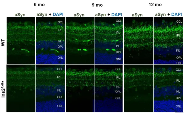
3. Results ... 23
3.1. Characterization of the WT and Ins2Akita population ... 23
3.2. Evaluation of synucleins profile in WT and Ins2Akita retinas ... 24
3.2.2 Are aSyn and bSyn presynaptic proteins in the retina? ... 31
3.2.3 Colocalization between the synucleins ... 37
3.3. Synaptic markers in the retina ... 45
4. Discussion and concluding remarks ... 49
viii
Abbreviations
AD Alzheimer’s Disease
AGEs Advanced Glycation Products
aSyn Alpha-synuclein
ALP Autophagy/Lysosomal Pathway
ATP Adenosine Triphosphate
Bax Bcl-2-associated X protein
BCA Bicinchoninic Acid
BSA Bovine Serum Albumin
bSyn Beta-synuclein
CNS Central Nervous System
CT Camera Temperature
DAPI 4',6-diamidino-2-phenylindole
DDC Dopa Decarboxilase
DLB Dementia with Lewy Bodies
DM Diabetes Mellitus
DME Diabetic Macular Edema
DO Dopamine
DR Diabetic retinopathy
ER Endoplasmic Reticulum
ERG Eletroretinography
FELASA Federation of European Laboratory Animal Science Associations)
GABA γ-aminobutyric acid GCL Ganglion Cell Layer
GLUT2 Glucose Transporter 2
gSyn Gamma-synuclein
IHC Immunohistochemistry
INL Inner Nuclear Layer
IPL Inner Plexiform Layer
L-DOPA L-3,4-dihydroxyphenylalanine
LB Lewy bodies
MODY MSA
Maturity-Onset Diabetes of the Young Multiple System Atrophy
mtDNA Mitochondrial DNA
NAC NPDR
Non-B-amyloid component
Nonproliferative Diabetic Retinopathy
NSF N-enthylmaleimide-Sensitive Factor
ix
O/N Over night
ONL Outer Nuclear Layer
OPL Outer Plexiform Layer
OT Object temperature
PBS Phosphate-buffered saline
PBS-T PBS-Tritton
PFA Paraformaldehyde
PD Parkinson’s disease
PDD Parkinson’s Disease Dementia PDR Proliferative Diabetic Retinopathy
PKC Protein kinase C
PNS Peripheral Nervous System
REM Rapid Eye Movement
ROS Reactive oxygen species
RPE Retinal Pigment Epithelium
RT Room Temperature
SDS Sodium Dodecyl Sulfate
SNARE NSF-Attatchment Protein Receptor
SNAP-25 SynaptopSome-Associated Protein
STZ Streptozotocin syn Synucleins TBS TBS-T TFAM TH Tris-Buffered Saline
Tris-Buffered Saline – Tween 20 Mitochondrial Transcription Factor A Tyrosine Hydroxylase
TUNEL Terminal deoxynucleiotidyl transferase nick and labeling
UPS Ubiquitin Proteasome System
VAMP 2 Vesicle-Associated Membrane Protein 2
VEGF Anti-Vascular Endothelial Growth Factor
WB Western Blot
x
Index of figures
Fig. 1.1. Common intersecting pathways underlying DR microvascular complications. ... 3 Fig. 1.2. Schematic enlargement of the retina, representing the nuclear layers. ... 5 Fig. 1.3. Responses of retinal photoreceptors, bipolar and ganglion cells to darkness and
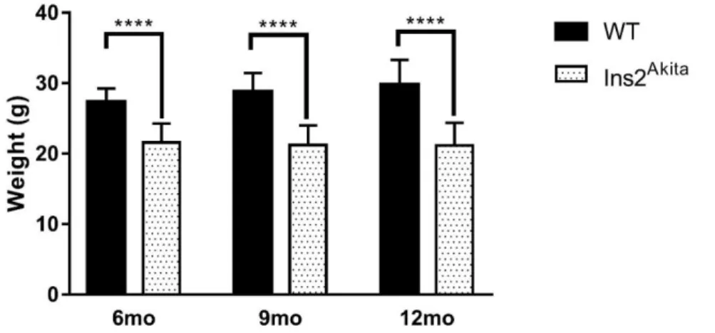
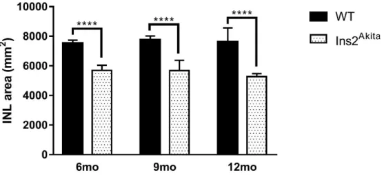
illumination in the respective field surround. ... 7 Fig. 1.4. Common intersecting pathways underlying PD pathogenesis. ... 8 Fig. 1.5. Schematic structure of synuclein proteins. ... 12 Fig. 1.6. Diabetes progression and molecular markers in the STZ and Ins2Akita diabetic retinopathy model. ... 15 Fig. 3.1. Glycemia levels of WT and Ins2Akita mice sacrificed between September 2016 and August 2017. ... 23 Fig. 3.2. Weight levels of WT and Ins2Akita mice sacrificed between September 2016 and August 2017. ... 23 Fig. 3.3. INL area (mm2) of WT and Ins2Akita mice. ... 24 Fig. 3.4. Immunohistochemistry for aSyn in retinal sections of WT and Ins2Akita mice of 6, 9 and 12-months old. ... 25 Fig. 3.5. Quantification of the amount of cell bodies containing aSyn in the GCL of 6, 9 and 12-months old WT and Ins2Akita mice retinas. ... 26 Fig. 3.6. Quantification of the amount of cell bodies containing aSyn in the INL of 6, 9 and 12-months old WT and Ins2Akita mice retinas. ... 26 Fig. 3.7. Immunohistochemistry for bSyn in retinal sections of WT and Ins2Akita mice of 6, 9 and 12-months old. ... 27 Fig. 3.8. Immunohistochemistry for gSyn in retinal sections of WT and Ins2Akita mice of 6, 9 and 12-months old. ... 28 Fig. 3. 9. Assessment of aSyn levels in 6, 9 and 12-months old WT and Ins2Akita protein extracts from whole retina. ... 29 Fig. 3.10. Assessment of bSyn levels in 6, 9 and 12-months old WT and Ins2Akita protein extracts from whole retina. ... 30 Fig. 3.11. Evaluation of the profile of presynaptic marker Synaptophysin with ageing and diabetes and its colocalization with aSyn in the same conditions. ... 32 Fig. 3.12. Immunohistochemistry with colocalization between bSyn and Synaptophysin in retinal sections of WT and Ins2Akita mice of 6, 9 and 12-months old. ... 33 Fig. 3.13. Evaluation of the profile of presynaptic marker Syntaxin 1A with ageing and diabetes and its colocalization with aSyn in the same conditions. ... 35 Fig. 3.14. Immunohistochemistry with colocalization between bSyn and Syntaxin 1A in retinal sections of WT and Ins2Akita mice of 6, 9 and 12-months old. ... 36 Fig. 3.15. Evaluation of the colocalization of aSyn and bSyn in retinal sections of WT and Ins2Akita mice of 6, 9 and 12-months old. ... 37
xi Fig. 3.16. Immunohistochemistry with colocalization between aSyn and gSyn in retinal sections of WT and Ins2Akita mice of 6, 9 and 12-months old. ... 39 Fig. 3.17. Evaluation of the profile of dopaminergic amacrine cells with ageing and DR and the colocalization of TH with aSyn in the same conditions. ... 40 Fig. 3.18. Immunohistochemistry with colocalization between aSyn and PKC-α in retinal sections of WT and Ins2Akita mice of 6, 9 and 12-months old. ... 41 Fig. 3.19. Immunohistochemistry with colocalization between bSyn and PKC-α in retinal sections of WT and Ins2Akita mice of 6, 9 and 12-months old. ... 42 Fig. 3.20. Immunohistochemistry with colocalization between gSyn and PKC-α in retinal sections of WT and Ins2Akita mice of 6, 9 and 12-months old. ... 42 Fig. 3.21. Immunohistochemistry with colocalization between aSyn and Calbindin in retinal sections of WT and Ins2Akita mice of 6, 9 and 12-months old. ... 43 Fig. 3.22. Immunohistochemistry with colocalization between bSyn and Calbindin in retinal sections of WT and Ins2Akita mice of 6, 9 and 12-months old. ... 44 Fig. 3.23. Immunohistochemistry with colocalization between gSyn and Calbindin in retinal sections of WT and Ins2Akita mice of 6, 9 and 12-months old. ... 44 Fig. 3.24. Assessment of SNAP-25 levels in 6, 9 and 12-months old WT and Ins2Akita protein extracts from whole retina. ... 46 Fig. 3.25. Assessment of Rab3a levels in 6, 9 and 12-months old WT and Ins2Akita protein extracts from whole retina. ... 47 Fig. 3.26. Assessment of Caspase 3 levels in 6, 9 and 12-months old WT and Ins2Akita protein extracts from whole retina. ... 48
xii
Index of tables
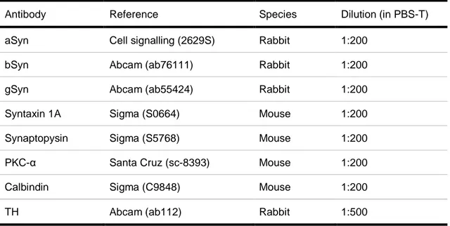
Table 2.1. Description of primary antibodies used for IHC. ... 18
Table 2.2. Description of secondary antibodies used for IHC. ... 19
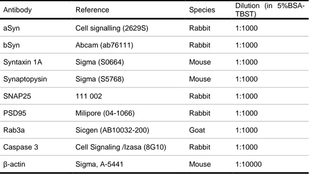
Table 2.3. Description of primary antibodies used for Western Blot. ... 20
Qual o papel das sinucleínas na neurodegeneração da retina?
1
1. Introduction
1.1. Diabetes mellitus
Diabetes mellitus (DM) is a chronic disease characterised by hyperglycaemia that is becoming a global issue mainly due to changes in people’s alimentary and exercise habits as well as ageing. The World Health Organization estimated that the number of people with diabetes has risen from 108 million in 1980 to 422 million in 2014, mostly due to sedentary lifestyles, lack of physical activity and obesity (1).
Diabetes can be classically classified in type 1 DM and type 2 DM but there is also gestational diabetes, Latent Autoimmune Diabetes in Adults and Maturity-Onset Diabetes of the Young (MODY). Type 1 DM, known as the insulin-dependent type, is characterised by the deficient insulin production due to the destruction of the islets of Langerhans in the pancreas and requires insulin administration. Type 2 DM, on the other hand, is the most common type and results from hyperglycaemia and insulin resistance (2).
DM is responsible for significant macro- and microvascular complications. Macrovascular complications include cardiovascular disease, stroke, and peripheral vascular disease. Microvascular complications include damage in the nervous and renal systems (neuropathy and nephropathy, respectively) and eye damage including increased risk for glaucoma, cataracts and, the most threatening ocular implication, diabetic retinopathy (DR) (3,4).
1.2. Diabetic retinopathy
Diabetic retinopathy is a progressive complication of diabetes and the leading cause of irreversible vision loss in working age adults (20-70 years old) (5).
The onset of diabetic complications such as DR is directly related to the duration of diabetes and the quality of glycaemic control (6). Since it may be asymptomatic until vision loss occurs and its diagnostics is based on direct ophthalmoscopy analysis or medical history upon complaint, DR is usually late diagnosed. However by 20–25 years of diabetes almost 90% of patients present some forms of retinopathy (7,8).
1.2.1. Proliferative and non-proliferative DR
Diabetic retinopathy can be classified into two stages, according to its severity: a less-severe form is non proliferative DR (NPDR) and a severe-form proliferative diabetic retinopathy (PDR) (2).
Non proliferative DR shows as early signs microaneurisms that arise from the retinal capillaries, leading to haemorrhages caused by the release of erythrocytes from said microaneurisms (9). These can be visible through the observation of some leakage by fundus photography, documenting the retina, and fluorescein angiography, a technique that examines the circulation of the retina and the choroid using a fluorescent dye and special camera (10).
Qual o papel das sinucleínas na neurodegeneração da retina?
2 Proliferative DR, which may occur up to 50% of type 1 diabetes patients and up to 10% of type 2 diabetes ones (11,12), is characterized by further ischemia resulting in the formation of new blood vessels that, untreated, extend into the vitreous cavity of the eye with the possibility of haemorrhage and consequent tractional retinal detachment. Furthermore, the formation of new blood vessels in other chambers of the eye may lead to block the outflow of the aqueous humour causing neovascular glaucoma. Altogether, these events lead to vision loss (13,14).
Both non proliferative and proliferative DR can lead to another change: the diabetic macular edema (DME), that can affect up to 20% of type 1 diabetes patients and up to 25% of patients with type 2 diabetes (15). It is characterized by increased vascular permeability and breakdown of the blood-retinal barrier, culminating in leakage from plasma from the macula, responsible for the major part of visual function, causing the swelling of the central retina. This event is followed by the formation of exudates from deposition of the lipid and lipoprotein content from the plasma, disrupting the light path in the macula and, ultimately, vision loss (9,13,15).
1.2.2. Retinal microvascular dysfunction
Microvasculature of the retina is considered by many the main site of pathology associated with DR. Although its biochemical mechanisms are not fully elucidated, it is known that continuous exposure of the retina to hyperglycaemia leads to metabolic abnormalities, including the activation of several pathways, accumulation of advanced glycation end products (AGEs), protein kinase C (PKC) activation and increase in oxidative stress (16,17).
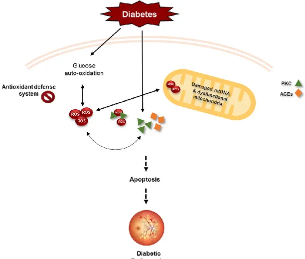
In DR, increased AGEs and PKC are reported within retinal capillaries. Increased AGEs are associated with increased inflammation while PKC activation leads to increased vascular permeability, alterations on blood flow and stimulation of neovascularization due to its relation with the vascular endothelial factor (VEGF) which is suspected to be a primary peon in the induction of vascularization in diabetes (Fig. 1.1) (18,19). Metabolic abnormalities combined with auto-oxidation of glucose and an impaired antioxidant defence system result in the production of reactive oxygen species (ROS) (Fig. 1.1) (20).
Under pathological conditions, the excessive bioavailability of ROS, as a result of increased production and/or decreased removal of ROS, will further increase the production of PKC and damage proteins, lipids and DNA (21). Mitochondria and its DNA (mtDNA) are one of the targets of its damaging effects (20,22).
ROS, by activating matrix metalloproteinases, damages the mitochondrial membrane changing its permeability allowing apoptotic induced factors to be released in the cytosol and subsequently activate the apoptosis machinery (Fig. 1.1). Thereby, retinal capillary and non-capillary cells undergo accelerated apoptosis. In early stages of DR, pericytes start to undergo accelerated death which is followed by the loss of endothelial cells resulting in pericyte ghosts, acellular capillaries and microaneurysms (23). Moreover, damages to the mtDNA impairs its transcription, further interfering with its homeostasis and compromising the mitochondrial machinery, namely the electron transport chain resulting in an increased ROS production (24).
Qual o papel das sinucleínas na neurodegeneração da retina?
3 Overall, microvascular complications result in an increase of vascular permeability, alterations of blood flow and neovascularization, which are the hallmarks of DR. This leads to edema, ocular haemorrhage and eventual retinal vessel closure (16,22).
Fig. 1.1. Common intersecting pathways underlying DR microvascular complications. Diabetic
environment increases the production of ROS and also metabolic abnormalities leading to capillary cell apoptosis and ultimately development of diabetic retinopathy. PKC, AGEs and the production of inflammatory mediators feed the metabolic abnormalities and continuous ROS production as well as oxidative damage to mitochondria, accelerating apoptosis. Adapted from (17).
1.2.3. Neurodegeneration in DR
However, even though DR was for many years seen as solely a microvascular disease caused by significant alterations in the retinal vasculature and blood barrier of the retina, recent evidence suggests that neurodegeneration occurs prior the microvascular complication onset, through deficits in the neural retina (5,26).
It is thought that alteration in the levels of various neurodegenerative metabolites’ and neurodegenerative factors as well as a decrease in neurotrophic factors damage the retinal neurons in early stages of the disease (27).
Qual o papel das sinucleínas na neurodegeneração da retina?
4 Apoptosis markers were identified in higher levels in diabetic retinas. Increased levels of pro-apoptotic protein Bax (Bcl-2-associated X protein) was found in diabetic retinas by several studies (28). Other resesarchers have shown a correlation between TUNEL (terminal deoxynucleiotidyl transferase nick end labeling)-positive cells and increased Caspase 3 levels in neuronal retinas of diabetic rats (29–31). In recent studies, clinical tools like multifocal and flash electroretinography (ERG), contrast sensitivity, color vision and short-wavelength automated perimetry indicate that neurons are vulnerable to damage, shortly after the onset of diabetes (32). Degeneration of retinal neurons has been reported on amacrine and ganglion cells (33,34). Studies in animal models further show photoreceptors’ death (35,36) and abnormalities in horizontal and bipolar synaptic terminals (36,37).
In the retina, glia and neurons share a close interaction with retinal vasculature to maintain a normal retinal function. It has been shown that apoptosis of neurons and activation of glial cells may cause oxidative stress and initiate vasoregretion (38), here suggesting a link between neurodegeneration and microvascular changes in diabetic retinopathy (39,40)
1.2.4. Prevention and treatment methods
The prevention methods for DR combine glucose and blood pressure control to normal levels, considerably retarding the progression of the disease. Further prevention and treatment methods usually apply to more severe forms of DR, relying on retinal laser photocoagulation to slow the formation of new vessels and the progression of vision loss (41). More advanced retinal disease, such as late stages of proliferative DR and macular edema, that combine vitreous haemorrhages and detachment of the retina upon new vessel formation, benefits from intravitreal anti-VEGF injections, vitrectomy, focal laser and argon laser treatment (42,43).
1.3. The retina functional architecture
The retina is a layered structure that includes both sensory neurons and intricate neural circuits that respond to light and perform the first stages of image processing before traveling through the optic nerve into the brain (44).
It is composed of vascular cells (pericytes and endothelial cells), microglia, macro glial cells (Müller cells, astrocytes) and neurons (17).
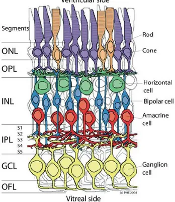
There are, essentially, five major neuronal cell classes distributed between the nuclear layers of the retina. Rod and Cone photoreceptors are integrated in the Outer Nuclear Layer (ONL) followed by horizontal, bipolar and amacrine cells that share the Inner Nuclear Layer (INL) and ganglion cells that, along with a subtype of amacrine cells, is found in the Ganglion Cell Layer (GCL) (Fig. 1.2). All cells communicate with each other electrically, through gap junctions and chemically, using neurotransmitters (45,46).
In between the nuclear layers, the Outer Plexiform Layer (OPL) and the Inner Plexiform Layer (IPL) assure the communication between neuronal cell populations of different layers through
Qual o papel das sinucleínas na neurodegeneração da retina?
5 synapses (Fig. 1.2). The plexiform layers can also be divided into strata, depending on the connections that take place in each one. The IPL is divided in 5 strata, whereas the OPL is traditionally divided in 3 (45). Strata 1 and 2 of the IPL comprise synapses between OFF-bipolar cells and retinal ganglion and amacrine cells as strata 3-5 contain synaptic connections between ON-bipolar cells and retinal ganglion and amacrine cell bodies (47).
ON- and OFF-type cells are so called depending on the stimulation. ON-cells are stimulated by a spot of light brighter than the background, occurring an ON-discharge. When the cells are stimulated by light darker than the background, OFF-cells are activated and an OFF-discharge occurs(47,48).
Fig. 1.2. Schematic enlargement of the neuronal retina. Outer Nuclear Layer (ONL), containing the
photoreceptors rodes and cones; the Inner Nuclear Layer (INL), containing the horizontal, bipolar and amacrine cells and the Ganglion Cell Layer (GCL) containing the ganglion cells; and the plexiform layers: Outer Plexiform Layer (OPL), containing the presynaptic terminals of the photoreceptors, bipolar and horizontal cells and Inner Plexiform Layer (IPL), containing the presynaptic terminals of amacrine, bipolar and ganglion cell bodies (in Webvision: Simple Anatomy of the Retina).
1.3.1. Processing of visual information
Photoreceptors rods and cones are the first response to photons each having specific roles in the retina. Rods show an elevated sensitivity to light and are therefore responsible for dim-light vision. Cones, although less sensitive to light than rods, exhibit a higher sensitivity to a specific light wave consequently engaging bright-light, high acuity color vision (49,50).
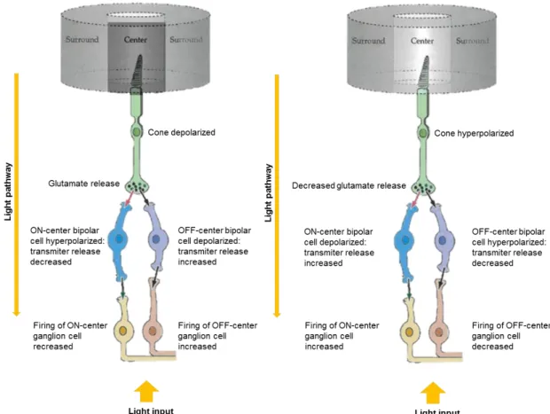
The processing of the visual information begins with the conversion of light into depolarizing spikes of neurotransmitter glutamate by photoreceptors into glutaminergic ON-center bipolar cells in
Qual o papel das sinucleínas na neurodegeneração da retina?
6 the OPL and hyperpolarizing spikes into OFF-center bipolar cells, mediated by horizontal cells (Fig. 1.3) (47,50).
Bipolar cells can be divided into two major classes, rod and cone bipolar cells and further divided into depolarizing bipolar cells (the ON-type) and hyperpolarizing bipolar cells (the OFF-type). Rod bipolar cells are ON bipolar cells and contact primarily with rod photoreceptors, whereas cone bipolar cells, which can be either ON- or OFF-type, mostly synapse with cone photoreceptors. Moreover, ON- and OFF-bipolar cells also contact with retinal ganglion and amacrine cells within the IPL (Fig. 1.3) (51,52).
When depolarized, bipolar cells release glutamate into ganglion cells but when hyperpolarized they decrease its release what increases or decreases ganglion cell firing rate, respectively (Fig. 1.3) (52).
Regarding the amacrine cells, there are more than 30 known types of these, that communicate with different neurons using different neurotransmitter, playing several roles in the retina (53). When responsible for the excitation of ganglion cells, they act in two ways, both relying on γ-aminobutyric acid (GABA) and glycine neurotransmitters: 1) direct feedforward inhibition from amacrine cells onto retinal ganglion cells, or 2) feedback inhibition, where amacrine cells are intermediates between bipolar and ganglion cells. In the end, the retinal ganglion cells send the message to the brain (54,55). Amacrine cells are classified by the width of their connection and layer of the IPL they are in and also by the neurotransmitter they use. The AII amacrine cells are the most studied amacrine cells and participate predominantly in the vertical flow of information in the photoreceptor-bipolar-ganglion cell chain, transmitting both rod and cone-driven signals within the ON- and OFF-pathways (54,56). Other examples include the A8, A17, A19 and A20 amacrine cells, intermediates in several neuronal cell chains of the retina, not forgetting the dopaminergic amacrine cells (56).
Qual o papel das sinucleínas na neurodegeneração da retina?
7
Fig. 1.3. Responses of retinal photoreceptors, bipolar and ganglion cells to darkness and illumination in the respective field surround. Depending on light availability, cone photoreceptors either hyperpolarize or
depolarize, decreasing or increasing glutamate release rates, respectively, leading to hyperpolarization of depolarization of ON- and OFF-center biplar cells, thus mediating ganglion cell’s firing rate and vision adaptation to light. Adapted from Webvision: Bipolar Cell Pathways in the Vertebrate Retina.
1.4. Parkinson’s Disease
Parkinson’s disease (PD) is the second most common neurodegenerative disease in middle-aged and elderly people. It is characterized by the loss of dopaminergic (dopamine-producing) neurons in the substantia nigra pars compacta of the brain and the aggregation of alpha-synuclein (aSyn) in association with other proteins such as ubiquitin in Lewy bodies (LBs) in the remaining nigral neurons, impairing optimal neuron functioning (57).
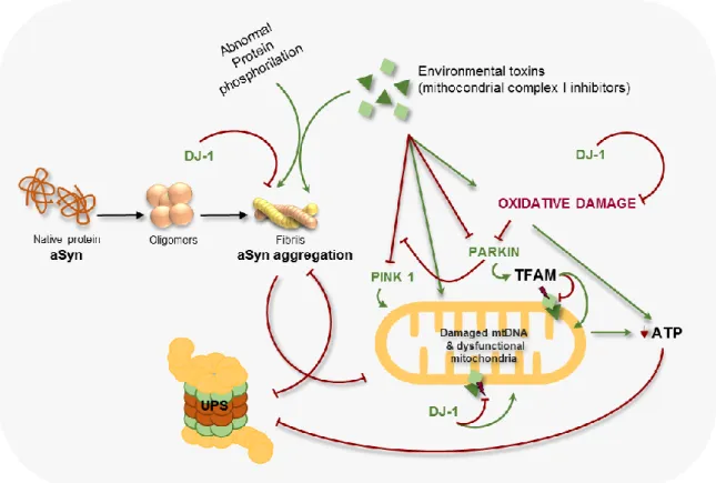
Epidemiological studies reveal that a small percentage of <10% of PD are familial cases of PD while the majority of cases are sporadic (58). This complex disease has multiple factors involved in its pathogenesis. Ageing seems to be the most potent risk for PD but other risk factors have shown to be important, such as predisposing factors, diabetes and exposure to environmental toxins like pesticides. Studies from both environmental factors and familial PD-related genes helped identifying the several pathways involved in the events leading to the death of dopaminergic neurons due to mitochondrial dysfunction, oxidative damage and protein accumulation (59).
Qual o papel das sinucleínas na neurodegeneração da retina?
8 Pathogenic mutations in the familial PD-linked genes aSyn, Parkin, DJ-1 and PINK1 as well as environmental factors have been associated with abnormalities in mitochondrial structure, function and protection system, oxidative damage, abnormal protein aggregation and protein phosphorylation compromising dopaminergic neuronal function and survival. (Fig. 1.4) (60). These mutated proteins are thought to bind to lipids and increase mitochondrial, lysosomal and vesicular membrane permeability, leading to mitochondrial dysfunction, impairment of the respiratory chain, decrease ubiquitin proteasome system (UPS) activity, increased calcium influx, ion homeostasis disruption and activation of Caspase 3 (Fig. 1.4). This cycle of events leads to cell death, by release of the apoptosis machinery (61,62).
The main manifestations of PD are motor changes such as resting tremor, bradykinesia, postural instability and cogwheel rigidity and, especially in early stages, symptomatic relief is available through dopamine (DO) restoring therapies. Moreover, non-motor manifestation such as constipation, rapid eye movement sleep (REM sleep) behaviour disorder, depression, cognitive disturbances and visual impairment have been rising awareness (63).
Fig. 1.4. Common intersecting pathways underlying PD pathogenesis. aSyn undergoes aggregation due
to pathogenic mutations which in turn compromise ubiquitin proteasome function (UPS) and cause mitochondrial dysfunction. Mitochondrial dysfunction and oxidative damage lead to a decrease in ATP which may compromise the UPS function promoting abnormal protein aggregation. Parkin increases mitochondrial biogenesis by activating mitochondrial transcription factor A (TFAM) and blocks PINK1-induced mitochondrial dysfunction, while pathogenic mutations in the protein and oxidative damage severely compromise its protective function. DJ-1 protects against oxidative stress, functions as a chaperone to block aSyn aggregation and protects against
Qual o papel das sinucleínas na neurodegeneração da retina?
9
mitochondrial dysfunction. PINK1, when not compromised by pathogenic mutations, seems to protect against mitochondrial dysfunction. Adapted from (64).
1.4.1. Visual impairment in PD
Decreased visual acuity, reduced colour vision, deficits in vision-spatial orientation and contrast sensitivity are commonly found in patients with PD (65).
The pathological changes that have been reported in the eye in PD refer mostly to the retina and include cell loss related to reductions in retinal dopamine, with a visible thinning of the retinal fiber layer observed (66,67). Dopamine is a key neuromodulator in the brain and is highly present in the retina, as a chemical messenger for light adaptation, modulating photoreceptor activity, organizing ganglion and bipolar cell receptive fields and coupling horizontal cells and the amacrine lateral system (68). It is synthetized and released by the dopaminergic amacrine cells, upon light input and circadian clock, and activates D1 and D2 dopamine receptors distributed throughout the retina. When reaching the horizontal cells it uncouples their gap junctions blocking the communication between cells. Similar occurs for the amacrine cells themselves, considering that dopamine is thought to lead to the uncoupling of gap junctions of AII amacrine cells. In this way dopamine controls the light signal that reaches the ganglion cells, decreasing their sensitivity which is important to avoid saturation to light stimulation (54,69). Therefore, dopamine release reduction would be catastrophic.
In the brain, dopamine reduction in subcortical regions involved in eye movement, like basal ganglia and substancia nigra pars reticulata, could also be implicated in visual impairment of PD patients (70).
1.5. The synuclein family
The synucleins (syn) are a family of small and highly conserved proteins, very closely related: aSyn, beta-synuclein (bSyn) and gamma-synuclein (gSyn), that are encoded by the SNCA, SNCB and SNCG genes, respectively (Fig. 1.5). The first 42 aminoacids are identical between the synucleins but studies show that aSyn and bSyn and more closely related to each other (71).
These proteins are divided in three domains: residues between 1 and 60 make up the N-terminal which contains four lysine-rich repeats highly conserved motif similar to the lipid-binding motifs in lipoproteins that form a α-helice in the presence of lipids, allowing lipid-binding capacity to these proteins; residues between 61 and 95 form the central domain which has a high hydrophobic aminoacid content responsible for the amyloidogenic properties of the protein and, finally, residues 96-140 make up the C-terminus which is thought to confer chaperone-like activity to the synucleins (Figure 1.5) (72).
Qual o papel das sinucleínas na neurodegeneração da retina?
10
1.5.1. aSyn
aSyn is a 14kDa protein that has been the center of focus in a group of neurodegenerative disorders called α-synucleinopathies, which includes both familial and sporadic PD, PD dementia (PDD), Dementia with Lewy bodies (DLB), multiple system atrophy (MSA), among others (73).
aSyn is located in the central nervous system (CNS), predominantly in the presynaptic terminals of neurons, where synaptic vesicles constantly approach in order to release their neurotransmitter content, being later on recycled to clear the active zone (74). Even though its role in the brain is not fully known, it has been speculated that aSyn is involved in synaptic signaling and membrane trafficking, being suggested to play a role in both exo- and endocytosis of synaptic vesicles (74).
Regarding the role of aSyn on synapses, its role in the assembly of the soluble N-ethylmaleimide-sensitive factor (NSF) attachment protein receptor (SNARE) complex is suggested. The SNARE complex is an assembly of plasma membrane proteins, namely syntaxin and synaptopsome-associated protein 25 (SNAP-25), with vesicle-synaptopsome-associated membrane protein 2 (VAMP2), that bind vesicles to the plasma membrane, undergo fusion and stimulate neurotransmitter release (75). Upon binding to synaptic vesicles during docking and priming, aSyn undergoes conformation changes folding into an amphipathic a-helix. As a result, this synuclein promotes the SNARE complex assembly by bounding to the SNARE-protein synaptobrevin-2/VAMP2 during synaptic exocytosis (73,75,76).
Moreover, aSyn may play a complementary role in endocytosis, facilitating membrane retrieval in order to maintain the membrane structure and facilitate further neurotransmitter release although the exact mechanisms are not fully understood (77). aSyn was described as being able to sense membrane curvature and stabilize it by binding its N-terminal region as well as residues between 65-97 region to lipid membranes and applying a double anchor mechanism by which aSyn tethers two vesicles to one another or to the plasma membrane, facilitating exo- and endocytosis (74).
1.5.1.1 aSyn toxicity
In its native state, aSyn is thought to predominantly exist as an unfolded monomer in equilibrium between cytosolic and membrane-bound states. However, duplication and triplication of the SNCA gene, post-translational modifications such as Ser129 and Ser87 phosphorylation (Figure 2), truncation and glycation as well as disease related mutations are thought to change this protein’s aggregation dynamics (78). All known clinical mutations, such as A30P, E46K, H50Q, G51D, A53T and A53E are present in the N-terminal, emphasizing the importance of this domain in the aggregation of aSyn (Fig. 5).
Evidence suggests that the principle mechanism behind α-synucleinopathies is the misfolding of aSyn into aggregates in intracellular bodies, beginning with the formation of relatively soluble oligomers that can self-assemble into insoluble fibrils, resulting in the formation of deposits (79). This leads to neuroinflammation, neurodegeneration and cell death (80).
Increased aSyn levels are thought to disrupt neurotransmitter release through a decrease in size and mobility of the synaptic-vesicle recycling-pool (81). Overexpression of aSyn stabilizes the
Qual o papel das sinucleínas na neurodegeneração da retina?
11 SNARE complex in the membrane, inhibiting vesicle fusion and, therefore, neurotransmitter release (82) such as dopamine release (83).
Not only can aggregated aSyn interfere with neurotransmitter release but it can also affect its synthesis. Dopamine synthesis consists on the conversion of tyrosine into dopamine in two steps: tyrosine is firstly converted into L-3,4-dihydroxyphenylalanine (L-DOPA), mediated by phosphorylated tyrosine hydroxylase (TH), following L-DOPA conversion into dopamine by DOPA decarboxylase (DDC) (84). However, aSyn binds to the dysphosphorilated TH, maintaining its inactive form and, subsequently, causing a decrease in its enzymatic activity and dopamine synthesis (85).
Overall, this protein has been found to interact with mitochondria, mitochondria-endoplasmic reticulum (mitochondria-ER) and ER-Golgi networks and with the ubiquitin proteasome system (86).
Mitochondria are essential for the synthesis of adenosine triphosphate (ATP), regulation of calcium, lipid metabolism and, overall, neuronal survival (87). ATP is synthesized via oxidative phosphorylation complexes, which are present in the inner membrane of mitochondria: ubiquinone oxidoreductase Complex I, succinate dehydrogenase Complex II, ubiquinol–cytochrome c oxidoreductase Complex III, cytochrome c oxidase Complex IV and ATP synthase Complex V mitochondrial (88). Mitochondrial complex I is responsible for catalyzing the first step of the electron transport chain which is the main source of ROS (89).
Studies revealed that aggregated aSyn binds to the inner membrane of mitochondria, altering its normal function by associating with complex I (90). This events lead to cytochrome c release and a consequent increase of Ca2+ uptake and ROS levels which ultimately leads to cell death (91). Furthermore, wild type aSyn and A53T mutant are proposed to promote an up-regulation of mitophagy, the delivery of damaged and dysfunctional mitochondria to the lysosome, culminating in an abnormal mitochondrial activity, increased ROS levels and further mitochondrial degradation levels (92–94). However, is not only aSyn that promotes ROS levels to rise. Increased ROS, and consequently oxidative stress, is suggested to promote aSyn aggregation, creating a never ending cycle of degeneration (95).
Moreover, mitochondria communicates with the ER to regulate several cellular processes (96). Alterations in this mitochondria-ER communication can cause deregulation of Ca2+ homeostasis resulting in protein misfolding, metabolic alterations and apoptosis (97). ER stress, which is usually caused by the accumulation of misfolded proteins within the ER, is associated with the development of several neurodegenerative diseases. By either interacting with ER chaperones or affect ER function while compromising ER membranes integrity and exposing portions for the ER lumen to the cytosol, Insoluble aSyn aggregates could generate ER stress (98). Actually, aSyn was reported to inhibit trafficking in the ER-Golgi network in yeast models, increasing protein accumulation in the ER, cell toxicity and cell loss and exacerbating ER stress (99). Moreover, similar to what was seen in yeast cells, vesicle accumulations due to trafficking impairment have been observed in neurons before Lewy body formation (100). Furthermore, vesicle accumulations are often found in proximity to Lewy bodies in later stages of disease (101).
Qual o papel das sinucleínas na neurodegeneração da retina?
12 Ultimately aSyn is degraded both by the UPS and by the autophagy/lysosomal pathway (ALP). However, some mutations such as the A30P and A53T may cause a failure in the release of aSyn and clogging of the autophagy translocation machinery thus accumulating aSyn (102).
1.5.2. bSyn and gSyn
bSyn is slightly smaller than aSyn but shares some of the synuclein features, including the location, being mainly found in the CNS (Fig. 1.5). Even though it is not as prone to aggregate as aSyn due to the lack of 11 aminoacids in its NAC domain, studies suggest that when exposed to toxins such as metal ions and pesticides, bSyn tends to fibrillate (103). Furthermore, when co-expressing with aSyn, it leads to an increase in cytotoxicity in yeast and forms heterodimers in both yeast and cell lines (104).
gSyn is the least conserved and the smallest of the three proteins (Fig. 1.5) (71). This synuclein is a putative marker for breast cancer (105). In the nervous system, it can be found mainly in the peripheral nervous system (PNS), including in primary sensory neurons, sympathetic neurons and motor neurons (106) but can also be found in other tissues such as ovarian and breast cancer (71). Although its function is likewise not known, exogenous expression of the protein potentiates the metastatic ability of breast tumours (107).
Fig. 1.5. Schematic structure of synuclein proteins. aSyn, bSyn and gSyn share high homology. The
N-terminal region is highly conserved between the three proteins. Major differences are observed in the acidic C-terminal, with bSyn and gSyn having a shorter size when compared to aSyn (adapted from (108)).
1.5.3. Synucleins in the eye
All the members of the synuclein family can be found ocular tissues. aSyn and bSyn are both located in synapse-rich IPL of the retina showing similar distribution patterns although bSyn is also found in the INL. gSyn, on the other hand, can be found in the GCL which is mainly composed of ganglion cells (109).
To date, few have dedicated to the task of describing in detail the exact neuronal population where these proteins are located.
Qual o papel das sinucleínas na neurodegeneração da retina?
13 A study conducted by Martínez-Navarrete et al. in several animal models, namely in rodent (rat and mouse), bovine, and primate (human and monkey) retinas, not only reports the presence of aSyn in the presynaptic terminals of retinal neurons in the IPL (110), in agreement with the findings of Surguchov et al. (76), but also in the OPL due to the colocalization with synaptophysin (110), a synaptic-vesicle transmembrane protein involved in the regulation of vesicular exocytosis in the CNS, that can be found in both plexiform layers of the retina (111). The same group claims that aSyn was further localized in the somata and dendrites of both GABAergic and glycinergic amacrine cells and showed high levels in the RPE in all vertebrate tested (110).
gSyn localization as well as physiological and pathological role are very poorly known. However, this synuclein was found by Martínez-Navarrete et al in cultured bovine retinal pigment epithelium (RPE) cells (110). Moreover, studies in rodent and human retinas, using suitable markers for ganglion cells, such as Brn-3 and Thy1-1 family proteins, were able to detect colocalization between these markers and gSyn antibody. gSyn was even suggested as a specific marker for that neuronal population (112). This synuclein was also implicated in glaucoma and even though a lot is yet to describe about its role in eye disease, has been found in the optic nerve of glaucomatous patients, in a subset of glial cells identified as possibly reactive astrocytes, which did not happen for healthy controls (106). Furthermore, in Alzheimer’s disease (AD) patients, a study revealed a decrease in gSyn levels, with no differences occurring in the levels of the other two synucleins, though the reasons behind this event remain unknown (109).
1.6. Common pathophysiology between PD and DR
Recent evidence points to a common pathophysiology in the visual impairment of PD patients and in DR, as an effect of a disruption in the dopaminergic system in both diseases. Dopamine is an essential neuromodulator in the brain and is highly present in the retina. In addition to regulating motor, cognition, and retinal function, dopamine present in the retina modulates light-adapted vision through the activation of selective receptors and retinal pathways (113,114). In fact, studies suggest that injecting the diabetic mice with dopamine-restoring and dopamine-activating drugs already used in PD can restore dopamine levels in dopaminergic amacrine cells and significantly improve retinal function (115).
Moreover, another study in a rodent PD model suggested that the administration of antidiabetic drugs proved to have a neuroprotective effect on retinal and nigrostriatal neurons in PD patients. This further suggests a common pathophysiology between these two pathologies (116).
Moreover, there is a resemblance between the diabetic retina and the environment underlying aSyn aggregation and, subsequently, PD. Hyperglycemia activates several pathways that, along with the impairment of the antioxidant defence system, result in an excessive bioavailability of ROS (20) that damages the mitochondrial membrane and mtDNA, compromising the mitochondrial machinery, namely the electron transport chain resulting in a continuous and increasing ROS production that leads to release of the apoptosis machinery (23). Likewise, in PD, increased oxidative stress and
Qual o papel das sinucleínas na neurodegeneração da retina?
14 inflammation processes as well increased glucation potentiate aSyn aggregation, a hallmark of PD (117). On the other way around, aSyn is believed to bind to the inner mitochondrial membrane, where it associates with mitochondrial complex I, culminating in increased ROS production, Ca2+ levels and release of cytochrome c, leading to cell death (90,91).
The synuclein family members, which play a major role in PD due to the formation of aggregates in brain tissues and consequent relation with neurodegeneration, are also highly expressed in the retina (118). Triple knockout of aSyn, bSyn and gSyn in mice leads to altered synapse structure and physiology and age-dependent neuronal dysfunction and decreased survival. Importantly, these animals developed retinal dysfunction and age dependent blindness, a strong indicative that synucleins play an important role in retina function (119).
Furthermore, aSyn aggregation was, as previously mentioned, also described in other degenerative diseases such as AD (120) but also in ageing retinas (121). Although LBs are the hallmark lesions of PD, aSyn-positive LBs also occur frequently in the brains of many AD patients (122). Also, within ageing, progressive accumulation of potentially toxic protein aggregates and the dysfunction of the ubiquitin proteasome system, eventually contribute to neuronal degeneration (121,123).
Altogether these data suggest a possible role of the synucleins in DR’s pathology.
1.7. Ins2
Akitaas a diabetic model
The most commonly used diabetes animal models include rodents, dogs and primates with diabetes induced by chemical toxins such as streptozotocin. Streptozotocin enters the pancreatic β-cells via a glucose transporter, glucose transporter 2 (GLUT2), and causes alkylation of DNA. DNA damage induces later on the formation of superoxide radicals. Consequently, hydrogen peroxide and hydroxyl radicals are also generated. As a result of the streptozotocin action, β-cells are destroyed (124).
Still, due to the creation of strain-dependent resistance to streptozotocin, studies have been developed in order to describe a better model for early diabetic retinal complications in diabetes.
The Ins2Akita, a C57BL/6 mutant heterozygous mouse, is a relatively recent and improved model for type 1 diabetes complications’ studies, such as DR. It consists of a point mutation in the insulin 2 gene, which replaces a cysteine with tyrosine at the seventh amino acid of the A chain of the insulin 2 gene product (125,126). This spontaneous mutation causes a conformational change in the protein, leading to its accumulation in the ER of pancreatic β-cells, triggering the unfolded protein response and consequently β-cell death. Loss of β-cells in the pancreas results in systemic hypoinsulinemia and hyperglycaemia, which are significant after only 4 weeks with significant as well as significantly less weight (Fig. 1.6) (126). Importantly, these mice develop diabetes complications including diabetic neuropathy (126–128).
Although the Ins2Akita mice show a few signs of proliferative DR like indices of neovascularization and new capillary bed formation (129), this model is particularly strong in retinal pathologies
Qual o papel das sinucleínas na neurodegeneração da retina?
15 characteristic of early, nonproliferative DR, such as increased frequency of apoptotic retinal neurons, microaneurysms, vascular damage, and increased vascular leakage, accompanied by vision loss (126,130,131).
When compared to other models, such as the streptozotocin(STZ)-induced diabetic rats, the Ins2Akita mice mimic several outcomes of diabetes established in the STZ-model, namely retinal complications such as increased vascular permeability, increased acellular capillaries and vascular inflammation (Fig. 1.6) (126). Moreover, increased levels of apoptosis markers such as Caspase 3 was reported in both models (Fig. 1.6) (126,132). However, the Ins2Akita model shows some earlier the other models such as the acute STZ-model. Thinning of the inner layers of the retina were reported in the Ins2Akita model after little more than 5 months of diabetes onset, more than two months earlier than in STZ mice, suggesting a degeneration and loss of horizontal, bipolar and amacrine cells (Fig. 1.6) (133,134).
Overall, the Ins2Akita has several advantages over other models. This heterozygotic mouse model breeds well, presents stable insulin-deficient diabetes that can be maintained at a noncatabolic state without exogenous insulin and shows a mechanism of diabetes onset that does not involve systemic immunologic alterations making it possible to evaluate the metabolic impact on the retina (126).
Fig. 1.6. Diabetes progression and molecular markers in the STZ and Ins2Akita diabetic retinopathy
model. The Ins2Akita mice present several retinal complications. By one month these mice show a higher blood
glucose concentration and higher levels of Caspase 3 in the retina and considerably less weight than controls. By 2 months old swollen processes in the retina are noticed and shortly after increased vascular permeability. Thinning of the Inner Nuclear and Inner Plexiform Layers of the retina are considerably increased in the Ins2Akita
model after little more than 5 months of diabetes onset, suggesting a degeneration and loss of horizontal, bipolar and amacrine cells. By 7 months old, increased acellular capillaries and vascular inflammation occur.
Qual o papel das sinucleínas na neurodegeneração da retina?
16
1.8. Scientific question and aims
Diabetes is a chronic disease responsible for significant macro- and microvascular complications such as Diabetic Retinopathy. Recently, DR was described as a neurodegenerative disease, where degeneration of neurons occurs prior to the microvascular onset. PD, the second most common neurodegenerative disease in the world, and DR are proposed to share a common pathophysiology in visual impairment due to the disruption of the dopaminergic system as well as to a similar hostile environment that, in PD, culminates in aSyn aggregation, toxicity and neuronal death. aSyn is the main focus not only in PD but also in other neurodegenerative diseases and can be found in ocular tissues. Its precise location in the retina and pathophysiological role are not fully described though it is suggested that it is crucial for vision to occur.
The aim of this study is to do a description of the synucleins distribution pattern in the retina and to establish a correlation between the synucleins profile and the progression of diabetic retinopathy, using a type 1 diabetes mice model, the Ins2Akita, as well as Wild Type controls in different stages of the disease.