Development of an
antisense-mediated
exon skipping
therapeutic strategy
for Mucolipidosis II
Regina Filipa Machado Vilela
Dissertação de Mestrado apresentada à
Faculdade de Ciências da Universidade do Porto em
Biologia Celular e Molecular
2017
D ev elop men t o f an an tise n se -med iated ex o n sk ip p in g th era p eu tic strateg y f o r Muc o lip id o sis II Regin a F ilipa M ac h ado V ile la FCUP INSA 2.º CICLOantisense-mediated
exon skipping
therapeutic strategy
for Mucolipidosis II
Regina Filipa Machado Vilela
Mestrado em Biologia Celular e Molecular
Departamento de Biologia 2017
Orientadora
Doutora Sandra Catarina da Conceição Alves, Investigadora Auxiliar,
Instituto Nacional de Saúde Doutor Ricardo Jorge
Coorientadoras
Professora Doutora Maria João Prata Martins Ribeiro, Professora Associada com Agregação,
Faculdade de Ciências da Universidade do Porto Doutora Liliana da Silva Matos,
Investigadora Pós-Doutoramento,
Eu, Regina Filipa Machado Vilela, aluna com o número 201201434 do Mestrado de Biologia Celular e Molecular da edição de 2016/2017, declaro que sou a autora da totalidade do trabalho experimental descrito,
assim como do texto apresentado, não apresentando texto plagiado e tendo tomado conhecimento das consequências de uma situação de plágio.
Dissertação apresentada à Faculdade de Ciências da Universidade do Porto para candidatura ao grau de Mestre em Biologia Celular e Molecular.
Dissertation presented to the Faculty of Sciences of the University of Porto for application for the Master’s Degree in Cell and Molecular Biology.
Agradecimentos
Porque este trabalho é fruto da participação de várias pessoas, gostaria de aproveitar esta oportunidade para expressar o meu agradecimento a todos que de alguma forma contribuíram para que este projeto e toda a minha formação fossem possíveis:
À Doutora Sandra Alves, minha orientadora, por me ter aceitado no seu grupo de investigação e por toda a dedicação ao longo deste ano, permitindo que desenvolvesse este projeto da melhor forma possível e melhorasse os meus conhecimentos e capacidades na área da Biologia Molecular. Obrigada por todas as sugestões e conselhos e por sempre se ter preocupado comigo e com o meu percurso académico.
À Professora Doutora Maria João Prata, minha co-orientadora, por me ter inicialmente apresentado à Doutora Sandra e por se ter mostrado sempre disponível para me ajudar em todas as etapas deste Mestrado, especialmente na elaboração desta dissertação. Obrigada também por todo o conhecimento transmitido durante as aulas das diversas disciplinas que lecionou e que em muito contribuíram para a minha escolha desta área de estudo.
À Liliana, também minha co-orientadora, por me ter sempre acompanhado a nível laboratorial, por me ter ensinado todas as técnicas necessárias para a realização deste projeto e por estar sempre disponível para esclarecer qualquer dúvida que tivesse. Um obrigada muito especial por toda a confiança e por me deixares aprender ao meu próprio ritmo e sem pressões.
A todo o grupo de investigação em Doenças Lisossomais de Sobrecarga do Instituto Nacional de Saúde Doutor Ricardo Jorge (INSA), por me terem acolhido da melhor forma possível e por me terem sempre apoiado ao longo de todo este percurso. Obrigada por todos os conselhos e partilha de experiências que tanto me ajudaram a crescer ao longo deste ano, tanto a nível pessoal como profissional.
Ao Paulo Gaspar, por aceitar contribuir para este projeto ao nível da realização dos ensaios enzimáticos e por se mostrar sempre disponível para esclarecer qualquer dúvida sobre este assunto.
Ao Professor Doutor José Pissarra e à Professora Doutora Conceição Santos, diretores do Mestrado de Biologia Celular e Molecular nestes dois anos letivos, assim como a todos os professores deste Mestrado, por todo o esforço e dedicação e pelo apoio dado a todos os alunos ao longo deste percurso. Obrigada por todo o conhecimento transmitido e por nos ajudarem a tornarmo-nos melhores cientistas e melhores pessoas.
A todos os professores que de alguma forma fizeram parte da minha vida escolar e académica, obrigada por terem contribuído para a pessoa que sou hoje e por me ajudarem a alargar os meus horizontes cada vez mais. Um agradecimento especial à educadora Sara e à professora Cecília que foram incansáveis nos meus primeiros anos e tanto se esforçaram por compreender e adaptar os programas escolares às minhas necessidades para que nunca perdesse a curiosidade e a vontade de aprender sempre mais.
À Dra. Edna Carvalho, minha psicóloga, um obrigada muito especial por me ter ajudado a ultrapassar medos e ansiedades e por ter contribuído para que hoje seja uma pessoa mais confiante e feliz. Lembrarei sempre os seus conselhos com carinho e amizade.
A toda a minha família, por todo o incentivo e força que sempre me deram e por terem sido as pessoas mais orgulhosas de todas as minhas conquistas. Um agradecimento especial aos meus dois avôs, Afonso e Adelino, por se revezarem em inúmeras boleias de casa para o comboio e do comboio para casa, permitindo que continuasse a morar junto da minha família apesar de estudar a várias dezenas de quilómetros de casa. Sem o vosso esforço, tudo isto não teria sido possível.
E por último, aos primeiros, os meus pais, Alda e Pedro. Eles que sempre acreditaram nas minhas capacidades e me apoiaram em todas as minhas escolhas a nível académico, incentivando-me a dar sempre o meu máximo e tentar fazer mais e melhor. Obrigada por inventarem mil e um planos em família para me distrair quando estava mais stressada e ansiosa e obrigada por me ensinarem a ver sempre o lado positivo de todas as coisas. O meu mais sincero obrigada por todo o vosso amor e carinho desde o meu primeiro dia e por estarem sempre presentes em todos os momentos da minha vida. E por fim, obrigada por me ensinarem que o mais importante da vida é simplesmente ser feliz.
Abstract
Lysosomal Storage Disorders (LSDs) are a group of rare inherited metabolic diseases caused by the malfunction of the lysosomal system, resulting in the accumulation of undegraded substrates inside the lysosomes and leading to severe and progressive pathology. Among them is Mucolipidosis II (ML II), one of the most severe LSDs, which is caused by the total or near total deficiency of the GlcNAc-1-phosphotransferase, a key enzyme for the correct trafficking of lysosomal hydrolases to the lysosome. One of the most frequent ML II causal mutations is a dinucleotide deletion on exon 19 of the GNPTAB gene (c.3503_3504delTC), which disrupts the reading frame and prevents the production of an active GlcNAc-1-phosphotransferase. Despite broad understanding of the molecular causes behind this and other LSDs, the same progress has not been observed in the development of new therapies, with current treatments being mostly symptomatic and presenting several limitations.
In recent years, the RNA molecule became one of the most promising targets for therapeutic intervention in diseases associated with genetic mutations. One strategy that has been developed involves the use antisense oligonucleotides (AOs) to induce the skipping of specific exons bearing disease causing mutations with the purpose of altering the splicing pattern, the mature RNA and ultimately the final protein product. This has previously been applied to several disorders with encouraging results but has yet to be thoroughly explored for the treatment of LSDs and specifically ML II.
Acknowledging this, this project intended to design and develop an RNA-based therapeutic approach through the use of AOs capable of inducing the skipping of exon 19 of the GNPTAB gene and consequently circumvent the effects of the most common ML II causal mutation and ultimately ameliorate patients’ phenotype.
An initial in silico analysis was conducted to study the effects of this therapy on the
GNPTAB transcript and protein product to ensure it would result in an in-frame protein and
that no important domains would be removed. The target sequence was also explored to select the most promising locations for AOs aimed at inducing the skipping of exon 19 and four molecules were designed. ML II patient fibroblasts were then transfected with the selected AOs at different concentrations and their GNPTAB transcripts were analyzed. All four molecules produced successful exon-skipping to some extent, with the best results observed with the AO directed at a region of exonic enhancers in the second half of the exon and the one targeting the 5’ splice site. These two molecules were then tested at higher doses and the first proved to be the most effective, generating large amounts of the transcript
missing exon 19 and also reducing the levels of full-length transcript at the highest concentrations.
Following RNA analysis, and because this enzyme is involved in the transport of lysosomal hydrolases to the lysosome, we evaluated the effects of this therapeutic approach at biochemical level by testing the effects of the most efficient AO on the levels of activity of some of these enzymes inside fibroblasts. Fibroblasts were again transfected with this molecule and enzymatic assays were conducted to measure lysosomal enzyme activity. However, no significant changes were observed. Still, this is merely an indirect measurement of the GlcNAc-1-phosphotransferase activity and therefore it is not possible by now to affirm whether or not its activity was restored, at least partially, after removal of exon 19.
Globally, this work explored the possibility of an innovative therapeutic strategy for ML II through the skipping of the mutation harboring exon 19 of the GNPTAB gene and confirmed the ability of these AOs for RNA splicing modulation. Further studies are needed to verify the effect of this approach on the levels of GlcNAc-1-phosphotransferase activity and in the amelioration of the patients’ phenotype, but these results provide the basis for future research regarding the treatment of this disorder through the targeting of the aberrant transcript and may ultimately contribute to better quality of life for ML II patients and their families.
Keywords: Lysosomal Storage Disorders; Mucolipidosis II; GlcNAc-1-phosphotransferase; splicing modulation; antisense oligonucleotides; exon skipping
Resumo
As Doenças Lisossomais de Sobrecarga (DLS) são um grupo de doenças metabólicas hereditárias raras, crónicas e progressivas causadas por um mau funcionamento do sistema lisossomal, resultando na acumulação de substratos não degradados no interior dos lisossomas. A Mucolipidose tipo II (ML II) é uma das patologias mais graves deste grupo, sendo causada pela perda de atividade da GlcNAc-1-fosfotransferase, uma enzima essencial para o correto transporte das hidrolases lisossomais para o lisossoma. Uma das mutações mais frequentes em doentes com ML II é a deleção de um dinucleótido no exão 19 do gene GNPTAB (c.3503_3504delTC) que altera a grelha de leitura do mRNA e impede a produção de uma GlcNAc-1-fosfotransferase ativa, comprometendo assim o correto transporte das enzimas lisossomais. Apesar do vasto conhecimento das causas moleculares responsáveis por esta e outras DLS, o mesmo progresso não tem sido observado no desenvolvimento de novas terapias, sendo os tratamentos atualmente disponíveis principalmente sintomáticos e com várias limitações.
Nos últimos anos, a molécula de RNA tornou-se um alvo promissor para a intervenção terapêutica em inúmeras doenças genéticas. Uma das estratégias aplicadas recorre ao uso de oligonucleótidos antisense para induzir o skipping de exões contendo mutações patogénicas tendo por objetivo alterar o padrão de splicing, o mRNA maduro e por último o produto proteico. Esta abordagem foi já aplicada a outras doenças com resultados promissores mas ainda não foi explorada para o tratamento das DLS e mais especificamente da ML II.
O objetivo principal deste projeto foi, portanto, o desenvolvimento de uma abordagem terapêutica envolvendo a manipulação da molécula de RNA, através do uso de oligonucleótidos antisense capazes de induzir o skipping do exão 19 do gene GNPTAB e, consequentemente, evitar os efeitos desta mutação e melhorar o fenótipo dos doentes.
Inicialmente foi realizada uma análise in silico para estudar os efeitos desta terapia ao nível do transcrito e do produto proteico do gene GNPTAB para garantir que esta abordagem resultaria numa proteína in-frame e que não seriam removidos domínios relevantes. A sequência alvo foi também explorada de forma a escolher as localizações mais promissoras para os oligonucleótidos antisense com o objetivo de induzir o skipping do exão 19 e quatro moléculas foram selecionadas. Fibroblastos de doentes foram então transfetados com os oligonucleótidos antisense escolhidos a diferentes concentrações e os seus transcritos do gene GNPTAB foram analisados. Todas as quatro moléculas produziram o skipping do exão 19 com sucesso, sendo os melhores resultados observados com o
oligonucleótido dirigido à região de enhancers exónicos na segunda metade do exão e com a molécula direcionada ao 5’ splice site. Estes dois oligonucleótidos antisense foram então testados a doses mais elevadas e o primeiro provou ser o mais eficiente, originando quantidades elevadas de transcrito sem o exão 19 e também reduzindo os níveis do transcrito completo nas concentrações superiores.
Após a análise de RNA, e uma vez que esta enzima participa na via de transporte das hidrolases lisossomais, foram avaliados os efeitos desta abordagem terapêutica ao nível bioquímico, testando os efeitos do oligonucleótido antisense mais eficiente nos níveis de atividade de algumas destas enzimas no interior dos fibroblastos. Os fibroblastos foram novamente transfetados com este oligonucleótido e foram realizados ensaios enzimáticos para quantificar a atividade de diversas enzimas lisossomais, não tendo sido observadas variações significativas. Contudo, estes ensaios são apenas uma análise indireta, não sendo por isso possível concluir se existe ou não recuperação, pelo menos parcial, da atividade da enzima GlcNAc-1-fosfotransferase após a remoção do exão 19.
Globalmente, este trabalho explorou a possibilidade de uma estratégia terapêutica inovadora para ML II através do skipping do exão 19 do gene GNPTAB e confirmou a capacidade destes oligonucleótidos antisense para a modulação do splicing de RNA. De futuro, são necessários mais estudos para verificar o impacto desta abordagem nos níveis de atividade da GlcNAc-1-fosfotransferase e na recuperação do fenótipo dos doentes com esta mutação. No entanto, os resultados obtidos oferecem uma base para novos estudos relativos ao tratamento desta doença através da manipulação do transcrito aberrante e podem, em última instância, contribuir para uma melhor qualidade de vida dos doentes com ML II e das suas famílias.
Palavras-chave: Doenças Lisossomais de Sobrecarga; Mucolipidose II;
Table of contents
Agradecimentos ... 9 Abstract ...11 Resumo ...13 Table of contents ...15 Table Index ...19 Figure Index ...21 Abbreviations ...27 Chapter 1 ─ Introduction...311.1. Lysosomal Storage Disorders ...33
1.1.1. Mucolipidosis II ...36
1.1.2. Biochemical diagnosis and molecular diagnosis ...39
1.1.3. Available treatments for LSDs and ML II ...40
1.2. Splicing therapies ...41
1.2.1. Splicing reaction ...41
1.2.2. Splicing signals and regulatory elements ...42
1.2.3. Alternative Splicing ...44
1.3. Antisense Oligonucleotides ...45
1.3.1. Chemical modifications: three generations of oligonucleotides ...45
1.3.2. Oligonucleotides for splicing modulation ...47
Chapter 2 ─ Objectives ...51
Chapter 3 ─ Materials and Methods ...55
3.1. Bioinformatical analysis ...57
3.1.1. Sequence alignment of GNPTAB protein products ...57
3.1.2. Prediction of protein domains within the GNPTAB protein product ...57
3.1.3. Prediction of glycosylation sites within the GNPTAB protein product ...58
3.1.4. Scoring of 5’ and 3’ splice sites and prediction of enhancers within GNPTAB exon 19 ...58
3.2. Cell culture and establishment of stable fibroblast cell lines ...58
3.3.1. Extraction of genomic DNA ...59
3.3.2. Amplification of exon 19 of the GNPTAB gene ...60
3.3.3. Evaluation of PCR amplification by agarose gel electrophoresis ...60
3.3.4. Sequencing of amplification products ...60
3.4. Analysis of endogenous splicing pattern in patient fibroblast cells ...61
3.4.1. Extraction of total RNA ...61
3.4.2. Synthesis of cDNA ...62
3.4.3. Amplification of GNPTAB transcript cDNA ...62
3.5. Design and synthesis of antisense oligonucleotides for the skipping of GNPTAB exon 19...63
3.6. Transfection of healthy control and ML II patient fibroblasts with antisense oligonucleotides ...65
3.6.1. Cell counting and plating ...65
3.6.2. Transfection ...66
3.7. Evaluation of in vitro GNPTAB gene exon 19 splicing pattern modulation with antisense oligonucleotides ...67
3.8. Quantification of lysosomal enzyme activity in healthy control and ML II patient fibroblasts after splicing pattern modulation with antisense oligonucleotides ...68
Chapter 4 ─ Results ...71
4.1. Bioinformatical analysis ...73
4.1.1. Sequence alignments of GNPTAB protein products ...73
4.1.2. Prediction of protein domains within the GNPTAB protein product ...75
4.1.3. Prediction of glycosylation sites within the GNPTAB protein product ...78
4.1.4. Scoring of 5’ and 3’ splice sites of GNPTAB gene exon 19 ...81
4.1.5. Prediction of enhancers within GNPTAB gene exon 19 ...81
4.2. Confirmation of the presence of the GNPTAB gene exon 19 c.3503_3504delTC mutation in patient fibroblast cells ...84
4.3. Analysis of GNPTAB gene exon 19 splicing pattern in fibroblast cells ...85
4.4. Exon-skipping antisense oligonucleotide in vitro therapeutic approach for patients affected by ML II ...88
4.4.1. Evaluation of transfection efficiency with eGFP plasmid in control fibroblasts ...88
4.4.2. Splicing modulation of exon 19 with antisense oligonucleotides in healthy control and ML II patient fibroblasts ...89
4.4.2.1. Experiments in healthy control fibroblasts ...89
4.4.2.3. Experiments in ML II Patient 2 fibroblasts ...94
4.5. Quantification of the activity of several lysosomal enzymes in fibroblasts...97
4.5.1. Enzyme levels prior to GNPTAB exon 19 splicing modulation with antisense oligonucleotides ...97
4.5.2. Enzyme levels following GNPTAB exon 19 splicing modulation with antisense oligonucleotides ...98
Chapter 5 ─ Discussion ... 101
Chapter 6 ─ Conclusion and Future Perspectives ... 113
References ... 119
Table Index
Table 1 – Classification of Lysosomal Storage Disorders (LSDs). Adapted from Filocamo &
Morrone, 2011; Boustany, 2013. ...34
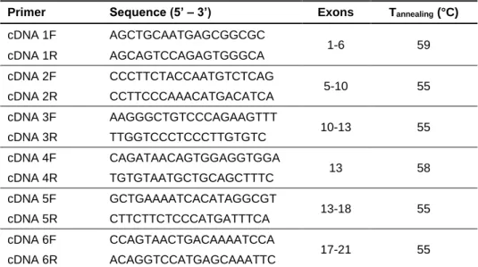
Table 2 – Sequences and annealing temperatures of primers used in the cDNA amplification
of GNPTAB gene. ...63
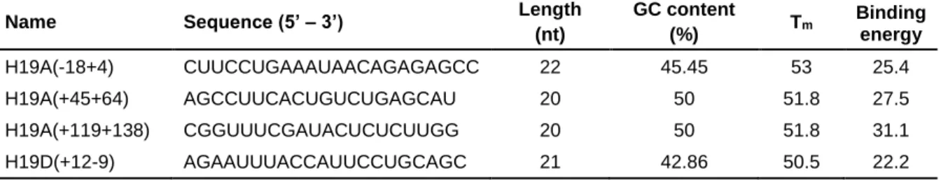
Table 3 – Sequences of antisense oligonucleotides selected for transfection in ML II patient
fibroblasts. Nomenclature as described by Mann et al., 2002. ...65
Table 4 – Composition of each enzyme activity assay and respective incubation period at
37ºC. ...69
Table 5 – Protein domains predicted by Pfam 30.0 for the GNPTAB protein product. n/a –
not applied. ...75
Table 6 – Protein domains predicted by UniProt for the GNPTAB protein product. ...76 Table 7 – Transmembrane domains predicted by UniProt for the GNPTAB protein product. 77 Table 8 – Metal binding sites predicted by UniProt for the GNPTAB protein product. ...77 Table 9 – Calcium binding region predicted by UniProt for the GNPTAB protein product. ....77 Table 10 – Secondary structure of the GNPTAB protein predicted by UniProt software
product. ...78
Table 11 – N-glycosylation sites predicted by NetNGlyc 1.0 Server for the GNPTAB protein
product. ...79
Table 12 – N-glycosylation sites predicted by GlycoEP for the GNPTAB protein product. Only
Table 13 – O-glycosylation sites predicted by GlycoEP for the GNPTAB protein product.
Only positions 1066-1250 are shown. ...80
Table 14 – Putative enhancers predicted by ESEfinder 3.0 for exon 19 of the GNPTAB gene
containing the c.3503_3504delTC mutation and 50bp of the flanking introns. Positions start at the beginning of the input sequence and not the beginning of exon 19. 1-50: intron 18; 51-216: exon 19; 217-266: intron 19. ...82
Table 15 – Exons of the GNPTAB transcript covered by each primer set and corresponding
amplicon size. ...86
Table 16 – Enzyme activity of lysosomal enzymes in fibroblasts of controls and LSDs
patients. Due to small sample amount of the Fabry cell line, some enzymatic assays could not be performed for these fibroblasts. ...97
Table 17 – Enzyme activity of lysosomal enzymes in fibroblasts of controls and ML II Patient
1 after transfection with the H19A(+119+138) AO. Plus sign (+) indicates the treatment with Lipofectamine 2000 + AO, while minus sign (-) indicates the treatment with just Lipofectamine 2000. ...98
Figure Index
Figure 1 – Newly synthesized lysosomal enzymes are transported from the endoplasmic
reticulum into the Golgi apparatus where a mannose-6-phosphate (M6P) marker is added through the action of two enzymes: GlcNAc-1-phosphotransferase transfers GlcNAc-1-P to mannose residues of the peptide, while N-acetylglucosamine-1-phosphodiester α-N-acetylglucosaminidase (“uncovering enzyme”) removes the GlcNAc residue to expose the M6P modifications. Subsequently, the lysosomal enzymes bind to M6P receptors and are targeted to the lysosomes. Adapted from Coutinho et al., 2012. ...36
Figure 2 – Schematic representation of the GlcNAc-1-phosphotransferase. The enzyme is
composed of two α subunits (blue), two β subunits (green) and two γ subunits (red). α and β subunits are responsible for the catalytic activity of the enzyme, while the γ subunits appear to facilitate and increase their action. Adapted from Pohl et al., 2011. ...37
Figure 3 – Motifs and domains of the α/β-precursor protein obtained from the GNPTAB
gene. Blue line indicates the position for cleavage by the site-1 protease, while red lines indicate the location of N-terminal dileucine motif and C-terminal arginine/isoleucine/arginine (RIR) motif, respectively. While in the wild-type protein product (A) both motifs are present, the C-terminal motif is missing from the mutant protein (B) due to the frameshift caused by the c.3503_3504delTC mutation, preventing its exit from the endoplasmic reticulum and subsequent cleavage into mature α and β subunits. Adapted from Franke et al., 2013; Qian et al., 2013. ...38
Figure 4 – Schematic representation of the spliceosome assembly and splicing process. U1
snRNP initially binds to the 5’ splice site (ss), while the splicing factor 1 (SF1) binds to the branch point (A) and the U2 auxiliary factor heterodimer (U2AF 65/35) binds to the 3’ ss and polypyrimidine tract (Py). Next, U2 snRNP binds to the branch point, excluding SF1 from the complex and interacts with U1 snRNP. U5, U4 and U6 snRNPs are then recruited as a preassembled complex with U6 snRNP binding to the 5’ss and interacting closely with U2 snRNP, displacing the U1 and U4 snRNPs and bringing the 5’ss and the branch point into close proximity for the first step of the splicing reaction. The spliceosome complex undergoes further conformational changes, resulting in the release of the intronic sequence and junction of the two exons, to produce the mature mRNA. Adapted from Chen et al., 2009; Will & Lührmann, 2011; Kornblihtt et al., 2013. ...42
Figure 5 – Core splicing signals within the pre-mRNA molecule. The consensus sequences
are as indicated with N being any nucleotide, R being a purine and Y being a pyrimidine. The 5’ splice site (ss) is located at the upstream intron junction with the consensus sequence AGGURAGU, while the 3’ss includes the polypyrimidine tract and the dinucleotide AG at the downstream intron junction. The branch point is defined by a highly conserved adenosine surrounded by seven poorly conserved nucleotides located 20-40 nucleotides upstream of the 3’ss. Adapted from Matlin et al., 2005; Kornblihtt et al., 2013. ...43
Figure 6 – Cis-acting splicing regulatory elements. Intronic splicing silencers (ISSs) and
exonic splicing silencers (ESSs) repress the inclusion of the target exon in the mature mRNA, while intronic splicing enhancers (ISEs) and exonic splicing enhancers (ESEs) promote exon inclusion. Adapted from Matlin et al., 2005; Kornblihtt et al., 2013. ...43
Figure 7 – Representation of the most common forms of alternative splicing. A: cassette
exon; B: mutually exclusive exons; C: alternative 5' splice sites; D: alternative 3' splice sites; E: intron retention; F: alternative promoters; G: alternative polyadenylation. Adapted from Matlin et al., 2005. ...45
Figure 8 – Chemical modifications of antisense oligonucleotides. First generation
oligonucleotides are characterized by a phosphorothioate (PS) backbone, obtained through the replacement of an oxygen atom with a sulfur atom. Second generation oligonucleotides contain a methyl or methoxyethyl group attached to 2’ position of the ribose. Finally, third generation oligonucleotides carry modifications in the furanose ring structure. Adapted from Chan et al., 2006. ...46
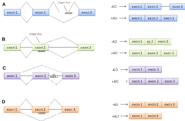
Figure 9 – Different methods of antisense-mediated splicing modulation. A: blocking of
cryptic splice site within the intronic sequence; B: blocking of cryptic splice site within the exonic sequence; C: inclusion of alternatively spliced exon; D: exclusion of exon or “exon skipping”. Adapted from Hammond & Wood, 2011. ...48
Figure 10 – Location of antisense oligonucleotide targets on exon 19 of the GNPTAB gene
Figure 11 – Example of Neubauer chamber used for cell counting. The Neubauer chamber
contains two distinct chambers, composed of 9 quadrants, each with a volume of 0,1 mm3 or 10-4 mL. ...66
Figure 12 – Alignment in Clustal Omega software of amino acid sequences for the full length
GNPTAB protein product and truncated protein resulting from the skipping of exon 19. Due to
the long size of the protein sequence, only exons 14 to 21 are shown in this picture. protein: full length protein product; skip19: truncated protein. ...74
Figure 13 – Alignment in Clustal Omega software of amino acid sequences encoded by
exon 19 of the GNPTAB gene in different animal species. ...74
Figure 14 – Protein domains predicted by Pfam 30.0 for the GNPTAB protein product. Color
codes and exact positions are depicted in Table 5. ...75
Figure 15 – Protein domains predicted by InterPro for the GNPTAB protein product. ...76 Figure 16 – Secondary structure of the GNPTAB protein predicted by UniProt software.
Green: beta strand; pink: turn; blue: helix. ...77
Figure 17 – N-glycosylation sites predicted by NetNGlyc 1.0 Server for the GNPTAB protein
product. ...79
Figure 18 – Graphic representation of the putative enhancers predicted by ESEfinder 3.0 for
exon 19 GNPTAB gene containing the c.3503_3504delTC mutation and 50bp of the flanking introns. Arrows indicate the location of the donor and acceptor splice sites. ...83
Figure 19 – Putative enhancers predicted by RESCUE-ESE on exon 19 of the GNPTAB
gene containing the c.3503_3504delTC mutation and 50bp of the flanking introns. Positions start at the beginning of the input sequence and not the beginning of exon 19. 1-50: intron 18; 51-216: exon 19; 217-266: intron 19. ...83
Figure 20 – Schematic figure of primer annealing for amplification of exon 19 of the
Figure 21 – Agarose gel electrophoresis of PCR amplification of GNPTAB gene exon 19 and
flanking intronic regions in control and patient fibroblasts. L: ladder; B: negative control (blank); HC: healthy control; P1: ML II Patient 1; P2: ML II Patient 2. ...85
Figure 22 – Schematic representation of annealing locations of all six primer sets for
amplification of different fragments of the GNPTAB transcript. ...86
Figure 23 – Agarose gel electrophoresis of amplification products of the GNPTAB transcript
in patient and control fibroblasts. A: primer set 1; B: primer set 2; C: primer set 3; D: primer set 4; E: primer set 5; F: primer set 6. L: ladder; B: negative control (blank); HC: healthy control; P1: ML II Patient 1; P2: ML II Patient 2. ...87
Figure 24 – Fluorescence microscopy images of healthy control fibroblasts transfected with
eGFP. The strong green fluorescence visible in the cells confirms the efficiency of
Lipofectamine® 2000 in transfecting cultured fibroblasts. ...88
Figure 25 – Agarose gel electrophoresis of amplification products of the GNPTAB transcript
(exons 17-21) in control fibroblasts after transfection with AOs. L: ladder; B: negative control (blank). Ø: control sample without AO. H19A(-18+4): directed to the 3’ss; H19D(+12-9): directed to the 5’ss; H19A(+45+64): directed to ESEs in the first half of exon 19; H19A(+119+138): directed to ESEs in the second half of exon 19...89
Figure 26 – Agarose gel electrophoresis of amplification products of the GNPTAB transcript
(exons 17-21) in control fibroblasts after transfection with AOs. L: ladder; B: negative control (blank). Ø: control sample without AO. H19D(+12-9): directed to the 5’ss; H19A(+119+138): directed to ESEs in the second half of exon 19. ...91
Figure 27 – Agarose gel electrophoresis of amplification products of the GNPTAB transcript
(exons 17-21) in ML II Patient 1 fibroblasts after transfection with AOs. L: ladder; B: negative control (blank). Ø: control sample without AO. H19A(-18+4): directed to the 3’ss; H19D(+12-9): directed to the 5’ss; H19A(+45+64): directed to ESEs in the first half of exon 19; H19A(+119+138): directed to ESEs in the second half of exon 19...92
Figure 28 – Agarose gel electrophoresis of amplification products of the GNPTAB transcript
(exons 17-21) in ML II Patient 1 fibroblasts after transfection with AOs. L: ladder; B: negative control (blank). Ø: control sample without AO. H19D(+12-9): directed to the 5’ss; H19A(+119+138): directed to ESEs in the second half of exon 19...93
Figure 29 – Agarose gel electrophoresis of amplification products of the GNPTAB transcript
(exons 17-21) in ML II Patient 2 fibroblasts after transfection with AOs. L: ladder; B: negative control (blank). Ø: control sample without AO. H19A(-18+4): directed to the 3’ss; H19D(+12-9): directed to the 5’ss; H19A(+45+64): directed to ESEs in the first half of exon 19; H19A(+119+138): directed to ESEs in the second half of exon 19...94
Figure 30 – Agarose gel electrophoresis of amplification products of the GNPTAB transcript
(exons 17-21) in ML II Patient 2 fibroblasts after transfection with AOs. L: ladder; B: negative control (blank). Ø: control sample without AO. H19D(+12-9): directed to the 5’ss; H19A(+119+138): directed to ESEs in the second half of exon 19...96
Abbreviations
2’-OMe 2’-O-methyl 2’-MOE 2’-O-methoxyethyl α-MM α-methylmannoside AG adenine/guanine AO antisense oligonucleotide ARSB arylsulfatase B AS alternative splicingATP adenosine triphosphate
BBB blood-brain barrier
BLAST Basic Local Alignment Search Tool
cDNA complementary DNA
CNS central nervous system
CR4 conserved region 4
del deletion
DLS Doenças Lisossomais de Sobrecarga
DMD Duchenne muscular dystrophy
DMAP DNA methyltransferase-associated protein
DMEM Dulbecco's Modified Eagle's Medium
DNA deoxyribonucleic acid
ERT enzyme replacement therapy
ESE exonic splicing enhancer
ESS exonic splicing silencer
ExPASy Expert Protein Analysis System
FBS fetal bovine serum
GAG glycosaminoglycan
GB γ-subunit binding region
GC guanine/cytosine
GlcNAc N-acetylglucosamine
GlcNAc-1-P N-acetylglucosamine-1-phosphate
HC healthy control
hnRNP heterogeneous nuclear ribonucleoprotein
HSCT hematopoietic stem cell transplantation
ISS intronic splicing silencer
LNA locked nucleic acid
LSD Lysosomal Storage Disorders
M6P mannose 6-phosphate
ML mucolipidosis
MPS mucopolysaccharidosis
mRNA messenger RNA
MU methylumbelliferyl
NCBI National Center for Biotechnology Information
ORF open reading frame
PBS phosphate buffered saline
PCR polymerase chain reaction
PenStrep penicillin/streptomicin
PMO phosphoroamidate morpholino oligomer
PNA peptide nucleic acid
pre-mRNA precursor mRNA
PS phosphorothioate
Py polypyrimidine tract
RIR arginine/isoleucine/arginine
RNA ribonucleic acid
RNase ribonuclease
RT-PCR reverse transcriptase polymerase chain reaction
SF1 splicing factor 1
SMA spinal muscular atrophy
SNP single nucleotide polymorphism
snRNA small nuclear RNA
snRNP small nuclear ribonucleoprotein
SR serine/arginine
SRSF serine/arginine-rich splicing factor
SRT substrate reduction therapy
ss splice site
TC thymine/cytosine
Tm melting temperature
TTR transthyretin
UDP-GlcNAc uridine diphosphate N-acetylglucosamine
Chapter 1
1.1. Lysosomal Storage Disorders
Lysosomal Storage Disorders (LSDs) are a group of rare inherited metabolic diseases caused by the malfunction of the lysosomal system, ultimately resulting in the accumulation of undegraded substrates inside the lysosomes (Filocamo & Morrone, 2011; Lieberman et al., 2012). As a consequence, multiple organelles and processes might be affected, such as signaling, vesicle trafficking, autophagy, calcium homeostasis and inflammatory response (Futerman & van Meer, 2004; Ballabio & Gieselmann, 2009; Platt et al., 2012), leading to cell death and tissue damage. These damages translate into a wide variety of clinical manifestations ranging from nearly asymptomatic individuals to serious and progressive clinical manifestations involving several organs and systems and generally resulting in death in the first few years of life (Platt et al., 2012). Despite being rare disorders individually, LSDs present a significant frequency when considered as a whole ranging from 7.6 cases per 100 000 births in British Columbia (Applegarth et al., 2000) to 26.9 cases per 100 000 births in the United Arab Emirates (Al-Jasmi et al., 2012), showcasing a strong variability in incidence between different ethnicities and populations. In the Portuguese population, the frequency of these disorders is estimated to be 25 cases per 100 000 births (Pinto et al., 2004), whereas other European countries have revealed a LSD incidence around 12-14 cases per 100 000 births (Poorthuis et al., 1999; Dionisi-Vici et al., 2002; Poupětová et al., 2010).
Encompassing about 50 distinct disorders, the classification of LSDs can be quite challenging and complex. While it has been traditionally associated with the nature of the accumulated substrate, in recent years, the molecular defect responsible for each disorder has taken on a more important role for the grouping of these diseases, allowing for the inclusion of other disorders that could not be properly organized in the traditional system (Table 1) (Filocamo & Morrone, 2011; Boustany, 2013). Nonetheless, the traditional classification is still widely used for disorders such as mucopolysaccharidoses (accumulation of mucopolysaccharides), sphingolipidoses (accumulation of sphingolipids) and oligosaccharidoses (accumulation of oligosaccharides) (Ballabio & Gieselmann, 2009), and the measurement of accumulated substrate is still quite useful as a biomarker for both diagnosis and therapy follow-up in LSDs (Cox, 2005).
Disorder Gene Enzyme deficiency Accumulated substrates Sphingolipidoses
Fabry disease GLA α-Galactosidase A Globotriasylceramide Farber lipogranulomatosis ASAH1 Acid ceramidase Ceramide
Gaucher disease GBA Glucosylceramidase Glucosylceramide Niemann-Pick disease (A/B) SMPD1 Sphingomyelinase Sphingomyelin GM1-gangliosidosis (I, II and III) GLB1 GM1-β-galactosidase GM1 ganglioside, keratan
sulphate, oligos, glycolipids GM2-gangliosidosis (Sandhoff) HEXAB β-Hexosaminidase A + B GM2 ganglioside, oligos GM2-gangliosidosis (Tay-Sachs) HEXA β-Hexosaminidase A GM2 ganglioside,
oligos, glycolipids GM3-gangliosidosis ST3GAL5 GM3 synthase GM3 ganglioside Krabbe disease GALC β-Galactosylceramidase Galactosylceramide Metachromatic leucodystrophy ARSA Arylsulphatase A Sulphatides Sphingolipid-activator deficiency PSAP Sphingolipid activator Glycolipids
Mucopolysaccharidoses
MPS I (Hurler, Scheie,
Hurler/Scheie) IDUA α-Iduronidase
Dermatan sulphate, heparan sulphate MPS II (Hunter) IDS Iduronate sulphatase Dermatan sulphate,
heparan sulphate MPS IIIA (Sanfilippo A) SGSH Heparan sulphamidase Heparan sulphate MPS IIIB (Sanfilippo B) NAGLU Acetyl α-glucosaminidase Heparan sulphate MPS IIIC (Sanfilippo C) HGSNAT Acetyl CoA; α-glucosaminide
N-acetyltransferase Heparan sulphate MPS IIID (Sanfilippo D) GNS N-acetyl
glucosamine-6-sulphatase Heparan sulphate MPS IVA (Morquio A) GALNS Acetyl
galactosamine-6-sulphatase
Keratan sulphate, chondroitin 6-sulphate MPS IVB (Morquio B) GLB1 β-Galactosidase Keratan sulphate MPS VI (Maroteaux–Lamy) ARSB Arylsulphatase B Dermatan sulphate
MPS VII (Sly) GUSB β-Glucuronidase Dermatan sulphate, heparan sulphate, chondroitin 6-sulphate MPS IX (Natowicz) HYAL1 Hyaluronidase Hyluronan
Oligosaccharidoses (glycoproteinoses)
Aspartylglicosaminuria AGA Glycosylasparaginase Aspartylglucosamine Fucosidosis FUCA1 α-Fucosidase Glycoproteins, glycolipids,
Fucoside-rich oligos α-mannosidosis MAN2B1 α-Mannosidase Mannose-rich oligos β-mannosidosis MANBA β-Mannosidase Man (β1 → 4) GlnNAc Schindler disease NAGA N-acetylgalactosaminidase Sialylated/asialoglycopeptides,
glycolipids Sialidosis (mucolipidosis I) NEU1 Neuraminidase Oligos, glycopeptides
Glycogenoses
Glycogenosis II (Pompe) GAA α-Glucosidase Glycogen
Lipidoses
Wolman disease LIPA Acid lipase Cholesterol esters
Non-enzymatic lysosomal protein defect
GM2 gangliosidosis
(GM2 activator deficiency) GM2A GM2 activator protein GM2 ganglioside, oligos Krabbe disease PSAP Saposin A Galactosylceramide
Metachromatic leucodystrophy PSAP Saposin B Sulphatides Gaucher disease PSAP Saposin C Glucosylceramide
Transmembrane protein defect
Sialic acid storage disease SLC17A5 Sialin Sialic acid Cystinosis CTNS Cystinosin Cystine Niemann-Pick type C1 NPC1
Niemann-Pick type C1 protein (proton-driven
transporter)
Cholesterol and sphingolipids
Niemann-Pick type C2 NPC2
Niemann-Pick type C2 protein (proton-driven
transporter)
Cholesterol and sphingolipids Danon disease LAMP2 Lysosome-associated
membrane protein 2
Cytoplasmatic debris and glycogen Mucolipidosis IV MCOLN1 Mucolipin Lipids
Lysosomal enzyme protection defect
Galactosialidosis CTSA Protective protein cathepsin
A (PPCA) Sialyloligosaccharides
Post-translational processing defect
Multiple sulphatase deficiency SUMF1 Multiple sulphatase Sulphatides, glycolipids, GAGs1 Trafficking defect in lysosomal
enzymes
Mucolipidosis II alpha/beta
Mucolipidosis III alpha/beta GNPTAB
GlcNAc-1-phosphotransferase
(α/β subunits)
Oligos, GAGs1, lipids
Mucolipidosis III gamma GNPTG
GlcNAc-1-phosphotransferase
(γ subunit)
Oligos, GAGs1, lipids Polypeptide degradation defect
Pycnodysostosis CTSK Cathepsin K Bone proteins
Neuronal ceroid lipofuscinoses (NCLs)
NCL 1 PPT1 Palmitoyl protein
thioesterase (PPT1) Lipofuscin, saposins A and D NCL 2 TPP1 Tripeptidyl peptidase 1
(TPP1)
Lipofuscin, subunit c of ATP synthase
NCL 3 CLN3 CLN3, lysosomal
transmembrane protein
Lipofuscin, subunit c of ATP synthase
NCL 4 DNAJC5 DnaJ homologue subfamily
C member 5
Lipofuscin, subunit c of ATP synthase
NCL 5 CLN5 CLN5, soluble lysosomal
protein
Lipofuscin, subunit c of ATP synthase
NCL 6 CLN6 CLN6, transmembrane
protein of ER
Lipofuscin, subunit c of ATP synthase
NCL 7 MFSD8 CLN7, lysosomal chloride
channel
Lipofuscin, subunit c of ATP synthase
NCL 8 CLN8 CLN8, transmembrane
protein of ER
Lipofuscin, subunit c of ATP synthase
NCL 10 CTSD Cathepsin D Lipofuscin, saposins A and D
NCL 11 GRN Progranulin ─
NCL 12 ATP13A2 P type ATPase ─
NCL 13 CTSF Cathepsin F ─ NCL 14 KCTD7 Potassium channel tetramerization domain-containing protein 7 ─ 1 GAGs: Glycosaminoglicans
1.1.1. Mucolipidosis II
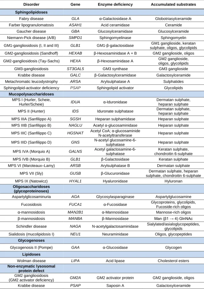
Like many other glycoproteins, soluble lysosomal proteins are synthesized in the endoplasmic reticulum and are co-translationally glycosylated on certain asparagine residues. After synthesis and glycosylation, lysosomal enzymes enter the Golgi apparatus where a mannose 6-phosphate (M6P) marker is added to the peptide, allowing for its binding to the M6P receptors in the Golgi and subsequent targeting to the lysosomes (Kornfeld, 1986). This modification is performed by two Golgi enzymes in sequential action. Initially, the enzyme N-acetylglucosamine-1-phosphotransferase (GlcNAc-1-phosphotransferase) transfers N-acetylglucosamine-1-phosphate (GlcNAc-1-P) to mannose residues in the peptide, after which a second enzyme, N-acetylglucosamine-1-phosphodiester α-N-acetylglucosaminidase (also known as “uncovering enzyme”) removes the GlcNAc residue to expose the M6P modifications, which can then be recognized by M6P receptors (Figure 1) (Qian et al., 2010).
Figure 1 –Newly synthesized lysosomal enzymes are transported from the endoplasmic reticulum into the Golgi apparatus where a mannose-6-phosphate (M6P) marker is added through the action of two enzymes: GlcNAc-1-phosphotransferase transfers GlcNAc-1-P to mannose residues of the peptide, while N-acetylglucosamine-1-phosphodiester α-N-acetylglucosaminidase (“uncovering enzyme”) removes the GlcNAc residue to expose the M6P modifications. Subsequently, the lysosomal enzymes bind to M6P receptors and are targeted to the lysosomes. Adapted from Coutinho et al., 2012.
Total or near-total deficiency of GlcNAc-1-phosphotransferase impairs the formation of this M6P marker, resulting in the nearly complete absence of lysosomal targeting in many cell types and tissues and the secretion of most lysosomal enzymes (Braulke et al., 2008). This defect is clinically recognized as mucolipidosis II (ML II or I-cell disease; MIM# 252500), an autosomal recessive disease often resulting in death in the first decade of life (Reitman et al., 1981b). Another type of mucolipidosis, mucolipidosis III (ML III or pseudo-Hurler polydystrophy; MIM# 252600), is a milder disease in which GlcNAc-1-phosphotransferase activity is reduced rather than absent (Honey et al., 1982).
The GlcNAc-1-phosphotransferase enzyme is an hexameric complex composed of two α, two β and two γ subunits, with α and β exerting the catalytic activity of the enzyme, whereas γ chains facilitate the folding of the catalytic subunits and maintain them in the right configuration, while also regulating their activity (Figure 2) (Bao et al., 1996; Coutinho et al., 2012).
The α and β subunits are encoded by GNPTAB, a gene located on chromosome 12, composed of 21 exons (Tiede et al., 2005), while the γ subunits are encoded by GNPTG, a gene located on chromosome 16, composed of 11 exons (Raas-Rothschild et al., 2000). Defects in GNPTAB result in ML II alpha/beta and ML III alpha/beta whereas mutations in
GNPTG have only been found in ML III gamma patients. Being encoded by the same gene,
the α and β subunits are synthesized as a single precursor protein in the endoplasmic reticulum, which is later cleaved in the Golgi apparatus by the site-1 protease to produce mature α and β subunits (Marschner et al., 2011; De Pace et al., 2013). Transport of the α/β-precursor protein to the Golgi depends on the presence of two sorting motifs in the protein
Figure 2 – Schematic representation of the GlcNAc-1-phosphotransferase. The enzyme is composed of two α subunits (blue), two β subunits (green) and two γ subunits (red). α and β subunits are responsible for the catalytic activity of the enzyme, while the γ subunits appear to facilitate and increase their action. Adapted from Pohl et al., 2011.
sequence: an N-terminal dileucine motif and a C-terminal dibasic arginine/isoleucine/arginine (RIR) motif (Figure 3A) (Franke et al., 2013). The absence of one or both of these domains inhibits the exit of the α/β-precursor from the endoplasmic reticulum and its subsequent cleavage, preventing the formation of mature α and β subunits and therefore impairing GlcNAc-1-phosphotransferase activity.
To date 142 pathogenic mutations in GNPTAB that cause ML II and ML III alpha/beta are listed in The Human Gene Mutation Database (Stenson et al., 2017). Almost all ML II patients have nonsense, frameshift, or splice-site mutations in GNPTAB, and these mutations are usually associated with a more severe clinical course. Conversely, ML III alpha/beta patients carry missense mutations or at least one hypomorphic allele that is not associated with a complete loss of functionality and the clinical signs and symptoms of ML III alpha/beta patients are milder and more slowly progressive than in ML II (Braulke et al., 2013). Among these multiple mutations described in the GNPTAB gene, the two-nucleotide deletion c.3503_3504delTC on exon 19 is considered the most common among ML II patients, having been previously studied by our research group (Encarnação et al., 2009; Coutinho et al., 2011). Since it involves the deletion of two nucleotides, this mutation causes a frameshift in the transcript, leading to an altered amino acid sequence, as well as to a premature stop codon (Plante et al., 2008). The synthesized α/β-precursor peptide lacks the C-terminal domain, preventing its exit from the endoplasmic reticulum and subsequent cleavage into mature α and β subunits (Figure 3B) (De Pace et al., 2013). This impairs the
A
B
Figure 3 – Motifs and domains of the α/β-precursor protein obtained from the GNPTAB gene. Blue line indicates the position for cleavage by the site-1 protease, while red lines indicate the location of N-terminal dileucine motif and C-terminal arginine/isoleucine/arginine (RIR) motif, respectively. While in the wild-type protein product (A) both motifs are present, the C-terminal motif is missing from the mutant protein (B) due to the frameshift caused by the c.3503_3504delTC mutation, preventing its exit from the endoplasmic reticulum and subsequent cleavage into mature α and β subunits. Adapted from Franke et al., 2013; Qian et al., 2013.
assembly of a functional GlcNAc-1-phosphotransferase enzyme and consequently the formation of the M6P marker in lysosomal enzymes, which results in an almost complete absence of hydrolases inside the lysosomes and accumulation of undegraded substrates (Kudo et al., 2006). This leads to a phenotype characterized by severe psychomotor retardation, coarse facial features, gingival hypertrophy, short stature, hepatosplenomegaly, joint contractures, osteopenia and cardiopulmonary complications that usually lead to death in the first decade of life (De Pace et al., 2013; Velho et al., 2015).
1.1.2. Biochemical and molecular diagnosis
Since the measurement of GlcNAc-1-phosphotransferase activity usually requires a very laborious assay that involves the radioactive [32P]UDP-GlcNAc substrate and is only available in a very small number of facilities, an indirect diagnosis of ML II and ML III is more commonly established by the assessment of lysosomal hydrolases (Alegra et al., 2014). Given the impaired sorting of multiple enzymes to the lysosomes in these patients, the biochemical confirmation of these disorders is usually attained indirectly through the detection of activity of lysosomal enzymes in plasma or in cultured fibroblasts. In general, a ten to twentyfold increase in serum lysosomal enzymes, together with decreased intralysosomal levels in fibroblasts, is indicative of those disorders (Kornfeld & Sly, 2001). In addition, the measurement of lysosomal enzymes activity may be used in the research field for the evaluation of the efficiency of therapeutic approaches that are being investigated both
in vitro and in vivo.
Nonetheless, molecular diagnosis is essential to confirm the presence of these disorders, with sequencing of the GNPTAB and GNPTG coding regions detecting disease-causing mutations in over 95% of patients. In general, ML II and ML III can be distinguished based on clinical criteria and disease progression but the identification of mutations in the
GNPTAB and GNPTG genes constitutes an important tool for the correct classification of
these disorders. In fact, the distinction between ML III alpha/beta and ML III gamma is only possible based on molecular diagnosis, since the two forms of ML III have the same clinical and biochemical characteristics. Moreover, prenatal diagnosis of at-risk pregnancies is dependent on prior identification of disease-causing mutations in the family (Braulke et al., 2013).
1.1.3. Available treatments for LSDs and ML II
Although the biochemical and molecular causes behind most LSDs have been largely disclosed, similar advances have not been made in the field of therapy. Currently, no definitive cure is available for this kind of disorders, so most available treatments focus on improving life expectancy and quality of life through multiple strategies (Boustany, 2013; Parenti et al., 2013). An obvious approach for treatment of these disorders is the restoration of enzyme activity, which can be achieved by different kinds of procedures. One possibility is hematopoietic stem cell transplantation (HSCT), where the patient’s tissues can be repopulated with the donor’s healthy cells, which will then produce the functional enzyme at normal levels and hopefully ameliorate the symptoms of the disease (Parenti et al., 2013). However, this sort of treatment has only proved helpful for a limited number of conditions, patients must be in their early childhood for this procedure to be effective, and the rate of associated complications and mortality is significant (Malatack et al., 2003; Cox, 2015). Another therapeutic approach is enzyme replacement therapy (ERT), in which a recombinant form of the target enzyme is administered to patients to be then taken up by the patient’s cells, where it is targeted to the lysosome to replace the defective enzyme and degrade the accumulated substrate (Parenti et al., 2013). Nonetheless, despite good results in improving the function of multiple organs, this form of therapy is unable to have any effect on the central nervous system (CNS) since the exogenous enzyme cannot cross the blood–brain barrier (BBB), limiting its application (Boustany, 2013; Parenti et al., 2013). Yet another common option for LSD treatment is substrate reduction therapy (SRT), which aims to reduce the production of the accumulated substrate so that patients with residual enzyme activity can successfully degrade it and avoid excessive storage in the lysosome (Bruni et al., 2007). For that purpose, this therapy uses small-molecule inhibitors to target specific steps in the biosynthetic pathways of the accumulated substrate and block or decrease the synthesis of these materials. Since the drugs used here are composed of very small molecules, they are able to cross the BBB and successfully reach the CNS, making it a better option for multiple disorders that involve the CNS (Parenti et al., 2013).
Unfortunately, in the specific case of ML II, many of these therapies are unsuitable given the nature of the affected enzyme and accumulation of multiple substrates inside the lysosomes. On one hand, ERT is not an option as the defective enzyme is not a lysosomal enzyme that can be internalized by the cells and targeted to the lysosome, and SRT also does not fit this disorder since various substrates are accumulated within the lysosomes due to a near total absence of hydrolytic enzymes in this organelle. Furthermore, although HSCT
has been reported in the past for the treatment of ML II (Grewal et al., 2003), no benefits were observed in the musculoskeletal system and the survival rate is quite low (Lund et al., 2014).
Acknowledging this, some innovative therapeutic approaches have been developed in recent years, including the usage of pharmacological chaperones that promote the correct folding and trafficking of defective proteins (Sawkar et al., 2006), the combination of these chaperones with ERT (Boustany, 2013), the induction of ribosomal read-through of nonsense mutations allowing for the production of a full-length protein (Parenti et al., 2013), and even gene therapy through the introduction of the correct gene sequence into the affected cells so that the functional enzyme can be produced (Bruni et al., 2007; Cox, 2015). Considering the impact of these disorders in both patients and families, there is an urgent need to invest in the development of new therapeutic strategies for their treatment in order to ameliorate the disease phenotype, offering to patients the best possible clinical outcome together with minimal side effects and improved quality of life.
1.2. Splicing therapies
1.2.1. Splicing reactionThe expression of eukaryotic genes requires a sequential series of finely regulated steps, amongst which one of the most important is splicing. In the process of splicing, non-coding introns are removed from the precursor messenger RNA (pre-mRNA) molecule and the coding sequences (exons) are joined together to produce the mature mRNA that will be exported to the cytoplasm and translated into protein (Matera & Wang, 2014). The splicing reaction is carried out by the spliceosome, a large RNA and protein complex, constituted by five small ribonucleoprotein complexes (snRNPs) and more than a hundred proteins (Matera & Wang, 2014). The first step of spliceosome assembly is the recognition of the 5’ splice site (ss) by the U1 snRNP, followed by the binding of splicing factor 1 (SF1) to the branch point and of the U2 auxiliary factor heterodimer (U2AF 65/35) to the polypyrimidine tract (Py) and 3’ss, originating the E complex (Chen & Manley, 2009; De Conti et al., 2013). SF1 is then replaced by the U2 snRNP at the branch point, originating the A complex and allowing for the interaction between U1 snRNP and U2 snRNP across the exon (Chen & Manley, 2009; Matera & Wang, 2014). Afterwards, the U4, U5 and U6 snRNPs are recruited as a preassembled complex, leading to the formation of the B complex. The interaction between
U4 and U6 is then disrupted and the U6 snRNP base pairs with the 5’ss, displacing U1 snRNP from its initial location and releasing it from the complex, along with the U4 snRNP (Will & Lührmann, 2011). At the same time, U6 snRNP also interacts extensively with U2 snRNP, bringing the 5’ss and the branch point into close proximity and allowing for the first step of splicing to take place, which originates the C complex containing the free upstream exon and the intron-exon lariat intermediate (Matera & Wang, 2014). This complex undergoes further ATP-dependent conformational changes and completes the second step of the splicing reaction, releasing the lariat intron and joining the coding exons to form the mature mRNA, while U2, U5 and U6 snRNPs are also released from the complex and recycled for future splicing reactions (Figure 4) (Matera & Wang, 2014).
1.2.2. Splicing signals and regulatory elements
Although the spliceosome orchestrates pre-mRNA processing with great complexity and fidelity, this is a fairly flexible mechanism under strong regulation by both cis-acting and
trans-acting elements. The 5’ and 3’ splice sites and the branch point are present in every
Figure 4 – Schematic representation of the spliceosome assembly and splicing process. U1 snRNP initially binds to the 5’ splice site (ss), while the splicing factor 1 (SF1) binds to the branch point (A) and the U2 auxiliary factor heterodimer (U2AF 65/35) binds to the 3’ ss and polypyrimidine tract (Py). Next, U2 snRNP binds to the branch point, excluding SF1 from the complex and interacts with U1 snRNP. U5, U4 and U6 snRNPs are then recruited as a preassembled complex with U6 snRNP binding to the 5’ss and interacting closely with U2 snRNP, displacing the U1 and U4 snRNPs and bringing the 5’ss and the branch point into close proximity for the first step of the splicing reaction. The spliceosome complex undergoes further conformational changes, resulting in the release of the intronic sequence and junction of the two exons, to produce the mature mRNA. Adapted from Chen et al., 2009; Will & Lührmann, 2011; Kornblihtt et al., 2013.
intron and therefore are known as core splicing signals (Wang & Burge, 2008). The 5’ss (donor site) is located at the upstream intron junction with the consensus sequence AGGURAGU, while the 3’ss (acceptor site) includes the 10-20 nucleotide long polypyrimidine tract and the dinucleotide AG at the downstream intron junction (Reed, 1996; Faustino & Cooper, 2003; Kornblihtt et al., 2013). The branch point is located 20-40 nucleotides upstream the 3’ss and consists of a highly conserved adenosine surrounded by a poorly conserved sequence of 7 nucleotides (Figure 5) (Reed, 1996; Kornblihtt et al., 2013).
Although this core splicing signals are highly conserved in less complex organisms, they tend to be less conserved in higher eukaryotes (Warf & Berglund, 2010), thus requiring the involvement of other cis-elements for the correct recognition of introns and exons. These elements can be enhancers or silencers depending on their role in promoting or inhibiting exon inclusion, respectively, and can also be divided into intronic or exonic elements depending on their location. As such, intronic splicing enhancers (ISEs) and exonic splicing enhancers (ESEs) favor the inclusion of the exon into the mature mRNA molecule, while intronic splicing silencers (ISSs) and exonic splicing silencers (ESSs) repress exon inclusion (Figure 6) (De Conti et al., 2013).
Figure 5 – Core splicing signals within the pre-mRNA molecule. The consensus sequences are as indicated with N being any nucleotide, R being a purine and Y being a pyrimidine. The 5’ splice site (ss) is located at the upstream intron junction with the consensus sequence AGGURAGU, while the 3’ss includes the polypyrimidine tract and the dinucleotide AG at the downstream intron junction. The branch point is defined by a highly conserved adenosine surrounded by seven poorly conserved nucleotides located 20-40 nucleotides upstream of the 3’ss. Adapted from Matlin et al., 2005; Kornblihtt et al., 2013.
Figure 6 – Cis-acting splicing regulatory elements. Intronic splicing silencers (ISSs) and exonic splicing silencers (ESSs) repress the inclusion of the target exon in the mature mRNA, while intronic splicing enhancers (ISEs) and exonic splicing enhancers (ESEs) promote exon inclusion. Adapted from Matlin et al., 2005; Kornblihtt et al., 2013.
ESEs are for the most part recognized by serine/arginine (SR) proteins, which also contain domains for protein-protein interaction (Wang & Burge, 2008; De Conti et al., 2013), while proteins associated with ISEs are still poorly characterized. In the case of silencer elements, ESSs and ISSs are recognized by members of the heterogeneous nuclear ribonucleoprotein (hnRNP) protein family, which act through different mechanisms, such as blocking interactions between spliceosome elements (Spellman & Smith, 2006) or promoting inhibitory RNA structures (Fisette et al., 2010). Nonetheless, the activity of a certain regulatory element often depends on its location within the pre-mRNA, a property called “context dependence” (Wang & Burge, 2008). Additionally, splicing can be affected in multiple aspects by other factors, such as phosphorylation, subcellular localization, ratio between SR proteins and hnRNPs and RNA secondary structure (Matlin et al., 2005; Jensen et al., 2009; Warf & Berglund, 2010).
1.2.3. Alternative Splicing
Aside from the so-called constitutive splicing process, alternative splicing (AS) also occurs, with some sequences altering between inclusion and exclusion into the mature mRNA (Faustino & Cooper, 2003; Revil et al., 2010; Liu et al., 2012). Since most AS events occur in the coding sequences of the mRNA transcript, they have consequences in the sequence of encoded proteins, affecting their structure and biological activity on multiple levels (Stamm et al., 2005). In that sense, AS events can be classified into five basic groups: cassette exons, mutually exclusive exons, alternative 5’ splice sites, alternative 3’ splice sites and intron retention (Figure 7A to Figure 7E) (Gamazon & Stranger, 2014; Sveen et al., 2016). Aside from these main types of AS, alternative transcripts can also arise from the usage of alternative promoters or alternative polyadenylation, which provide additional variability to mRNAs produced from a single gene (Figure 7F and Figure 7G) (Matlin et al., 2005; Roy et al., 2013). Furthermore, AS can also affect gene expression through removal or addition of regulatory elements that control mRNA stability, localization and translation (Faustino & Cooper, 2003). Lastly, not only the sequence of the transcript can be altered, but also its abundance, which is associated with several development processes, such as sex determination, tissue-differentiation and muscle contraction (Black, 2003), as well as with a number of different diseases (Love et al., 2015; Sveen et al., 2016).
1.3. Antisense Oligonucleotides
In the last two decades, many approaches have been explored in the field of RNA-based therapeutics in order to manipulate splicing of pre-mRNA or promote degradation of a mutant mRNA. Concerning the modulation of splicing, although multiple methods can be employed, the vast majority involves the usage of antisense oligonucleotides (AOs). AOs are short synthetic oligonucleotides that bind to target RNAs through standard Watson-Crick base pairing (Bennett & Swayze, 2010). AOs can act via different mechanisms and, in the case of splicing modulation, they are designed to target splice sites or splicing regulatory elements, blocking the binding of splicing factors and spliceosome components, which results in alterations in the splicing pattern and the final protein product (Hammond & Wood, 2011; Evers et al., 2015).
1.3.1. Chemical modifications: three generations of oligonucleotides
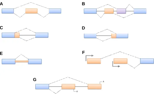
Initially, AOs were just synthetic unmodified DNA or RNA, which despite delivering some promising results, would prove to be quite ineffective in biological systems due to their susceptibility to degradation by nucleases, poor affinity for their target mRNA, multiple
off-Figure 7 – Representation of the most common forms of alternative splicing. A: cassette exon; B: mutually exclusive exons; C: alternative 5' splice sites; D: alternative 3' splice sites; E: intron retention; F: alternative promoters; G: alternative polyadenylation. Adapted from Matlin et al., 2005.
A B
C D
E F