2017
UNIVERSIDADE DE LISBOA
FACULDADE DE CIÊNCIAS
DEPARTAMENTO DE BIOLOGIA VEGETAL
Understanding the cross-talk between microbiota, host fitness,
and the environment using Egyptian mongoose (Herpestes
ichneumon) as a model
André da Conceição Pereira
Mestrado em Microbiologia Aplicada
Dissertação orientada por:
Doutora Mónica Vieira Cunha
i
ACKNOWLEDGEMENTS
Este é, até hoje, um dos momentos mais importantes da minha vida. Esta dissertação simboliza a longa caminhada feita durante dois longos anos. Quero agradecer em primeiro lugar, e antes de mais nada, à minha orientadora Doutora Mónica Vieira Cunha pela grande oportunidade que me deu de integrar o grupo de investigação que lidera no cE3c-FCUL. Durante este longo ano, deu-me um voto de confiança e proporcionou-me todo o apoio material, financeiro e humano para conseguir realizar com sucesso a minha dissertação. Quero agradecer também o apoio financeiro do cE3c (Ref. UID/BIA/00329/2013; 2015-2017). Agradeço também ao Doutor Rogério Tenreiro, líder do laboratório de Microbiologia e Biotecnologia, por me ter acolhido no seu laboratório e disponibilizado o suporte necessário para a realização do meu trabalho. Ao Doutor Carlos Fonseca e Doutor Victor Bandeira, da Universidade de Aveiro, agradeço o material biológico fornecido e os respetivos dados bio-ecológicos. Agradeço, de igual forma, à Cláudia Ramalho, à Filipa Antunes Silva, à Doutora Lisete Fernandes e à Doutora Ana Tenreiro, por todos os conselhos profissionais dados durante este ano. Agradeço também todo o apoio pessoal e profissional a todos os colegas de laboratório, nomeadamente, Beatriz Ramos, Tiago Baeta, Ana Reis, Ana Soares, Marta Lourenço, Inês Santos, Ana Catarina Rocha, João Melo, Pedro Teixeira, Alexandra Lança e Mariana Nascimento. Tenho também de agradecer do fundo do meu coração à minha família mais próxima: à minha madrinha, ao meu padrinho, ao meu primo e colega biólogo Rui Miguel, ao meu primo Caco pelas parvoíces próprias das idades que ajudam a descontrair nos momentos mais tensos e a minha querida prima Fátima Patrícia. Claro que não me posso esquecer do meu pai, que desde cedo me criou e me proporcionou todo o suporte pessoal, emocional e financeiro para seguir os meus sonhos e os meus estudos. Pai, sem ti nada disto teria sido possível. Aos amigos que ganhei no mestrado, em especial, a Ana Vinagre, a Ana Fraga, ao Jorge Figueiredo e a Rita Franciosi, pelos jantares de escárnio e maldizer, onde podemos desabafar sobre todos os problemas pessoais e profissionais ao longo destes dois anos. Por fim, mas tão importante como os restantes, ao meu grupo de amigos mais próximo – que sem eles não teria conseguido finalizar este manuscrito e que, depois de finalizado, tanto prazer de conquista me dá – em particular, a minha querida Adriana Carago, a minha grande amiga Calu Maria e a minha amiga de infância Beatriz Peres.
ii
Understanding the cross-talk between microbiota, host fitness,
and the environment using Egyptian mongoose (Herpestes
ichneumon) as a model
André da Conceição Pereira
2017
This thesis was fully performed at Microbiology and Biotechnology Lab
(LMB-BioISI|Bugworkers|TecLabs) and Centre for Ecology, Evolution and Environmental
changes (cE3c), under the direct supervision of Professor Mónica Vieira Cunha, in the
scope of the Master in Applied Microbiology of Faculdade de Ciências da Universidade
de Lisboa.
iii
ABSTRACT
Gut microbiota is the complex and diverse community of bacteria, archaea, fungi, protozoa, and viruses present in the gastrointestinal tract of animals. Once established, this ecosystem is relatively stable, but responsive to a variety of effects, namely host diet, health, genetics, sex, and reproductive status, and also the habitat. Increasing importance has been attributed to host gut-microbe interactions due to implications in the immune system and ecological features, such as behavior, however, the microbiota of many carnivores remains unknown. In this work, the gut microbiota of Egyptian mongoose, a medium-size mammalian carnivore, with opportunistic feeding behavior, ranging in distribution throughout the African continent, but also in Mediterranean Middle East, southern Turkey, and the Iberian Peninsula, was thoroughly investigated using a wide range culture-based approach. The aims of this work were: [1] the characterization of the core gut microbiota of Egyptian mongoose population; [2] the investigation of sex- and age class-related differences of gut microbiota; and [3] the analysis of the relationship between bio-environmental features and gut microbiota of these specimens.
Fecal samples from ten males and ten females sampled in mainland Portugal were enriched in Buffered Peptone Water, in both aerobic and anaerobic conditions. Part of the enriched samples was pasteurized and inoculated into YCFA P solid medium, under both aerobic and anaerobic conditions. The remaining part was inoculated into YCFA under anaerobic conditions and into YCFA, MacConkey, PDA supplemented with chloramphenicol solid media, ESBL chromogenic medium, with and without antibiotic supplement, and Brilliance ESBL solid medium. Selected isolates were grouped into different morpho-physiological types (MT) based on Gram character, catalase and oxidase activities, and endospore formation. A bacterial isolate belonging to each MT, in each media, for each mammal host, was selected for molecular fingerprinting using Random Amplified Polymorphic DNA (RAPD) with M13 and PH primers. Strain relationships were analyzed by hierarchical numerical methods with Pearson correlation coefficient and Unweighted Pair Group Method with Arithmetic mean (UPGMA) clustering. One isolate from each cluster was randomly selected for 16S rDNA gene sequencing. Fungi isolates with different morphology were selected for genomic identification through Internal Transcribed Spacer (ITS) region sequencing.
In this study, we generated for the first-time extended baseline information on the microbiome of mongoose, enabling the exploitation of microbial community differences between sexes and exploring the influence exerted by the biological and environmental context of each host in its microbiota signature. Looking at each individual host as a habitat with its own community, the II, VI, MT-VII, MT-IX and MT-XI types may be considered the core gut microbiota community and the remaining morpho-physiological types can be considered part of the intra-specific individual microbiota community. Additionally, we perceived that the majority of individuals possess MT-II ESBL-producing bacteria.
iv Higher microbial load was present in fecal samples from female hosts in rich medium under anaerobic conditions, both for total and sporobiota bacterial community. The bacterial microbiota of both males and females was dominated by Gram-positive bacteria, mainly of the phylum Firmicutes, with bacilli isolates prevailing, in particular, Enterococcus spp. and Bacillus spp. The growth of putative
Escherichia coli was only registered in female host samples. The specimens analyzed in this study
revealed high Proteobacteria/Bacteroidetes ratio, a feature that may be related to a carnivorous or scavenger dietary regime and with highly efficient energy harvest. Filamentous fungi were exclusively detected in fecal samples from male hosts and their genus identified as Pseudozyma and Naganishia (Basidiomycota phylum), Penicillium (Ascomycota phylum) and Mucor (Mucoromycota phylum). Although the number of surveyed specimens is limited, considerable similarity between adult and juvenile microbiota was found, which contrasted with sub-adult’s, probably due to higher proximity and interaction between the first two groups, since this species social behavior includes protection of the cubs and juveniles, leading to similar diet and easier host-to-host transmission of microbiota.
This work sets the ground for more comprehensive studies on the microbiota of Mediterranean wild carnivores, including sympatric threatened species. Future studies using culture-independent methods will improve our knowledge of this species microbiome and lead to a better understanding of its bio-ecology.
v
RESUMO
O trato gastrointestinal (GI) dos vertebrados é um ecossistema complexo que serve de habitat para uma enorme variedade e diversidade de microrganismos, maioritariamente bactérias, mas também árqueas, fungos, protozoários e vírus. Carnívoros, têm um sistema gastrointestinal complexo, onde a maior concentração bacteriana se encontra no intestino grosso, dominada por aeróbios restritos. Em vertebrados, uma vez desenvolvido o microbiota intestinal, a sua composição é relativamente estável, sendo possível sofrer variações devido a dieta, sistema imunitário, genética, sexo e estado reprodutivo do hospedeiro, bem como devido ao habitat. Independentemente destas variações, a maioria dos vertebrados tem um microbiota intestinal composto por membros dos filos Firmicutes, Bacteroidetes e Proteobacteria.
O sacarrabos, Herpestes ichneumon (Linnaeus, 1758), é um mamífero carnívoro da família Herpestidae. Este mamífero tem uma alimentação oportunista, mas primordialmente constituída por coelhos. Tem como distribuição geográfica o continente africano, tendo-se expandido até ao Mediterrâneo Oriental. Na Península Ibérica, é considerado tradicionalmente uma espécie introduzida durante as Invasões Muçulmanas, mas um estudo recente baseado em DNA mitocondrial sugere que sofreu uma dispersão natural durante as flutuações marítimas do Pleistocénico Tardio. Esta espécie era restrita ao sul do rio Tejo, tendo-se vindo a expandir por todo o território nacional, havendo registo da sua presença a norte do rio Douro, estando ausente apenas no noroeste do território de Portugal continental. Esta expansão foi motivada por mudanças do uso da terra em ecossistemas dominados por vegetação arbustiva, limpezas florestais, práticas agrícolas e alterações climáticas. Em indivíduos adultos amostrados em Portugal, foram encontradas evidências de dimorfismo sexual, particularmente no que concerne ao tamanho corporal, tendo-se também encontrado diferenças no tamanho corporal de indivíduos de diferentes regiões.
O estudo do microbiota de animais selvagens apenas recentemente ganhou importância, tendo sido negligenciado devido a falta de financiamento específico e a dificuldades técnicas na obtenção e manutenção das amostras. Uma vez que o microbiota tem sido progressivamente reconhecido como fundamental na ecologia dos mamíferos, neste trabalho investigou-se o microbiota intestinal de 20 espécimes de sacarrabos, incluindo 10 machos e 10 fêmeas, amostrados em Portugal continental, explorando abordagens de cultura microbiológica, usando-se para o efeito um espetro alargado de condições de crescimento. Os objetivos deste trabalho foram: (1) caracterizar o microbial nuclear (core
microbiota) da população de sacarrabos; (2) investigar as diferenças do microbiota intestinal
relativamente ao sexo e à classe etária; (3) analisar a relação entre as características bio-ecológicas e o microbiota intestinal desta espécie. As amostras foram enriquecidas em água peptonada, divididas e incubadas em paralelo em condições de aerobiose e anaerobiose. Parte destes enriquecimentos foi pasteurizado e inoculado em meio sólido YCFA P. A parte restante do enriquecimento foi inoculada nos meios sólidos YCFA, MacConkey, PDA suplementado com cloranfenicol, e ainda nos meios cromógénicos ESBL com (ESBL w/ AS) e sem (ESBL w/o AS) suplemento de antibióticos, e Brilliance
vi ESBL, ambos utilizados para deteção de bactérias produtoras β-lactamases de largo espectro (ESBL). O número de unidades formadoras de colónias por mililitro foi determinado para todas as condições. Cinco colónias bacterianas de diferentes morfologias obtidas nos diferentes meios foram repicadas e caracterizadas através de testes morfo-fisiológicos, nomeadamente, coloração de Gram, coloração de endósporos e testes da catalase e oxidase. Leveduras e fungos filamentosos foram observados macroscopicamente, ao nível da cor, e microscopicamente, ao nível das hifas e esporos. Os indivíduos foram organizados em diferentes tipos fisiológicos. Um isolado pertencente a cada tipo morfo-fisiológico (MT), proveniente de cada meio de cultura, e obtido de cada hospedeiro, foi selecionado para genotipagem, utilizando os primers M13 e PH. A relação entre os diferentes isolados foi analisada utilizando métodos hierárquicos numéricos com o coeficiente de correlação de Pearson e o método de aglomeração Unweighted Pair Group Method with Arithmetic mean (UPGMA), estabelecendo-se uma percentagem de semelhança de 70 para a formação de clusters. Um isolado de cada cluster foi selecionado para sequenciação do gene que codifica para 16S rRNA e um isolado de cada levedura e fungo filamentoso com diferentes morfologias foram selecionados para sequenciação da região entre o domínio D1/D2 do gene que codifica para 26S rRNA e a região Internal Transcribed Spacer (ITS), respetivamente. As sequencias obtidas foram comparadas com sequencias publicamente disponíveis na base de dados GenBank através do programa BLASTN no servidor do NCBI. A diversidade das amostras foi analisada através do cálculo de índices de diversidade e de estimadores de riqueza especifica não-paramétricos. Procurou-se identificar igualmente associações entre o microbiota e dados bio-ecológicos do hospedeiro e caraterísticas do seu habitat.
De modo a se recuperar o maior numero de bactérias cultiváveis, utilizou-se dois meios de cultura não-seletivos (YCFA e ESBL w/o AS). Em condições de aerobiose, registou-se maior numero de CFU/g de peso fresco de fezes e maior diversidade bacteriana em YCFA e ESBL w/o AS, tendo-se detetado, respetivamente, uma média de 2,8x10⁹ e 3,3x10¹² CFU/g de peso fresco de fezes, de bactérias aeróbias em cada um dos meios e sete e treze tipos morfo-fisiológicos. Os resultados registados em YCFA são semelhantes aos reportados noutros estudos focados em amostras de humanos, ruminantes (vaca, ovelha e cabra), suínos (porco doméstico) e carnívoros (urso pardo). Os resultados obtidos em ESBL w/o AS são semelhantes aos reportados em estudos com humanos. As ligeiras diferenças reportadas nos diversos estudos devem-se, possivelmente, a diferenças decorrentes das porções do GI selecionadas para estudo e de caraterísticas individuais dos espécimes amostrados, mas também a diferenças nos meios de cultura utilizados. Em anaerobiose, verificou-se o crescimento médio de 5,5x10⁹ CFU/g de peso fresco de fezes em YCFA. Um estudo anterior que fez uso do mesmo meio de cultura (YCFA) para caraterização do microbiota de humanos, e nas mesmas condições de incubação, reportou o crescimento de cerca de 72% da população bacteriana detetada por métodos independentes de cultura (metagenómica), o que sugere que este meio de cultura é adequado para capturar a riqueza e diversidade bacterianas presentes no trato gastrointestinal.
vii Para se estudar o esporobiota, foi utilizado o meio YCFA agar suplementado com glucose, maltose, cellobiose e taurocholato de sódio (YCFA P), tendo sido detetado, em média, 5,1x10⁵ e 4,2x10⁸ CFU/g de peso fresco de fezes, de bactérias formadoras de endósporos, em condições de aerobiose e anaerobiose, respetivamente. Tanto quanto se conseguiu apurar, nenhum outro estudo reportou a comunidade esporulante do trato GI.
Relativamente aos meios de cultura seletivos usados, o meio de cultura MacConkey permite o crescimento seletivo de bactérias gram-negativas. Detetou-se, em média, 8,0x10⁹ CFU/g de peso fresco de fezes, de bactérias da família Enterobacteriaceae neste meio de cultura, sendo estes resultados ligeiramente diferentes de estudos anteriores focados noutras espécies de carnívoros, nomeadamente urso pardo, para o qual foi reportado cerca de 108 cópias de genes/g de peso fresco de fezes. Em amostras do íleo de porcos domésticos e, em humanos, foi detetado 107 CFU/g de peso fresco de fezes. Os fabricantes dos dois meios de cultura cromogénicos seletivos utilizados descrevem a possibilidade de deteção e isolamento de bactérias gram-negativas produtoras de ESBL. Os resultados obtidos no presente estudo sugerem uma redução clara do crescimento bacteriano na presença dos suplementos com atividade antimicrobiana fornecidos, cuja atividade é exercida sobre várias espécies bacterianas. Não foi assim possível confirmar a presença de E. coli produtoras de ESBL, uma vez que o seu crescimento, em condições seletivas por adição do suplemento, foi inibido. Verificou-se também falta de seletividade destes meios de cultura para isolamento específico de bactérias gram-negativas, uma vez que se registou um crescimento de cerca de 99% de bactérias gram-positivas nestas condições. No meio de cultura Brilliance, registou-se a menor taxa de crescimento microbiano, com um crescimento de 100% de isolados pertencentes ao género Pseudomonas, de acordo com os resultados de sequenciação do gene 16S rRNA.. De acordo com estes resultados, não foi detectado nenhum género de
Enterobacteriaceae que pareça possuir capacidade de produzir ESBL. Estudos anteriores demonstraram
a existência de Enterobacteriaceae produtoras de ESBL em animais selvagens de Portugal continental, bem como de outros paises, em particular em mamíferos da familia Herpestidae. A existência de contaminações cruzadas de bactérias fecais que circulam entre a população humana e animais selvagens e a possibilidade de transmissão de bactérias resistentes entre estas duas comunidades são também realçadas por vários estudos.
No que diz respeito à caraterização do micobiota intestinal, utilizou-se PDA suplementado com cloranfenicol, tendo sido registado, em média, cerca de 1,08x10⁸ CFU/g de peso fresco de fezes, de fungos, dos quais cerca de 5,0x10⁷ CFU/g de peso fresco de fezes correspondem a leveduras e 5,8x10⁷ CFU/g de peso fresco de fezes a fungos filamentosos.. Do que foi possível aferir da consulta da bibliografia disponível, este resultado é o mais alto registado no tracto GI, tendo os outros estudos registado um máximo de 106 CFU/g de peso fresco de fezes e 107 cópias de genes/g de peso fresco de fezes noutras espécies de mamiferos.
Relativamente ao efeito do sexo no microbiota, registou-se maior carga microbiana em fêmeas em YCFA e em YCFA P, e em anaerobiose. Registou-se também em fêmeas a presença presuntiva de E.
viii
coli em meio ESBL sem suplemento de antibiótico, não tendo sido observado o crescimento desta
espécie em amostras de machos. Seis das oito amostras que registaram crescimento bacteriano em Brilliance são provenientes de fêmeas. Em termos de diversidade microbiana, registou-se um maior número de isolados do MT-II em meio ESBL com antibiótico e de isolados MT-IX, aquando da soma das percentagens de todos os meios, também em amostras de fêmeas. Em contraste, registou-se a presença de fungos filamentosos somente em amostras provenientes de machos. Estudos anteriores realizados com amostras fecais de chimpazés, macacos e lémures, demostraram diferenças significativas entre sexos nas comunidades bacterianas respetivas. Por outro lado, um estudo realizado em humanos demonstrou maior riqueza e diversidade de fungos em amostras de fêmeas.
A sequenciação do gene 16S rDNA de 139 isolados selecionados demonstrou que o microbiota bacteriano do sacarrabos é dominado por bactérias gram-positivas (76%), do filo Firmicutes (68%), nomeadamente da classe Bacilli (50%), dos géneros Enterococcus (18%) e Bacillus (14%). Os membros do filo Firmicutes são normalmente os mais abundantes no trato GI dos vertebrados, sendo responsáveis, sobretudo, pela degradação de proteínas. Membros da família Bacillaceae são frequentemente associados a amostras de solo e ar, sendo por vezes considerados transientes no trato GI originários de plantas e raízes utilizadas como alimento. O trato GI de vertebrados é possivelmente o maior reservatório de Enterococcus, sendo considerados patogénicos oportunistas. Estudos recentes identificaram, em Portugal, Enterococcus spp. provenientes de diversos ambientes, incluindo o trato GI de animais selvagens, estando este género associado a mamíferos com uma alimentação predominantemente carnívora. A elevada percentagem de bactérias do filo Proteobacteria e a baixa percentagem de Bacteroidetes no nosso estudo, indica um rácio elevado de Proteobacterias/Bacteroidetes, o qual é normalmente associado a animais com um regime alimentar carnívoro ou detritívoro, de que são exemplos a chita, o diabo da Tasmânia, a hiena e o urso polar. Este rácio está também associado a um armazenamento energético altamente eficiente.
A sequenciação da região entre o domínio D1/D2 do gene que codifica para 26S rRNA e a região ITS dos 6 diferentes fungos isolados permitiu identificar os géneros Penicillium, Naganishia, Pseudozyma e Mucor.
Relativamente aos índices de diversidade calculados (índice de Shannon, índice de Simpson e índice de equitabilidade das espécies derivado do índice de Shannon), registou-se, ao nível do género bacteriano, valores muito semelhantes entre as três comunidades (fêmeas, machos e total da população de sacarrabos), sendo todas elas comunidades bem balanceadas e com elevado nível de equitabilidade. Adicionalmente, os estimadores não-paramétricos de riqueza especifica demonstram uma taxa de complementaridade de 100%, sugerindo que todos os 21 géneros detetados no estudo, correspondem ao total de géneros teoricamente existentes na comunidade. Tendo todos estes parâmetros em conta, conclui-se que o painel e número de isolados selecionados para sequenciação do 16S rRNA é adequado ao propósito deste estudo de caracterizar o microbiota nuclear da população de sacarrabos.
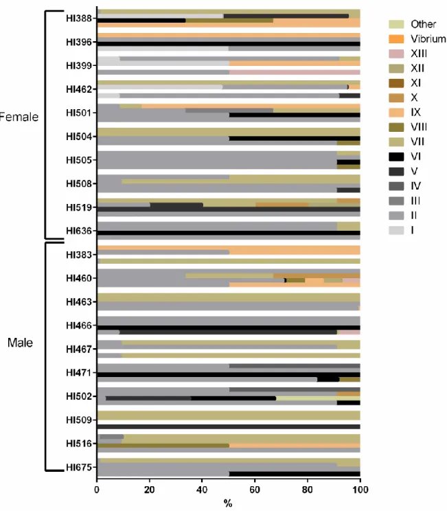
ix A análise de variações individuais no microbiota nuclear (core microbiota) foi possibilitada pela seleção prévia ao estudo de individuos com o mesmo regime alimentar (com base na análise do conteúdo estomacal presente no momento da morte), localização geográfica, condições edafoclimáticas e habitat (avaliado pelo uso da terra). Registaram-se variações do microbiota nuclear entre indivíduos, com valores de riqueza e diversidade compreendidos entre 4,6x10¹¹ e 8,8x10¹² CFU/g e 3 e 7 MT, respetivamente, à semelhança do que já foi previamente reportado noutros animais selvagens e em humanos. Analisando cada hospedeiro como um habitat com a sua própria comunidade, os tipos morfo-fisiológicos MT-II, MT-VI, MT-VII, MT-IX e MT-XI podem ser considerados o microbiota intestinal nuclear e os restantes tipos morfo-fisiológicos podem ser considerados parte da comunidade microbiota individual intraespecífica. Também, detetamos que a maioria dos hospedeiros possuía bactérias produtoras de ESBL pertencentes ao tipo morfo-fisiológico II.
Registou-se similaridade do microbiota intestinal de indivíduos adultos e juvenis, em contraste com o microbiota de indivíduos sub-adultos, provavelmente devido a fatores de carácter comportamental, uma vez que esta espécie possui padrões de proteção e alimentação dos indivíduos mais jovens, zonas de marcação de território através da excreção anal de fluidos e defecação comunitária em latrinas. Estes fatores podem promover a transmissão fecal-oral intraespecífica de microrganismos entre indivíduos da mesma comunidade (adultos e juvenis).
Este estudo visou, pela primeira vez, a caracterização extensiva da composição microbiana do trato GI de sacarrabos, permitindo a análise das diferenças na comunidade microbiota entre sexos e a análise da influencia exercida pelo contexto biológico e ambiental em cada hospedeiro na sua assinatura microbiota Apesar da elevada diversidade microbiana capturada por recurso a métodos clássicos de cultura, estudos futuros baseados em métodos independentes de cultura (metagenómica) poderão complementar a informação aqui reunida, conduzindo a um melhor entendimento da comunidade microbiana presente no sacarrabos e da bioecologia da espécie; uma espécie altamente adaptativa, em franca expansão no território e cuja presença exerce efeitos em cascata na estrutura e organização das comunidades dos ecossistemas mediterrânicos. Acresce que a natureza comparativa deste estudo, relativamente aos contributos que o sexo do hospedeiro pode ter sobre o seu microbiota, ajudam a melhorar o entendimento sobre os aspetos ecológicos e adaptativos desta espécie, reforçando a importância de se considerar o microbioma como um componente fundamental da biologia do hospedeiro e um elemento chave necessário para compreender a ecologia dos mamíferos.
Palavras-chave: Sacarrabos, Microbiota Intestinal, Identificação & Diferenciação, Carnívoros, Aptidão
x
TABLE OF CONTENTS
ACKNOWLEDGEMENTS ... i ABSTRACT ... iii RESUMO ... v TABLE OF CONTENTS ... xLIST OF FIGURES ... xii
LIST OF TABLES ... xiii
ABBREVIATIONS ... xiv
CHAPTER I – INTRODUCTION ... 1
1.1. Gut microbiota: introduction ... 1
1.1.1. Gut microbial community and diet ... 1
1.1.2. Gut microbial community and host health ... 2
1.1.3. Gut microbial community, host habitat and genetics ... 2
1.1.4. Gut microbial community, host sex and reproductive status ... 2
1.2. Herpestes ichneumon: biology and ecology ... 3
1.3. Objectives of the present work ... 4
CHAPTER II - MATERIALS AND METHODS ... 4
2.1. Egyptian mongoose specimens ... 4
2.2. Bacteriological culture ... 5
2.3. Purification and presumptive identification of isolates ... 7
2.4. Molecular identification and molecular fingerprinting of bacterial isolates ... 8
2.5. Molecular identification of fungi ... 9
2.6. Homology searches for genome-based identification of isolates ... 10
2.7. Diversity analysis of the samples under study ... 11
2.8. Data analysis ... 11
CHAPTER III – RESULTS ... 12
3.1. Comparison of bacterial burden and diversity of morpho-physiological types between mongoose host sexes ... 12
3.2. Detection of mycobiota community ... 15
3.3. Differences between media selectivity and detection of diversity ... 16
3.4. Host individuals as representatives of single communities ... 17
3.5. Molecular identification and molecular fingerprinting of bacterial isolates ... 20
3.6. Interaction between microbiota and bio-environmental features ... 28
xi CHAPTER V – CONCLUDING REMARKS AND PERSPECTIVES ... 44 CHAPTER VI – BIBLIOGRAPHY ... 46 CHAPTER VII – APPENDIXES ……….………..………....53
xii
LIST OF FIGURES
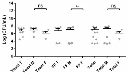
Figure 1.1 – Egyptian mongoose (Herpestes ichneumon) adult (A) and cub (B), in Mediterranean maquis. ... 3 Figure 2.1 – Brilliance ESBL medium differentiating pigmentation characteristics (manufacturer available information). ... 7 Figure 2.2 – Flowchart used for differentiation of the purified bacterial isolates into different morpho-physiological types using morphological (cell morphology, Gram and endospore staining) and biochemical tests (catalase and oxidase tests). ... 8 Figure 3.1 – Microbial load (expressed as Log CFU/ml) of mongoose cultivable bacteria grown in selective and non-selective media. (A) Comparison between four different media/conditions: YCFA incubated under aerobiosis (YCFA w/O2), YCFA supplemented with sodium taurocholate and incubated under aerobiosis (YCFA P w/O2), YCFA incubated under anaerobiosis (YCFA w/o O2), and YCFA supplemented with sodium taurocholate and incubated under anaerobiosis (YCFA P w/o O2). (B) Comparison between lactose non-fermenting (LNF) bacteria and lactose-fermenting (LF) bacteria in MacConkey medium. (C/D) Comparison between Extended-spectrum beta-lactamases (ESBL) Chromogenic medium (C) without (ESBL w/o AS) and (D) with (ESBL w/ AS) ESBL antibiotic supplement with the results present by colony color/type.. ... 13 Figure 3.2 – Comparison of microbial load (expressed as log CFU/ml) in female and male mongoose samples inoculated in ESBL Chromogenic medium without (gray bars) and with (white bars) ESBL antibiotic supplement. ... 14 Figure 3.3 – Comparison of bacterial load (expressed as Log CFU/ml) between (A) chromogenic media and (B) non-selective media. ... 15 Figure 3.4 – Comparison of microbial load (expressed as Log CFU/ml) between yeast and filamentous fungi (FF) in the PDA medium with chloramphenicol. ... 16 Figure 3.5 – Percentage of isolates belonging to each morpho-physiological type in each media/condition in Egyptian mongoose individuals. ... 16 Figure 3.6 – Percentage of isolates belonging to each morpho-physiological type in each Egyptian mongoose individual in 4 different media/conditions: YCFA incubated under aerobiosis, YCFA supplemented with sodium taurocholate and incubated under aerobiosis, YCFA incubated under anaerobiosis, and YCFA supplemented with sodium taurocholate and incubated under anaerobiosis. 18 Figure 3.7 – Percentage of isolates belonging to each morpho-physiological type in each Egyptian mongoose individual between ESBL Chromogenic medium without and with ESBL supplement of antibiotics. ... 19 Figure 3.8 – Percentage of the sum of isolates belonging to each morpho-physiological type discovered in all media in each Egyptian mongoose individual……….………20 Figure 3.9 - Bacterial isolates identification and differentiation by a hierarchical numerical analysis….21 Figure 3.10 - Gut microbiota abundance of Egyptian mongoose population at different taxonomical levels………...27 Figure 3.11 – Two principal component (PC) plots depicting the dispersal area of microbiota data of each Egyptian mongoose specimens clustered corrosponding to age class. (A) PC1xPC2 and (B) PC1xPC3 planes……….32 Figure 3.12 – Two principal component (PC) plots depicting the dispersal area of microbiota data of each Egyptian mongoose specimens clustered corresponding to sex. (A) PC1xPC2 and (B) PC1xPC3 planes……….33 Figure 3.13 – Dendrogram representing the relationship between the Egyptian mongoose specimens in terms of microbiota. ………..34 Figure 3.14 – Two principal component (PC) plots depicting the dispersal area of biological data of each Egyptian mongoose specimens. (A) PC1xPC2 and (B) PC1xPC3 planes. ………..35 Figure 3.15 – Two principal component (PC) plots depicting the dispersal area of environmental data of each Egyptian mongoose specimens. (A) PC1xPC2 and (B) PC1xPC3 planes. ………..36
xiii
LIST OF TABLES
Table 2.1 – List of Egyptian mongoose specimens studied in this work. Sex, age class, stomach
content at death, georeferenced location, and land-use data are indicated. ... 6 Table 2.2 – Taxonomic threshold similarity values (%) for bacteria and fungi. ... 10 Table 2.3 – Diversity measurements: diversity indices and non-parametric estimators of species richness... 11 Table 3.1 – Diversity measurements for male, female, and the total population of Egyptian
mongoose. ... 28 Table 3.2 – Microbiota PCA explanatory variables for each principal component (PC) and their
explanatory value. ... 30 Table 3.3 – Biological and environmental PCA explanatory variables for each PC and their explanatory value. ... 31 Supplementary Table 7.1 – Matrix of microbiota variables (OTUs) based in presence/absence of every hierarchical bacterial level………..53 Supplementary Table 7.2 – Matrix of 26 biological variables related to sex, age class, reproductive status, stomach content at time of death, and different body measurements……….56 Supplementary Table 7.3 – Matrix of 17 environmental variables related to georeferenced location, land-use, climatic data, road net, river net, and population data……….57 Supplementary Table 7.4 – Statistical significant results from data represented in Figures 3.6, 3.7 and 3.8……….58 Supplementary Table 7.5 – Information on the 16S rDNA nucleotide sequences of a selected group of bacterial isolates………...61 Supplementary Table7. 6 – Information on the ITS nucleotide sequences of a selected group of fungi isolates………....80
xiv
ABBREVIATIONS
°C – degrees Celsius µL – Microliter
ANOVA – Analysis of Variance
BLASTN – nucleotide-nucleotide Basic Local Alignment Search Tool bp – Base pair
BW – Body Weight CaCl2 – Calcium Chloride CFU – Colony Forming Unit DNA – Deoxyribonucleic Acid
ESBL – Extended-Spectrum β-Lactamases
ESBL w/ AS – ESBL chromogenic medium with antibiotic supplement ESBL w/o AS – ESBL chromogenic medium without antibiotic supplement FF – Filamentous Fungi
g – Grams
GI – Gastrointestinal Tract h – hour
HBL – Head and Body Length HD – Head Diameter
HW – Hearth Weight IgA – Immunoglobulin A
ITS – Internal Transcribed Spacer K2HPO4 – Dipotassium Phosphate
KESC – Klebsiella, Enterobacter, Serratia, Citrobacter group KW – Kidney Weight
L – Liter
LF – lactose Fermenting
xv M – Mol
mg – Milligrams
MgSO4.7H2O – Magnesium Sulfate Heptahydrate min – Minutes
mL – Milliliter mM – Millimolar
MT – Morpho-physiological Type NaCl – Sodium Chloride
NaHCO3 – Sodium Bicarbonate
NCBI – National Center for Biotechnological Information ng – Nanograms
NP – Neck Perimeter
OTU – Operational Taxonomic Unit PC -Principal Component
PCA – Principal Component Analysis PCR – Polymerase Chain Reaction
PDA w/ CHLO – PDA medium supplemented with chloramphenicol PFI – Perivisceral Fat Index
RAPD – Random Amplified Polymorphic DNA RHFL – right hind foot length
RHLL – Right Hind Leg Length rpm – Revolutions per minute
rRNA – Ribosomal Ribonucleic Acid s – Seconds
SCFA – Short Chain Fat Acids SFI – Subcutaneous Fat Index SH – Shoulder Height
xvi SW – Spleen Weight
TBE – Tris-Boric acid-EDTA TE – Tris-EDTA
TL – Tail Length
UPGMA – Unweighted Pair Group Method with Arithmetic mean V – Volt
v/v – Volume/volume w/ O2 – in aerobiosis w/o O2 – in anaerobiosis w/v – Weight/volume
YCFA – YCFA agar medium
1
CHAPTER I – INTRODUCTION
1.1. Gut microbiota: introduction
The vertebrate gastrointestinal (GI) tract is a complex ecosystem that is the habitat of an enormous density and diversity of microorganisms, containing mostly bacteria, but also archaea, fungi, protozoa, and viruses (1, 2). In carnivores, microbial density and diversity differ within gut sections, with the main concentration of bacteria being found in the large intestine (approximately 5x10¹⁰ CFU/g wet weight of feces), dominated by strict anaerobes (3). At birth, vertebrates begin to be colonized with microorganisms and, for humans it is known that individual microbiota is acquired during the first year of life, stabilizing its composition later on (4), being usually dominated by members of Firmicutes, Bacteroidetes and Proteobacteria phyla (5). However, there are a variety of effects that can alter this equilibrium (6).
1.1.1. Gut microbial community and diet
Host diet has such a deep effect on the gut microbiota that resulted in an evolutionary divergence between carnivores and herbivores, leading to two distinct gut types, with an increase in microbiota diversity from carnivores to herbivores (5, 7). Nevertheless, gut physiology is, as well, a predictor of the gut microbiota landscape, influencing distribution along the GI tract, such as the dichotomy that is observed between foregut fermenters vs. hindgut fermenters herbivores (5, 7).
For most mammals, diet can vary drastically, in time and space, across season and habitat, so that the composition of the gut microbiota, as well as it functionality, are likely to fluctuate across season and habitat in direct response to these dietary changes (8). Gut microbiota that can quickly shift activity, by changing its composition in response to changes in host dietary intake can lead to improved nutritional flexibility and increased fitness host (9). However, if a poor microbiota response to short-term changes in the diet occurs, the limited food and nutrient availability can affect host health and immune system (8), leading to decreased host fitness.
Microbial communities from animals are mostly composed of r-selected organisms, that can rapidly use the accessible nutrients in the gut and quickly multiply (7), giving a selective advantage to the host, increasing host fitness, ultimately leading to the persistence and survival of this microbiota in the host, and increasing microbiota fitness. Because of this mutually beneficial interaction, gut microbiota and the host coevolved, this is, they reciprocally adapted to each other as interacting species (1, 10). Thus, host adaptation to a defined diet gives chance to gut microbes to evolve and to adapt to the host gut and environment (11). This coevolution has been shown by the discovery of patterns of community similarity that match the mammalian phylogeny, with some lineages co-diversifying with their mammal hosts (5).
Additionally, gut microbiota plays a role in host energy uptake, breaking down fibers, carbohydrates, and proteins otherwise not digestible by the host, producing short-chain fatty acids
2 (SCFA) that provide up to 70% of an animal’s daily energy intake (12). In addition, they also reduce the pH of the intestinal lumen, facilitating nutrient absorption and preventing accumulation of toxic metabolic by-products, producing vitamins, and regulating xenobiotic metabolism (6). Alterations in gut microbial community composition thus alter the interactions between microbes, subsequently affecting energy production and host nutrition (6), and ultimately affecting the host fitness.
1.1.2. Gut microbial community and host health
The existence of an adaptive immune system in vertebrates permits a greater level of complexity of their microbiota (13). The gut microbiota has been shown to modulate the host immune system by attenuating inflammatory responses and increasing resistance to pathogenic bacteria, in fish, rodents, mice, piglets and humans, assisting in the development and maturation of the host intestinal mucosal and systemic immune systems, in mice and humans, in the development and function of the brain and modulating behavior, in humans (6).
The symbiosis, developed from coevolution, is based on a molecular exchange linking microbial signals that are recognized by host receptors to arbitrate valuable outcomes for both host and microbes (14). Furthermore, the adaptive immune system is recognized to shape microbial community composition in the gut. This system mediates tolerance to the gut microbiota through IgA production (15). Additionally, Toll-like receptors are important in their ability to evaluate the composition of the microbiota, and they also mediate the host tolerance to symbiotic microbes and the immune responses to pathogens (2).
1.1.3. Gut microbial community, host habitat, and genetics
Microbiota attained primarily in life are inherited from the mother or from social contacts, but the microbial composition is also influenced by host genetics (16). Host species that live in more interconnected social groups, with high frequencies of social contact, are expected to have less inter-individual variation in gut microbial community structure than those that are geographically isolated (17). Populations that become geographically isolated should develop distinct gut microbial communities because they are exposed to distinct microbial taxa pools, thus they are colonized by distinct gut microbial communities, normally with lower taxonomic and/or functional diversity (6).
More recently, host genetics has been associated with the taxonomic structure of the gut microbiota, since intraspecific differences have been observed (18), however individual phylotypes are also promiscuous, as they have been found in multiple host species (5, 7).
1.1.4. Gut microbial community, host sex, and reproductive status
Information about the influence of host sex and reproductive status on microbial community composition is still lacking, however, in mammals, these two parameters have been linked to variations
3 in gut microbial community composition, for example in chimpanzees (19), black howler monkeys (20), rufous mouse lemurs (16), Verreaux’s sifakas (17), and humans (21, 22).
Regarding microbiota, two essential concepts can be considered: core microbiota and individual microbiota. The core microbiota is the number and the identity of bacteria that are shared among different individuals of the same species; in contrast, the individual microbiota is the transient gut inhabitants that fluctuate, depending on the genetics, habitat, health, diet, among other factors (23). The common core bacteria are conserved during the mutual coevolution of the species and its intestinal microbes (23). These consortia of microorganisms are important to investigate the mechanisms underlying microbe–microbe and microbe-host interactions.
1.2. Herpestes ichneumon: biology and ecology
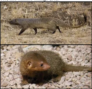
The study of wildlife microbiota has recently gained attention, but few studies are still available due to technical difficulties in obtaining appropriate samples and the lack of specific funding (3). To study the microbiota and their interaction with host fitness and the environment, we used Egyptian mongoose, Herpestes ichneumon (Linnaeus, 1758), as a model (Figure 1.1). This species is a medium-sized mammalian carnivore from the Herpestidae family, with an opportunistic feeding behavior, consuming mostly rabbits, but also reptiles, other small mammals, amphibians, birds, crayfish, eggs or carrion (24). Despite being mostly African, ranging extensively throughout the continent, it has expanded into the Mediterranean Middle East and southern Turkey (25). In Iberia Peninsula, it was conventionally considered as an introduced species during the Muslim Invasions (26, 27). However, a recent study based on mitochondrial DNA suggested that the Egyptian mongoose naturally dispersed into the Iberian Peninsula during the Late Pleistocene sea-level fluctuations (28). It was restricted to the south of the Tagus River (29), nonetheless, in the last three decades, it gradually expanded into central and north-eastern regions (30). This expansion was mostly driven by a land-use change in shrub-dominated ecosystems, forest clearing, agricultural practices, and climate change (31).
Egyptian mongoose has a home-range of about 3 km², habiting locals with understory vegetation in coastal, lacustrine, and riparian habitats, avoiding humid forests and extreme deserts. In Europe, it is found in Mediterranean maquis. Listed as Least Concern, the species is widespread, common, and present in many protected areas. There are no major threats to this species across its range, although, on the Iberian Peninsula, incidental and deliberate poisoning is a localized threat; also, in Portugal, mongoose hunting is legal (32). Evidence of both sexual and
A
B
Figure 1.1 – Egyptian mongoose (Herpestes ichneumon) adult (A) and cub (B), in Mediterranean maquis.
4 regional dimorphism in body size of Egyptian mongoose adults in Portugal have been found. This dimorphism probably results from differences in feeding habits leading to southern male adults being larger and heavier (33). H. ichneumon exhibit variability in social organization, ranging from solitary individuals to groups, which show cooperative tendencies, particularly in areas with abundant food resources. The exclusive home-range use of males in high-density populations suggests the existence of a polygynous mating system, with is accomplished due to the spatial distribution of females, in combination with the absence of paternal care behavior (24, 29, 34-36).
1.3. Objectives of the present work
Several aspects of mongoose’ biology remain ill-defined. In this study, we thoroughly investigated the gut microbiota of Egyptian mongoose sampled in South Portugal, since the gut microbiota is being progressively acknowledged as a fundamental component of mammals’ ecology. The aims of this work were: [1] the characterization of the core gut microbiota of Egyptian mongoose population; [2] the investigation of sex- and age class-related differences of gut microbiota; and [3] the analysis of the relationship between bio-environmental features and gut microbiota of these specimens. To accomplish these objectives, the fecal samples were cultured in selective and non-selective media, under aerobiosis and anaerobiosis conditions, in order to capture the most representative diversity of gut microbiota. Purification and presumptive identification of microbial isolates followed, using morpho-biochemical tests and grouping the isolates in morpho-physiological types. The purified bacterial isolates were submitted to 16S rRNA- and Internal Transcribed Spacer-based molecular identification and molecular fingerprinting based on Random Amplification of Polymorphic DNA (RAPD). At the bacterial genus level, a diversity analysis of the samples under study was performed. We compared sex- and age class-related differences (genus-based phylotypes) and tested the effect of individual bio-environmental features on the microbiota of each individual specimen.
CHAPTER II - MATERIALS AND METHODS
2.1. Egyptian mongoose specimens
Twenty Egyptian mongoose carcasses, both male (n=10) and female (n=10), from predator control hunted by shotgun, were donated for scientific purposes and were selected based on sex, age class (inferred from dentition), stomach content at the time of death, and land-use (Table 2.1). The carcasses were subjected to necropsy by a veterinarian; no sign of putrefaction or disease were detected. This information was obtained from University of Aveiro, which developed ecological studies with the same specimens (33). The selected specimens had an age class distribution of 16 adult, two subadult, and two juvenile.
These animals were originated from south of Tagus River (Baixo Alentejo), wherein similar land-use, predominated by agroforestry, mixed forests, shrubs and agriculture, was confirmed. The selected animals had the same stomach content at death, namely mammal and egg items.
5
2.2. Bacteriological culture
After collection at necropsy, samples from the terminal portion of the large intestine of Egyptian mongoose specimens were kept frozen at -20°C until utilization, to preserve both aerobic and anaerobic species and to avoid potential loss of bacterial viability and composition changes. The samples were divided into two equal parts, that were homogenized in buffered peptone water (1 g stool per 10 mL) and incubated at 37°C for 24 h with orbital shaking (150 rpm), under aerobic and anaerobic conditions. Anaerobic atmosphere was accomplished by completing with 20% of the final volume of mineral oil.
Next, 1 mL of enriched fecal sample from aerobic and anaerobic conditions were pasteurized at 80°C for 12 min, serially diluted and plated onto YCFA agar (12.5 g/L agar, 10 g/L casitone, 2.5 g/L yeast extract, 4 g/L NaHCO3, 1 g/L cysteine, 0.45 g/L K2HPO4, 0.45 g/L KH2PO4, 0.9 g/L NaCl, 0.09 g/L MgSO4.7H2O, 0.09 g/L CaCl2, 1 mg/L resazurin, 10 mg/L haemin, 10 µg/L biotin, 10 µg/L cobalamin, 30 µg/L p-aminobenzoic acid, 50 µg/L folic acid, 150 µg/L pyridoxamine, 33 mM acetate, 9 mM propionate, 1 mM isobutyrate, 1 mM isovalerate, 1 mM valerate, 50 µg/L thiamine and 50 µg/L riboflavin), supplemented with 2 g/L each of glucose, maltose, and cellobiose and 0.1% of sodium taurocholate (YCFA P) (37), and incubated for 72 h at 37°C, under aerobic and anaerobic conditions, respectively. The rest of the enriched fecal sample, incubated under anaerobic conditions, was serially diluted and 100 µL was plated onto YCFA agar supplemented with 2 g/L each of glucose, maltose, and cellobiose (YCFA) (37), and incubated for 72 h, at 37°C, in anaerobic conditions. Anaerobic conditions were accomplished using AnaeroGenTM 3.5L anaerobic atmosphere generation systems (Thermo Scientific). Additionally, the rest of the enriched fecal sample incubated under aerobic conditions was serially diluted and 100 µL was plated onto YCFA and also onto selective media: MacConkey solid medium (12 g/L agar, 5 g/l bile salts, 10 g/l lactose, 0.075 g/l neutral red, 20 g/l peptone, 5 g/l sodium chloride); Potato Dextrose Agar supplemented with chloramphenicol (PDA+CHLO) (Biokar diagnostic, Noack Group); ESBL chromogenic medium (Conda, Pronadisa), with (ESBL w/ AS) and without (ESBL w/o AS) ESBL supplement of antibiotics (Conda, Pronadisa); and Brilliance ESBL medium (Brilliance) (Oxoid). All media were incubated at 37°C, under aerobic conditions for 24 h, except YCFA and PDA solid media that were incubated for 72 h.
6 Table 2.1 – List of Egyptian mongoose specimens studied in this work. Sex, age class, stomach content at death, georeferenced location, and land-use data are indicated.
ID Sex Age1
Stomach content at death2 Georeferenced location Land-use3
Mammals Reptiles Invertebrates Eggs District Latitude Longitude Urban Agroforestry Shrubs Vineyards
and Orchards Coniferous Mix
forests Agriculture HI383 Male Adult 100 0 0 0 Beja 37,824 -7,377 0 233 0 0 0 125 42 HI388 Female Subadult 100 0 0 0 Beja 37,824 -7,377 0 233 0 0 0 125 42 HI396 Female Adult 100 0 0 0 Beja 37,824 -7,377 0 135 93 0 0 172 0 HI399 Female Adult 100 0 0 0 Beja 37,720 -8,097 0 135 93 0 0 172 0 HI460 Male Juvenile 2 100 0 0 0 Beja 37,824 -7,377 0 0 250 0 4 0 132 HI462 Female Adult 96 4 0 0 Beja 37,824 -7,377 0 135 93 0 0 172 0 HI463 Male Adult 0 0 29 0 Beja 38,107 -7,205 93 17 0 170 0 0 110 HI466 Male Adult 100 0 0 0 Beja 38,107 -7,205 0 127 116 0 0 0 157 HI467 Male Adult 77 20 3 0 Beja 37,824 -7,377 0 127 116 0 0 0 157 HI471 Male Adult 0 0 0 100 Beja 37,824 -7,377 0 127 116 0 0 0 157 HI501 Female Adult 100 0 0 0 Beja 37,824 -7,377 0 135 93 0 0 172 0 HI502 Male Juvenile 2 100 0 0 0 Beja 37,824 -7,377 0 0 0 0 0 0 400 HI504 Female Subadult 95 0 0 0 Beja 37,824 -7,377 0 233 0 0 0 125 42 HI505 Female Adult 65 35 0 0 Beja 37,824 -7,377 0 127 116 0 0 0 157 HI508 Female Adult 97 3 0 0 Beja 38,287 -8,224 0 127 116 0 0 0 157 HI509 Male Adult 89 0 0 0 Beja 38,107 -7,205 76 0 4 0 123 8 189 HI516 Male Adult 100 0 0 0 Beja 37,824 -7,377 76 0 4 0 123 8 189 HI519 Female Adult 100 0 0 0 Beja 37,824 -7,377 0 135 93 0 0 172 0 HI636 Female Adult 88 0 12 0 Beja 37,824 -7,377 0 135 93 0 0 172 0 HI675 Male Adult 100 0 0 0 Setúbal 38,107 -7,205 0 127 116 0 0 0 157
1Each specimen was assigned to one of four age classes: adults over one year of age, sub-adults between nine and twelve months, juveniles type II between five-and-a-half and nine
months. Skulls with completely developed definitive dentition were assigned to the adult class. Skulls with 40 definitive teeth, but with some still growing, were assigned to the sub-adult age class. Animals whose skulls presented all 40 teeth, but including at least one milk tooth, were assigned to the juvenile type II class (33).
2(personal information from Victor Bandeira)
3Number of hectares of each habitat type (urban, rice fields, agroforestry, shrubs, inland water bodies, vineyards and orchards, coniferous, broadleaved and mix forests and agriculture
areas) were retrieved from Corine Land Cover (2006) with spatial resolution of 250m. This variable was represented by mean values of the 2×2 km grid cell, considering the home-range of the Egyptian mongoose (33).
7
2.3. Purification and presumptive identification of isolates
For purification of isolates, five isolated colonies from each different morphology were picked from different dilutions of all media. The colonies that were picked up were re-streaked twice to confirm purity. The purified individual colonies were assessed in terms of shape, pigmentation, and opacity. Bacteria were characterized in terms of gram character, endospore formation and the presence of catalase and cytochrome c oxidase enzymes.
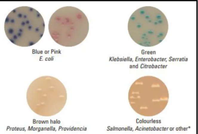
ESBL Chromogenic medium can differentiate between E. coli that grow as pink colonies and bacteria belonging to the Klebsiella,
Enterobacter, Serratia, and Citrobacter (KESC)
group that grow as dark blue colonies (information available from the manufacturer, Figure 2.1.). Brilliance ESBL medium can differentiate between E. coli that grow as blue or pink colonies and KESC bacteria group that grow as green, green/blue or even brownish-green colonies (38). Moreover, Proteus, Morganella, and Providencia group grow as tan-colored colonies with brown halo (38). Finally,
Pseudomonas aeruginosa colonies can also be differentiated, once they exhibit pyocyanin-related
green-brown pigmentation (39). Colorless colonies may be Salmonella spp., Acinetobacter spp. or others (information provided by the manufacturer).
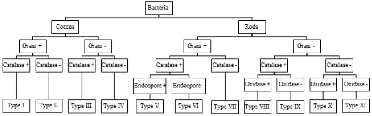
The isolated fungi were stained using lactophenol cotton blue and different morphological aspects were characterized. For filamentous fungi isolates, hyphal septation and spores color, morphology, and septation were assessed. For yeast isolates, we evaluated cell morphology and division. Considering the previous characterization, all isolates were grouped into morpho-physiological types (MT): MT-I to MT-XI for bacterial isolates (Figure 2.2); MT-XII for yeasts; MT-XIII for filamentous fungi. Additionally, MT-Vibrium and MT-Others were created, the first to place a vibrium-like isolate and the second to place unidentifiable isolates.
Figure 2.1 – Brilliance ESBL medium differentiating pigmentation characteristics (manufacturer available information).
8
Figure 2.2 – Flowchart used for differentiation of the purified bacterial isolates into different morpho-physiological types using morphological (cell morphology, Gram and endospore staining) and biochemical tests (catalase and oxidase tests).
2.4. Molecular identification and molecular fingerprinting of bacterial isolates
With the objective to analyze the intraspecific polymorphism present in the bacterial population, molecular fingerprinting was based on random amplified polymorphic DNA (RAPDs). A bacterial isolate from each MT, from each solid media, from each mongoose host, was selected. Total DNA extraction was performed by direct boiling at 95°C for 15 min of 2 to 3 colonies in 250 µL of TE 1 M, pH 8.0, centrifuged, the supernatant was collected to a clear microtube and stored at -20°C.
For single primed PCR fingerprinting, an initial screening to select the most appropriate primer was performed using four different primers. Seven isolates (one from each morpho-physiological type) were tested, including two isolates of MT-II and one isolate of MT-VII, MT-V, MT-VIII, MT-IX, and MT-IV. The tested primers can be classified into three groups: primers directed to regions containing mini-satellite from M13 bacteriophage – M13 (5’-GAG GGT GGC GGT TCT-3’) (40); random primers – OPC19 (5’-GTT GCC AGC C-3’) (41) and 1281 (5’-AAC GCG CAA C-3’) (42); and universal primers for 16S rRNA gene – PH (5’-AAG GAG GTG ATC CAG CCG CA-3’) (43).
PCR amplifications were performed in a Biometra Uno II Thermal Cycler, using a total volume of 15 μl and including 0.2 mM of primer (Invitrogen), 7.5 µL of NZYTaq II 2x Green Master Mix (NZYTech), 5 µL of DNA (from different dilutions of the DNA extract). DNA dilutions were decided from empirical analyses of DNA extract concentration run in 0.8% (w/v) agarose gel.
After preliminary analyses, the selected primers for molecular fingerprinting were M13 and PH. PCR cycling conditions for M13 consisted of 94°C for 5 min, followed by 40 cycles of 60 s at 94°C, 3 min at 40°C, 120 s at 72°C, plus an additional step at 72°C for 7 min, for chain elongation. The PCR cycling conditions for PH consisted of 95°C for 3 min followed by 35 cycles of 30 s at 94°C, 30 s at 35°C, 3 min at 72°C, plus an additional step at 72°C for 5 min.
The PCR products were resolved by 1.5% (w/v) agarose gel (NZYTech) containing 0.03 µL/mL of GreenSafe Premium (NZYTech) in 0.5 X TBE buffer (44.5 mM Tris, 44.5 mM boric acid, and 1 mM
9 EDTA) (Invitrogen), at 90 V for 4 h. DNA was visualized under UV light and photographed with Alliance 4.7 system (UVITEC Cambridge).
To obtain a measure of reproducibility, each PCR batch included a randomly selected duplicate, with a total number of 18 isolates for M13 and 22 isolates for PH. The similarity between each pair of duplicates was based on the dendrogram computed with Pearson correlation coefficient and the unweighted pair group method with arithmetic average (UPGMA) as the agglomerative clustering algorithm (software package BioNumerics version 4.0 – Applied Maths). The reproducibility value was determined as the average value for all pairs of duplicates. Strain relationships, based on the molecular characters presented as fingerprints, were analyzed by hierarchical numerical methods with Pearson correlation similarity and UPGMA clustering, using 70% similarity as the cutoff value for cluster formation.
Molecular identification was based on 16S rRNA gene sequence analysis. At least, one isolate from each dendrogram cluster resulting from RAPD-fingerprints was randomly selected for 16S rRNA gene sequencing. A PCR was performed using as forward primer 63f (5′-CAG GCC TAA CAC ATG CAA GTC-3′) and as reverse primer 1387r (5′-GGG CGG WGT GTA CAA GGC-3′) (44), in a final volume of 25 µL with 0.2 mM from each primer (Invitrogen), 12.5 µL NZYTaq II 2x Green Master Mix and 5 µL of DNA (from a 1:100 dilution of DNA extract). This set of primers allow the amplification of all hypervariable region (V1-V9). The PCR amplification program consisted of 1 cycle of 5 min, 95°C, followed by 30 cycles of 45 s, 95°C; 45 s, 55°C; 120 s, 72°C, and a final step of 7 min, 72ºC. PCR products with expected size (approximately 1500 bp) were observed in a 1.5% agarose gel, in 1 X TBE buffer (89 mM Tris, 89 mM boric acid, and 2 mM EDTA), run at 90 V for 1.5 h, using GreenSafe Premium (0.003% (v/v)). DNA was quantified using a Qubit fluorometer (Invitrogen), following manufacturer’s instructions. Samples were commercially sequenced by Sanger sequencing technique using 63f primer (GATC Biotech AG). Since the Taq DNA polymerase used has a mutation rate of 10- 5, the reproducibility of the originated sequences was assessed through the comparison of duplicate sequences that were re-sequenced and taking into consideration that, for financial reasons, only one strand was sequenced.
2.5. Molecular identification of fungi
Fungi isolates with different morphology were selected for genomic identification through sequencing. Yeast DNA was extracted using the direct boiling method previously described, and filamentous fungi DNA was extracted using NZY Plant/Fungi gDNA Isolation kit (NZYTech), following the manufacturer instructions.
Amplification of the D1/D2 domain region of the 26S rRNA gene in yeast (45) and the Internal Transcribed Spacer (ITS) region in filamentous fungi were performed (46). For yeast isolates, a PCR was performed using NL-1 (5’-GCA TAT CAA TAA GCG GAG GAA AAG-3’) and NL-4 (5’-GGT
10 CCG TGT TTC AAG ACG G-3’) primers (47), and for filamentous fungi, a PCR was performed using ITS5 (5’-GGA AGT AAA AGT CGT AAC AAG G-3’) and ITS4 (5’-TCC TCC GCT TAT TGA TAT GC-3’) primers (48). In both cases, a final volume of 25 µL was used containing 0.2 mM of each pair of primers (Invitrogen), 12.5 µL NZYTaq II 2x Green Master Mix and 5 µL of DNA (from a 1:100 dilution of DNA extract). The PCR amplification program consisted of 1 cycle of 3 min at 95°C, followed by 35 cycles of 30 s at 94°C, 30 s at 55°C, 30 s at 72°C and a final step of 10 min at 72ºC. PCR products with expected size (approximately 650 bp and between 600 and 800 bp, respectively) were observed in a 1.2% agarose gel, in 1 X TBE buffer, at 90 V for 1 h, using GreenSafe Premium and extracted using QIAquick Gel Extraction Kit, according to manufacturer’s handbook.
DNA was quantified using a Qubit fluorometer, following manufacturer’s instructions. For Sanger sequencing, samples were prepared adding with 20 to 80 ng/µL of PCR product to 5 µM of NL-4 and ITSNL-4 primers, for yeast and filamentous fungi, respectively, and commercially sequenced.
2.6. Homology searches for genome-based identification of isolates
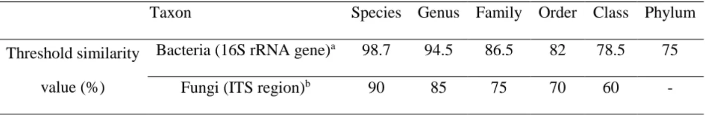
Electropherograms were manually analyzed and corrected when necessary; undetermined nucleotides were designated as N. The 16S rRNA gene, D1/D2 domain region and ITS gene sequences were compared with those available in the GenBank databases using the BLASTN program through the National Center for Biotechnology Information (NCBI) server. Comparisons were performed using the default parameters. Sequences were annotated with taxonomic information from the top three best matches displaying the same nucleotide pairwise identity. The criteria used for bacteria and fungi identification are represented in Table 2.2 (49-51). A failure to identify phylotypes was defined as a 16S rRNA gene sequence similarity score lower than 75% and an ITS sequence similarity score lower than 60% with sequences deposited in GenBank at the time of analysis.
Table 2.2 – Taxonomic threshold similarity values (%) for bacteria and fungi.
Taxon Species Genus Family Order Class Phylum
Threshold similarity value (%)
Bacteria (16S rRNA gene)a 98.7 94.5 86.5 82 78.5 75
Fungi (ITS region)b 90 85 75 70 60 -
aas in (42, 43).
11
2.7. Diversity analysis of the samples under study
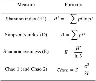
Diversity analyses were performed through the calculation of diversity indices (Shannon index, Simpson index and Species evenness index derived from Shannon index) and the determination of nonparametric estimators (Chao 1 and Chao 2) of species richness (Table 2.3) (52).
Table 2.3 – Diversity measurements: diversity indices and non-parametric estimators of species richness.
Measure Formula
Shannon index (H’) 𝐻′= − ∑ 𝑝𝑖 ln 𝑝𝑖
Simpson’s index (D) 𝐷 = ∑ 𝑝𝑖2
Shannon evenness (E) 𝐸 = 𝐻′ ln 𝑆 Chao 1 (and Chao 2) 𝐶ℎ𝑎𝑜 = 𝑆 +𝑎
2 2𝑏
Where pi=ni/N; S is the number of OTUs in the sample; N is the total number of isolates from a given OUT in the sample; ni is the number of isolates in an OTU; a is the number of OTUs recorded only once; b is the number of OTUs recorded only twice. Chao 1 analyses species abundance data and Chao 2 analyses species incidence data.
2.8. Data analysis
Considering culture assays, results from CFU counts are displayed as means of values of at least ten independent experiments with respective standard deviation. All variables were tested for normality using D’Agostino-Pearson test (α=0.05). When comparing two conditions, a t-student test (Mann-Whitney test, α=0.05) was performed. When comparing multiple conditions, a non-parametric ANOVA (Kruskal-Wallis test, α=0.05) with a Dunn’s Multiple Comparison post-test was performed. When comparing multiple host communities, a two-way ordinary ANOVA (α=0.05) with a Tukey's Multiple Comparison post-test was performed. All statistical analyses were performed using GraphPad Prism software.
For microbiota and bio-environmental data integration, we performed a Principal Component Analysis (PCA) using available information for all 20 Egyptian mongoose specimens. A matrix using 72 microbiota operational taxonomic units (OTUs), based on presence/absence of every hierarchical bacterial level (Supplementary Table 7.1), was normalized using the standard score. The normalized matrix was used to perform an initial PCA. Scatter projection diagrams were obtained both for sex and age class. A dendrogram was then made based on the normalized Euclidean distance derived from the projection matrix using UPGMA. The cophenetic correlation coefficient was calculated and a 2-way Mantel test (Mantel, 1967) was performed to measure the faithfulness of the dendrogram compared to
12 the pairwise distances of the original unmodeled matrix. Two others PCAs were made using a matrix with 26 biological and 17 environmental variables (Supplementary Table 7.2 and 7.3). These matrices were normalized using the standard score and PCAs were performed. Scatter projection diagrams were obtained for the clusters originated from the previous microbiota dendrogram. The explanatory variables of each Principal Component (PC) were selected if the correlation coefficient between the variable and the PC were |0.5|. If a variable had this behavior with more than one PC, this variable was used as an explanatory variable for the PC with the higher correlation coefficient. All these analyses were made using NTSYSpc software (version 2.20d; Exeter Software, Setauket, NY, USA).
CHAPTER III – RESULTS
3.1. Comparison of bacterial burden and diversity of morpho-physiological types
between mongoose host sexes
Culture-dependent methods were used to investigate gut microbiota diversity in 20 fecal samples of Egyptian mongoose specimens. A rich medium for bacterial growth was used, with (YCFA P) and without (YCFA) 0.1% de sodium taurocholate supplementation. In both cases, samples were incubated under aerobic (w/ O2) and anaerobic (w/o O2) conditions.
Microbial load in these four media/conditions (Figure 3.1 A) were compared; in male samples, an average of 2.6x10⁸ CFU/mL, 1.6x10⁴ CFU/mL, 2.9x10⁸ CFU/mL, and 8.5x10⁵ CFU/mL were found in YCFA w/ O2, YCFA P w/ O2, YCFA w/o O2 and YCFA P w/o O2, respectively. In female samples, we registered a mean of 3.0x10⁸ CFU/mL, 8.7x10⁴ CFU/mL, 8.0x10⁸ CFU/mL, and 4.1x10⁷ CFU/mL in YCFA w/ O2, YCFA P w/ O2, YCFA w/o O2 and YCFA P w/o O2, respectively.
Comparing the microbial load in the four media/conditions (Figure 3.1 A), a significant higher microbial load was registered in YCFA w/ O2 and YCFA w/o O2, when comparing with YCFA P w/ O2 andYCFA P w/o O2, respectively. Additionally, a significant lower microbial load was registered in YCFA P w/ O2 than in YCFA P w/o O2 and in YCFA w/ O2 when comparing with YCFA w/o O2.
Regarding sex-related differences (Figure 3.1 A), no significant differences were found in the microbial load using YCFA w/ O2 and YCFA P w/ O2 as growth conditions, but female hosts had a higher microbial load than males in YCFA w/o O2 (p-value=0.0410) and YCFA P w/o O2 (p-value=0.0288).
A panel of selective growth media was used to detect specific groups of bacteria. The microbial load in MacConkey medium had no significant difference between sexes, both for lactose non-fermenting (LNF) bacteria, lactose non-fermenting (LF) bacteria, and the sum of both types (Figure 3.1 B), with a mean of 2.1x10⁸, 1.8x10⁸, and 3.9x10⁸ CFU/mL registered for male mongooses, respectively, and a mean of 8.1x10⁸, 3.9x10⁸, and 1.2x10⁹ CFU/mL, respectively, in female mongooses.
13
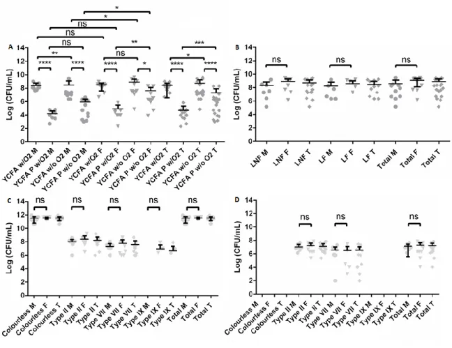
Figure 3.1 – Microbial load (expressed as Log CFU/mL) of mongoose cultivable bacteria grown in selective and non-selective media. (A) Comparison between four different media/conditions: YCFA incubated under aerobiosis (YCFA w/O2), YCFA supplemented with sodium taurocholate and incubated under aerobiosis (YCFA P w/O2), YCFA incubated under anaerobiosis
(YCFA w/o O2), and YCFA supplemented with sodium taurocholate and incubated under anaerobiosis (YCFA P w/o O2). (B)
Comparison between lactose non-fermenting (LNF) bacteria and lactose-fermenting (LF) bacteria in MacConkey medium.
(C/D) Comparison between Extended-spectrum beta-lactamases (ESBL) Chromogenic medium (C) without (ESBL w/o AS) and (D) with (ESBL w/ AS) ESBL antibiotic supplement with the results present by colony color/type. Results from male (M), female
(F), and in total (T) are presented. Horizontal bars represent the mean and error bars represent the standard deviation from 10 (male and female) and 20 (total) independent values. Statistical analysis was performed using a t-student test (Mann-Whitney test, α=0.05). ns – non-significant value≥ 0.05), * - significant value=0.01 to 0.05), ** - very significant (p-value=0.001 to 0.01), *** - extremely significant (p-value=0.0001 to 0.001), **** - extremely significant (p-value< 0.0001).
Additionally, two selective media for detection of extended-spectrum beta-lactamases (ESBL) producing gram-negative bacteria, namely the ESBL Chromogenic medium with ESBL antibiotic supplement (ESBL w/AS) and the ready-to-use Brilliance ESBL medium (Brilliance), were used. As well, we decided to compare the differences resulting from the addition of the ESBL antibiotic supplement, incubating in ESBL Chromogenic medium without this supplement (ESBL w/o AS).
In ESBL chromogenic medium, metallic blue colonies belonged to morpho-physiological type (MT) II, light blue colonies belonged to MT-VII, and pink colonies belonged to MT-IX, being putative
E. coli. Colorless colonies were very diverse, fitting to different MT, namely MT-I, M-III to MT-VI,