“Losanga” decorated imitations of italic late republican black gloss
tableware from South-Western Iberia: A multi-analytical/
microchemical characterization
☆
Nick Schiavon
a,⁎
, Vincenzo Soria
b, Ana Margarida Arruda
b, Massimo Beltrame
a, José Mirão
aaHercules Laboratory, University of Évora, Évora, Portugal b
UNIARQ, Centro de Arqueologia, University of Lisbon, Lisbon, Portugal
a b s t r a c t
a r t i c l e i n f o
Article history: Received 15 August 2015 Accepted 13 October 2015 Available online 20 October 2015 Keywords: Roman pottery SW Iberia Portable XRF BSEM + EDS XRD
Italic black gloss tableware
The micro-chemical/mineralogical composition of samples of grey-paste imitations of Italic Late Republican black gloss tableware displaying a particular kind of lozenge-shaped decoration (“Losanga pottery”) from Portuguese and Spanish archaeological sites in SW Iberia has been analysed by BSEM + EDS,μXRD, Powder XRD, Portable XRF andμRaman spectroscopy. “Losanga” decorated ceramics have been found throughout the Western Mediter-ranean. Most of the sherds display a green-brown to greyish-black engobe at the surface resembling the gloss found in Attic pottery from Classical Greece. The overall chemical, mineralogical and fossiliferous homogeneities of the ceramic paste show common features (low K-feldspar/plagioclase ratio, high Ca content, abundance of well-preserved fragments of foraminifera microfossils) that indicate lowfiring conditions in the kiln ranging from 650 to 900 °C. With respect to the ceramic body, analytical results confirm an enrichment in the surface gloss layer of iron, potassium and aluminium and a depletion in silicon and calcium; the veryfine grain size of the surface coating suggests elutriation of iron oxide-rich clays as confirmed by the presence of magnetite, maghemite and goethite inμ-XRD scan. Chemical and mineralogical data also suggest that the firing process was performed in a 600–850 °C temperature range, adopting the well-known technique of alternating oxidizing and reducingfiring conditions largely employed at the time. The analytical results, while compatible with the archaeological hypothesis of a common provenance of the raw materials for pottery production from the Guadalquivir valley workshops cannot be considered conclusive due to the similarity in the geological substrate in the two SW Iberian regions under study.
© 2015 Elsevier B.V. All rights reserved.
1. Introduction
In the study of Roman ceramics and pottery materials, micro-analytical techniques combining chemical, microscopical and mineralog-ical analyses have proved very useful to shed light on raw material prov-enance and production technology[1–11]. Due to their widespread and ubiquitous distribution across the entire geographical width of the Roman influenced territories, amphorae fragments have received the most attention by researchers. In terms of socio-historical significance, though, tableware pottery production, albeit less studied, is of no less im-portance due to its wide distribution in the Mediterranean and because it may provide precious information on common trade routes and imported technologies across the Roman Empire[3,12–17].
With the term Italic black gloss ware (also known as Ceramica
Campana, or Céramica Campaniense or “Ceramique Campanienne”
according to its geographical distribution) archaeologists identify a specif-ic group and type of tableware production developed between the IV and I century BC mainly in the Italic peninsula characterized by a red, creamy or greyish clay ceramic paste often associated with a black thin superficial engobe/gloss layer[3,12,15,17–19]. During the 2nd and 1st century BC, the massive commercialization of Italic black gloss tableware all around the Mediterranean basin led to imitative processes embedded with social and economic meanings in different regions of the Roman territories in-cluding SW Iberia[15,17]. These ceramics have been typologically regarded as tableware“imitations” of the Italic black gloss prototypes, supposed to be locally (in the sense of not imported from the Italic Penin-sula) produced during the embryonic phase of interaction[15]. In fact, the craft imitative phenomenon during the Late Republican period in the pro-vincial contexts is supposed to be the consequence of processes driven by the imports of exogenous goods stored in amphorae. Similarly, the growth of tableware imitation is due to the imports of Italic black gloss ta-bleware during the last two centuries before the Common Era. Within the Italic black gloss production, a special typology of high socio-historical sig-nificance is represented by the so-called “Losanga-style” tableware,
☆ Selected papers presented at TECHNART 2015 Conference, Catania (Italy), April 27–30, 2015.
⁎ Corresponding author.
E-mail address:[email protected](N. Schiavon).
http://dx.doi.org/10.1016/j.microc.2015.10.017
0026-265X/© 2015 Elsevier B.V. All rights reserved.
Contents lists available atScienceDirect
Microchemical Journal
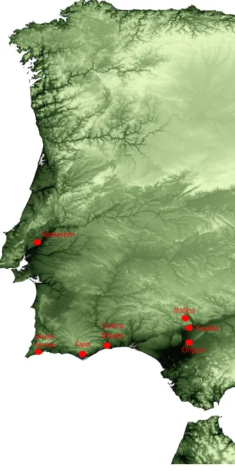
characterized by the presence of a decoration of stamped arcs joining four trefoils (Fig. 1). It is generally accepted that the“Losanga” decora-tion in pottery objects appeared in the 1st century BC[14]and had a major production centre in the Roman colony of Cales, in the northern part of modern Campania region in Italy[20,21];“Losanga” pottery objects have been found throughout the Western Mediterranean including sites on the Atlantic African Coast[22]. In this study, samples of“Losanga” tableware from four archaeological sites in Southern/Cen-tral Portugal (Faro, Castro Marim, Monte Molião and Santarém) dated from the middle of the 1st century BC to the second half of the 1st century AD[17,19]and from 3 sites in the Seville area (Seville: Patio de Banderas; Italica: Pajar de Artillo; Orippo: Cortijo de Tixe) in the An-dalusian region (Southern Spain) dated from the 1st century BC[12,16, 23]were selected for microchemical, microscopical and mineralogical analyses (Figs. 1, 2). A common feature of both Andalusian and Portu-guese sites (which explains their role as important consumption centres of the time) lies in their proximity to the sea and to well-established ancientfluvial routes (respectively along the Guadalquivir, Tagus and Guadiana river valleys). The Guadalquivir area has long been recognized as an important centre for ceramic production[10,13, 23,24]as evidenced by the identification of several ceramic workshops with raw materials (clays and temper) for pottery manufacturing com-ing from the Quaternaryfluvial sedimentary deposits of the Guadalqui-vir River. The importance of the GuadalquiGuadalqui-virfluvial valley as a major ceramic production area in the Baetica province of the Hispania Penin-sula during Roman occupation has been recently confirmed also by archaeometric analytical data on Haltern 70 amphorae[9]. It is worth noting that the current archaeological investigation in the Guadalquivir valley has not yet recognized the production of the tableware typology investigated in the current study. This state of affairs has led archaeolo-gists to postulate a predominant unidirectional East to Westflux of “Losanga” tableware produced in the Guadalquivir area from Baetic to Lusitania. In this vein, the assumption of a shared origin of the typolog-ically similar“Losanga” tableware pottery fragments found in Portu-guese sites with the Andalusian ones, though, has, so far, been based only on ceramological evidences[12]as well as on a sort of a “depen-dency” in terms of dynamism in the distribution of goods from the Andalusian region[25], following the attribution of a catalyst role to that region as expressed in the concept of the“Círculo del Estrecho”
[26]. Major and trace-elements' chemical compositions of potsherds from Roman amphorae in the Sado and Tagus river valley[4,5]have indeed been compared with coeval pottery fragments from the Guadal-quivir area: these studies identified similarities but also differences linked to changes in the geological/mineralogical terrain of the raw materials used in pottery manufacturing.
The combined micro-analytical approach adopted in this study intends to add important data to the above mentioned debate by pro-viding BSEM + EDS,μXRD, PXRF and μRaman spectroscopy data to com-pare the detailed chemical and mineralogical compositions of both the grey paste and the dark surface slip of selected samples from the inves-tigated sites with the aim to establish for thefirst time a possible corre-lation between composition and typology, and between the Portuguese and Andalusian assemblages and to determine imported versus local manufacturing practices.
2. Materials and methods
31 samples of“Losanga style” tableware pottery (Fig. 1) were select-ed for analysis by BSEM + EDS, Portable XRF and Micro and Powder XRD. Resin inglobated polished blocks were analysed by BSEM-EDS using a HITACHI S3700N interfaced with a Quanta EDS microanalysis system. The Quanta system was equipped with a Bruker AXS XFlash® Silicon Drift Detector (129 eV Spectral Resolution at FWHM/MnKα). Standardless PB/ZAF quantitative elemental analysis was performed using the Bruker ESPRIT software. The operating conditions for EDS
Fig. 1.“Losanga” decorated tableware pottery from SW Iberia.
analysis were as follows: BSEM mode (BSEM), 20 kV accelerating voltage, 10 mm working distance, and 120μA emission current. The de-tection limits for major elements (NNa) were in the order of 0.1 wt.%.
μ-XRD and Powder XRD (PXRD) were used to identify crystalline phases. A Bruker AXS D8 Discovery XRD with the Da Vinci design with a Cu Kα source operating at 40 kV and 40 mA and a lynxeye 1-dimensional detector was used. Scans were run from 3 to 75° 2θ, with 0.05° step and 1 s measuring time by point. PXRD was used for bulk analysis whileμ-XRD was used to investigate the black slip. The XRD system was reconfigured and a Goebel mirror and a 300 μm beam-collimator were employed.
For major and minor elemental analyses a Portable XRF was used for quick and non-destructive examination. The instrument used was a Brüker TRACER III-SD handheld portable XRF spectrometer equipped with a Rhodium tube. Archaeological samples were analysed six times each with the same analytical condition. Six repetitions were consid-ered sufficient, especially for ceramic samples, to obtain representative results for K, Ca, Fe, Mn, Rb, Sr, Zr, Y and Nb and to reduce problems related to grain size and mineralogy[7]. Flat (or almostflat) sections were selected for analysis to avoid interference caused by the presence of a slip, surface contaminant[8]and/or surface roughness[7]. Operat-ing conditions were 40 kV and 35μA using a filter (green filter, 12 mm Al + 1 mm Ti + 6 mm Cu) with 300 s live-time-counts. Charac-teristic radiations were collected by a silicon drift detector (SDD) with an energy resolution of 149.68 eV at Mn Kα FWHM. The beam spot is elliptical in shape with dimension 3 by 4 mm (7 mm2). The equipment
also possesses a camera, allowing the visualization of the analysed area and thus the capture of the image and the spot of analysis. S1 PXRF software (v. 3.8.30) was used to record the spectra, and ARTAX software (v. 5.3.0.0) for thefirst spectra evaluation and for the selection of the ref-erence standards.
Quantifications were performed converting count rates as the ratio of the Kα Rh peak following the guideline of Speakman and Shackley
[27]and using a“calibration macro” developed by Bruker, loaded in Excel environment. The calibration macro allows the selection of the spectra lines, in this case K Kα, Ca Kα, Mn Kα, Fe Kα, Rb Kα, Sr Kα, Y Kα, Zr Kα and Nb Kα, and of inter-element corrections that compensate for the complex X-Ray physics occurring during excitation, notably matrix effects caused by inter element excitation and self absorption. To create the calibration 15 reference materials certified by the U.S. Geological Survey (USGS) were used; 7 were micro-analytical reference samples mounted in epoxy resin (BIR-1G, GSC-1G, GSE-1G, NKT-1G, BCR-2G, BHVO-2G, TB-1G) and 8 were powdered reference samples (BCR-2, W-2A, GSP-2, SBC-1, AGV-2, COQ-1, QLO-1A, SGR-1B, BCR-2)
Fig. 3. Portable XRF data. Ternary diagram Rb–Y–Sr. All samples (but one from Santarem — ST20) show similar compositions.
Table 1
Concentration (wt.% for K2O, CaO, Fe2O3and ppm for Rb, Sr, Y, Zr and Nb) and standard deviation of the archaeological sample from Seville (SV), Italica (ITA), Orippo (OR), Castro Marim (CM), Santarém (ST), Monte Molião (MM), Faro (FA), and Castro Marim (CM).
Samples K2O CaO Fe2O3 Mn Rb Sr Zr Y Nb
wt.% St.dev wt.% St.dev wt.% St.dev ppm St.dev ppm St.dev ppm St.dev ppm St.dev ppm St.dev ppm St.dev 2— SV 3.996 0.381 11.027 0.533 1.005 0.187 748.768 41.957 65.662 5.982 521.595 10.160 200.695 5.408 30.265 2.135 13.200 4.330 3— SV 3.628 0.266 12.185 0.336 0.959 0.247 582.624 36.581 69.842 9.748 669.955 30.687 197.077 4.049 27.568 2.071 10.315 3.048 4— SV 3.194 0.247 7.960 0.302 1.948 0.149 343.873 19.519 64.752 6.729 166.078 16.447 180.967 3.959 23.520 1.426 20.997 4.877 5— SV 4.549 0.146 5.449 0.298 2.283 0.160 1130.729 46.999 87.731 9.706 719.726 20.857 187.300 2.411 82.373 11.179 4.237 2.420 6— SV 5.159 0.126 5.200 0.235 1.863 0.171 863.487 22.805 69.808 6.863 570.299 35.024 207.224 4.529 28.393 3.853 11.698 2.721 7— SV 3.098 0.113 11.165 0.315 0.918 0.294 595.597 51.398 63.038 9.329 454.909 27.059 212.681 3.417 32.929 0.830 11.517 3.445 8— SV 6.247 0.220 7.907 0.445 1.905 0.101 719.755 90.330 89.100 4.492 968.304 40.169 192.281 2.920 65.458 3.493 n.d n.d. 9— SV 4.232 0.121 9.493 0.329 1.640 0.184 785.928 14.822 74.979 8.727 588.498 15.178 222.324 2.086 34.347 1.648 9.991 0.956 10— SV 4.773 0.157 6.943 0.289 2.005 0.167 498.705 34.780 66.573 6.553 326.143 17.210 182.249 4.989 10.884 4.009 17.047 4.319 11— SV 2.716 0.194 8.501 0.218 2.268 0.114 2097.862 52.875 64.042 3.374 270.616 16.689 202.083 1.317 27.611 0.619 15.215 2.136 12— SV 4.344 0.273 6.853 0.480 2.352 0.065 434.203 68.340 101.021 4.015 322.423 15.914 169.154 1.249 22.723 0.524 14.366 1.083 13— ITA 4.824 0.203 5.807 0.244 1.619 0.069 2576.995 59.597 72.940 4.900 982.622 27.309 198.067 3.467 33.917 1.663 4.031 3.995 14— OR 4.290 0.176 9.065 0.239 1.220 0.085 1249.896 48.198 73.752 3.512 712.894 15.707 194.295 1.392 26.256 0.404 9.775 1.523 15— OR 5.785 0.345 8.946 0.493 0.628 0.157 930.840 31.789 54.277 5.099 807.518 6.327 204.498 3.498 29.470 0.954 9.857 2.330 16— OR 3.167 0.671 11.562 0.937 1.071 0.073 943.678 39.113 61.561 5.742 894.865 33.137 211.472 3.004 34.464 2.583 6.560 3.196 17— OR 4.870 0.209 7.500 0.383 1.423 0.178 460.857 41.760 80.509 6.040 514.507 11.570 185.816 3.908 32.948 3.928 13.984 2.797 18— OR 5.422 0.363 5.844 0.570 1.991 0.161 704.993 7.644 80.105 9.247 545.041 7.374 201.145 2.133 32.167 1.997 17.479 2.741 19— CM 5.900 0.239 7.308 0.400 1.454 0.207 577.129 53.998 90.335 10.215 348.669 8.650 193.374 5.546 32.710 0.701 12.762 4.396 20— ST 5.101 0.144 3.154 0.280 3.366 0.120 115.115 25.880 227.866 10.953 142.122 11.635 173.494 2.722 31.753 0.978 12.216 2.442 21— ST 5.999 0.199 5.546 0.213 1.844 0.099 569.733 16.258 92.138 4.773 487.434 20.216 198.252 2.740 34.912 0.644 9.053 4.444 22— ST 6.535 0.212 6.303 0.233 0.704 0.252 594.721 45.356 70.909 10.791 426.652 23.258 203.796 3.129 30.085 0.213 9.340 3.313 23— ST 5.653 0.238 5.764 0.329 1.796 0.111 708.620 49.608 88.620 7.254 338.726 14.090 182.962 5.036 28.544 1.826 16.220 4.209 24— ST 3.648 0.107 8.050 0.303 2.445 0.186 728.933 22.068 72.278 10.274 415.608 29.023 253.555 1.872 31.116 1.181 49.649 2.876 25— ST 3.977 0.300 9.026 0.428 1.495 0.092 773.662 37.512 83.173 4.046 380.878 8.454 186.436 4.645 26.422 1.965 19.148 4.447 26— MM 3.220 0.186 10.152 0.342 2.284 0.106 632.601 60.217 77.114 5.493 402.775 23.588 189.602 3.020 27.420 2.093 22.530 2.069 27— MM 2.447 0.138 6.864 0.265 3.294 0.150 1646.575 14.106 132.799 10.280 370.455 16.989 181.306 2.544 41.805 1.189 19.855 2.667 28— FA 7.228 0.165 5.765 0.130 1.789 0.093 957.865 21.185 73.880 4.126 561.278 12.931 200.301 4.346 32.485 1.577 11.124 3.225 29— FA 6.284 0.357 3.382 0.579 2.398 0.126 1404.744 39.768 89.530 6.586 463.083 12.569 200.061 2.598 39.176 1.945 11.753 1.955 30— CM 6.599 0.338 6.119 0.474 1.507 0.165 1017.040 46.066 79.448 6.322 527.135 21.295 191.826 1.811 31.868 4.766 14.512 3.004 31— CM 3.934 0.164 7.918 0.359 2.276 0.126 1999.498 85.139 63.003 3.785 345.560 10.794 232.263 2.863 34.228 1.501 40.946 1.212 32— CM 4.413 0.280 6.826 0.413 2.468 0.107 1245.596 59.200 68.308 4.518 414.771 8.894 205.039 4.071 30.549 0.725 18.912 3.887
transformed into pressed pellets of 4 mm each. Previous studies on obsidian demonstrated that powdered and solid reference samples can be used for empirical calibration and data checking. To evaluate the accuracy of the calibration each of the 15 reference standard mate-rials were measured six times and concentrations were calculated using the calibration. For each element the relative standard deviations (%RSDs) were plotted against certified values[28]showing that as con-centration decreases, %RSD increases. For the chemical elements under consideration %RSDs obtained were higher than 20% for concentrations: K2Ob 0.626 wt.%; Mn b 176 ppm; Rb b 4.92 ppm; Sr b 32.3 ppm;
Yb 4.8 ppm; Zr b 100 ppm; and Nb b 7.9 ppm. For CaO and Fe2O3
%RSD is less than 10%.
Micro-Raman spectroscopy was used to further complement BSEM + EDS andμ-XRD in investigating the surface black gloss. Two different modes of investigation were used: a) laser beam focused on powdered engobe material obtained by carefully scratching the surface of selected samples and b) laser beam focused directly on the black surface of the sherds. The RAMAN used was an HORIBA Xplora with an Olympus BX41 microscope; the laser was a He–Ne laser operating at 638 nm; two objectives of 10 × and 100 × were used to focus the laser beam on the samples. The backscattered light was dispersed by using a grating of 600 lines/mm with an exposure time of 5 s with 10 scans, and 1200 lines/mm with an exposure time of 10 s with 15 scans
to get sufficiently informative spectra. The sample viewing system consisted of a colour television camera attached to the microscope. After each spectrum had been recorded, a visual inspection was per-formed in order to detect any superficial change caused by the laser. The HORIBA LabSpec Suite package was used for spectra acquisition and manipulation; the obtained spectra were identified through a com-parison with the library of standard spectra of the software or with the reference spectra in the literature.
3. Results and discussion 3.1. X-rayfluorescence
Chemical data obtained by PXRF on 31 samples, 17 from Spain (11 from Patio de Banderas, 1 from Pajar de Artillo, 5 from Cortijo de Tixe) 14 from Portugal (6 from Santarém, 4 from Castro Marim, 2 from Faro and 2 from Monte Molião) are presented inTable 1. Notwithstanding the fact that the main elements present were known to be Si and Al coming from the alumino-silicate fraction of the clays and sands (quartz, phyllosilicates and feldspars: see XRD section below) used in ceramic manufacturing, for the purpose of comparing the composition of the potsherds from Spanish and Portuguese; selected elements for analysis were Ca, K, Fe (expressed in oxides %) and Mn, Rb, Sr, Y, Zr and Nb (expressed in ppm). Of the selected elements, CaO is the most abundant ranging from 3.18% (sample ST20 from Santarém) to 12.18%
Fig. 5. BSEM + EDS ceramic paste. General view of ceramic paste from Patio de Banderas site (Seville) showing bimodal grain size distribution in ceramic microfabric. Note abun-dance of calcareous microfossil fragments.
Table 2 XRD Results.
SEV2 SEV4 SEV5 SEV7 SEV10 SEV11 ITA12 CM18 ST19 ST20 ST21 ST23 MM26 FA29 CM32
Quartz +++ +++ +++ +++ +++ +++ +++ +++ +++ +++ +++ +++ +++ +++ +++ Plagioclase +/++ +/++ +/++ ++ +/++ ++ +/++ +/++ ++ ++ +/++ +/++ +/++ +/++ +/++ K-feldspar +/++ +/++ +/++ +/++ +/++ +/++ ++ + + +/++ +/++ +/++ +/++ +/++ Feldspathoids − − − − − − − − − − − − − − Calcite +++ +++ +++ +++ +++ +++ +++ ++ ++ +++ +++ +++ +++ +++ +++ Dolomite ++ ++ vtg/? vtg/? ++ ++ vtg/? +/++ vtg/? + ++ ++ vtg/? Illite ++ ++ ++ ++ ++ ++ ++ ++ ++ ++ ++ ++ ++ ++ ++ Rutile vtg + Diopside − − − + − − − − − − − − Analcime − − − − − − − − − − − − Hematite − − vtg − − − − + +/vtg − − − +/vtg Smectite − − − − + − − − − + − − Maghemite − − − − − − − − − − +/vtg +/vtg Amphibole − − ? − − − − − − − − −
+++: abundant, ++: present, + small amount, vtg: traces, ? doubts in presence,−: undetected. Notes:
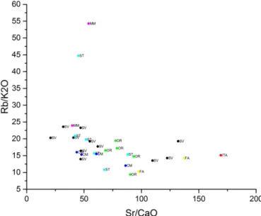
In sample 26, the smectite pattern is, in fact, the smectite–chlorite (PDF 07-0027) pattern. Fig. 4. Portable XRF data. Binary diagram Rb/K2O–Sr/CaO.
in sample SEV3 in Patio de Banderas site. According to the classification proposed by Maniatis & Tite[29]which sets 6% in weight of CaO as the percentage threshold to distinguish between calcareous and non-calcareous ceramics, 13 out of 17 Spanish samples and 9 out of 13 samples from Portugal can be considered as calcareous. Variations in CaO content may be ascribed to differences in the raw materials used (clays and sands), to intentional addition by the pottery workshops to produce objects with specific purposes[6,30,31]and/or to circulation of Ca-rich solutions within the pore spaces of the pottery fragments dur-ing their burial history[1]. The widespread presence (see BSEM + EDS section below) in both Spanish and Portuguese potsherds under inves-tigation of calcite (rarely dolomite) grains and of well preserved calcar-eous microfossils (foraminifera of the genus Globigerina) as main constituents of the temper fraction is supportive of thefirst hypothesis mentioned above. With a view to highlight compositional similarities between Spanish and Portuguese sites, a not so easy task bearing in mind the homogeneity of the used raw materials (estuarine river sediments) which in turn reflects the fairly homogeneous geological background across the Southern Portugal and Spain investigated sites, selected elemental binary and ternary plots have been used. Except for one sample from Santarém (ST20) and one from Monte Molião (MM26), both the ternary diagram Rb–Y–Sr (Fig. 3) and the Rb/K2O–
Sr/CaO binary diagram (Fig. 4) suggest a geochemical compatibility of the raw materials used in both Spanish and Portuguese samples. Sample
ST20 in fact distinguishes itself for the high Rb content of 227 ppm versus an average value of 69 ppm in the Spanish samples.
3.2. X-ray diffractometry
XRD results from 16 samples (6 from Patio de Banderas, 1 from Italica, 4 from Santarém, 2 from Castro Marim, 1 from Monte Molião and 2 from Faro) are summarised inTable 2.
Mineralogically, XRD spectra are fairly homogeneous across Andalu-sian and Portuguese samples. Quartz is confirmed as the main crystal-line phase present (abundant to very abundant) together with calcite, feldspars and phyllosilicates (mainly illites with minor illite/smectite and in one case, sample 10 from Patio de Banderas in Seville, smectite). Plagioclase feldspars are always more abundant than K-feldspars, this being especially true in Andalusian samples. Minor dolomite peaks are detectable in both Portuguese (Santarém, Monte Molião but not Faro and Castro Marim) and Patio de Banderas sherd fragments. With regards to kilnfiring temperature, the near absence of newly formed“high temperature” mineral phases such as diopside, gehlenite and mullite (diopside has been found only in two samples -sample SEV 7 from Patio de Banderas and sample ST20 from Santarém, together with the presence of well defined peaks belonging to carbonates (calcite and dolomite) and the absence of melting features in phyllosilicates
Fig. 9. BSEM + EDS ceramic paste. Illitic laths splitting along cleavage planes due to dehydroxilation processes duringfiring processes. Hematite crystals also visible (bright). Fig. 8. BSEM + EDS ceramic paste. Planktonic foraminifera fragment with well preserved fossil microstructure from Patio de Banderas site (Seville).
Fig. 7. BSEM + EDS ceramic paste. Scattered micron-size monazite grains within ceramic microfabric from Santarem site.
Fig. 6. BSEM + EDS ceramic paste. General view of ceramic paste showing bimodal size in ceramic microfabric.
typical of high temperaturefiring such as the development of mullite filled nanopores [10] suggests low firing conditions in the kilns (b900 °C). The widespread abundance of illite as the dominant clay mineral species is also in line with this hypothesis and may be used to fix the kiln temperature in the range N600 °C b950 °C [2,32]. Ti (rutile) and Fe oxides (hematite and maghemite) are present in all sam-ples as accessory minerals as well as Cu-oxides (cuprite). The Fe-oxides may represent newly formed phases during thefiring process and their coexistence is compatible with pottery manufacturing technique involving atmosphere changes duringfiring from oxidizing to reducing conditions[10].
3.3. Scanning-electron microscopy and micro-analysis
BSEM + EDS analysis has been used to describe the micro-texture of the ceramic paste and its relation with the black engobe surface layer and to identify the presence of individual mineralogical markers which due to their scarcity were undetectable by Portable XRF and XRD analyses.
3.3.1. Ceramic body
Under BSEM the ceramic paste in all samples displays afine-grained microfabric with a bimodal grain-size distribution (Figs. 5,6). While thefine-grained groundmass is constituted by micron-size laths of illite and plagioclase feldspars, the main temper components are mineral grains of quartz and calcite with (in decreasing order of abundance)
titanite, zircon (often zoned), ilmenite, rutile, monazite, hematite and clinopyroxene mineral grains as accessory constituents. In Portuguese samples from Santarém (sample 19) and Castro Marim (sample 18) and in three Andalusian samples (sample 8 from Patio das Banderas in Seville city centre, sample 12 from Italica and sample 16 from Orippo), small inclusions of gold are also found in the temper fraction whereas in two samples from Seville (samples 1 and 7), scattered Pb-rich grains are present. The detection of Au grains in Roman pottery from the Gua-dalquivir river valley region is not unexpected as gold can be present in alluvial placer deposits used as a pottery raw material source[11]. Pyrite, (FeS2) and chalcopyrite (CuFeS2) grains were found only in
sam-ples from Santarém and Castro Marim. Common temper components, albeit found only as scattered micron-size mineral grains, are also grains of monazite ((Ce,La,Y,Th)PO4;Fig. 7), a phosphate accessory mineral
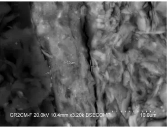
typically found influvial deposits in areas characterized by granitic geological substrates such as is the case in the SW Iberian areas under investigation[33]. Microfossils (planktonic foraminifera of the genus Globigerina) are also commonly found with their microstructure still well preserved (Fig. 8): the widespread presence of calcite grains to-gether with the preservation of the fossil outline and microstructure confirms XRD results in suggesting low firing temperatures (between 750 and 900 °C). The lowfiring temperature in the kiln used to produce the“Losanga” pottery under investigation is further confirmed by the absence of bright reaction rims formed by Ca-rich silicate phases (such as diopside, gehlenite and anorthitic plagioclases) which typically form in Ca-rich ceramics whenfired at higher temperatures[2]. In terms of microfabric porosity in the temper,fissures at quartz grain bound-aries as a result of shrinkage of clay minerals during drying and expan-sion of quartz crystals fromα to β phases during firing[10,34]are present, particularly in samples from Andalucia where secondary poros-ity typically with rounded pores can also be seen. Illitic laths while maintaining their sheet-like typical habit, show separation along basal cleavage planes (Fig. 9), a feature that has been interpreted as a result of de-hydroxylation during low temperaturefiring processes[10]. 3.3.2. Black gloss
A fairly continuous and homogeneous vitreous superficial engobe (thickness 13–16 μm) can be seen on both external and internal parts of all (but one) ceramic fragments (Fig. 10). With respect to the ceramic body, black gloss layers show Fe, K and Al enrichment and Si and Ca depletion with scattered micron-size Fe and Fe–Ti inclusions (Fig. 11). Thefine grain size of the surface dark coating suggests the application by elutriation of iron oxide-rich clays rather than a varnish as confirmed by the presence of magnetite (Fe3O4), maghemite (γ-Fe2O3), and
goethite (α-FeO(OH) in μ-XRD spectra which indicate highly reducing atmosphere andfiring temperature not exceeding 850 °C). μ-Raman spectroscopy confirmed the presence of magnetite and hematite miner-al species.
4. Conclusions
The chemical and mineralogical analyses of the tableware Losanga style fragments from Portuguese and Andalusian sites reveal the follow-ing common features: a) high Mg-calcite (rarely dolomite) content and high plagioclase/alkali feldspar ratio confirmed both by μ-XRD plots and by Portable XRF (high Sr and low Rb); b) abundance of well pre-served planktonic (Globigerinae) foraminifera microfossil fragments; c) widespread presence of illitic as a mineral marker in the groundmass of the ceramic paste; and d) presence of minor mineral markers in the ceramic paste such as monazite (Ce,La,Y,Th)PO4, pyrite FeS2,
chalcopy-rite CuFeS2, cuprite Cu2O and gold.
The mineralogical data of the ceramic paste, in particular the presence of calcite grains, the good preservation of calcitic microfossils and the near absence of secondary newly formed minerals such as diop-side, indicate a low technological process involving lowfiring tempera-ture in the kiln (b900 °C). Small differences between Portuguese and
Fig. 11. BSEM + EDS engobe. Micron-size Fe-rich inclusions. Fig. 10. BSEM + EDS engobe. Interface engobe–ceramic paste.
Andalusian ceramic fragments (rare presence in one Santarém sample of diopside indicating higherfiring T) could be related to variable quality standards amongst local workshops.
The multidisciplinary approach adopted in this study which combined archaeological evidence with detailed microchemical, microscopical and mineralogical analyses provided a fast and non-destructive (or micronon-destructive) tool to add“hard” data towards an-swering a long lived and still very much alive debate in the archaeology of“Campaniense” pottery production in SW Iberia.
The fairly homogeneous nature of the geological substrate and of the raw materials available in the Portuguese and Spanish sites investigated makes it hard to draw unequivocal conclusions on a common local production of the potsherds in the Guadalquivir. Further research will focus on both expanding the number of investigated sites and refining trace element data using other techniques such as ICP-MS in order to better define trade routes, production technology transfer mechanisms in this important region of the Roman Empire.
Acknowledgements
The authors would like to thank Prof. Enrique Garcia Vargas for the authorization to sample pottery fragments from the Patio de Banderas site and Dr. Concepción San Martín Montilla Head of the Departamento de Conservación e Investigación of Seville's Archaeological Museum del Museo Archeologico di Sevilla for access to pottery objects from Italica-Pajar de Artillo e Orippo-Cortijo de Tixe.
References
[1] J. Buxeda, I. Garrigós, V. Kilikoglou, P.M. Day, Chemical and mineralogical alteration of ceramics from the Late Bronze Age kiln at Kommos, Crete: the effect on the formation of a reference group, Archaeometry 43 (2001) 349–371.
[2] G. Cultrone, C. Rodríguez Navarro, E. Sebastián, O. Cazalla, M.J. De la Torre, Carbonate and silicate phase reactions during ceramicfiring, Eur. J. Mineral. 13 (2001) 621–634.
[3] P. Mirti, P. Davit, Technological characterization of Campanian pottery of type A, B and C and of regional products from ancient Calabria (southern Italy), Archaeometry 43 (1) (2001) 19–33.
[4] M.I. Dias, M.I. Prudêncio, On the importance of using scandium to normalize geochemical data preceding multivariate analyses applied to archaeometric pottery studies, Microchem. J. 88 (2008) 136–141.
[5] M.I. Dias, M.I. Prudêncio, M.A. Gouveia, M.J. Trindade, R. Marques, D. Franco, J. Raposo, C.S. Fabião, A. Guerra, Chemical tracers of Lusitanian amphorae kilns from the Tagus estuary (Portugal), J. Archaeol. Sci. 37 (2010) 784–798.
[6] G. Cultrone, E. Molina, C. Grifa, E. Sebastián, Iberian ceramic production from Basti (Baza, Spain):first geochemical, mineralogical and textural characterisation, Archaeometry 53 (2011) 340–363.
[7] N. Forster, P. Grave, N. Vickery, L. Kealhofer, Non-destructive analysis using PXRF: methodology and application to archaeological ceramics, X-Ray Spectrom. 40 (5) (2011) 389–398.
[8] R.J. Speakman, N.C. Littlea, D. Creelb, M.R. Millerc, J.G. Iñañeza, Sourcing ceramics with portable XRF spectrometers? A comparison with INAA using Mimbres pottery from the American Southwest, J. Archaeol. Sci. 38 (12) (2011) 3483–3496.
[9] B.F.O. Costa, A.J.M. Silva, A. Ramalho, G. Pereira, M. Ramos Silva, X-ray compositional microanalysis and diffraction studies of Haltern 70 amphorae sherds, X-Ray Spectrom. 41 (2012) 69–74.
[10]G. Cultrone, E. Molin, A. Arizzi, The combined use of petrographic, chemical and physical techniques to define the technological features of Iberian ceramics from the Canto Tortoso area (Granada, Spain), Ceram. Int. 40 (2014) 10803–10816.
[11] F. Nocete, R. Sáez, M.R. Bayona, J.M. Nieto, A. Peramoa, P. López, J.I. Gil-Ibarguchi, N. Inácio, S. García, J. Rodríguez, Gold in the southwest of the Iberian Peninsula during the 3rd millennium BC, J. Archeol. Sci. 41 (2014) 691–704.
[12] J.J. Ventura Martínez, La cerámica Campaniense en Andalucía Occidental(PhD thesis from Universidad de Sevilla) 1990.
[13]R. de Almeida, Ánforas del Guadalquivir en“Scallabis” (Santarém, Portugal): una aportación al conocimiento de los tipos minoritarios, Collecció Instrumenta, 28, Publicacions i Edicions de la Universitat de Barcelona, Barcelona, 2008.
[14]J. Principal, A. Ribera, El material más apreciado por los arqueólogos. La cerámica fina. La cerámica de barniz negro, Manual de cerámica romana. Del mundo Helenístico al Imperio Romano, Alcalá de Henares: Editores: Comunidad de Madrid, Museo Arqueológico Regional : Colegio de Doctores y Licenciados en Filosofía y Letras y en Ciencias de la Comunidad de Madrid 2013, pp. 43–146.
[15]J. Pimenta, V. Soria, H. Mendes, Cerâmicas de verniz negro itálico e imitações em pasta cinzenta de Monte dos Castelinhos— Vila Franca de Xira, CIRA Arqueologia, 32014 86–121.
[16] M.J. Ramos Suárez, E. García Vargas, Las imitaciones de vajilla de barniz negro itálico en el Bajo Guadalquivir, Comer a la moda— Imitaciones de vajilla de mesa en Turdetania y la Bética occidental durante la antigüedad (s. VI A.C.–VI D.C.), Collecció INSTRUMENTA, 46, Publicacions i Edicions de la Universitat de Barcelona, Barcelona 2014, pp. 239–269.
[17] V. Soria, Cerâmica campaniense a pasta cinzenta em território português, VI Encuentro de Arqueología del Suroeste Peninsular. Villafranca de los Barros, 4–6 octubre de 2012, Villafranca de los Barros: Editor Ayuntamiento de Villafranca de los Barros 2014, pp. 1361–1388.
[18] N. Lamboglia, Per una classificazione preliminare della ceramica campana, in Atti del I Congresso Internazionale di Studi Liguri, Bordighera (1952) 139–206.
[19] C. Alves, R. Mataloto, V. Soria, As produções de imitação da campaniense itálica em pasta cinzenta no Sul do território actualmente português, Actas del II Congresso Internacional da SECAH— Ex Officina Hispana 2 (Braga, 3–6 Abril 2013), tomo I 2014, pp. 113–124.
[20] L. Pedroni, Produzione e diffusione della ceramica calena“media”: problemi e ipotesi di lavoro, la ceramica de vernís negre del s s. II i I a.C.: centres productors mediterranis i comercialització a la Peninsula Ibérica, Mataró 2000, pp. 345–362.
[21] L. Pedroni, Ceramica calena a vernice nera, produzione e diffusione, Petruzzi Editore, 2001.
[22] J.P. Morel, La céramique à vernis noir du Maroc: une révision, Lixus. Actes du colloque de Larache (8–11 novembre 1989), École Française de Rome, Rome 1992, pp. 217–233.
[23] E. García Vargas, R. Almeida, H. González Cesteros, Los tipos anfóricos del Guadalqui-vir en el marco de los envases hispanos del siglo I a.C. Un universo heterogéneo entre la imitación y la estandarización, SPAL 20 (2011) 185–283.
[24]P. Berni Millet, Las ánforas de aceite de la Bética y Sua presencia el la Catalunia romana, Publicacions Universitat de Barcelona, Barcelona, 1998.
[25] E. de Sousa, A.M. Arruda, A gaditanização do Algarve, Mainake 32 (2) (2010) 951–974.
[26] M. Tarradell, Los fenicios en Occidente: nuevas perspectivas, Los Fenicios, Barcelona, 1967.
[27] R.J. Speakman, M.S. Shackley, Soil science and portable XRF in archaeology: a response to Frahm, J. Archaeol. Sci. 40 (2) (2013) 1435–1443.
[28] W. Horwitz, L.R. Kamps, K.W. Boyer, Quality assurance in the analysis of foods and trace constituents, J. Assoc. Off. Anal. Chem. 63 (1980) 1344–1354.
[29]Y. Maniatis, M.S. Tite, Technological examination of Neolithic–Bronze Age pottery from central and southeast European and from the Near East, J. Archaeol. Sci. 8 (1981) 59–76.
[30]R.J. Hoard, M.J. O'Brien, M. Ghazavy Khorasgany, V.S. Gopalaratnam, A material-science approach to understanding limestone-tempered pottery from the Midwest-ern United States, J. Archaeol. Sci. 22 (1995) 823–832.
[31] N.S. Müller, V. Kilikoglou, P.M. Day, G. Vekinis, The influence of temper shape on the mechanical properties of archaeological ceramics, J. Eur. Ceram. Soc. 30 (2010) 2457–2465.
[32] N. Schiavon, G.A. Mazzocchin, F. Baudo, Chemical and mineralogical characterisation of weathered historical bricks from the Venice lagoonal environment, Environ. Geol. 56 (2–3) (2008) 767–776.
[33] N. Schiavon, G.A. Mazzocchin, The provenance of sand in plasters from Roman wall paintings in the NE of Italy: a chemical mineralogical approach, Open Miner. J. 3 (2009) 32–39.
[34] V. Kilikoglou, G. Vekinis, Y. Maniatis, P.M. Day, Mechanical performance of quartz-tempered ceramics: part I, strength and toughness, Archaeometry 40 (1998) 261–279.