Sara Teresa Neves da Silva
Dissertation presented to obtain the Ph.D degree in Structural
Biochemistry
Instituto de Tecnologia Química e Biológica António Xavier | Universidade Nova de
Lisboa
Structural insights into the human
multifunctional protein RuvBL2
Photo taken after the oral dissertation and discussion, on the 27th November
2017. From left to right: Sabine Gorynia, Édouard Bertrand, Pedro Matias (supervisor), Sara Silva, Tiago Bandeiras (co-supervisor), Sandra Macedo-Ribeiro, Professor Maria Arménia Carrondo (president of the jury) and Teresa Santos-Silva.
Sara Teresa Neves da Silva
Dissertation presented to obtain the Ph.D degree in Structural
Biochemistry
Instituto de Tecnologia Química e Biológica António Xavier | Universidade Nova de Lisboa
Oeiras, November, 2017
Structural insights into the
human multifunctional protein
RuvBL2
The work described in the present dissertation concerns the study of the human multi-functional protein RuvB-Like 2. The main focus of this work was the elucidation of the crystallographic structure of this potential drug target, complemented by lower resolution techniques, in order to assess different conformations and elucidate the possible mode of action. These structural studies constitute the third chapter of this dissertation. The second chapter is constituted by functional analyses, necessary in order to gain an understanding of the characteristics of the target protein, both at the functional level, but especially with the aim of improving stability for the structural analyses.
This dissertation is therefore divided in four chapters. The first chapter includes a general introduction to the RuvB-Like area, including published results from all areas of study of these proteins. The purpose of this introduction is to review and correlate the results produced by the several groups studying RuvB-Like proteins, putting them in the cellular context. The second chapter includes an introduction to DNA-binding proteins, followed by a description of the results on DNA-binding properties of hsRuvBL1 and hsRuvBL2, and studies on the influence of tags on stability and oligomerization of hsRuvBL2. The third chapter focuses on structural analyses of hsRuvBL2, using X-ray crystallography, electron microscopy and small angle X-ray scattering. The final discussion aims to correlate the previously described results, and connect them with the state of the art, including data on the aggresome, produced with resource to protein produced during this work.
Acknowledgments
The work achieved in this dissertation was only possible due to the contribution of many people, to whom I am indebted:
I deeply thank Pedro Matias, my supervisor, for the support and guidance throughout these years, whenever help was needed or a question arose. For the support, understanding and patience during the most challenging times, which was deeply felt and appreciated, and for believing in my capacities.
I deeply thank my co-supervisor, Tiago Bandeiras, for the guidance and excellent teaching abilities. For the inspiration, provided during every scientific discussion, always with good mood and a joke, even during the (frequent) stressful times. For all the concern, patience and help, both visible and on the “backstage” during the most difficult times, which was always felt and appreciated.
Professor Maria Arménia, the Head of the Macromolecular Crystallography Unit, for always inspiring us to try new things and network with each other and with scientists outside the group, thus encouraging our scientific and personal development. For the concern and support throughout these years.
José Brito, for collecting the crystallographic data of hsRuvBL2 and for the help provided in the determination of the structure, always with attention to detail. For always finding the time to clarify my doubts, even more than once, with patience, great teaching skills and humour.
All my colleagues in the Macromolecular Crystallography Unit and in the Structural Biology for Drug Discovery group, for contributing to a fun and
collaborative working environment, where I learned much about work and life. In particular to Filipe Rollo, Bernardo Caniço, Adriana Temporão and Diana Silva, who arrived at the group while I was writing, for their friendship and support during this difficult process.
João Carita, for the large-scale production of biomass and help with cell disruption whenever needed.
Ricardo Coelho, for help provided during the (many) problems with the crystallization robot.
Bruno Correia and Colin Mcvey, for help with the implementation of the electrophoretic mobility shift assays.
Rocío Arranz, Carlos Óscar Sorzano and Roberto Melero, for the support provided in the electron microscopy experiments. Rocío Arranz and Jaime Benito, and the other members of the group of Structure of Macromolecular Assemblies and the Biocomputing unit, for receiving me in their groups.
Mark Tully, James Doutch and Robert Rambo, for support with the SAXS experiments.
Christine Ebel, for performing the analytical ultracentrifugation experiments and data analyses.
Phillipe Carpentier, for the support with the High Pressure cryocooling of crystals.
Hassan Berhali, for guidance during the controlled dehydration experiments at BM14.
Louise Bird, for the support with the high throughput cloning of c-Myc constructs.
Fundação para a Ciência e Tecnologia (FCT), Fundo Europeu de Desenvolvimento Regional (FEDER) (through COMPETE2020) and Instruct for funding.
A special thanks to my friends Diana Macedo, Pedro Silva and André Santos, the group formed in the beginning of our PhD classes from the geeks sitting in the front row, for the least productive, most fun working sessions.
Mariana, my best friend since we met when we were 3 years old, a big thank you for your support and for being so interested in my work and all things science. All my friends from outside the world of science, for their support and friendship.
A very special thank you to my family, my parents Belmira Teresa Pinguinhas and Manuel António Neves da Silva and my sister, Vera Teresa Neves da Silva, for your love, patience and support, which were a source of motivation.
Thesis publications
The work described in this dissertation resulted in two publications:
Zaarur N, Xu X, Lestienne P, Meriin AB, Mccomb M, Costello CE, Newnam GP, Ganti R, Romanova NV, Shanmugasundaram M, Silva STN, Bandeiras, TM, Matias PM, Lobachev KS, Lednev IK, Chernoff YO, Sherman MY; RuvbL1 and RuvbL2 enhance aggresome formation and disaggregate amyloid fibrils. EMBO J. 2015; 34: 2363–2382. doi:10.15252/embj
Silva STN, BritoJA, Arranz R, Sorzano CO, Ebel C, Doutch J, Tully M, Rambo R, Carazo JM, Carrascosa J, Matias PM, Bandeiras TM; Structure of human RuvB-Like 2 provides mechanism for coupling between ATP binding and mechanical action.
Dissertation Abstract
RuvB-Like transcription factors, RuvBL1 and RuvBL2, function in cell cycle regulation and development. They have been attributed the functions of chaperone, transcription regulator and helicase, sometimes in an ATP-dependent fashion, but just how these functions are regulated in each protein is still a mystery, that many groups have been working to understand. There is already a vast, albeit scattered amount of knowledge gathered about RuvBL1 and RuvBL2 proteins. However, the functions of these important proteins still need to be placed into the context of the various signalling pathways of which they are a part. This may be a daunting task, since the functions attributed to these proteins seem to be used in various combinations depending on the complex in which they are included. To add further complexity, these proteins can work separately or in complex, and in a way that seems to be regulated either by their oligomeric state, binding partners and/or post-translational modifications. RuvBL1 and RuvBL2 have been found to be associated with the aetiology of a number of cancers and other diseases, such as heart hyperplasia and ciliopathies. As such, with this work, our group strived to provide a contribution to this area, a contribution we hope will be useful particularly in the health and pharmaceutical industry areas, since the structure of human RuvBL2 determined during this thesis is a widely recognized potential drug target.
The path to a better understanding of any protein is through the observation of its structure, and that was the main purpose of this work: to obtain the atomic-resolution structure of the missing player in the human RuvB-Like family, RuvB-Like 2 (hsRuvBL2). We present the atomic structure of human RuvBL2 with a level of completion that provides novel insight into its biology. The
hsRuvBL2 structure resolves the mobile domain II, which is responsible for
binding may lead to domain II motion through interactions with the N-terminal loop and further show that inserted affinity tags affect hsRuvBL2 oligomerization and stability in solution. A comparison with its homolog hsRuvBL1 shows differences in surface charge distribution that may account for differences in regulation. Finally, single particle EM data reveals that single-stranded DNA can promote the oligomerization of monomeric hsRuvBL2. The structural information that was gathered in this work shows that hsRuvBL2 presents specific motifs at the surface level that allow hsRuvBL1 and hsRuvBL2 to be distinguished in the context of a signalling pathway. Furthermore, this was the first apo structure obtained for a eukaryotic RuvBL2. Comparison of our structure with the structures of ADP-bound truncated hsRuvBL2, and fungal ADP-ADP-bound ctRuvBL2 (Chaetomium
thermophilum), suggests a putative mode of action for the coupling of ATP binding
with a mechanical movement that could be the basis of ATP-dependent activities. Since crystallography provides a detailed, but somewhat static structural model, we used electron microscopy to gain some insight into the plasticity of the
hsRuvBL2 complex. Curious, although of still unknown biological relevance, is the
observation that hsRuvBL2 is also able to form heptamers.
We combined the obtained structural knowledge with DNA binding assays, since the DNA binding mode of RuvB-Like proteins is still a major focus of discussion, and largely unknown. Our results suggest that hsRuvBL2 must be monomeric at the onset of activity in order to bind DNA, since we could not observe a DNA electrophoretic shift in the presence of hexameric hsRuvBL2. While the low concentrations necessary to obtain monomeric hsRuvBL2 precluded the observation of DNA binding by electrophoretic mobility shift assay, electron microscopy allowed the observation that monomeric hsRuvBL2 associates into a ring, possibly a hexamer, in the presence of single-stranded DNA. EMSA assays further showed that hsRuvBL1 monomers are also able to bind ssDNA, and seem
to be able to confer to hexameric hsRuvBL2 the ability to bind ssDNA, or alternatively to form bridges between DNA and hexameric hsRuvBL2.
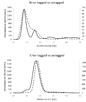
An analysis of the oligomerisation states of hsRuvBL2 with and without tags, shows that, as observed in yeast, tag presence and position interfere with the oligomerisation state. We also observed that they interfere with concentration-dependent oligomerisation state, such that C-terminally tagged hsRuvBL2 is always a hexamer, while the N-terminally tagged form varies its oligomerisation state depending on protein concentration.
Resumo da Dissertação
Os factores de transcrição da família RuvB-Like, RuvBL1 e RuvBL2, funcionam na regulação do ciclo celular e do desenvolvimento. A estas proteínas estão atribuídas as funções de chaperone, reguladores de transcrição e helicase, por vezes de forma dependente de ATP. No entanto, a forma como estas diferentes funções são reguladas é ainda um mistério, que diversos grupos tentam elucidar. Existe já um espólio vasto de informação sobre a RuvBL1 e RuvBL2. No entanto, as funções destas proteínas ainda têm de ser colocadas no contexto das diversas vias de sinalização das quais fazem parte. Esta pode ser uma tarefa árdua, já que as funções atribuídas a estas proteínas parecem ser usadas em diversas combinações, dependendo do complexo onde estão incluídas. A dificultar o processo, estas proteínas podem funcionar separadamente ou em complexo, e de uma forma que parece ser regulada pelo seu estado oligomérico, parceiros de ligação e/ou por modificações pós-translacionais. Tanto a RuvBL1 como a RuvBL2 estão associadas com a etiologia de vários cancros e outras doenças, como hiperplasia cardíaca e ciliopatias. Como tal, o nosso grupo pretendeu produzir uma pequena contribuição para a área de estudo destas proteínas, uma contribuição que esperamos que seja útil, em particular nas áreas de saúde e indústria, já que a estrutura da RuvBL2
humana determinada durante esta tese é um potencial alvo de compostos farmacológicos amplamente reconhecido.
O caminho para a compreensão do funcionamento de qualquer proteína passa pela análise da sua estrutura, e foi este o principal objectivo deste trabalho: a obtenção da estrutura de resolução atómica remanescente da família das RuvBLs, a RuvBL2 humana (hsRuvBL2). Aqui apresentamos a estrutura atómica da RuvBL2 humana suficientemente completa para providenciar nova informação quanto à sua biologia. A estrutura da hsRuvBL2 resolve o domínio II, um domínio móvel responsável por interacções proteína-proteína e regulação da actividade ATPase. Neste trabalho tentamos demonstrar de que forma a ligação de ATP pode levar à movimentação do domínio II através de interacções com o loop N-terminal, e demonstramos ainda que as tags de afinidade afectam a oligomerização e estabilidade da hsRuvBL2 em solução. Uma comparação com a homóloga
hsRuvBL1 demonstra diferenças na distribuição de cargas superficiais que podem
justificar algumas diferenças de regulação entre as duas proteínas. Finalmente, dados obtidos por microscopia electrónica revelam que a presença de ADN de cadeia simples promove a oligomerização de hsRuvBL2 monomérica. A informação estrutural recolhida neste trabalho demonstra que a hsRuvBL2 apresenta motivos específicos na sua superfície que permitem que seja distinguida da hsRuvBL1 no contexto de uma via de sinalização. Adicionalmente, esta foi a primeira estrutura apo obtida para uma RuvBL2 de um organismo eucariota. Uma comparação da estrutura aqui obtida com a estrutura da hsRuvBL2 truncada ligada a ADP, e com a estrutura da RuvBL2 de Chaetomium thermophilum, também ligada a ADP, sugere um potencial modo de acção que conecta a entrada de ATP com um movimento mecânico do domínio II que poderá ser a base de actividades dependentes de ATP. Visto que a cristalografia de raios-X providencia um modelo estrutural detalhado, porém algo estático, recorremos à microscopia electrónica para obter informação acerca da plasticidade do complexo hsRuvBL2. Observámos
que esta proteína é capaz de formar heptâmeros, um facto curioso mas de relevância biológica ainda indeterminada.
Combinámos o conhecimento estrutural obtido com ensaios de ligação ao ADN, já que o modo de ligação ao ADN das RuvBLs é ainda um foco de discussão, e pouco caracterizado. Os nossos resultados demonstram que a hsRuvBL2 tem de estar no estado monomérico no início da reacção, por forma a estabelecer ligação com o ADN, visto que não observámos variação na distância percorrida pelo ADN em gel na presença de hsRuvBL2 hexamérica, quando comparada com a distância percorrida pelo ADN isoladamente. Tendo em conta que as concentrações às quais a hsRuvBL2 é monomérica são demasiado baixas para permitir a observação de ligação por ensaios de variação de mobilidade electroforética em gel (EMSA), utilizámos microscopia electrónica para observar que a hsRuvBL2 monomérica se reorganiza em anéis na presença de ADN de cadeia simples. Os ensaios por EMSA mostraram ainda que monómeros de hsRuvBL1 são também capazes de se ligar ao ADN de cadeia simples, e parecem conferir à hsRuvBL2 hexamérica a capacidade de se ligar ao ADN, ou alternativamente podem actuar como pontes de ligação entre o ADN e o hexâmero de hsRuvBL2.
Uma análise das formas oligoméricas de hsRuvBL2 com e sem tags de afinidade demonstrou que a presença e posição das tags interferem com o estado de oligomerização. Observámos também que há uma influência na variação de estado oligomérico conforme a concentração de proteína: a hsRuvBL2 com tag C-terminal é sempre hexamérica, enquanto que a hsRuvBL2 com tag na extremidade N-terminal adquire diferentes estados oligomérico conforme a concentração a que se encontra.
Table of Contents
Chapter 1 - Introduction
1.1 A simplified view of the eukaryotic cell ... 4
The crowded cell 4
Eukaryotic cell compartmentalization 5
DNA packing and organization 8
1.1.3AAA+ PROTEINS 11
1.1.4HELICASES 16
1.1.5CHAPERONES 20
1.1.6RUVB AND RUVB-LIKE PROTEINS 25
1.1.7ROLES OF RUVBLS IN THE CELL AND CELL CYCLE 31
1.2 RuvBL1 and RuvBL2 in higher-order complexes ... 36
1.3 RuvBLs in development and disease ... 47
1.3.1DEVELOPMENT 47
1.3.2CANCER 49
1.3.3HYPOXIA 52
REFERENCES 55
Chapter 2 - Oligomerization and DNA-binding of RuvBL2;
Preliminary study of binding partner c-Myc
INTRODUCTION TO SSDNA-BINDING PROTEINS 69
2.1 hsRuvBL2 stability, oligomerization and DNA binding ... 72
2.1.1METHODS 72
Protein expression and purification 72
Differential scanning fluorimetry 73
Analytical ultracentrifugation 76
Small angle X-ray scattering 77
Electrophoretic mobility shift assays (EMSA) 78
Analysis of hsRuvBL2 binding to DNA by negative staining EM 79
2.1.2RESULTS AND DISCUSSION 80
Purification tags and nucleotides affect hsRuvBL2 stability and oligomerization
state 80
Stability of hsRuvBL2 oligomers varies with concentration 86 Insights into hsRuvBL1 and hsRuvBL2 binding to DNA 91
2.2 Preliminary study of RuvBL-interacting protein c-Myc ... 96
2.2.1METHODS 97
Construct design 97
Cloning procedures 99
Protein expression and purification 100
2.2.2RESULTS AND DISCUSSION 102
REFERENCES 104
Chapter 3 - Structure of human RuvBL2
3.1 Current structural knowledge on RuvB-Like proteins ... 108
3.2 Structure of RuvBL2 at 2.8 Å by X-ray crystallography ... 119
3.2.1METHODS 120
Protein production 120
Protein crystallization and crystal optimization 121
Data collection 127
Structure solution and refinement 128
3.2.2RESULTS AND DISCUSSION 131
Purification of RuvBL2 131
Crystallization of RuvB-Like 2 132
Initial attempts to solve the structure from low resolution data 135 Data collection of crystals diffracting to 2.8 Ångstrom 138
Atomic structure of full-length RuvB-Like 2 139
3.3 Structure of RuvBL2 by electron microscopy... 152
3.3.1METHODS 153
Protein production 153
Data collection - negative staining EM 155
Data processing and refinement of negative staining data 155
3.3.2RESULTS AND DISCUSSION 156
Low resolution structure of RuvBL2 by negative staining EM 156
REFERENCES 160
Chapter 4 - Discussion
4.1 Discussion ... 167
4.2 Concluding remarks and future perspectives ... 179
REFERENCES 183
Chapter 1
1.1 A
SIMPLIFIED VIEW OF THE EUKARYOTIC CELL... 4
The crowded cell 4
Eukaryotic cell compartmentalization 5
DNA packing and organization 8
1.1.3AAA+ PROTEINS 11
1.1.4HELICASES 16
1.1.5CHAPERONES 20
1.1.6RUVB AND RUVB-LIKE PROTEINS 25
1.1.7ROLES OF RUVBLS IN THE CELL AND CELL CYCLE 31
1.2 R
UVBL1
ANDR
UVBL2
IN HIGHER-
ORDER COMPLEXES... 36
1.3 R
UVBL
S IN DEVELOPMENT AND DISEASE... 47
1.3.1DEVELOPMENT 48
1.3.2CANCER 49
1.3.3HYPOXIA 52
This introduction aims to integrate relevant information presently available on RuvB-Like proteins with knowledge of the eukaryotic cell. The elucidation of the human RuvBL2 structure, determined in this work, will contribute to a better understanding of this protein family, which contains proteins that function both separately and in complex. Since they are deeply involved in such fundamental processes as development, chromatin remodelling and disease, including multiple cancer types, it is fundamental to understand their roles in the cell. In that way, one can follow the next step in regulating their activity with a better defined strategy in mind: the design of chemical compounds that will affect a particular activity, without interfering with their parallel workings inside the cell, a risk when targeting such a ubiquitous protein. Fortunately, this protein comprises discrete areas, targetable by multiple regulators, which may provide some clues as to where to begin, as long as it is possible to correlate the effect of targeting such areas to the outcome in the cell, and at the organism level.
1.1 A
SIMPLIFIED VIEW OF THE EUKARYOTIC CELL
The crowded cell
The eukaryotic cell environment is based on an aqueous moiety, with a high concentration of macromolecules (50-400 mg/mL), leading to macromolecular crowding effects, such as volume exclusion of reactants and reduced diffusion coefficients of macromolecules1. Such an overcrowded
environment precludes random movements and chance collisions as the basis for the protein-protein contacts needed for catalytic activities to occur. In fact, overcrowding promotes protein association and self-association of monomers2,3.
environment, and improves catalytic efficiency, through the formation of multi-subunit complexes composed of the enzymes that are part of a catalytic pathway. Many different proteins have been identified so far in such supramolecular assemblies, with varying degrees of binding affinities and complexity. RuvBL1 and RuvBL2 are two of those proteins that take part in various assemblies, either individually4–7 or as a heteromeric complex8–13.
Eukaryotic cell compartmentalization
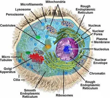
The function of a protein is strongly dictated by its sub-cellular location and by interactions with binding partners, which may also be correlated with the deposition of post-translational modifications (epigenetic markers). Additionally, the cell proteome distribution varies during the cell cycle, and a few genes are only transcribed for specific cell types. Furthermore, about one-third of its proteins are found in multiple organelles, which suggests roles in diverse and multiple pathways, such as is the case for RuvB-Like proteins. This wide distribution justified a brief description of the eukaryotic cell, for visual aid (Fig. 1.1).
The eukaryotic cell is highly compartmentalized, which confers functional segregation. It contains a true nucleus, delimited by a nuclear membrane. The inner membrane serves as an anchoring site for chromatin, and the outer membrane is continuous with the endoplasmic reticulum. Nuclear pores distributed across the membrane allow the exchange of macromolecules between the nucleus and the cytoplasm. The nucleoplasm supports the chromosomes and the nucleoli, and it is the place where transcription and replication events take place. The nucleoli are structures not bound by membranes, where ribosomes are synthesized, processed and assembled, a complex process controlled by nucleolar
substructures such as the fibrillar centre. The nucleoli also comprise proteins involved in stress responses and cell cycle regulation.
The cytoskeleton is composed of three types of fibres. The microtubules are the stiffest of the three, and are responsible for spindle formation during mitosis. Actin filaments are polarized, dynamic filaments with the ability to quickly assemble and disassemble, making them essential in the response to outside stimuli and in cell motility. As such, they are in direct contact with membrane-bound proteins and focal adhesions. The latter, also known as force-sensing and transducing complexes, are multi-subunit assemblies constructed by layers connecting actin filaments to the extracellular matrix. Functions of the different constituents of the focal adhesions start from receptor-matrix binding, linkage to actin and force transduction, intracellular signal transduction and actin
Figure 1.1 – Representation of the eukaryotic cell. Highlighted are the most relevant
organelles and cellular structures. Depicted from http://www.carolguze.com/text/102-7-eukaryoticcells.shtml.
polymerization and regulation. Two examples of components of focal adhesions are PI3Ks (phosphatidylinositol kinases) and Arp2/3 (actin related proteins 2 and 3). The Arp2/3 complex binds to the pointed ends of actin filaments, nucleating them, and facilitating the formation of actin-based protrusions, essential for motility. Finally, the intermediate filaments provide support to the cell and resistance to mechanical stress, and anchor the chromosomes to the inner part of the nuclear membrane, the lamina.
The cytoplasm comprises the cytosol, a semifluid liquid that contains mostly proteins, amino acids and ions, plus all non-nuclear organelles. The endoplasmic reticulum (ER) is a membrane-bound organelle, and the first step in the secretory pathway, together with the Golgi apparatus (GA)14. Vesicles are
small, enclosed organelles that are formed to create a dedicated environment to perform a variety of specialized functions, such as specialized metabolic reactions, transport, degradation of biomolecules and secretion. The centrosome is an organelle responsible for microtubule organization in the cell. It is composed of two centrioles, embedded in the pericentriolar matrix, and it is the microtubule organising centre (MTOC) of the cell during G1 and G2 phases of the cell cycle (see section 1.1.1). During mitosis the centrosome will mature into the spindle pole, and may form cilia by elongating microtubules15. The mitochondria are
involved in the production of energy in the form of ATP, are involved in cell death, signalling and differentiation, and are the only organelles to possess their own genome (in animals). Interestingly, it was recently shown that RuvBL2 possesses a mitochondrial targeting signal in one of it isoforms (isoform 3, Accession: NP_001308120.1), and indeed RuvBL2 and RuvBL1 were identified in mitochondria-enriched fractions. It has been suggested that, either by alternative splicing or alternative translation initiation, a form of RuvBL2 could be produced starting only at M46, thus activating this mitochondrial targeting signal. The
function of mitochondrial RuvBL2 isoform 3 is not yet clear, but it was shown to interact with the mitochondrial DNA polymerase gamma16.
Each cell is delimited by the plasma membrane, which provides a selective barrier with the outside environment. Proteins located in the membrane are responsible for cell to cell contacts, signal transduction, transport and adhesion to other cells or the extracellular matrix. Some proteins possess a signal peptide that promotes active transport out of the cell, and are thus secreted via the secretory pathway.
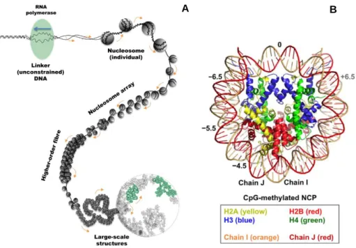
DNA packing and organization
Human chromosome sizes range between 50,000,000 and 300,000,000 bp.
Figure 1.2 – A) Structural organisation of genomic DNA. In order for the processing machinery to
access the DNA, a section of the genome has to be unconstrained (uncoiled), for which the surrounding will become supercoiled, up to a limit. From17. B) Crystallographic structure of the
nucleosome. Depicted from18.
In each cell, the DNA fibre is more than 2 meters long and can only be accommodated in the cell nucleus by iterative folding (Fig. 1.2A) into a condensed structure, the chromosome. Linear arrays of nucleosomes are coiled into a 30 nm fibre19, linked and stabilised by linker histone H1. The basic folding
unit - the nucleosome - is composed of 146 base pairs supercoiled in two turns around a histone octamer, composed of two H2A-H2B heterodimers and a H3-H4 tetramer (Fig. 1.2B), forming the fundamental unit of chromatin8,18,20.
Histones not only help in DNA packing, but can also be interchanged between different forms that bind more or less tightly to DNA, aiding in transcription regulation, together with DNA sequence and remodelling machinery. Histone post-translational modifications (PTMs) mark locations of double strand breaks and replication fork stalling, for further processing by the cellular repair machinery. After a DNA strand break or replication fork stall, canonical forms of H2A are replaced at the site of the break by H2AX, which contains a conserved SQ(E,D)(I,L,F,Y) motif at the carboxyl terminus, a common PI3K kinase (PIKK) phosphorylation motif. The serine residue is phosphorylated immediately after a double strand break, by PIKKs DNA-PKcs (DNA-dependent protein kinase catalytic subunit) and ATM (ataxia-telangiectasia mutated), forming a number of γ-H2AX foci that approximates the number of DSBs. H2AX phosphorylation triggers a series of molecular events that activate DNA repair response, and proteins such as BRCA1, Nbs1, Rad50 and Rad51 have been found to form foci that co-localize with γ-H2AX foci21,22. The consequence is an extensive
change in local chromatin composition, including acetylation, ubiquitination, potential H2A.Z deposition and eventually histone eviction, concomitant with end resection8. For example, the RuvBL-containing INO80 chromatin remodelling
complex is recruited by γ-H2AX to sites of replication fork stalling as cells enter the S-phase, and promotes efficient progression of replication by stabilising stalled replisomes. INO80 also functions in collaboration with SRCAP to replace
γ-H2AX with the H2AZ variant in case of DSBs23. This facilitates nucleosome
eviction prior to DNA strand repair, since H2AZ nucleosome structure and biochemical studies suggest that its association with DNA may be weaker than that of H2AX22. In yeast, Htz1 (homolog of human H2AZ) promotes deposition of
Gcn5 histone acetyltransferase, which acetylates histone H3 in response to UV stress, in Htz1-containing nucleosomes. Htz1 subsequently stabilises the interaction of Rad14 with damaged DNA, promoting nucleotide excision repair (NER) after UV irradiation24.
The mobility of a chromosomal locus varies with cell cycle stage and with chromatin status. DNA mobility, particularly at sites of double strand breaks (DSB), is not simply caused by Brownian motion, but is a controlled and regulated cellular process, promoted by enzymes. With about 53 different types of Snf2-type ATPases (enzymes that catalyse the hydrolysis of ATP to ADP with concomitant DNA/RNA remodelling) in humans, it is expected that each one will have different, even if maybe partially redundant, impacts on chromatin composition, mobility and structure which may, at least in the case of INO80, impact on chromatin dynamics. However, the activity of the DNA damage response (DDR) system and chromatin remodellers is not restricted to damaged sites, as checkpoint activation leads to an increase in chromatin mobility that is dependent on the INO80-dependent ATR (ataxia telangiectasia Rad3-related) kinase Mec1 and its downstream target kinase Rad538. In case of
difficult-to-repair DSBs and collapsed replication forks, DNA is relocalized from the nucleoplasm to the nuclear pore, an event that is dependent on the presence of histone H2A.Z, which implies an involvement of the RuvBL-containing SWR1 complex. In fact, depending on the DSB cause, chromatin re-localization may occur towards the nuclear pore or the membrane protein Mps3. This is dependent on the cell cycle stage, such that association of persistent DSBs with Mps3 (which suppresses recombination) requires Rad51 and end resection, which occurs only
in the S phase, while DSB association with the nuclear pore (which favours non-canonical recombination) occurs also in G1 phase8.
1.1.3
AAA
+ PROTEINSATPases associated with various cellular activities (AAA+ ATPases) are present in all domains of life, and eukaryotic genomes typically encode 50 – 100 different proteins belonging to this family25. The AAA+ ATPases fall within the
second major group of the P-loop NTPases, the ASCE (additional strand catalytic E, for the characteristic catalytic glutamate residue within the Walker B motif). Proteins of this superfamily use the energy obtained from ATP binding and hydrolysis to perform a variety of biochemical activities. Members of this superfamily include helicases, chaperones and regulatory components of proteolytic machines, which usually assemble into hexameric rings or helical structures (although pentameric, heptameric and octameric examples also exist)25.
As such, this superfamily comprises members involved in the normal maintenance of the cell, but also in stress response. The defining feature of the numerous members of this group is an ATP-binding module, which associates into arrays to originate a functional complex. ATP binding and hydrolysis events at the interface of neighbouring subunits lead to conformational changes within the AAA+ assembly that are responsible for the enzymatic activity of the complex26,27. The interface between protomers thus undergoes adjustments
according to the bound nucleotide state, while maintaining integrity of the complex. Interestingly, it was observed that of all the AAA+ ATPases analysed, RuvBL1 protomers had the largest surface contact area. In fact, the ATP binding pocket in RuvBL1 is so tightly occluded that it seemed to render impossible the exchange of the bound ADP, which could justify the observed low ATPase and
lack of helicase activities. It was suggested that a co-factor may be necessary to enable these activities, similarly to other members of this family25,28.
This large superfamily is characterized by the heterogeneity of its members’ structures and functions. However, there are some common structural features: all possess a core αβα nucleotide-binding domain and an adjacent α-helical domain, composed of two α-helical hairpins associated to form a left-handed superhelical structure (Fig. 1.3). The latter is a smaller domain, poorly conserved at the primary sequence level, but highly conserved structurally, such that it is a hallmark of the AAA+ ATPase superfamily. In these multimeric complexes, the ATP-binding site is located at the interface between neighbouring subunits: one protomer provides the main nucleotide binding pocket, composed of Walker A,
Figure 1.3– General structural features of the AAA+ ATPase core. A) The ATPase core is subdivided
into an αβα subdomain and a superhelical subdomain. B) Distribution of the canonical AAA+ motifs. C) Top and side views of the HsIU from Haemophilus influenzae. The superhelical “lid” domain (purple), locates to the outside of the ring structure. Additional helices (orange) form the I-domain, characteristic of HsIU-family proteins. From29.
Superhelical domain
Walker B, sensor I and sensor II motifs; the adjacent protomer contributes a conserved arginine (box VII, Second Region of Homology – SRH – or SRC motif)30. These conserved motifs all map to the AAA+ core αβα nucleotide fold,
except for sensor II, which is located in the helical subdomain, known as the AAA+ “lid”. An arginine “finger”, extending from one monomer to the adjacent one, completes the ATP binding pocket, and polarises the γ phosphate to facilitate hydrolysis. This important residue has also been shown to be responsible for oligomerization of hexameric ATPases, since its mutation abolished oligomerization, as well as ATP binding and hydrolysis and DNA translocation. Addition of wild-type monomers to the mutated ones resulted in dimer formation, since only part of the population was able to provide this residue to promote the interaction31,32. The sensor I motif is typically an Asn, but
can also be another polar residue, such as Ser, Thr or Asp; this motif participates in hydrolysis by coordinating the attacking water nucleophile, together with the Walker B residues. The Walker A motif has a consensus sequence GXXXXGK[T/S] (X represents any amino acid), in which the Lysine present on the P-loop (phosphate-binding loop) contacts directly the β and γ phosphates of ATP, making it critical for ATP binding. The Walker B motif consensus sequence is hhhhDE (h represents a hydrophobic amino acid). In this motif, the glutamate polarises a water molecule for in-line attack of the γ phosphate during ATP hydrolysis, and its mutation to glutamine or alanine abolishes this activity25,31.
These mutants can nevertheless be useful in order to obtain a “trapped” structure that may allow the observation of transient conformational states25,27,33,34. This
glutamate residue has a pivotal role in translation of nucleotide state to enzyme activation state. Indeed, analysis of several ADP and ATP bound AAA+ ATPases shows that two major conformational changes occur between the ATP and ADP complexes. The first major movement occurs in the glutamate sidechain itself, which rotates by approximately 100° in the ATP complex relative to its position in
the ADP complex, and forms a hydrogen bond with an asparagine residue in an adjacent strand. This “glutamate switch” thus traps the ATP-bound complex in a higher energy and inactive form, in contrast to the ADP-bound “active” form. The second major movement occurs in an area farther from the active site, and may involve loops that interact directly with the Glu-Asn pair, providing communication between the ATPase and ligand-binding sites. The known exceptions to date are the HslUV/Clp family proteases and the RuvB-Like helicases, in which the glutamate contacts a conserved threonine or serine residue instead of the Asn residue. This switch pair formation requirement upon ATP binding is suggested to be a way of repressing ATPase activity until there is ligand binding (a mechanism which is reversed in the cases where the ligand represses instead of activating)31.
Although AAA+ ATPases require ATP to perform activities that other enzymes can perform without, their ATP turnover is frequently poor in comparison with other ATPases such as hexokinase or even DNA helicases (turnover numbers are typically 0.1 to a few per second for AAA+ enzymes, versus several hundred per second for the “simpler” metabolic ATPases). Furthermore, the ATPase activity is often positively or negatively regulated by a ligand, and sometimes the same ligand can have opposite effects on different proteins. For instance, DNA binding leads to an increase in the ATPase activity of Replication Factor C, and yet represses ATPase activity of the Origin Recognition Complex. ATP hydrolysis suppression is necessary in complex enzymatic systems, with complex assembly pathways and multiple intermediate steps. ATPase activity is enabled at the point when ATP turnover is necessary for completion of the reaction or recycling of components. In this way, ATP binds during complex assembly, but its hydrolysis is suppressed until the complex is fully assembled31.
Sequence and structure comparisons show that AAA+ ATPases went through significant divergent events both before and after the emergence of the last common ancestor between bacteria, archaea and eukarya. Members of this superfamily can be subdivided into groups, clades and families, where the clade is defined by the distinct structural elements located within and around the AAA+ core. The five groups described are: Classic AAA ATPases (classical AAA clade or extended AAA group); helicases and clamp loaders (HEC); protease, chelatase, transcriptional activators and transport (PACTT); ExeA (type II secretory pathway ATPase); and signal transduction ATPases with numerous domains (STAND). RuvB-Like/Rvb proteins belong to the classical AAA group, which also includes NSF, CDC48, Pex, Bcs, proteasomal ATPase, katanin, Vps4, FtsH and Clp Domain 1 (d1) families. The last two families, as well as RuvBL, diverge from the others25,29. However, topological analysis of the ATP binding
pocket revealed RuvBL1 to have closer structural similarities with the HEC group (DnaA, Orc, CDC6 clade)25.
The great functional diversity of AAA+ proteins is a reflex of their diversity of accessory domains and co-factors, the diversity of their oligomeric assemblies and heterogeneity of residues that define key nucleotide-binding features. These variations define the specificity of AAA+ assemblies for different substrates and the mechanisms responsible for coupling conformational changes within the assembly. Interestingly, adaptor proteins have been identified which interact with the ATPase module to regulate its activity by increasing its target specificity, without necessarily compromising other activities of the ATPase. These adaptor proteins are an elegant way for the cell to strictly regulate the activity of AAA+ proteins by regulation of the adaptor proteins themselves. An example of an adaptor protein in higher eukaryotes is p47, which interacts with the AAA+ ATPase p97, regulating the p97-mediated fusion of Golgi and transitional endoplasmic reticulum membranes, as well as the growth of the
nuclear envelope. Interestingly, many adaptor proteins identified so far, despite their sequence variability, use the N-terminus of the ATPase as the interaction platform for binding and regulating the ATPase activity26.
1.1.4
H
ELICASESNucleic acid helicases are molecular motor proteins that couple the chemical hydrolysis of a triphosphate nucleotide with the unwinding of the energetically stable DNA or RNA duplexes. This process allows access to single stranded templates for proteins involved in transcription, replication, recombination and repair machineries. The mode of unwinding of each helicase depends on the structure and type of nucleic acid substrate. Helicases can thus be classified as DNA-DNA, DNA-RNA or RNA-RNA dependent NTPases. The interaction of helicases with each nucleic acid substrate may be regulated by other proteins that modulate their access to the substrate. Such regulatory mechanisms ensure that in vivo each helicase will be active only in the pathway and at the moment when they are required35. The first helicase was discovered in 1976 in Escherichia coli, and two years later another was found in an eukaryotic organism,
the lily flower. Since then, helicases have been found to be ubiquitous to all living organisms and many viruses36,37.
Most helicases share a few common biochemical features, namely nucleic acid binding, NTP binding and nucleic acid-stimulated hydrolysis of NTP. Helicases do not necessarily unwind nucleic acids, and those that do may or may not translocate along the strand. Most commonly, helicases bind preferentially to a ssDNA strand in a sequence-independent way, and from there they start to unwind the DNA double helix unidirectionally. A few exceptions preferentially bind dsDNA, such as RecBCD, simian virus 40 (SV40) large antigen and, notably, RuvB helicases36.
Helicase classification is made difficult by the complexity of existing systems, the variability of oligomerization states, nucleic acid specificity, translocation polarity, preference for single or double stranded DNA/RNA, and the addition of regulatory modules. The most recent classification system has been proposed by Singleton et al in 200738, which divides helicases into 6
superfamilies according to their most prevalent functional motifs and characteristics. All superfamilies have common motifs that mark them as helicases: the Walker A (G(x)4GKT) and Walker B (DExD/H) motifs, and the
“arginine finger”, that is involved in energy coupling. Superfamily 1 includes only bona fide monomeric ssDNA helicases, i.e., proteins that translocate along single stranded nucleic acids and catalyse the separation of double strands39.
Superfamily 2 is the largest, encompassing 10 intensively studied families, including DEAD-box RNA helicases, the RecQ-like and the Swi/Snf family (where the complexes SWR1, INO80 and RAD54 are included). Swi/Snf complexes are involved in several processes in the cell that are correlated with chromatin remodelling, such as replication, DNA repair, RNA polymerase regulation, cell cycle progression and tumour progression. The mechanisms employed by these large supramolecular complexes involve mostly histone manipulation, i.e., altering the structure of nucleosomes in order to change DNA accessibility. Chromatin remodelling complexes are composed of multiple subunits, including one member of the Swi/Snf family, and other accessory proteins. The INO80 complex, for instance, not only includes the SF2 helicase Ino80, but also the RuvBL1/2 dodecameric complex, and 12 other proteins40,41 (see section 1.3.2 for
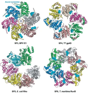
more information on chromatin remodelling complexes containing RuvBL proteins as one of the components). Superfamilies 3-6 include helicases that form hexamers and double hexamers: SF3 is composed entirely of viral helicases42, and
includes AAA+ proteins with multiple enzymatic activities, a 3’ to 5‘ processivity
origin binding domain, which allows viruses to bypass the host-cell regulatory mechanisms, and a characteristic C motif43 (Fig. 1.4); SF4 includes only helicases
with 5’ to 3’ polarity; SF5 is composed of Rho5, which while being closely related to SF4 helicases, was attributed to a different superfamily on the basis of differences in sequence (Fig. 1.4). It functions as transcription terminator, unwinding RNA:DNA duplexes and releasing newly-formed RNA transcripts44.
SF6 comprises only DNA helicases, unlike SF1-5, which contain both DNA and RNA helicases36. As in the case of SF3, members of this family have many
characteristics of AAA+ proteins (see section 1.1.3). However, SF6 members display also characteristic sensor 1 and 2 motifs (Fig. 1.4). Representative SF6 members include the mini chromosome maintenance (MCM) protein complex, which is the helicase component of the eukaryotic replicative holo-helicase CMG45, and prokaryotic RuvB (see section 1.1.6).
Figure 1.4 - Representative helicases from Superfamilies 3 to 6. Of notice is the general trend
Although the prokaryotic RuvB has been included into SF6, RuvB-Like proteins have not been allocated to any particular superfamily. Their structural characteristics support their inclusion in SF6, considering the presence of SF6-specific motifs Walker A and B, and sensor 1 and 2, as well as some of the biochemical characteristics known so far, namely their classification as AAA+ ATPase with multiple cellular functions and ssDNA specificity. Like MCM, RuvBLs interact with binding partners through the outer part of the ATPase core, in the latter case through the accessory domain II module46. Curiously, the Rho
helicase, the constituent member of SF5, was observed by EM to have an open “notched” conformation (Fig. 1.4), which is proposed to correspond to an open ring or absence of one subunit. This conformation was also observed by EM for RuvBL2 (unpublished result, see chapter 2, section 2.5.2). This diversity of characteristics among superfamilies creates a vast and complex number of systems, adapted to the particular needs of the cell.
Some proteins have been classified as helicases with basis on their sequence, displaying the characteristic helicase motifs, but do not perform the biochemical helicase activity. This is the case of SWI/SNF family, from SF2 (see section 1.2), which couples ATP binding and hydrolysis with chromatin remodelling through nucleosome rearrangements that alter chromatin accessibility47.
A genome-wide analysis of human helicases48 identified 64 RNA helicases
and 31 DNA helicases, including 5 RecQ members, KU70, 9 MCM (minichromosome maintenance) proteins, nucleolin, 2 chromodomain helicases, 2 DNA repair helicases, lymphoid-specific helicases, RuvB-Like 1 and 2, Pif1, Twinkle, BACH1, RecQ5 isoforms alpha, beta and gamma and regulator of telomere elongation helicase (RTEL1).
1.1.5
C
HAPERONESMost major processes that take place in the cell are performed by multi-component complexes, usually through energy-dependent conformational changes. Examples include complexes involved in chromatin remodelling, proteasomes and ribosomes. The complexity of such structures foretells the need for specific support in the assembly process. The help of “molecular matchmakers”, able to use the energy gained from ATP hydrolysis to induce conformational changes in one or both elements of a molecular pair, was initially described for protein-DNA complex formation49. However, since then this
concept has been expanded to include other types of complexes, such as multi-protein complexes50. Chaperones can thus be ascribed the function of promoting
the non-covalent assembly of other proteins or protein complexes. Chaperones may also interact with other proteins to prevent aggregation and/or promote proper folding, many times in an ATP-dependent manner. Many evolutionarily related chaperones belong to the AAA+ family, and use ATP-dependent conformational alterations in the ATPase core to perform their functions. Some AAA+ proteins still specialise in disassembly, remodelling or disaggregation, acting either in concert with a proteasomal machine for further target degradation, or by themselves51. Many of these proteins share a hexameric
structure and a conserved protein-processing pore. However, it has been observed that they do not necessarily exert their disassembling/remodelling functions through translocation-dependent unfolding, but may do so through other ATP-dependent interactions with the target protein (e. g. the σ54-activating enzymes)51.
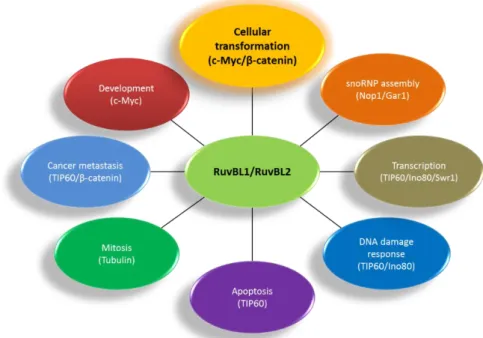
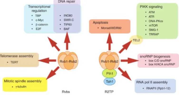
RuvBL proteins are fundamentally involved in various mechanisms that implicate a chaperone function. They are involved in the assembly of chromatin remodelling complexes INO80, SWR/SRCAP and TIP60, but not as the catalytic
subunit. In all cases the RuvBL1/2 complex seems to have a scaffolding function12.
However, as part of the larger SWR/SRCAP complexes, responsible for histone exchange, the RuvBL1/2 complex was found to have not only a scaffolding function, but also to be able to perform in vitro by itself the exchange of canonical histone H2A for the more labile H2A.Z52, another function added to the panoply
of activities already identified for these versatile proteins.
As part of the R2TP complex, RuvBL1/2 participates in the assembly of box C/D and box H/ACA snoRNPs. R2TP is involved in the assembly of core factors in both snoRNPs, but recruits specific factors in each family. These factors chaperone the core components as they are assembled, regulate the assembly and disassembly of pre-snoRNP intermediates and regulate the activity of intermediate subcomplexes53,54. In eukaryotes and archaea, box C/D snoRNAs
modify rRNAs and possibly mRNAs, and box H/ACA snoRNAs have motifs included in the RNA sequence of telomerase, a complex that works in the synthesis of telomeric DNA54. As a chaperone in snoRNPs assembly, the R2TP
complex regulates ribosome biogenesis and consequently controls cell proliferation.
In mammalian cells, R2TP interacts through RuvBL1/2 with intermediate H/ACA snoRNP core components and their co-chaperones, namely SHQ1 and dyskerin/NAP57, dissociating them for subsequent assembly in the holo-complex. The dyskerin interaction is suggested to make use of the RuvBL1/2 central channel, which seems fit to thread the unstructured dyskerin C-terminus. This tail is not necessary for binding to RuvBL1/2, but it is necessary for separation of dyskerin from SHQ1, which further suggests a mechanism of action whereby the complex “grabs” both proteins and follows with a rotational movement of the barrel-like structure, which could be responsible for the forceful separation of both proteins. This possibility may be supported by the fact that point mutations in the Walker A and Walker B motifs of RuvBL2 lead to severe defects in snoRNA
accumulation55. This model seems to be applicable also to C/D core proteins, since
some of their components possess similarly unstructured, highly charged tails, which are essential for yeast survival and for mammalian nucleolus maturation11,12. The biogenesis of box C/D snoRNPs is mediated by the
R2TP/HSP90 complex and other assembly factors. These assembly factors are as of yet uncharacterized, although pull-down assays performed in yeast have identified TAF9, NOP17 and BCD1 as intermediate factors, which are not present in the mature snoRNP. Of these, NOP17 and BCD1 have been shown to interact directly with RuvBL1 and RuvBL2, respectively56. Interestingly, interaction of
R2TP with box C/D snoRNPs was shown to decrease in poorly growing cells, as R2TP re-localizes from the nucleus to the cytoplasm, depending on the cell growth phase and nutrient condition57.
All core components of H/ACA snoRNPs also assemble with TERC (Telomerase RNA component) to form TERC-containing RNP, in a process involving the RuvBL1/2:dyskerin complex and dependent on the ATPase activity of RuvBL1. In addition to TERC, the Telomerase complex includes TERT (Telomerase reverse transcriptase) and the TERC-binding protein dyskerin. As part of the RuvBL1/2 complex, RuvBL1 was shown to interact directly both with TERT and with dyskerin. Telomerase holoenzyme adds DNA repeats to telomeres, nucleoprotein structures that protect the termini of chromosomes. In cancer cells, Telomerase activity is upregulated, conferring immortality by protecting chromosome ends indefinitely, contrary to the gradual loss of activity observed in normal cells. Both RuvBL1 and RuvBL2 were shown to be essential for Telomerase activity and for the accumulation of TERC and dyskerin, as depletion of RuvBL1, RuvBL2 or dyskerin leads to loss of TERC. Furthermore, the low-activity TERT:RuvBL1/2 complex seems to be highly abundant relative to the highly active TERT:TERC:dyskerin complex, which suggests that the former may also be a target for regulation, or that its assembly may require some time.
Interestingly, the TERT:RuvBL1/2 complex is especially abundant during S phase, a time-dependent association that may justify the cell cycle-dependence of Telomerase formation58. Additionally, since RuvBL1 and RuvBL2 are involved in
maintenance of dyskerin levels, they could also be targets of interest in congenital diskeratosis, a disease caused by low activity of dyskerin and decreased TERC levels, which results in low telomerase activity and much shorter telomere length59. It is further suggested that epigenetic modifications of RuvBL1 and
RuvBL2 may be an interesting venue of study in the context of Telomerase activity regulation, since it has been observed that, e. g., SUMOylation of RuvBL1 and RuvBL2 modifies their transcriptional activity and protein-protein interaction59–61.
Still as components of R2TP, RuvBL1 and RuvBL2 are involved in the assembly and stability of PIKKs, via the concerted interaction of the R2TP:HSP90/Prefoldin-Like complex, the TTT complex and the PIKKs12.
The RuvBL1/2 complex was also recently shown to have chaperone functions in protein homeostasis, under stress conditions as yet unidentified (other than heat shock or proteasome inhibition, which did not induce RuvBL expression). In this role, RuvBL1/2 complex was shown to promote the formation of an organelle, the aggresome, which functions as storage compartment for aggregated proteins, and possibly also in their proteasomal or autophagic degradation62. This organelle is formed as an alternative to the
ubiquitin-proteasome degradation pathway, when the accumulation of aggregates becomes too extensive63, and indeed overexpression of RuvBL1 and RuvBL2 protected
yeast cells exposed to proteotoxic heat stress. Furthermore, depletion of either RuvBL1 or RuvBL2 suppressed aggresome formation in mammalian cells expressing aggregate-forming synphilin – a substrate of the aggresome. To help elucidate the role of RuvBL1/2 complex in this process, we collaborated with the group of Michael Sherman, by providing RuvBL1/2 full-length complex, which
was used for interaction assays with synphilin (since this was the total of our contribution to this study, and occupied only a small portion of the total duration of my PhD, the RuvBL1/2 complex purification protocol is only briefly described in the discussion section). It was thus assessed that RuvBL chaperone activity was independent of R2TP complex (see sections 1.1.7 and 1.2), and that neither RuvBL1 nor RuvBL2 had the ability to promote aggresome formation by themselves, but that the RuvBL1/2 complex formation was necessary for this function. This is consistent with their dodecameric, barrel-like structure, as well as their inner channel charge distribution, which as a mix of positive and negative charges from both types of monomers, supports a possible association with a polypeptide chain. Co-immunoprecipitation followed by mass spectrometry identified both RuvBL1 and RuvBL2 as interacting with synphilin. Interestingly, this interaction occurs between the ankyrin repeat domain of synphilin, which is the domain responsible for its aggregation, and K372 of RuvBL1, which is located on the surface of the ATPase core and in close proximity to the central channel. This interaction is critical for assembly of the aggresome. In line with this, while in naïve cells, RuvBLs are distributed throughout the cytoplasm and nucleus, after proteasome inhibition they were recruited to protein aggregates and then transported to the aggresome. Interestingly, ATPase activity was significantly stimulated also by other amyloid-forming proteins, namely insulin and casein fibrils, an increase more noticeable in the domain II-truncated complex. In fact, even denatured BSA potently enhanced this activity, in contrast to the native form, which had no effect. Finally, it was concluded that the main effect of RuvBLs is actually on disaggregation, rather than recruitment of aggregates to the aggresome, since disassembly of aggresomes is strongly suppressed by RuvBL1 depletion62.
1.1.6
R
UVB
ANDR
UVB-L
IKE PROTEINSRuvBL1/Pontin/Rvb1/Tip49 was initially discovered in 1997 in rat, in association with the TATA-binding box protein (TBP), by Masato Kanemaki, from the group of Taka-Aki Tamura64,65. TBP is a general transcription factor that works
at the core of multiprotein transcription factors, necessary for recruitment of all classes of RNA polymerases, which binds at the TATA box promoter element66. In
1998 RuvBL1 was found as part of a complex with RuvBL2/Reptin/Rvb2/Tip48, associated with the RNA polymerase II holoenzyme, and later in 1999 RuvBL2 was found as part of a complex with RuvBL1 alone in human cells67.
RuvB-Like proteins are eukaryotic proteins with partial structural homology to bacterial RuvB (Resistance to UV) proteins (Fig. 1.5), the ATP-dependent motors of the RuvAB complex that drive branch migration of Holliday junctions formed during homologous recombination77. The RuvA, RuvB and
Table 1.1 – Different RuvBL names and their origins.
RuvBL1 RuvBL2 Name origin
TIP49 TIP48 TATA-binding protein (TBP)-interacting protein68,69
TIP49a TIP49b TBP-interacting protein67
Pontin52 Reptin52 Repressing Pontin526
TAP54α TAP54β TIP60-associated protein70
TIH1 TIH2 TIP49a/b homolog71
ECP54 ECP51 erythrocyte cytosolic protein72
NMP238 --- nuclear matrix protein73
Rvb1 Rvb2 RuvB homolog74
p50 p47 protein75
RuvC proteins are necessary for normal levels of cellular resistance to UV or ionizing radiation, or of mitomycin. Based on this partial homology, these proteins were immediately assumed to be ATP-dependent DNA helicases, and indeed they were shown to have helicase as well as ATPase activities in vitro67,78.
Interestingly, RuvB-Like amino acid sequences are highly conserved not only in eukaryotes but also in Archaea, highlighting them as some of the most conserved nuclear dynamics-related proteins. Phylogenetic analyses further showed that the analysed archaea possess only one RuvBL copy, belonging to the RuvBL2 branch. Moreover, bacterial RuvB was shown to be closest to RuvBL2 than RuvBL1. Together, these facts suggest that RuvBL2 is the common ancestor of the RuvBL family, from which RuvBL1 would diverge after the emergence of eukaryotes.67
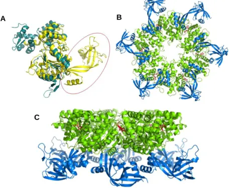
Figure 1.5 – Main structural features of RuvB-Like proteins. A - Structural alignment of human
RuvBL2 (yellow) and bacterial RuvB (blue). The red circle highlights the extra domain II in RuvBL proteins. B – Bottom view of the RuvBL1 hexamer. The central ATPase core (green) is formed by domains 1 and 3, and displays a central channel. The extra domain II from each monomer (blue) protrudes outwards, producing 6 mobile arms. C – Side view of RuvBL1 hexamer. ATPase core in green, with bound nucleotide in red. Domains 2 in blue.
A B
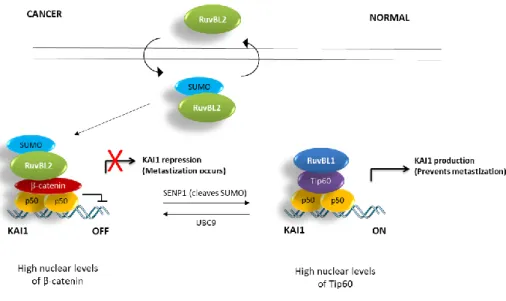
This raises the question for the need for two highly similar proteins, which often work together. It is possible that this need arose as organisms became more complex, in order to fine-tune more complex pathways, such as the Wnt/β-catenin signalling pathway, or pathways responsible for cell adherence (possibly during the transition to multicellularity), as can be observed in their antagonistic regulation of metastization processes by controlling the expression of metastasis Suppressor gene KAI161,79.
RuvB hexamers contain only an ATPase core, and associate to form double rings through the mediation of RuvA octamers previously primed through interaction with a Holliday junction80. Compared to prokaryotic RuvB,
RuvB-Like proteins comprise an extra domain II, located between domains I and III, encircled in fig. 1.5A (interestingly, domain II of RuvBL proteins locates spatially to roughly the same position occupied by RuvA in relation to RuvB80,
such that a double RuvB ring acquires conformation similar to a RuvBL1/2 double ring81–83). Domains I and III form an ATPase core, responsible for nucleotide
binding and hydrolysis (in green in figs. 1.5B and 1.5C) whereas domain II forms a mobile arm that protrudes from the more rigid core, such that in a ring the ATPase core faces a central channel and six mobile domains protrude outwards. RuvBLs are usually found in the cell as single or double hexameric rings, with a 1:1 proportion of each9,84. Whether each ring is constituted of a mixture of RuvBL1
and RuvBL2 or homomeric still under debate, since the resolution of the structures obtained by cryo-electron microscopy (cryo-EM) does not yet allow a clear distinction between RuvBL1 and RuvBL2. It is also not always clear whether the contact between rings occurs between the ATPase cores or through the domains II. However, it is now mostly accepted that the latter interaction is probably the most frequent, since both EM and crystallographic structures of dodecamers obtained to date show double hexamers interacting through the domains DII, as well as alternating RuvBL1 and RuvBL2 monomers in the
hexameric rings. Nevertheless, both proteins are able to form homohexamers in vitro28,85. Oligomer formation was shown to be dependent on the presence of the
Walker A and Walker B motifs. However, these were not necessary for binding to other proteins through domain II7.
RuvBL1 is encoded in chromosome location 3q21, a region of frequent rearrangements in leukaemia and solid tumours86,87. RuvBL2 is encoded in
location 19q13.33 of chromosome 19, and is physically linked with the breast
cancer-related CGB/LHB gene cluster88. This chromosome has more than double
Figure 1.6 - RuvBL2 protein expression data, identified by specific antibody CAB012432, from Abcam (product 36569). Top: Expression levels in several organs. Bottom: expression levels in
cancer vs. normal cells. Dark blue: high expression levels; light blue: medium expression levels. The bars indicate the proportion of high vs. normal levels of RuvBL2 identified in a total number of analysed samples, for each cancer type. From the protein atlas annotation project.