Michael Martins Vaz
Licenciado em Biologia Celular e Molecular
Sol-gel entrapped biosystems:
enzyme(s) and whole cells
Dissertação para obtenção do Grau de Mestre em Biotecnologia
Orientador: Susana Barreiros,
Professora Associada com Agregação, FCT/UNL
Co-orientadores: Isabel de Sá-Nogueira,
Professora Associada com Agregação, FCT/UNL
Eurico Cabrita,
Professor Auxiliar, FCT/UNL
Júri:
Presidente: Prof. Doutor Carlos Alberto Gomes Salgueiro
Arguente: Doutora Ana Sofia Diogo Ferreira
Vogal: Prof. Doutora Susana Filipe Barreiros
Setembro de 2014
i
Michael Martins Vaz
Licenciado em Biologia Celular e Molecular
Sol-gel entrapped biosystems:
enzyme(s) and whole cells
Dissertação para obtenção do Grau de Mestre em Biotecnologia
Orientador: Susana Barreiros,
Professora Associada com Agregação, FCT/UNL
Co-orientadores: Isabel de Sá-Nogueira,
Professora Associada com Agregação, FCT/UNL
Eurico Cabrita,
Professor Auxiliar, FCT/UNL
Júri:
Presidente: Prof. Doutor Carlos Alberto Gomes Salgueiro
Arguente: Doutora Ana Sofia Diogo Ferreira
iii
Copyright Michael Martins Vaz, FCT/UNL, UNL
A Faculdade de Ciências e Tecnologia e a Universidade Nova de Lisboa têm o direito, perpétuo e sem limites geográficos, de arquivar e publicar esta dissertação através de exemplares impressos reproduzidos em papel ou de forma digital, ou por qualquer outro meio conhecido ou que venha a ser inventado, e de a divulgar através de repositórios científicos e de admitir a sua cópia e distribuição com objetivos educacionais ou de investigação, não comerciais, desde que seja dado crédito ao autor e editor.
v
Acknowledgments
The accomplishment of this work was only possible with the help of all the people from the groups I worked with and also with the support of my family and friends.
For this I would like to acknowledge in first place to Professor Susana Barreiros for all the outstanding orientation in this work, for all the support and kind advices during these last months and for all the time spent towards the conclusion of this thesis. To Professor Isabel de Sá-Nogueira for orienting me in all the biologic procedures and for the availability. To Professor Eurico Cabrita for performing all the NMR experiments and for teaching me the basics of this technique.
To all my colleagues from laboratory 427, that were always available for any problem and for all the good mood that they bring to the workplace. To Francisca Mano, Luiza Silva, Sandra Abrantes and José Vieira, that were exceptional colleagues and friends during our thesis development.
To my colleagues from laboratory 327, Mário Ferreira and Lia Godinho, for all the help in the laboratory, availability to discuss issues (pertinent or not) and for their willingness during the time I was present. To Aristides Mendes for the friendship, discussions, motivation speeches and also for the willingness at all moments, good or bad.
To Ana Sofia Ferreira and Marta Corvo for helping with NMR related issues.
To Vanessa for being at my side in all the moments of this thesis, for all the love and support that really gave me motivation to keep carrying on the work.
To my sister Sónia, for all the support and for being herself. To my parents that gave me the possibility to study and to achieve this academic goal, for all the support they always gave me and for the love and comprehension they had all these years.
To all my friends, for just being there.
Finally I would like to acknowledge Fundação para a Ciência e a Tecnologia (FCT/MEC) that partially funded this work through projects PEst-OE/BIA/UI0457/2013 and PEst-C/EQB/LA0006/2013. To all of you (and for all whom I may have forgotten) my sincere gratitude,
vii
Abstract
In this work two different procedures to utilize the sol-gel technology were applied to immobilize/encapsulate enzymes and living cells.
CO2 has reached levels in the atmosphere that make it a pollutant. New methods to utilize this gas to
obtain products of added value can be very important, both from an environmentally point of view and from an economic standpoint. The first goal of this work was to study the first reaction of a sequential, three-step, enzymatic process that carries out the conversion of CO2 to methanol. Of the three
oxidoreductases involved, our focus was on formate dehydrogenase (FateDH) that converts CO2 to
formate. This reaction requires the presence of the cofactor β-nicotinamide adenine dinucleotide in reduced form (NADH). The cofactor is expensive and unstable. Our experiments were directed towards generating NADH from its oxidized form (NAD+), using glutamate dehydrogenase (GDH). The formation of NADH from NAD+ in aqueous medium was studied with both free and sol-gel entrapped GDH. This reaction was then followed by the conversion of CO2 to formate, catalysed by free or
sol-gel entrapped FateDH. The quantification of NADH/NAD+ was made using UV/Vis spectroscopy. Our results showed that it was possible to couple the GDH-catalyzed generation of the cofactor NADH with the FateDH-catalyzed conversion of CO2, as confirmed by the detection of formate in the medium,
using High Performance Liquid Chromatography (HPLC).
The immobilization of living cells can be advantageous from the standpoint of ease of recovery, reutilization and physical separation from the medium. Also dead cells may not always exhibit enzymatic activities found with living cells. In this work cell encapsulation was performed using Escherichia coli bacteria. To reduce toxicity for living organisms, the sol-gel method was different than for enzymes, and involved the use of aqueous-based precursors. Initial encapsulation experiments and viability tests were carried out with E. coli K12. Our results showed that sol-gel entrapment of the cells was achieved, and that cell viability could be increased with additives, namely betaine that led to greater viability improvement and was selected for further studies. For an approach to “in-cell” Nuclear Magnetic Resonance (NMR) experiments, the expression of the protein ctCBM11 was performed in E. coli BL21. It was possible to obtain an NMR signal from the entrapped cells, a considerable proportion of which remained alive after the NMR experiments. However, it was not possible to obtain a distinctive NMR signal from the target protein to distinguish it from the other proteins in the cell.
ix
Resumo
Neste trabalho foram utilizados dois procedimentos para imobilizar/encapsular enzimas e células vivas utilizando a tecnologia sol-gel.
O CO2 atingiu níveis na atmosfera que o tornam um poluente. Métodos para utilizar este gás na obtenção
de produtos de valor acrescentado podem ser muito importantes, do ponto de vista ambiental e económico. O primeiro objetivo deste trabalho foi estudar a primeira reação de um processo enzimático sequencial, em três passos, que realiza a conversão de CO2 em metanol. Das três oxidoredutases
envolvidas, o nosso foco foi a enzima formato desidrogenase (FateDH) que converte CO2 em formato.
Esta reacção requer o cofator β-nicotinamida adenina dinucleotide na sua forma reduzida (NADH). O cofactor é caro e instável. As nossas experiencias foram direcionadas para gerar NADH a partir da sua forma oxidada (NAD+), utilizando glutamato desidrogenase (GDH). A formação de NADH a partir de NAD+ em meio aquoso foi estudada com GDH livre e também imobilizada em sol-gel. Esta reacção foi secundada pela conversão de CO2 em formato, catalisada pela FateDH livre ou imobilizada em
sol-gel. A quantificação de NADH/NAD+ foi feita por espectroscopia UV/Vis. Os nossos resultados mostram que foi possível acoplar a formação do cofator NADH catalisada pela GDH com a conversão de CO2 catalisada pela FateDH, tal como confirmado pela detecção de formato no meio, utilizando
Cromatografia Líquida (HPLC).
A imobilização de células vivas pode ser vantajosa do ponto de vista da facilidade de recuperação, reutilização e separação física do meio. Além disso as células mortas podem não manifestar algumas das actividades enzimáticas que as células vivas exibem. Neste trabalho encapsularam-se células da bactéria Escherichia coli. Para reduzir a toxicidade do método sol-gel para os organismos vivos, utilizou-se um protocolo diferente do seguido com enzimas, envolvendo neste caso a utilização de soluções aquosas de precursores. Os primeiros ensaios de encapsulação e testes de viabilidade foram feitos com E. coli K12. Os nossos resultados mostram que as células foram encapsuladas e que a viabilidade celular podia ser aumentada com o recurso a aditivos, nomeadamente betaína que conduziu aos melhores resultados e foi selecionada para os estudos seguintes. Com vista a uma abordagem de Ressonância Magnética Nuclear (RMN) “in-cell”, expressou-se a proteína ctCBM11 em E. coli BL21. Obteve-se um sinal de RMN das células encapsuladas, uma quantidade considerável das quais se mantiveram vivas após os ensaios de RMN. No entanto, não foi possível obter um sinal característico da proteína alvo que permitisse distingui-la das outras proteínas da célula.
xi
Table of Contents
Acknowledgments ... v
Abstract ... vii
Resumo ... ix
List of Figures ... xiii
List of Tables ... xvii
List of Symbols and Abbreviations... xix
1. Introduction ... 1 1.1. CO2 as a pollutant ... 1 1.2. CO2 conversion to methanol ... 4 1.3. Biocatalysis ... 5 1.4. Oxidoreductases ... 7 1.5. Bioconversion of CO2 to methanol... 7 1.6. Ionic Liquids ... 10 1.7. Sol-gel method ... 12
1.8. The sol-gel approach for cell immobilization ... 14
1.9. Preserving cell viability within silica gels ... 18
1.9.1. The starting solution... 18
1.9.2. The gelation reaction ... 20
1.9.3. Gel ageing ... 21
1.10. Microorganisms used in the present work ... 22
1.10.1. Escherichia coli... 22
1.10.2. Carbohydrate Binding Modules ... 23
1.11. Nuclear Magnetic Resonance (NMR) ... 24
1.12. In cell NMR ... 26
1.13. Thesis Goals ... 28
2. Materials and methods ... 29
2.1. Sol-gel immobilization of enzymes ... 29
2.2. Quantification of NADH and CO2 reduction ... 30
2.3. Bacterial strains and growth conditions ... 31
2.4. Sol-gel immobilization of cells ... 32
2.5. Viability tests ... 32
2.6. Nuclear Magnetic Resonance (NMR) ... 33
xii
3.1. Enzymatic system ... 35
3.2. Cell immobilization ... 43
4. Conclusions and Future work ... 59
xiii
List of Figures
Figure 1.1 – Carbon cycle. Adapted from[2] ... 2 Figure 1.2 – Global carbon emissions and principal contributors The black line shows the high increase of carbon emission through time. Adapted from [3]..………...………..….2 Figure 1.3 – Carbon Dioxide phase diagram. Adapted from [10]………..…..3 Figure 1.4 – Methanol derived products. Adapted from [4].……….……..…….5 Figure 1.5 - Reaction catalysed by an oxidoreductase (dehydrogenase) using NAD(P)H as cofactor. Adapted from Gamenara, et al. [21]………...……….….7 Figure 1.6 - The enzyme coupled reaction to convert carbon dioxide to methanol. Adapted from [17]……...8 Figure 1.7 – CO2 reduction to methanol by co-immobilized enzymes in titania particles. Adapted from [24]………..…….9 Figure 1.8 – Addition of carbonic anhydrase to the three enzyme system. Adapted from [28]…..…...10 Figure 1.9 – Ionic liquid properties. Adapted from [36]………..…..12 Figure 1.10 – The principles of sol-gel technology. a) A thin film of sol is used to coat surfaces, b) The xerogel is formed through the drying of the gel. Adapted from [41]………...………13 Figure1.11 – Production of biofuels and other co-products from cyanobacteria and microalgae. Adapted from [43]………15 Figure 1.12 - Human fibroblasts on glass fibres and coated by the Biosil process. Adapted from [46]……….……16 Figure 1.13 - Left: stress induced response of native and recombinant bacteria. The recombinant bacteria express reporting proteins in addition to its native defense mechanisms. Right: single E. coli cell fluorescent response after induction by 1.2 mmolar mitomycin C. Confocal microscope images were taken after incubation times of 0, 140,260,340,420, and 480 min. Adapted from[50].………....…....17 Figure 1.14 - Evolution of 14C glucose incorporation in entrapped cells in the four gels aged one, fifteen
and thirty days. The error range is within 20- 30%. From left to right the bars represent, respectively, SiO2-PB, SiO2-Gly, SiO2-gelatin and SiO2-PVA. Adapted from [52]……… …18
Figure 1.15 - Cryogenic scanning electron microscopy examination (cryo-SEM) of Escherichia coli bacteria encapsulated inside different silica matrices (arrows identify cells): a) silica made from prehydrolyzed tetraethoxysilane, b) the same material after ageing, c) effect of glycerol addition, and d) effect of N-(3-triethoxysilylpropyl)gluconamide addition. Adapted from [67]………..……....22
xiv Figure 1.16 - Ribbon representation of the three-dimensional structure of CtCBM11.Adapted from
[77]………...…….….24
Figure 1.17 - Splitting of two energy levels from nuclei with Spin (I) = ½, through the action of a magnetic field (B0), with direction indicated (↑↑↑). N is the population of spins in the higher (Nα) and the lower (Nβ) energy states. Adapted from [84]……….………...25
Figure 1.18 – Chemical shift changes in “in cell NMR”. a) represent conformational changes, b) conformational changes and c) binding events that can be detected by chemical shift differences.Adapted from [85]………..………….27
Figure 2.1 - Experimental set-up for the reactions in aqueous medium………..…………...…..30
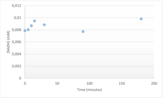
Figure 3.1 – NADH evolution over time in sodium phosphate buffer 0.1 M, pH 7, using 1.5mg (0.5 mg/mL) FateDH, with CO2 bubbling into the media, at room temperature………...…...35
Figure 3.2 – Absorbance spectrum of the cofactor present in the reaction medium………..36
Figure 3.3 - UV/Visible absorption spectra of NAD+ and NADH [93]………..……36
Figure 3.4 – Saturation of the absorbance by NADH concentration at 340 nm with 1.5 mg (0.5 mg/mL) GDH, 1.5 mM of NAD+ in sodium phosphate buffer 0.1 M, pH 7, at room temperature………....37
Figure 3.5 – NADH concentration over time, as generated with 1.5 mg (0.5 mg/mL) of free GDH in sodium phosphate buffer 0.1 M, pH 7, at room temperature………..…….38
Figure 3.6 – NADH concentration over time as generated with sol-gel immobilized GDH (amount of matrix containing 1.5 mg of enzyme) in sodium phosphate buffer 0.1 M, pH 7, at room temperature……….39
Figure 3.7 – NADH concentration variation over time in experiments where NADH was generated using 1.5 mg (0.5 mg/mL) of free GDH, using 20 mM of glutamate and 0.15 mM of NAD+. At 270 minutes, 1.5 mg (0.5 mg/mL) of free FateDH was added and CO2 bubbling through the solution was initiated. Reactions performed in sodium phosphate buffer 0.1M, pH 7, at room temperature………...39
Figure 3.8 - NADH concentration variation over time in experiments where NADH was previously generated using sol-gel entrapped GDH (amount of matrix containing 1.5 mg of enzyme). Sol-gel entrapped immobilized FateDH (amount of matrix containing 1.5 mg of enzyme) was used in sodium phosphate buffer 0.1 M, pH 7, with CO2 bubbling into the medium, at room temperature……….40
Figure 3.9 – HPLC analysis of the reaction medium recovered after performing the conversion of CO2 to formate. The upper figure shows a reference spectrum by addition of 20 ppm of formate. The lower figure shows the sample spectrum with magnification, where formate can be detected (peek 8)………..…..41
Figure 3.10 - Effect of phosphate on the rate of degradation of NAD(P)H. adapted from [97].. ... 42
Figure 3.11 – Images of the sol-gel matrix………...….…….43
Figure 3.12 – Growth curve of E. coli K12 in Minimal Medium with Glucose………44
xv Figure 3.14 – Variation of the viability percentage of encapsulated E. coli K 12 cells recovered from sol-gel matrices after encapsulation for 1, 15 and 30 days………...……..….46 Figure 3.15 – Evolution of the CFU count % of encapsulated cells in gels aged for 1, 15 and 30 days. The additives used were 10 % glycerol, 1% gelatin and 1% PVA. From left to right the bars represent, respectively, SiO2-PB, SiO2-Gly, SiO2-gelatin and SiO2-PVA. Adapted from
[52.]……….………..….47 Figure 3.16 – Viability percentage evolution of encapsulated E. coli K 12 cells in gels prepared with the addition of 100 mM of betaine (blue), 110 mM of glycerol (orange) and PBS (grey), and aged for 1, 3 and 7 days……….……47 Figure 3.17 –Growth curve of E. coli BL21 in M9 minimal media………...48 Figure 3.18 – SDS-PAGE gel from the expression of ctCBM11 in E. coli BL21, with an induction time of 2 and 17 hours. M - Low Molecular Weight Protein Marker (NZYTech). P - Insoluble fraction. S – Soluble fraction. Extracts run in a 12.5% SDS-PAGE, stained with Coomassie Blue. The arrow points toward ctCBM11……….…………...…49 Figure 3.19 – Two-dimensional 1H-15N HMQC spectrum from the encapsulated cells………….….50 Figure 3.20 – Two-dimensional 1H-15N HMQC spectrum from the encapsulated cells with cells concentrated 50x………...…….51 Figure 3.21 – Variation of the viability percentage of encapsulated cells before and after the NMR experiments. ... 52 Figure 3.22 – Optical microscopy image of the sol-gel matrix (magnification – 100x)………52 Figure 3.23 – Optical microscopy image of encapsulated cells not submitted to NMR experiment (magnification 100x)………..53 Figure 3.24 – Optical microscopy image of the sol-gel matrix with encapsulated cells after the NMR experiment (magnification 100x)……….…..53 Figure 3.25 – Bacterial distribution in the sol-gel matrix as observed by optical microscopy (a) and SEM (b) [56]………..…54 Figure 3.26 - Two-dimensional 1H-15N HMQC spectra. A – Non-encapsulated pellet. B – Encapsulated pellet. C -Overlap of the non-encapsulated pellet (blue) with the encapsulated pellet (red). D – Magnification of the overlap of the non-encapsulated pellet (blue) with the encapsulated pellet (red)………..….…….55 Figure 3.27 – Two-dimensional 1H-15N HMQC spectrum of ctCBM11. A – Isolated protein. B Overlap of the spectra from isolated ctCBM11 (blue) and the non-immobilized pellet (red). C - Magnification of the overlap of the spectra from ctCBM11 (blue) and the non-immobilized pellet (red)………....56
xvii
List of Tables
Table 1.1 – Classification of enzymes based on the chemical reaction catalysed. ... 6
Table 1.2 – Examples of oxidoreductases used in ionic liquids………...………11
Table 3.1 – Cell concentration before and after 24h of encapsulation………...………..44
xix
List of Symbols and Abbreviations
% (v/v) – Percentage volume/volume
[BMIM] [OTf] – 1 –Butyl 3-methylimidazolium trifluoromethanesulfonate [BMIM][BF6] - 1 –Butyl 3-methylimidazolium hexafluoroborate
[BMIM][Cl] - 1 –Butyl 3-methylimidazolium chloride [EMIM][EtSO4] – 1-Ethyl-3-methylimidazolium ethyl sulfate
[MIm] [Cl] – 1-Methylimidazolium chloride
[MIm][BF4] -1-Methylimidazolium tetrafluoroborate
[MMIM][MeSO4] – 1–Methyl –3–methylimidazolium methyl sulphate
ADH – Alcohol dehydrogenase
CAZY - Carbohydrate-Active enzymes database CBM – Carbohydrate Binding Module
CFU – Colony Forming Unit CO2 – Carbon Dioxide
DNA – Deoxyribonucleic acid FateDH – Formate dehydrogenase GDH – Glutamate dehydrogenase GFP – Green Fluorescent Protein HETCOR – Heteronuclear correlation
HLADH – Horse liver Alcohol dehydrogenase HPLC – High Performance Liquid Chromatography HRP – Horseradish peroxidase
HSQC - Heteronuclear single-quantum correlation spectroscopy IL – Ionic Liquid
xx LB – Luria Berthani medium
LUDOX – Colloidal silica MM – Minimal Medium
MMG – Minimal Medium with Glucose NAD+ - β-Nicotinamide adenine dinucleotide
NADH – β-Nicotinamide adenine dinucleotide, reduced disodium salt hydrate NMR – Nuclear Magnetic Resonance
OD600nm – Optical Density at 600 nanometres
PBS – Phosphate Buffer Saline
PMSF – Phenylmethylsulfonyl fluoride
SDS – PAGE - Sodium dodecyl sulfate polyacrylamide gel electrophoresis SEM – Scanning Electron Microscopy
SiNa – Sodium Silicate SM – Semimetal
SOFAST-HMQC - band-Selective Optimized-Flip-Angle Short-Transient - Heteronuclear multiple-quantum correlation spectroscopy
TEOS – Tetraethoxysilane TMOS – Tetramethylsilane TMS – Tetramethylsilane
1
1. Introduction
The sol-gel method can be a very versatile tool to encapsulate biomaterials. The sol-gel method was used in this work to address the problem of increasing amounts of CO2 released to the atmosphere, as a
technique to entrap enzymes capable of converting CO2 to useful compounds. The sol-gel method was
also used to encapsulate living cells, by providing a microenvironment adequate for maintaining cell viability and function while offering the advantages of immobilization for potential industrial applications. In particular, the goal was to develop a system that would allow the monitoring of cell viability/function using Nuclear Magnetic Resonance (NMR).
1.1. CO2 as a pollutant
Carbon dioxide (CO2) is a gas present in high amounts in the earth’s atmosphere and therefore important
for its balance. In the last decades the concentration of this gas in the atmosphere increased markedly, leading to changes in climate and in living conditions around the world. Some of these changes are felt in the so called global warming and, more recently in the cases of smog in some great cities like Paris or Beijing, due to the release of gases, including CO2, from car exhaust or factories. Due to its high
concentration in the atmosphere, CO2 can be considered a pollutant and therefore the topic of CO2
utilization as a source of chemicals becomes of great interest.
In nature CO2 is utilized by plants to perform photosynthesis. In this process the plants use atmospheric
CO2 and convert it to organic products with water and light [1]. In addition to the atmosphere, CO2 is
also present in the water, soils, and live beings, as seen in figure 1.1.
Since the industrial revolution in the 18th century, CO
2 emissions originating in human activity have
considerably increased (figure 1.2) due to the use of motorized vehicles (such as cars, trains or ships), the construction of factories, and other activities that involved, at some point, a high consumption of fossil fuels, such as coal or petrol. Another cause is also the growth of population, which led to a major growth of cities and to a reduction of green areas of the planet by deforestation that decreases the capacity to naturally convert CO2 from the atmosphere. Today the rate of fossil fuels consumption is
much higher than the capacity of the planet to regenerate those materials that take millions of years to be produced. This is actually a major concern and it is why the need of new ways to obtain fuels and, simultaneously, reduce gas emissions is such a hot topic.
2
Figure 1.1 – Carbon cycle [2]
Figure 1.2 – Global carbon emissions and principal contributors. The black line shows the high increase of carbon emission through time. Adapted from [3].
As referred above, CO2 emissions have progressively increased in the last few years leading to further
environmental problems, calling for a need to decrease those emissions. Several organizations and governments have issued legislation or directives to help and guide this effort (e.g. the Kyoto protocol) [1, 4]. So far these actions have not been as significant as hoped for, with major cases of gas pollution in some cities.
3 In order to control the emission of CO2, and reduce it, several processes can be followed. For instance
CO2 can be captured by geo-sequestration, a process that involves injecting CO2 at high pressure into
geological formations, in some cases with formation of stable carbonates [4].
CO2 can also be used in the production of liquid fuels [5]. Other approaches include post combustion
capture processes, using chemical absorption onto amine-based solvents. This, however, has some downsides, as the processes have great energy demands for regeneration, and the amount of CO2 that
can be captured, relative to the amount of compound used to bind it, may not be very high. In addition, the need for purification of the CO2 stream increases associated costs [1].
CO2 capture is more effectively implemented by industries with major CO2 emissions, such as factories
that produce ammonia [7].
At normal temperature and pressure, CO2 is a gas. But with increasing temperature and pressure, CO2
reaches its critical point (31 ºC and 73.8 bar; Figure 1.3), at which the liquid and vapor phases become indistinguishable. Above the critical point, CO2 becomes a supercritical fluid. In this particular region
CO2 has lower viscosity and higher diffusivity, similar to those of gases, and has a density and solvation
ability similar to those of liquids [8, 9].
4 The density and viscosity of a supercritical fluid can change markedly near the critical point by varying slightly pressure and temperature conditions [11], which allows the fractionation of a mixture of several components, e.g. through depressurization that decreases CO2 density and therefore its solvation ability.
This is behind the use of supercritical CO2 as a solvent for extraction. Many applications have been
implemented, such as decaffeination [7, 8], hops extraction, deacidification of oils, dry cleaning, or polymer impregnation [12]. CO2 can also be used as solvent for reaction, including enzyme catalysed
reactions [13].
In spite of these factors supercritical CO2 requires the use of high pressures, adequate safety equipment,
and its use must be pondered based on perceived advantages over other solvents.
1.2. CO2 conversion to methanol
Due to the fact that today CO2 is an environmental issue, it is important to find ways to reduce its
concentration in the atmosphere. CO2 is a very stable molecule and its transformation by chemical
processes is thermodynamically unfavourable. The approaches used for converting CO2 include
heterogeneous catalysis, namely by modifying copper catalysts in processes to produce methanol from CO; homogeneous catalysis in which various catalysts are used to reduce CO2 into CO [6,14];
electrochemistry, in which methods like the direct and indirect reduction and a two-step reduction are used [6,15]; photochemistry, in processes like the regeneration of NADPH-model complexes [6,15]; thermochemistry, in which very high temperatures (above 1000 ºC) and the use of metal oxide catalysts allowed the splitting of CO2 into CO [15]; and enzymatic catalysis, in which enzymes are used to
convert CO2 in a series of coupled reactions (this process will be discussed later) [6].
The conversion of CO2 can be directed to generate a product of added value, such as methanol. Methanol
is a platform chemical. It can be converted into chemicals, polymers, building materials and fuels [4, 16]. Figure 1.4 shows several chemicals that can be generated from methanol.
5
Figure 1.4 –Methanol derived products. Adapted from [4].
The Gibbs free energy and enthalpy (ΔH) values (at 25ºC and 1 bar) for the mostly used reactions for methanol production in industry are given below:
CO + 2 H2 → CH3OH ΔH = - 90.8 kJ/moL [3,6,14] ΔG = - 25.34 kJ/moL [17] (1)
CO2 + 3 H2 → CH3OH + H2O ΔH = - 49.6 kJ/moL [3,6,14] ΔG = 3.30 kJ/moL [17] (2)
CO + H2O → CO2 + H2 ΔH = - 41.2 kJ/moL [3] ΔG = - 28.6 kJ/moL [17] (3)
Eq.1 represents the hydrogenation of carbon monoxide, which is an exothermic and spontaneous reaction, as seen by the negative associated ΔH and ΔG values. Eq.2 represents the hydrogenation of CO2, which is not thermodynamically favorable. Eq.3 shows the water-gas shift reaction. As in the case
of Eq.1, it is driven by favorable ΔH and ΔG values, and uses the water produced upon CO2
hydrogenation, thus acting as the driving force for hydrogenation to occur.
There are many strategies to overcome the thermodynamic limitations in CO2 conversion, including the
6 1.3. Biocatalysis
In the present work, enzymatic catalysis is used, based on the utilization of CO2 as reactant for the
production of methanol.
Catalysis is one of the principles of Green Chemistry. The catalyst provides a reaction pathway involving lower activation energy, thus leading to higher reaction rates.
Enzymes are biological catalysts. Biocatalysis can be carried out with isolated enzymes, or with microorganisms that exhibit the catalytic activities of interest for a particular application.
The enzymes used in biocatalysis are divided in six categories, which are discriminated in table 1.1.
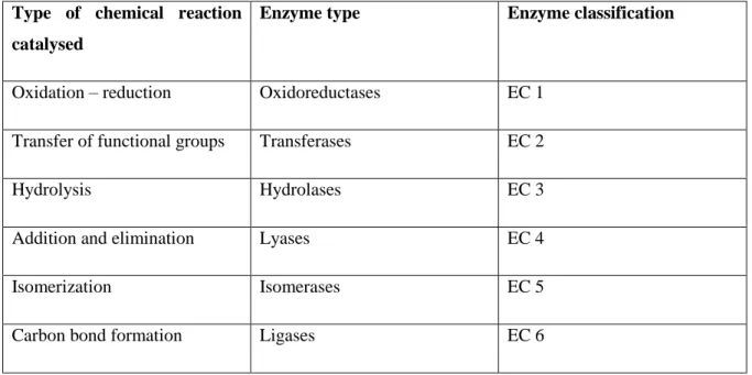
Table 1.1 – Classification of enzymes based on the chemical reaction catalysed.
Type of chemical reaction catalysed
Enzyme type Enzyme classification
Oxidation – reduction Oxidoreductases EC 1
Transfer of functional groups Transferases EC 2
Hydrolysis Hydrolases EC 3
Addition and elimination Lyases EC 4
Isomerization Isomerases EC 5
Carbon bond formation Ligases EC 6
When using microorganisms it is important to ensure that there are no interferences from the other enzymes expressed by the cells [18]. On the other hand, the use of isolated enzymes requires expression in recombinant strains, enzyme release and enzyme purification [19]. The latter is costly. But enzymes, being much less complex than cells, are easier to control as concerns experimental conditions such as temperature, or pH [6].
Hydrolases are the most used enzymes. One advantage is that they do not require co-factors, which are usually expensive. This is also the case with transferases, lyases and isomerases [6]. On the other hand, for many oxidoreductases to be able to function, a co-factor is needed, leading to higher associated costs as regards providing this species and regenerating it.
7 1.4. Oxidoreductases
The enzymes used in this work are all oxidoredutases, commonly known as oxidases or, in this case, dehydrogenases. Dehydrogenases use a cofactor to catalyse the electron transfer from one molecule to another (figure 1.5).
The oxidoreductases used in this work require NADH/NAD+ (NADH = reduced β-nicotinamide adenine dinucleotide). They are formed by two domains: the catalytic domain, and the NAD binding domain. The function of the latter is the binding of NAD+ or NADH in an active conformation.
The reduction of NADH by oxidoreductases follows a series of steps:
Cofactor and substrate binding to an enzyme;
Substrate reduction and cofactor oxidation;
Dissociation from the enzyme [20].
Figure 1.5- Reaction catalysed by an oxidoreductase (dehydrogenase) using NAD(P)H as cofactor. Adapted from Gamenara, et al. [21].
1.5. Bioconversion of CO2 to methanol
The conversion of CO2 to methanol can be accomplished through the action of three enzymes, namely
formate (FateDH), formaldehyde (FaldDH) and alcohol (ADH) dehydrogenase, whose action requires the presence of the cofactor NAD in its reduced form – NADH (figure 1.8).
8
Figure 1.6 – The enzyme coupled reaction to convert carbon dioxide to methanol. Adapted from [17]
Studies show that pH is a very important factor as, in the case of methanol production, values of pH lower than 6 favor methanol production, but higher values are unfavourable [22]. The study just referred also shows that this reaction should be conducted at low ionic strength and higher temperatures.
The introduction of the three-enzyme system was done in 1999 by Obert and Dave [17]. The authors used FateDH, FaldDH and ADH in free form in aqueous medium, as well as immobilized in a sol-gel matrix. The maximum relative yield of methanol per mole of NADH added (3 mol of NADH are consumed per mol of methanol produced) obtained by the authors were 21.0 %, with 100 µmol of NADH, and 91.2%, with 50 µmol NADH.
The need to regenerate the cofactor comes from the fact that cofactors are very expensive, with prices that can reach $1000/mol, limiting the process [23]. To overcome this problem, Zahab et al. [23] looked at the regeneration of NADH from NAD+ using the enzyme glutamate dehydrogenase (GDH), which acts on glutamate. The authors immobilized the cofactor on polystyrene particles, and obtained an accumulated yield of methanol of 127% (based on the total amount of NADH used by the authors in the experiments), after 11 cycles of reusing.
Other authors, like Sun et al [24], immobilized the multi-enzymatic system in titania particles (figure 1.6), with good yields of methanol.
NADH regeneration can also be made photochemically in the process of CO2 conversion to methanol.
One example is the use of the three dehydrogenases in combination with the photoreduction of methyl viologen through the photosensitization of zinc tetraphenylporphyrin tetrasulfonate in the presence of triethanolamine in aqueous media [25].
9
Figure 1.7 – CO2 reduction to methanol by co-immobilized enzymes in titania particles. Adapted from [24].
The conversion of CO2 into organic compounds can also be made by microorganisms [22, 26, 27].
Addo et al [28] used the three-enzyme system referred by Obert and Dave [17] and added the enzyme carbonic anhydrase (figure 1.7) in order to solve the problem of low CO2 solubility in aqueous media.
Carbonic anhydrase is a powerful catalyst for the hydration of carbon dioxide, converting it to bicarbonate. The study shows that the addition of this enzyme to the system can enhance the rate of carbon capture by the hydration of carbon to bicarbonate, which is the primary substrate for FateDH. In this system NADH regeneration is made with the use of an electrode, which makes possible to maintain high levels of NADH in the system.
10
Figure 1.8– Addition of carbonic anhydrase to the three enzyme system. Adapted from [28].
In our work, the objective was to introduce a fourth enzyme, GDH, for the regeneration of NADH, as done by Zahab et al. [23]. It was also envisaged to replace aqueous solutions with ionic liquids.
1.6. Ionic Liquids
Ionic liquids (ILs) are salts in the liquid state. These are made of anions and cations, whose asymmetry results in weaker ionic association, which leads to melting points below 100ºC [29–32]. To differentiate these compounds from regular salts, the designation “room temperature ionic liquid” (RTIL) can be used [12, 32].
ILs attracted attention due to their characteristics that conform to green principles, such as negligible vapor pressure, which facilitates recycling and reutilization [12,30]. The attractiveness of the ILs is very much related to the possibility of designing ILs to meet given specifications, due to judicious combination of anion and cation. One drawback is that the synthesis of many ILs cannot be considered green.
11 The water miscibility of ILs can vary from immiscible up to fully miscible [9, 29]. In any case, water-immiscible ILs can solubilize much higher water contents than most organic media. One species ILs dissolve well is CO2. CO2 has very low solubility in water [29], but in ILs CO2 solubility can be orders
of magnitude higher. CO2 being a substrate in the reaction that is the focus of this work, higher amounts
of CO2 in the medium favour its enzymatic conversion. The solubility of CO2 in ILs increases with
pressure, according to Henry’s law.
Many ILs have higher viscosity than water, and this can be a disadvantage because it makes mass transfer processes more difficult [30]. On the other hand, when CO2 is solubilized in high amounts in
ILs, it fluidizes the medium.
Figure 1.9 shows a summary of IL properties.
Looking at the use of oxidoreductases in ILs, there are many reports on their activity being preserved, whether free [32] or immobilized [34].
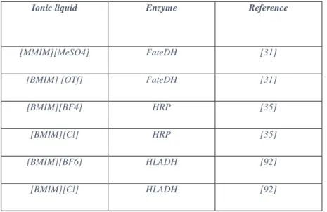
In table 1.2 some examples of oxidoreductases used in IL media are shown.
Table 1.2 – Examples of oxidoreductases used in ionic liquids.
Ionic liquid Enzyme Reference
[MMIM][MeSO4] FateDH [31]
[BMIM] [OTf] FateDH [31]
[BMIM][BF4] HRP [35]
[BMIM][Cl] HRP [35]
[BMIM][BF6] HLADH [92]
12
Figure 1.9 – Ionic liquid properties. Adapted from [36].
1.7. Sol-gel method
The principles of sol–gel technology for immobilization have been described in detail in many articles and reviews [37-40].
In aqueous solution a salt of a metal ion Mz+ is solvated by water molecules that can be deprotonated. For semimetals, SM (Si, B, Ge, etc.), the SM–O bond has a more covalent character, and hydrated species are found as SM(OH)n with Brönsted acid properties. In both cases, when hydroxyl functions are present, a condensation reaction can occur to form (semi)metal– oxygen–(semi)metal bonds.
The condensation is influenced by the nature of the metal ion, its concentration, the solution pH, and can lead to the formation of an infinite network of oxo-bridged (semi)metal chains that will correspond to the oxide phase. In the early stages of the condensation, colloidal species grow in solution and form the sol. These particles then continue to grow and/or aggregate to form a gel (figure 1.10).
13
Figure 1.10 – The principles of sol-gel technology. a) A thin film of sol is used to coat surfaces, b) The xerogel is formed through the drying of the gel Adapted from [41].
In some cases the condensation occurs too fast and instead of a gel, the particles form a precipitate. The process has been normally developed with metal alkoxides, which are molecules of general formula M(OR)N, where R is an organic group, mainly an alkyl chain. They do not possess M–OH hydroxylated species necessary for the condensation to proceed and need to be hydrolysed first.
It is possible to control the final structure of a gel if the nature and availability of hydroxylated species are adjusted, namely by pH modification. For example, using acidic condition we may obtain dense microporous networks, with pore size of less than 2 nm, and under alkaline condition we can obtain particulate mesoporous gels, with pore sizes between 2 and 50 nm.
A typical sol-gel reaction for the formation of silica is described as follows:
Si(OCH3)4 + 4H2O MEOH catalyst SiO2 + 4CH3OH + 2H2O
The most popular silicon alkoxides used are tetramethoxysilane [Si (OCH3)4; TMOS] and
tetraethoxysilane [Si (OC2H5)4; TEOS]. It is necessary to add water for the hydrolysis to occur, but
TMOS is not soluble in water, which also makes necessary the use of the alcohol, methanol, to ensure the homogeneity of the mixture. To speed up the reaction it is also possible to use catalysts such as acid, base, or a nucleophilic agent, such as dimethylaminopyridine or fluoride ion. After having mixed all
14 these components, a gel is formed within minutes that contains methanol, added as solvent and produced in the hydrolysis reaction.
In many cases, hydrolysis and condensation proceed during gel ageing and syneresis, with additional methanol and water being expelled. The gels can then be preserved it their wet state in closed flasks or they can be dried at room temperature or freeze-dried, forming xerogels. To avoid the cracking of silica networks it is possible to add a drying control additive, such as formamide or ethylene glycol. Alternatively, supercritical drying techniques allow the formation of highly porous, low-density aerogels.
1.8. The sol-gel approach for cell immobilization
In our work we focus on the immobilization of living cells in a sol-gel matrix. Our interest in such a system is the particular advantage that living organisms may have in some reactions, being able to produce some particular compound in an easier and cheaper way, compared with an enzymatic system. An approach in which the cells can live longer and still have good catalytic capability would be a great advantage to some industries.
In this work the emphasis was on developing a method through which the cells would maintain a good viability when immobilized, that further allowed the system to be studied using NMR.
This topic can be related to CO2 conversion by the phenomenon of photosynthesis. Photosynthesis is a
phenomenally efficient and highly sophisticated process that uses solar energy to convert CO2 to other
chemical species, generating oxygen in the process. The successful use of this mechanism would be a step forward to reduce the increasing atmospheric CO2 levels. Living organisms such as cyanobacteria,
which can assimilate CO2 and, through photosynthesis, can generate carbohydrates, would be very
useful as tools for making CO2 a reusable carbon source [42].
Cyanobacteria live almost in every ecosystem in the planet, playing a vital role in nutrient recycling. They are used as model organisms for fundamental research in photosynthesis and carbon and nitrogen fixation, and are used in biotechnology for producing food additives, nutritional and pharmaceutical compounds, and pigments, for biofuels and other products [42]. The production of biofuels and other co-products is shown in figure 1.11.
The utilization of such a system in this work was considered and relevant literature was consulted, as it came in line with the enzymatic conversion of CO2. However, the conditions required by these
organisms, such as the illumination or the handling of the cultures, led to the choice of another, simpler model. The chosen organism for this work was Escherichia coli.
15
Figure1.11 – Production of biofuels and other co-products from cyanobacteria and microalgae. Adapted from [43].
The main advances on the immobilization of live cells in sol-gel matrixes have been led by the groups of Coradin and Carturan. The first author based his method on the utilization of aqueous-based precursors, which will be discussed further, that reduce the impact of the alcohol toxicity on the cells. The second author uses a method called BIOSIL. This process has been used in the immobilization of various mammalian cells. The process is based on gas-phase alkoxysilane precursors that are passed over the wet cells. The precursors react with the water available in the system to form a silica scaffold over the cells (Figure 1.12) [44]. Cells submitted to this treatment have been shown to remain metabolically active. This method has been used to develop bio-artificial organs and cell-grafting [37,45].
16
Figure 1.12 - Human fibroblasts on glass fibres and coated by the Biosil process. Adapted from [46]
The versatility of silica matrices for cell immobilization is demonstrated by the development of biosensors, bioreactors and bio-artificial organs. These applications exploit specific features of the cell, such as the response to external stimuli or metabolite turnover. The environment set for the cells is chemically defined, but is not close to a native environment of the species used, and although the silica matrix is biologically inert, it is still not clear how the immobilization within a shrinking matrix affects the cell physiology and gene regulation [47].
Some studies were made in order to evaluate the cell physiology under these stress conditions and also cell viability. For this purpose E. coli is a common choice for researchers. One of the first E. coli reported encapsulations in a TMOS-derived silica matrix showed that the cells are randomly dispersed in the matrix and the enzymatic activity is retained within the wet gel, even in the presence of methanol [48].
A study from Premkumar et al. [49] reports the use of confocal fluorescence microscopy of fluorescent GFP-expressing E. coli to confirm that there was no visible cell growth in the porous silica matrices. Since then the idea of no cell growth in the matrix and the diffusion of molecular oxygen into the matrix to be accessible to the cells (which require it to the expression of GFP fluorescence), is accepted.
The effects of stress during the entrapment were also studied with fluorescence and luminescence reporters for heat shock, oxidative stress, fatty acid availability, peroxides and genotoxicity. All of these had a similar behaviour when induced in solution or in silica, but showed no background levels that indicated stress as result of the immobilization in TMOS-derived matrices (figure 1.13) [49].
17
Figure 1.13 - Left: stress induced response of native and recombinant bacteria. The recombinant bacteria express reporting proteins in addition to its native defense mechanisms. Right: single E. coli cell fluorescent response after induction by 1.2 mmolar mitomycin C. Confocal microscope images were taken after incubation times of 0, 140,260,340,420, and 480 min. Adapted from [50].
Cell viability is often determined by the number of colony forming units (CFU) resulting from crushed gels suspended in a saline solution, and this viability could be maintained for several months [51] Glycerol as an additive was shown to improve cell viability using 14C-labeled glucose incorporation
methods and CFU counting. The results obtained from the two methods have been shown to be homologous for samples of gel crushed after 15 and 30 days (figure 1.14).
The reasons for these results have been hypothesized to be differences in glucose metabolism for glycerol-containing and glycerol-free gels, determined by 13C NMR. The samples containing glycerol
led to lower concentration of unmetabolized glucose during one month, which indicates that the glucose metabolism is better supported [52].
It is also interesting to see that there are no significant differences reported for the viability of cells when encapsulation was performed over bacteria cultures in growth phase or in stationary phase [51]. The research on the subject of encapsulation in silica gels, namely for E. coli bacteria, shows that the cells can remain viable in the matrix and still respond to external stressors, that the matrix has not induced the stress response genes testes, and that the cells can be immobilized in a variety of silica matrices with good structural integrity.
18
Figure 1.14 - Evolution of 14C glucose incorporation in entrapped cells in the four gels aged one, fifteen
and thirty days. The error range is within 20- 30%. From left to right the bars represent, respectively, SiO2-PB, SiO2-Gly, SiO2-gelatin and SiO2-PVA. Adapted from [52].
1.9. Preserving cell viability within silica gels
To take forward a project based on sol-gel immobilization of cells it is important to evaluate three stages: the starting solution, the gelation reaction, and the ageing phase. The starting solution should be studied mainly in terms of chemistry: cytotoxicity of the reagents, pH, and ionic strength. The gelation reaction must be considered from a physico-chemical point of view, in particular reaction rate, temperature, or viscosity. The ageing phase is the most challenging as its success depends not only on parameters that are intrinsic to each component of the system, but also on the cell–matrix interface [41].
1.9.1. The starting solution
There are three aspects of the initial mixture than can have a strong impact on cell viability. One of these aspects is the reaction solvent, mainly methanol, which is highly toxic. Then comes the precursor cytotoxicity. As regards TMOS, it releases methanol when in contact with water upon hydrolysis. A third relevant aspect is the extreme conditions of pH or the use of other toxic chemicals. These problems led to the development of several protocols aiming at circumventing the negative effects perceived.
One approach was to perform the reaction in a two-step method, without the use of an alcohol as a solvent [53]. Here the silicon alkoxide is mixed with an aqueous HCl solution at pH 2 under sonication, to perform the hydrolysis step. The Si(OR)4–water mixture is initially a two-phase system, but
19 the pH favours a fast hydrolysis. It is then obtained a stable sol of silica particles. The cell suspension in a neutral pH buffer is then added, increasing the mixture pH to 7, triggering the gel formation.
Such a process is good for enzyme immobilization, but the fact that it releases alcohol during the hydrolysis makes it harder to use for the immobilization of living cells. Other processes tried to remove the alcohol from the mixture by flowing air during the hydrolysis [54] or with the use of rotavapor to remove harmful by-products after the hydrolysis [55]. In both cases, a continuous monitoring of the alcohol evaporation is needed, as well as the addition of water to avoid an early gel formation.
To avoid the presence of alcohol it is possible to start the reaction from aqueous-based molecular precursors. In the case of silica, these precursors are available as sodium or potassium silicate alkaline solutions. These solutions contain a high concentration of silicon species (above 5 M) consisting of silicic acid—Si(OH)4—and its oligomers—SixOy(OH)zn-. Since Si–OH groups are initially present,
hydrolysis is not required and condensation reactions can occur as soon as the pH is decreased to 8 or below, upon mineral or organic acid addition.
In spite of these benefits, the solutions referred above also have a drawback, namely the fact that the presence of sodium gives the starting mixture a high ionic strength, which needs to be taken in consideration in the immobilization of live organisms. For instance, algae survive in high salinity media but in general common bacteria do not.
One method to overcome this problem is to treat the silicate solution with an acid-exchange resin to replace Na+ by H+ [56]. As a result, an acidic silicate solution is obtained that can form gels upon
neutralization. It is also possible to form a gel using an aqueous suspension of preformed colloidal particles. The two conditions are that the pH/ionic strength are compatible with gel formation, and that no precipitation and no detrimental interactions exist between the particles and the cell membrane. This method permitted the use of other materials to form gels, such as boehmite [57], ferrihydrite [58], and, in the case of yeast cells, in titanium oxide [59] wet networks.
These procedures can be combined in many ways. For instance, silica nanoparticles and sodium silicate can be advantageously mixed in the starting solution [60]. Silicate condensation leads to a cementation of the colloidal network, whereas silica nanoparticles allow the preparation of a silica solution at high concentration while avoiding excess sodium [61].
Silicates may also be treated by an ion- exchange resin before being mixed with colloidal silica [62]. The combination of silicate and functional alkoxides was also described to control the porosity of the matrices and/or the cell material interface, especially in the case of microalgae and plant cells [63].
20 1.9.2. The gelation reaction
After the addition of the cell suspension, the condensation reaction occurs. Here the cells experience a rapid change (gelation occurs between 2 and 10 minutes) in their chemical and physical environment. This is why it is important to immobilize the cells during their growth phase, where they are more likely to adapt to their new conditions.
There are no studies relating the process of gel formation to cell viability in these systems, so it is difficult to realize what specific effect is responsible for the observed variations in cell viability. A first reason is that survival rates are usually reported after a minimum of 24 h after encapsulation, when significant gel ageing has already occurred. Moreover, when short-term data are provided, their interpretation is not straightforward.
For instance, 1 h after encapsulation, the viability of E. coli is decreased to 65% in silica gels but in the presence of glycerol it remains near 80% [60]. This effect was attributed to a protective effect of glycerol that surrounds the bacteria and decreases their interaction with the gel surface. Another additive found to increase E. coli viability in sol-gel was glycine betaine [64]. The results for this additive were similar to the ones with glycerol, after ageing, but do not reveal modifications in the silica gel mechanical stability, specific surface or transport properties. This may be due to the localization of betaine near the cell surface in cytoplasm to preserve the water bound and avoid unfolding. Also this additive operates at much lower concentration than glycerol.
E. coli viability may also be improved by the acidification of the sodium silicate using citric acid instead of hydrochloric acid [65]. The reason for this effect may be that the organic acid slows down the condensation reaction kinetics, reducing the physical stress of the cells, and also that citric acid promotes the production of glycerol that can act as a protecting agent.
Another study used strong interactions between the cell surface and the material, allowing a favourable deposition of silica on the cell surface [66]. In this study an enzyme called silicatein, found in sponges, was used, by preparing genetically modified E. coli to express it on its surface. In the presence of a low amount of silicates, it was observed that silica condensation occurs in close proximity to the cells.
21 1.9.3. Gel ageing
With the ageing of the gel, more condensation occurs, that leads to syneresis and network reorganization and densification. This makes the cell surface more similar to the inorganic surface, resulting in an increased mechanical constraint.
Regarding this subject some studies were performed in order to see the impact of ageing in cell viability. One study used cryogenic scanning electron microscopy (cryo-SEM) [67] with E.coli. Figure 1.15 compares encapsulated cells using a prehydrolyzed TEOS solution after ethanol evaporation at different ageing times. In Fig. 1.15A, bacterial cells protrude from a porous matrix and cell wall– silica direct contacts are difficult to see. Moreover the internal structure of the cells is clearly visible. Upon ageing (Fig. 1.15B), empty shells are observed within the pores of a denser network with clear cell–matrix contact. In 1.15 C and D, we can see that the incorporation of protecting agents like glycerol (C) and N-(3-triethoxysilylpropyl)gluconamide (D) result in well-defined and thick cavities around a nicely preserved bacterial cell, which can be explained by the decrease in the size of the interface between the cell wall and the silica network.
The direct impact of the mechanical properties has only been addressed once, in the case of encapsulation of diatom cells in silica [68]. In this study, three situations were considered. The material with low silica concentration resulted in weak gels from which diatom cells escaped within a few days. With high silica concentration, the result was robust gels in which cells died fast. And with an intermediate silica concentration, the results showed that the cells could be observed up to 2 months. Even under favourable conditions, the different silica concentrations resulted in different cell viability, suggesting that the mechanical constraint imposed by the silica has in impact on cell survival.
In this work the process used for the encapsulation of E. coli cells was based on the one described by Nasif et al [60] in which aqueous based precursors are used to form the sol-gel matrix. This method was used due to its simplicity, as there is no need to remove alcohol formed by the hydrolysis that occur with typical precursors, and also for the reduced toxicity for the organisms encapsulated. Also some additives were used to enhance cell viability within the sol-gel matrix.
22 .
Figure 1.15 - Cryogenic scanning electron microscopy examination (cryo-SEM) of Escherichia coli bacteria encapsulated inside different silica matrices (arrows identify cells): a) silica made from prehydrolyzed tetraethoxysilane, b) the same material after ageing, c) effect of glycerol addition, and d) effect of N-(3-triethoxysilylpropyl)gluconamide addition. Adapted from [67].
1.10. Microorganisms used in the present work
1.10.1. Escherichia coli
E. coli are gram negative, facultative, anaerobic, rod-shaped bacteria that commonly live in the lower intestine of warm blooded organisms. These bacteria are normally harmless but and are part of the intestinal flora, being very important for the host by producing vitamin K12 [69] and preventing the colonization of the intestine by pathogenic bacteria [70]. In spite of this, some E. coli serotypes (a subdivision for E. coli based on some major surface antigens) can be pathogenic for the host, causing food poisoning.
These bacteria have been used throughout the years in the laboratory to perform a wide range of research. The main reasons for their intensive use are their low cost and the fact that the bacteria are easy to grow and manipulate. These bacteria hold an important role in biotechnology as they are very good host to produce heterologous proteins [33]. In fact many proteins, like insulin, are produced by
23 expression in E. coli. This makes E. coli one of the most important model organisms, in which a great majority of recombinant DNA methods are tested. E. coli have been used in areas such as bioremediation, biofuels production, production of immobilized enzymes [33] or vaccine development [71].
As these bacteria are much studied, there is also a great variability of strains characterized, which can be found in nature. Many have also been created in laboratories around the world. For the particular case of this work, two strains will be mentioned: the K12 and BL21 strains.
E. coli K12 was first isolated in 1922 from a patient with diphtheria at Stanford University [72]. Since then the strain was widely used and modified by different scientists, creating different lineages. The strain-used derivatives are mostly from the E. coli K 12 used by Clifton in 1944, in his studies of the nitrogen metabolism [73]. This strain was F+, meaning that the strain possessed the fertility factor F that
permits genes to be transferred by conjugation to bacteria not possessing this factor, and lysogenic for phage lambda. It was one of the first organisms (and E. coli strain) having its genome fully sequenced in 1997 [74].
The BL21 strain is a derivative from the B strain that was found by Felix d’Herelle in the Institut Pasteur in Paris, around 1918 [75]. The original name used then was Bacillus coli, from which Delbruck and Luria, later around 1942, in their study of bacteriophages T1 and T7, based themselves to name the strain as E. coli B [76]. Although no strain from d’Herelle was deposited in a database, it was passed through several groups until it reached Delbruck. The strain used by Delbruck and Luria later gave rise to derivative strains, namely BL21. The strain used in this work was BL21 (DE3), which possesses a copy of the T7 RNA polymerase for the pET expression system.
1.10.2. Carbohydrate Binding Modules
Carbohydrate binding modules (CBM) are generally found in glycoside hydrolases that catalyse the degradation of the plant cell wall. This is important in nature due to their role in recycling plant cell wall fixed carbon [77]. They act by mediating the contact of the enzyme with the substrate, enhancing the activity of the catalytic module against insoluble polysaccharides [78, 79]. That makes CBMs very important in potentiating the ability of cellulases and hemicellulases to degrade plant cell wall polysaccharides.
These modules have been grouped in 39 families based on primary structure similarity (afmb.cnrs-mrs.fr/ CAZY) [77]. Also we can find three types of CBM, called type A, B and C. Type A CBMs interact with the flat surfaces of crystalline polysaccharides and have a planar hydrophobic carbohydrate-binding site [80, 82]. Type B CBMs bind to single polysaccharide chains, accommodating these ligands in extended clefts [81, 82]. Type C CBMs bind to small sugars [82].
24 In this work we used a CBM from family 11 from the organism Clostridium thermocellum Lic26A-Cel5E. This enzyme contains GH5 and GH26 catalytic domains that display β-1,4 and β-1,3–1,4-mixed linked endoglucanase activity, respectively [77]. The molecular weight of this particular protein is about 19.6 kDa. The structure of ctCBM11 is shown in figure 1.16.
Figure 1.16 - Ribbon representation of the three-dimensional structure of CtCBM11.Adapted from [77].
1.11. Nuclear Magnetic Resonance (NMR)
Nuclear Magnetic Resonance (NMR) spectroscopy is a widely used technique. In simple terms the technique allows the study of samples placed in a magnetic field and subjected to an appropriate radiofrequency, by the absorption of energy by the nuclei of that sample. It is used to determine compound structures and to perform qualitative and quantitative analyses.
The nuclei have positive charges and many of them behave like they were spinning. Typically the spins can have 2 orientations, the one that is more favourable (which can be referred to as the low-energy state) and the one that is precisely 180º in the opposite direction (the high-energy state) [53]. This spin is characterized by the spin quantum number (I) and can have 2I + 1 different orientations in relation to an axis. The values of I can be 1/2, 1, 3/2 …, with no spin for I=0[83, 84].
The most studied proton studied by NMR is the hydrogen proton 1H. In order to prevent an interference by a peek related to the solvent used, it is common to use deuterated solvents, which contain 2H instead of 1H. This particular proton is also used to obtain spectra from various nuclei, such as 13C or 15N [83, 84].
25 The distribution of nuclei in the different energy states when submitted to any radiofrequency energy follows the Boltzmann distribution. In eq.4 Nupper represent the population of nuclei (number of nuclei) in the high-energy state, being Nlower the population in the low-energy state. K represents the Boltzmann constant and T is the temperature in Kelvin. Figure 1.17 shows the application of this equation in which the population in the lower state is always slightly larger than in the upper state. This small excess in the lower state enables the NMR technique [93].
Nupper/(Nlower) = e^(-∆E/kT) (4)
Figure 1.17 - Influence of the magnetic field B0 in the separation of nuclear energy levels for spin I= ½. There is also shown the relative population in each energy state assuming 2 million protons in the sample. Adapted from [93]
The spectra is analysed by the chemical shift of a nucleus, which is expressed in ppm. To do so the sample resonance frequency is compared to a standard. The shift can be influenced by some factors like the chemical environment.
26 1.12. In cell NMR
The in cell NMR techniques use two features from NMR spectroscopy that makes it a good method to investigate biological macromolecules [85]. This technique can be used in physiological or “near- physiological” conditions and has the necessary sensitivity to detect changes in the chemical shift of a NMR-active nucleus.
“In-cell NMR” is not intended to resolve the structures from molecules, but can provide information about the localization of the macromolecule in its natural environment by analysing the chemical shift changes in these conditions. These changes can be post-translational modifications, conformational changes, or binding events. They can be detected by the change in the resonance frequencies of the affected nuclei [85]. This is summarized in figure 1.18.
The analysis is made by detecting the differences between the in-cell spectra and the in vitro spectra. To study the cause of the interactions it is possible, for example, to simulate the in vivo conditions in vitro and add the suspected interaction molecules to an in vitro sample. This method was used by Dedmon et al. and resulted in the finding that the bacteria protein FlgM, unfolded in vitro, is actually partially folded in the E. coli cytoplasm [86].
The chemical-shift differences in some NMR spectra can also be used to detect and characterize interactions of proteins with drug molecules. This is an interesting application as screening tool for the pharmaceutical industry.
As seen above, one disadvantage of traditional in vitro NMR is that the interactions observed in these conditions do not always occur in the same way in vivo. Some reasons for this difference can be the difficulty of the molecule to cross the cellular membrane, its fast metabolization, its binding to other cellular components with higher affinity than to its intended target, or differences in the target-protein conformation between its in vitro and in vivo states.
Regarding cell survival with this technique, the high cellular density can cause problems through oxygen starvation and limitation of the amount of available nutrients. This problem has been addressed by using modified NMR tubes to allow a continuous flow of media [85]. However this makes it difficult to ensure that the cells stay in the tube during the experiment. To solve this problem, low-melting agarose was used to immobilize the cells in the tube [85].
27
Figure 1.18 – Chemical shift changes in “in cell NMR”. Adapted from [85].
The downside of the “in cell NMR” technique is the apparent low sensitivity of the equipment, since there is a need to overcome a threshold of protein concentration in order to obtain a signal. In spite of this difficulty, this technique remains an important approach to study protein interactions inside the cell as it is a non-invasive method.
a) Conformational changes b) Conformational changes c) Binding events that can be detected by chemical shift differences.


![Figure 1.9 – Ionic liquid properties. Adapted from [36].](https://thumb-eu.123doks.com/thumbv2/123dok_br/15771676.1075988/34.892.112.680.110.482/figure-ionic-liquid-properties-adapted-from.webp)

![Figure 1.16 - Ribbon representation of the three-dimensional structure of CtCBM11.Adapted from [77]](https://thumb-eu.123doks.com/thumbv2/123dok_br/15771676.1075988/46.892.189.712.274.600/figure-ribbon-representation-dimensional-structure-ctcbm-adapted.webp)

![Figure 2.1 - Experimental set-up for the reactions in aqueous medium. Adapted from [94]](https://thumb-eu.123doks.com/thumbv2/123dok_br/15771676.1075988/52.892.152.590.541.778/figure-experimental-set-reactions-aqueous-medium-adapted.webp)