development of the ear, neural crest
and pharyngeal system in mice
Filipa Pontes de Moraes
Dissertation presented to obtain the PhD degree in Biology at the Instituto de Tecnologia Química e Biológica, Universidade Nova de Lisboa
Oeiras 2010
Acknowledgements
First of all I would like to thank Professor Dr. Moisés Mallo for giving me the opportunity to work in his lab and supervise my PhD thesis. Thank you for being always present and for provide the first important boost of energy that triggered me to pursue a PhD in your lab. Your high level of scientific expertise, and the freedom you gave me to pursue my goals set the background in which I am built today.
A todos os actuais e ex-membros do laboratório do Moisés Mallo no Instituto Gulbenkian de Ciência: Marta Carapuço, Inês, Emília, Vanessa, Diogo, Pedro, Cristina, Filipa, Ana, Marta Costa, Tânia, Vitória, Natália, e Jennifer obrigada por todo o apoio e companheirismo.
Um obrigado especial à Marta Carapuço que me ajudou bastante nos primeiros passos de Biologia de Desenvolvimento. Obrigada Tânia pela partilha nestes últimos anos. Nati gracias pelo companheirismo e energia, a tua forma de saborear a vida é inspiradora. Marta Costa, tu és das pessoas que mais me orgulho de ter conhecido, a tua força e inteligência têm sido inspiradoras. Muito obrigada pela leitura crítica da tese, pela eterna disponibilidade e pela grande amizade. Jennifer, my dear friend, thank you so much! For your focused mind, for critical reviewing the thesis, for helping me in such a way... I cannot describe it here. Recently you have been the light of my days. “You are so close I can almost taste it!”.
A todo o pessoal dos servicos técnicos do IGC sem os quais este trabalho não seria possivel. Manuel Rebelo e Dolores Bonaparte do Biotério, Ana Nóvoa e Joana Bom da Unidade de transgénicos, João Garcia do Serviço de Informática e toda a Unidade de Imagiologia celular principalmente ao Gabriel Martins por todo o apoio e entusiasmo contagiante.
À Fundação para a Ciência e Tecnologia agradeço o apoio financeiro essencial a preparação desta tese (Apoio financeiro da FCT e do FSE no âmbito do Quadro Comunitário de Apoio, SFRH/BD/18620/2004).
Ao Professor António Coutinho, o seu apoio crucial em momentos chave do meu PhD abriu-me muitas janelas de oportunidades. “Prof. Coutinho tem dois minutos?” Muito obrigada por todos os minutos de conversa “roubada” que sempre se revelou desafiante e inspiradora.
Thank you to my thesis committee Jocelyne Demengeot and Joaquín León for the helpful discussions and kind advice.
To Anne Eichmann who gave me the possibility to learn new techniques and to work in such rich and friendly environment. Thank you Liz Jones for all the training and friendship.
Charles Little, your generosity and enthusiasm touched me a lot!
Um obrigado especial à Ana Tavares que sempre se mostrou disponível para discutir o meu trabalho contribuindo com discussões científicas saudáveis e motivantes.
Obrigada à Leonor Saúde por me ter sempre apoiado com energia positiva e bons conselhos.
A todos os amigos que fiz durante a minha estadia no “planeta“ IGC: Célia, ai senhora! Que bons momentos de puro disparate! Susana e Dinis, obrigada pela força e disponibilidade, Boston é um must! Santiago estás no coração, amigo! Margarida “cheia de dengo”, tenho saudades. Patrícia, estejas onde estiveres sinto-te sempre presente, obrigada pelo apoio incondicional. Paulo e Fátima obrigada pelas conversas e pela paciência. Gracias Beatriz pela eterna disponibilidade. Daniel, estavas lá e não me deixaste cair, sei que posso contar sempre contigo, obrigada. Obrigada Rita pela revisão critica dos textos, pela força e amizade. Raquel obrigada pelo companheirismo nestes últimos anos, és
uma pessoa muito especial para mim! Mariana “‘bora ao Avante?”, obrigada pela energia e força. Catch, sabendo-te longe mas à distância de um click, sentia-te perto... Obrigada pela paciência e por todas as conversas, foste uma ajuda preciosa.
Laura amiga, este último ano foi de prova para ambas. Tem sido muito bom saber que posso contar contigo sem preconceitos, com ternura e com uma extraordinária compreensão. Obrigada pela companhia, por todo o apoio e generosidade e por me ajudares a percorrer este e muitos outros caminhos;) Ao João agradeço todos os anos de companheirismo e por se mostrar sempre disponível para ajudar. E sabes uma coisa? Sei que vais conseguir!
Agradeço ao Paulo por me ter proporcionado momentos de pura gargalhada neste último ano. “Tu vais chumbar!!”
Aos amigos do teatro e da dança Catarina, Rita, Elisa, Pêpê, Zé, Sofia N. e Peter M. que me proporcionaram momentos de pura criação essenciais para manter uma “insanidade saudável”.
Aos meus pais obrigada pelo apoio que me deram. Ao Luis e à Isabel obrigada pelo interesse e pela força.
A ti Rui, obrigada por teres estado sempre presente e por me aceitares como sou. Obrigada pela ternura, e pela força inspiradora que és. Tem sido bom ter-te por perto.... sou mais feliz ao teu lado.
“E a liberdade para o professor o que é?
É a liberdade de se ser plenamente aquilo que se é. Outro dia, um amigo que estava numa terra do interior e que me perguntava o que devia fazer por lá. Mandei-lhe dizer: faça favor de ser o que é e de se tornar contagioso!”
Table of Contents
Summary ...vii
Resumo ...xi
Abreviations ...xv
Index of Figures ...xvii
Index of Tables ...xix
Chapter 1: General Introduction ...1
A. Development of the pharyngeal arches ...3
B. Embryonic arteries and heart outflow tract ...24
C. Tbx1 in mouse embryonic development and in congenital heart disease ....44
D. The mammalian ear development ...48
E. Summary aims and goals ...63
Chapter 2: Tbx1 is required for proper neural crest migration and to stabilize spatial patterns during middle and inner ear development ...65
Abstract ...68
Introduction ...69
Material and Methods ...72
Results ...73
Discussion ...89
Acknowledgements ...96
Chapter 3: Bmp2 is required for migration but not for induction of neural crest cells in the mouse ...97
Abstract ...100
Introduction ...100
Material and Methods ...103
Results ...104
Discussion ...115
Chapter 4: Imaging methods to study aortic arch development in mouse
embryos ...121
Abstract ...124
Introduction ...124
Results and Discussion ...127
Acknowledgements ...144
Material and Methods ...145
Chapter 5: A role for the absence of vascular smooth muscle cells in the regression of the first and second embryonic arch arteries ...151
Abstract ...154
Introduction ...155
Results ...157
Discussion ...165
Material and methods ...168
Acknowledgements ...169
Chapter 6: General Discussion ...171
Goals ...173 Conclusions ...173 Discussion ...174 References ...189 Curriculum Vitae ...219 Publications ...223 Appendix ...227
Summary
Development of the vertebrate head involves complex interactions between tissues derived from the three germ layers. Significantly, alterations in the development of this region of the embryo are often associated with a wide variety of human congenital birth defects. Some of them are inherited disorders such as Treacher Collins, Branchio-oto-renal and DiGeorge syndromes. During embryonic development, morphogenesis of the craniofacial area occurs sequentially with the formation of a variety of transient structures, which then undergo complex sequential reorganization to form the adult structures. One of these transient structures is the pharyngeal system, which is formed by a series of individual pharyngeal arches (also called branchial arches), separated by ectodermal clefts and endodermal pouches. Each of the arches is composed by a mesenchymal core, which is formed by mesodermal and neural crest-derived cells, surrounded by epithelia provided by the ectoderm (in the outside) and endoderm (in the inside). In addition, branchial arches contain a cranial nerve and are transversed by a pharyngeal arch artery. Upon differentiation, the pharyngeal apparatus gives rise to structures as diverse as the facial skeleton, the heart outflow tract or organs like the thymus or the thyroid. In addition to the pharyngeal apparatus, other structures are also involved in the formation of head structures. Most notably for the purpose of this thesis, the inner ear of vertebrates derives from another transient embryonic structure, the otic vesicle, which gives rise to the organs of the hearing and equilibrium.
The main focus of this thesis was to further understand the morphogenetic events of the cell populations and the genetic players involved in the development of craniofacial area and the pharyngeal system.
Chapter 1 presents a general introduction to the pharyngeal arch region in mouse embryos and the tissues involved in the correct morphogenesis of this system. A particular section is given to the formation of the mammalian ear development. The work described in Chapter 2 involves characterization of the ear phenotype
of null mutant embryos for the Tbx1 gene. TBX1 is the major genetic determinant for the pharyngeal arch-derived defects observed in individuals with DiGeorge syndrome. The middle ear skeleton components were strongly affected in null embryos due to a combination of underdeveloped branchial arches and misrouting of neural crest cells. The inner ears of Tbx1-/- animals are hypoplastic.
The strong inner ear phenotypes observed in these mutants appears to be caused by the inability of the Tbx1-/- embryos to keep properly segregated
functional domains in the otocyst (Moraes et al., 2005).
Chapter 3 describes neural crest development in the absence of the Bmp2 gene. Bone morphogenetic protein (BMP) signalling is essential for neural crest development in several vertebrates. Using several markers and a transgenic reporter approach, this chapter shows that neural crest cells are induced in Bmp2 null mutant embryos, but that these cells fail to migrate out of the neural tube and this defect in migration is not due to apoptosis. The data also suggest that the molecular mechanisms for neural crest migration diverge among species (Correia et al., 2007).
Chapter 4 outlines the development of new imaging tools to study vascular development in mammalian embryos, in particular the arteries that pass through pharyngeal arches. We produced double transgenic reporter mouse lines with endothelial and neural crest cells expressing the fluorescent GFP and RFP proteins, respectively. These transgenic mouse lines, Tie2-GFP and HtPA-RFP, when introduced into the Tbx1 null background, confirmed the phenotype of these mutants already described by other methods in Chapter 2. In addition, this chapter reports the development and optimization of a staining technique that enables the analyses of the vasculature of the entire embryo in 3 dimensions. Using this tecnique, the endothelial cells (anti-PECAM) and smooth muscle cells (anti-SMA) from embryonic arch arteries were observed by confocal microscopy. As a proof of principle we evaluated the phenotype of specific mutants with abnormal aortic arch development, such as Tbx1 and Gbx2 KO embryos. We were also able to show that the impaired development of embryonic arch arteries
was more severe in Tbx1+/-;Gbx2-/- mutants than in Gbx2-/- embryos. These
observations suggested that the decreased dosage of Tbx1 in Gbx2 mutant background leads to a cumulative effect in the development of the aortic arteries. Chapter 5 describes the study of smooth muscle recruitment around the embryonic arch arteries. In wild type embryos the cranial arch arteries (first and second) that regress during development, never become associated with vascular smooth muscle cells (VSMCs). In contrast, the caudal arch arteries (third, fourth and sixth) that are maintained at E10.5, recruit VSMCs to their walls shortly after their formation. The embryonic arteries of two mutant strains that have abnormal development in this area were analysed. In particular, I demonstrated that the persistent second arch artery found in mutants for the endothelin receptor A is associated to the presence of VSMCs and that the prematurely regressing fourth arch artery of Pbx1 mutants fails to recruit vascular smooth muscle cells before its regression. Finally, I show that forced differentiation of VSMCs in the rostral arches (using the transgenic embryo HtpA-Myoc::Mkl2) is sufficient to stabilize their normally regressing arteries. The data presented in this Chapter indicates that the physiological regression of the first and second arch arteries results from their failure to recruit VSMCs, most likely because these arches are not populated by cardiac neural crest cells, which are the source of VSMCs in this area of the embryo.
Finally, in Chapter 6 I present a general discussion integrating some of the most relevant results obtained during the course of this work.
Overall, this work describes and interprets the ear defects in the absence of Tbx1, the impaired neural crest development in Bmp2 null mutants, and shows a clear advancement in our understanding of neural crest-derived vascular smooth muscle cells in the remodelling of the embryonic arch arteries in mammalian embryos.
Resumo
O desenvolvimento da estrutura craniofacial nos vertebrados envolve interacções complexas entre os tecidos originários das três camadas germinais: endoderme, mesoderme e ectoderme. É de salientar que alterações no desenvolvimento desta região no embrião estão frequentemente associadas a um vasto espectro de defeitos congénitos nos humanos: alguns dos quais correspondem a doenças hereditárias, tais como os síndromas Branchio-oto-renal, síndrome de Teacher Collins, e o síndrome de DiGeorge. Durante o desenvolvimento embrionário, a morfogénese da região craniofacial ocorre com a formação de um vasto número de estruturas transitórias, as quais sofrem uma complexa reorganização até à formação das estruturas no adulto. Uma destas estruturas é o sistema faríngeo, o qual é constituído por uma série de arcos faríngeos (também designados por arcos branquiais) separados entre si externamente por ectoderme, e internamente por endoderme. Cada arco faríngeo consiste num centro de tecido mesênquimatoso composto por células derivadas da mesoderme e da crista neural. Para além do mesênquima, apresentam um componente nervoso e um vaso sanguíneo (arco aórtico ou artéria embrionária). A diferenciação do aparelho faríngeo dá lugar a estruturas tão diversas como o esqueleto facial, o tracto de saída do coração ou a órgãos como o timo ou a tiróide. Para além do sistema faríngeo, outros componentes constituem a estrutura craniofacial dos vertebrados. No contexto desta tese será evidenciada a formação do ouvido interno, responsável pela audição e equilíbrio. O ouvido interno é proveniente da vesícula ótica (ou otocisto), outra estrutura transitória e presente em estádios embrionários no desenvolvimento dos vertebrados.
O principal objectivo deste trabalho foi o de aprofundar o conhecimento sobre a formação das estruturas craniofaciais dos vertebrados, bem como os eventos morfogenéticos associados ao desenvolvimento do sistema faríngeo. Nomeadamente o estudo das diferentes populacões celulares e determinantes genéticos envolvidos.
No Capítulo 1 é apresentada uma introdução sobre a formação do sistema faríngeo nos embriões de ratinho. Um enfoque particular é dado na secção sobre o desenvolvimento do ouvido nos mamíferos.
O trabalho descrito no Capítulo 2 envolve a caracterização do fenótipo do ouvido de embriões mutantes para o gene Tbx1. TBX1 é o maior determinante genético de anomalias provocadas por deformações dos arcos faríngeos. Tais anomalias são geralmente encontradas em pacientes com o síndrome de DiGeorge. Os ossículos do ouvido médio encontram-se severamente afectados devido ao desenvolvimento insuficiente dos arcos branquiais assim como a alterações na trajectória de migração da crista neural. Os ouvidos internos dos animais Tbx1
-/-são hipoplásicos. As severas malformações observadas ao nível do ouvido interno parecem ser originadas pela incapacidade de nestes mutantes os diferentes grupos de células que constituem regiões funcionais distintas no otocisto se manterem separados (Moraes et al., 2005).
O Capítulo 3 descreve o desenvolvimento da crista neural na ausência do gene Bmp2. A via de sinalização BMP é essencial ao desenvolvimento da crista neural em várias espécies de vertebrados. Através da caracterização molecular destes mutantes e da criação de uma linha de ratinhos transgénicos, neste capítulo é descrito que as células da crista neural dos embriões mutantes para Bmp2 são induzidas: no entanto, estas células são incapazes de se destacar do tubo neural e migrarem para as diversas regiões do embrião. Estes resultados também sugerem que os mecanismos moleculares para a migração da crista neural divergem entre as diferentes espécies de vertebrados (Correia et al., 2007). O Capítulo 4 descreve novas ferramentas para estudar o desenvolvimento vascular nos embriões de mamífero, em particular o desenvolvimento das artérias embrionárias (ou arcos aórticos) presentes no sistema faríngeo. Foram produzidas linhas transgénicas de ratinhos com células endoteliais que expressam GFP (proteína verde fluorescente) e com células da crista neural que expressam RFP (proteína vermelha fluorescente). Estas linhas transgénicas (Tie2-GFP e HtPA-RFP), quando introduzidas no background genético mutante
para Tbx1, confirmam o fenótipo vascular previamente descrito por outros grupos. Também validam as alterações observadas na trajectória da migração da crista neural destes mutantes, já previamente descritas no Capítulo 2. Neste capítulo relata-se também o desenvolvimento e optimização de uma técnica de imunofluorescência, a qual permite a análise tridimensional do sistema vascular do embrião de ratinho. As células endoteliais (observadas com anti-PECAM) e as células do músculo liso (observadas com anti-SMA), que constituem os arcos aórticos, foram observadas por microscopia confocal. De modo a validar esta técnica como uma ferramenta fiável na análise dos diversos fenótipos vasculares, avaliámos o fenótipo de determinados mutantes com anomalias ao nível do desenvolvimento dos arcos aórticos, tais como os embriões mutantes para Tbx1 e Gbx2. Também mostramos que as deformações dos arcos aórticos eram mais severas nos mutantes Tbx1+/-;Gbx2-/- do que as deformações observadas nos
embriões Gbx2-/-. Esta análise sugere que um baixo nível de Tbx1 no background
genético mutante de Gbx2 leva a um efeito cumulativo no desenvolvimento dos arcos aórticos.
O Capítulo 5 contém o estudo do recrutamento das células do músculo liso em volta dos arcos aórticos. Na estirpe selvagem (WT), os vasos aórticos mais anteriores (presentes no primeiro e segundo arcos faríngeos) regridem durante o desenvolvimento e nunca se encontram associados às células do músculo liso vascular (VSMCs). Pelo contrário, os arcos aórticos posteriores (presentes no terceiro, quarto e sexto arcos faríngeos), que se mantêm durante o desenvolvimento, têm capacidade de recrutar VSMCs. Estas células do músculo liso que se encontram na zona faríngea posterior do embrião, envolvem os vasos endoteliais (arcos aórticos) logo após a sua formação. Foram analisadas duas linhas de ratinhos mutantes que apresentam anomalias no desenvolvimento dos arcos aórticos. Em particular, é descrito que o segundo arco aórtico, anormalmente persistente e que se encontra em mutantes para o receptor de endotelina A, está associado à presença de células de músculo liso vascular e que nos embriões mutantes para o gene Pbx1, o arco aórtico que regride
prematuramente nestes embriões não consegue recrutar VSMCs. Finalmente, é observado que a diferenciação forçada de VSMCs no arcos faríngeos anteriores (usando os embriões transgénicos HtpA-Myoc::Mkl2) é suficiente para estabilizar as artérias, que normalmente regridem. Os resultados apresentados neste capítulo indicam que a regressão fisiológica do primeiro e segundo arcos aórticos resulta da sua incapacidade de recrutar VSMCs, sendo bastante provável que esta incapacidade resulte do facto de que estes arcos não se encontrem numa zona composta por crista neural cardíaca, que é a fonte de VSMCs na região faríngea do embrião.
Finalmente, o Capítulo 6 corresponde a uma discussão geral que integra alguns dos resultados mais relevantes obtidos durante o curso deste trabalho.
Em resumo, este trabalho descreve e interpreta os defeitos na formação do ouvido causados pela ausência de Tbx1, as anomalias no desenvolvimento da crista neural em embriões mutantes para Bmp2 para além de mostrar um claro avanço na nossa compreensão do papel das células do músculo liso vascular (originárias da crista neural cardíaca) na remodelação vascular nos embriões de mamífero.
Abreviations
AP Antero-posterior BMP Bone morphogenetic protein
cDNA Complementary DNA
CNS Central nervous system
CVG Cochleo-vestibular ganglion
DNA Deoxyribonucleic acid
DGS DiGeorge syndrome
Dlx Distal-less related
dUTP deoxyuridine triphosphate nucleotides DV Dorso-ventral
E Embryonic day
EAA Embryonic arch arteries EAM External acoustic meatus
ECE Endothelin converter enzyme Eph Ephrin
EMT Epithelial to mesenchymal transition
ErbB4 Tyrosine kinase-type cell surface receptor HER4 ET-1 Endothelin-1
ET-A Endothelin receptor A FGF Fibroblast growth factor
FGFR FGF receptor
Gbx2 Gastrulation brain homeobox-2 GFP Green fluorescent protein
Ht-PA Human tissue plasminogen activator
Hox Homeobox genes
KO Knock-out
Lfng Lunatic fringe
ML Medio-lateral
Myoc Myocardin
Mkl2 (MRTF-B) Myocardin like 2 (Myocardin related transcription factor-B)
NC Neural crest
N-CAM Neural cell adhesion molecule 1 NCC Neural crest cell
OFT Outflow tract
PA Pharyngeal arch
Pax Paired-box PBS Phoshate buffer saline
PBT Phosphate buffer saline – Tween20 0.1% Pbx1 Pre-B-cell leukemia transcription factor 1 PCR Polymerase chain reaction
PDGF Platelet-derived growth factor PFA Paraformaldehyde PTA Persistent truncus arteriosus RA Retinoic acid
Raldh2 Retinaldehyde dehydrogenase 2-RA synthesizing enzyme
RNA Ribonucleic acid
RFP Red fluorescent protein SMA Smooth muscle actin
SHH Sonic hedgehog
Srf Serum response factor
Tbx T box
TGFβ Transforming growth factor beta TUNEL Terminal dUTP nick-end Labelling YFP Yellow fluorescent protein
VEGF Vascular endothelial growth factor VSD Ventricular septal defect VSMC Vascular smooth muscle cell
Index of Figures
Figure 1.1 The pharyngeal arches ... 4
Figure 1.2 Neural crest induction, specification and delamination. ... 13
Figure 1.3 Migratory streams of neural crest ... 15
Figure 1.4 Representation of the vasculature of the mammalian embryo at E9.5. ... 26
Figure 1.5 Development of the pharyngeal arch arteries (or aortic arches) in the mouse embryo. ... 29
Figure 1.6 Cardiac neural crest contribution for the heart outflow tract and aortic arch artery remodelling. ... 32
Figure 1.7 The mammalian ear ... 48
Figure 1.8 The middle ear development. ... 50
Figure 1.9 Development of the otic vesicle. ... 51
Figure 1.10 A compartment-boundary model of ear morphogenesis. ... 57
Figure 1.11 Morphogenesis of the mouse inner ear. ... 59
Figure 1.12 Sensory organ formation in the mouse inner ear. ... 62
Figure 2.1 Middle ear phenotype of Tbx1 mutants. ... 74
Figure 2.2 Analysis of the neural crest of Tbx1-/- mutant embryos. ... 77
Figure 2.3 Phenotypic analysis of the inner ears of Tbx1-/- animals. ... 78
Figure 2.4 Analysis of otocyst patterning in Tbx1-/- embryos at E9.0-E9.5. ... 83
Figure 2.5 Analysis of otocyst patterning in Tbx1-/- embryos at E10.5. ... 86
Figure 2.6 Expression of Tbx1 during early development. ... 88
Figure 3.1 Neural crest cell production in Bmp2-/- embryos. ... 106
Figure 3.2 Transgenic analysis of neural crest cell production in Bmp2-/- embryos. ... 111
Figure 3.3 TUNEL analysis of Bmp2-/- embryos. ... 112
Figure 3.4 Analysis of anterior–posterior patterning in the neural tube of Bmp2 mutant embryos. ... 114
Figure 4.1 Imaging of GFP vessels of wt and Tbx1 mutant mouse embryos. ... 128
Figure 4.2 Production of a fluorescent reporter mouse line for migratory neural crest cells. ... 132
Figure 4.3 Imaging of endothelial cells and neural crest cells in transgenic mouse embryos. ... 134
Figure 4.4 Imaging recruitment of vascular smooth muscle cells to the embryonic arch arteries (EAA) in whole mouse embryos. ... 137 Figure 4.5 Imaging embryonic arteries and vascular smooth muscle cells in whole
embryos. ... 141 Figure 4.6 Normal embryonic arteries seen in Gbx2 mutant embryos and absence of the
fourth arch artery in Tbx1+/-;Gbx2-/- embryos at E11.5. ... 142 Figure 5.1 Recruitment of VSMCs to the embryonic arch arteries. ... 158 Figure 5.2 Persistent second arch artery in ET-A null mutants is covered by VSMCs. ... 160 Figure 5.3 Absence of VSMCs in the fourth arch artery of Pbx1 null embryos. ... 162 Figure 5.4 HtpA-Myoc::Mkl2 transgenic embryos contain a persistent second arch artery
covered by VSMCs. ... 164 Figure 6.1 Schematic representation of cardiac neural crest migration and differentiation
Index of Tables
Table 1.1 Derivatives of the pharyngeal arches (PA) ... 6 Table 1.2 Main cell types derived from neural crest ... 8 Table 1.3 Vascular Derivatives of the caudal pharyngeal arches ... 30 Table 1.4 Genes involved in formation and remodelling of aortic arch arteries ... 38
Chapter 1:
General Introduction
A. DEVELOPMENT OF THE PHARYNGEAL ARCHES
The pharyngeal system is a transient embryonic complex that is present in all vertebrates. It comprises the pharyngeal arches, also known as branchial arches, which are serially arranged pairs of swellings that appear along the sides of the embryonic head. They give rise to structures of the face and neck, contributing to the adult oro-pharyngeal apparatus with primary functions of breathing and feeding (Noden, 1983a; Noden, 1983b). Cells from all germ layers contribute to the development of the arches (Fig. 1.1). Each arch contains a core of paraxial mesoderm and an aortic arch artery, which are surrounded by neural crest cells (NCC). The arches are externally covered by ectoderm and internally by pharyngeal endoderm. Ectodermal clefts and endodermal pharyngeal pouches separate the arches in areas where ectodermal and endodermal cells are in direct contact (Graham, 2008). Significantly, each of these different embryonic cell types gives rise to distinct derivatives (Le Douarin et al., 2004). The ectoderm generates the epidermis and also forms thickenings called placodes which give rise to some of the sensory neurons of the cranial ganglia (D'Amico-Martel and Noden, 1983; Couly and Le Douarin, 1990). The endoderm gives rise to the pharyngeal and middle ear epithelium and the taste buds. It also forms the thymus as well as two endocrine glands: the thyroid and the parathyroid (Le Douarin and Jotereau, 1975). The mesoderm-derived mesenchyme contributes to the musculature and endothelial precursors of the pharyngeal arches (Couly et al., 1992; Trainor et al., 1994). The neural crest can give rise to a wide variety of tissues such as neurons, glial and Schwann cells of the peripheral nervous system, cartilages and bones of the head, melanocytes, connective tissue in muscle and glands and vascular smooth muscle cells of the arch arteries (Couly et al., 1993; Le Douarin et al., 1993; Jiang et al., 2000; Etchevers et al., 2001).
Figure 1.1 The pharyngeal arches
(A) Side view of a mouse embryo at E9.5 showing the first (l), second (ll) and third pharyngeal (branchial) arches (B) Drawing representing the neural crest in yellow migrating from the hindbrain to the pharyngeal arches. The rhombomeres, are labelled R1 through to R7. The pharyngeal arches are labelled PA 1-4. The eye, midbrain and otic vesicle (ov) are also highlighted. (C) A longitudinal section through the pharyngeal arches showing the different embryonic populations that contribute to the arches. The ectoderm, which surrounds the outer face of the arches is green, while the endoderm which forms the inner surface of the pharynx is labelled red. The neural crest is shown in yellow and the mesodermal core of the arches is blue. (D) A transverse section through the second pharyngeal arch showing the relationships of the arch components to each other (adapted from Graham and Smith, 2001).
At a certain stage in its development, each pharyngeal arch contains progenitors of cartilage and bone, skeletal and smooth muscle, an aortic arch and a cranial nerve. The development of the system gives rise to structures as diverse as skeletal and muscle components of the face, the heart outflow tract or organs like the thymus or the thyroid. Although the distinct pharyngeal arches generate the same basic tissues, each arch also has its own identity and contributes to specific structures. The first most anterior arch forms structures including the malleus, incus and tympanic ring of the middle ear, maxilla, mandible and temporal bone, jaw muscles and portions of the trigeminal nerve; the second contributes to some of the bones and muscle of the neck, the stapes of the middle ear, the styloid process of the temporal bone, the facial nerve, muscles of facial expression and the stapedial artery. The third arch forms the body and the greater horn of the hyoid bone, the thymus, and the glossopharyngeal nerve; the fourth pharyngeal arch gives rise to the thyroid cartilage, superior branch of the vagus nerve (X); the sixth arch forms the cartilages and the muscles of the larynx and the laryngeal portion of vagus nerve (X). In addition all these most posterior arches (3rd to 6th) give rise to the aortic arteries connected to the heart. The derivatives of the pharyngeal arches are summarized in Table 1.1.
Table 1.1 Derivatives of the pharyngeal arches (PA)
PA Skeletal derivatives
(neural crest and mesoderm) Cranial nerves (neural tube) Muscles (mesoderm) Arch arteries (mesoderm)
1st Maxilla, mandible and temporal bone, malleus of the middle ear, also Meckel´s cartilage Maxillary and mandibular divisions of trigeminal nerve (V) Jaw muscels; muscle of the ear and soft palate
The aortic arch regress to maxillary branch of the carotid artery (to the ear, nose, and jaw
2nd Stapes, styloid process of temporal bone, lesser horn of the hyoid bone
Reichert´s membrane
Facial nerve (Vll) Muscles of facial expression; jaw and upper neck muscles
The aortic arch regresses to the stapedial artery, arteries to the ear region
3rd Greater horn of the hyoid bone, thymus, inferior parathyroid gland
Glossopharyngeal nerve (lX)
Stylopharyngeus (to elevate the pharynx)
Common carotid artery; root of internal carotid
4th Laryngeal cartilages Thyroid cartilage, superior parathyroid gland
Superior laryngeal branch of vagus nerve (X)
Constrictions of pharynx and vocal cords
Right 4th aortic arch: Subclavian artery
Left 4th aortic
arch: aortic arch
6th
Ring of cartilage around the trachea, cartilage of the larynx Recurrent laryngeal of vagus nerve (X) Intinsic muscles of larynx Right 6th aortic arch: pulmonary artery. Left 6th aortic arch: Pulmonary artery and ductus arteriosus (Adapted from Gilbert, 2003)
The development of this system involves tightly co-ordinated interactions between different embryonic cell types to generate a normal morphology, when the correct pharyngeal arch development is compromised all of the organs that it gives rise to are affected. Significantly, alterations in the development of this region of the embryo are often associated with a wide variety of human congenital birth defects, and they are evident in a number of inherited disorders such as Treacher Collins, Branchio-oto-renal and DiGeorge syndromes (Poswillo, 1988; Jerome and Papaioannou, 2001).
Here I will review the contribution of the cell populations and corresponding genetic factors involved in the correct development of the mammalian pharyngeal region. I will emphasize the role of neural crest giving an overall introduction about this important cell population characteristic of all vertebrates.
A.1 Neural crest
The neural crest was initially described in the nineteenth century by Wilhelm His as a novel longitudinal strip of cells dorsal to spinal cord lying between the dorsal ectoderm and the neural tube. This mesenchymal population was later named neural crest by Marshall in 1879 (Hall, 1999) and has been considered one of the most interesting tissues of the vertebrate embryo for a variety of reasons:
1) In evolutionary terms the neural crest has been crucial for the generation of the vertebrate head, as Gans and Northcutt pointed out on their significant paper “Neural crest and the origin of vertebrates: a new head” (Gans and Northcutt, 1983), which clearly made the point that many of the features that distinguish the vertebrates from their closest relatives have their origin in the neural crest;
2) This cell population displayed an unusual and striking degree of motility and spread throughout the body;
3) Finally these cells diversify into a wide variety of cell types, including neurons and glial cells of the peripheral nervous system, melanocytes, smooth muscle
and many of the skeletal and connective tissue of the head (Le Douarin and Kalcheim, 1999; Table 1.2).
Table 1.2 Main cell types derived from neural crest
Neurons Peripheral nervous system (PNS): sensory, sympathetic and enteric
ganglia
Glial cells PNS satellite glial cells and Schwann cells of peripheral nerves
Pigment cells Skin melanocytes
Mesenchymal cells Cartilages and bone
Odontoblasts Dermis
Connective tissue in muscles and glands Meninges of the forebrain
Adipocytes
Vascular smooth muscle cells (VSMCs) (Adapted from Dupin et al., 2007)
A.1.1 Induction of Neural crest
Neural induction (also called neurulation) occurs when a thickening of ectoderm is segregated from the rest of the embryonic ectoderm and originates the precursor region of the nervous system called the neural plate. This neural plate will invaginate and the neural folds elevate and close, forming the neural tube. Neural crest induction is the step in which a group of cells in the ectoderm receives signals that will instruct them to become specified as neural crest precursors. These cells arise at the interface between the neural plate and the surface ectoderm of the embryo; from there they delaminate and migrate along specific routes to many destinations in the vertebrate embryo (Selleck and Bronner-Fraser, 1996). The process of induction of neural crest cells has been extensively studied in different model organisms. The prevailing view is that interactions
between the neural plate and the surrounding non-neural ectoderm specify the neural crest (Moury and Jacobson, 1990; Selleck and Bronner-Fraser, 1995; Mancilla and Mayor, 1996;). Studies in several model organisms sometimes produced conflicting results in the effort to characterize the molecular mechanisms that induce neural crest formation. One of the problems when analysing this process has been separating neural crest induction from “neural induction” due in part to the close spatiotemporal association of both processes. Moreover, many of the same signals, (such as BMPs, FGFs and Wnts) implicated in neural crest formation, were also key molecules for the induction of neural tissue itself. It has been suggested recently that induction of neural plate and neural crest occur independently (Wawersik et al., 2005).
The bone morphogenetic proteins (BMPs) were among the first factors implicated in the earliest processes of neural crest development (Liem et al., 1995). BMPs are secreted proteins of the transforming growth factor beta (TGF-β) superfamily and are involved in dorsoventral patterning during early development. Experiments in chick suggested that Bmp4 and Bmp7, expressed in the epidermis were implicated in neural crest induction (Liem et al., 1995). Inhibition of BMP signalling in the ectoderm leads to neural induction; when BMP is added to explants of neural epithelial tissue, BMP it is able to substitute for the crest-inducing activity of the non-neural ectoderm. Experiments in Xenopus animal caps (Marchant et al., 1998) and genetic analysis in zebrafish (Barth et al., 1999), suggest that neural crest induction could be explained by a response of the ectoderm to intermediate levels of BMPs: epidermis forms where BMP is high, neural plate forms where BMP signalling is low. An alternative model is that the border between neural plate and non-neuroectoderm is a competency zone – a region where neural crest can be induced (Bastidas et al., 2004). This model predicts that there are separate, sequential signals for the formation of neural crest. However, the role of BMPs in neural crest induction is not entirely clear. In chick embryos, Bmp4-soaked beads implanted in the prospective neural plate are not able to alter the specified neural fate (Streit et al., 1998). More recently,
another study showed that induction of neural crest cells in vitro was not only dependent of BMP signals but also depends on the simultaneous activity of other factors (Garcia-Castro et al., 2002). In addition, studies performed in chicken embryos suggested that BMPs are required for the migration rather than the induction of neural crest cells (Sela-Donenfeld and Kalcheim, 1999). Furthermore, experiments in Xenopus demonstrate that BMP signalling alone is not sufficient for neural crest induction in vivo and there is a direct requirement for a Wnt signal for this process to occur (LaBonne and Bronner-Fraser, 1998). Mouse studies have shown that null mutants for Bmp5 are viable and display morphologic alterations in the ear, cartilage and bone elements (Kingsley et al., 1992). On the other hand, mutations in Bmp4 or Bmp7 do not produce obvious defects in cranial neural crest development (Dudley et al., 1995; Luo et al., 1995; Winnier et al., 1995). Even mouse embryos mutant for both Bmp5 and Bmp7 also present no defects in neural crest formation and migration. Nevertheless, Bmp5 and Bmp7 have been reported to be required in a redundant manner for the survival of posmigratory cells, as demonstrated by their increased cell death in Bmp5;Bmp7 double mutants (Solloway and Robertson, 1999). In addition Bmp2, which is expressed in a small region of the surface ectoderm, is essential for the formation of migratory neural crest cells (Kanzler et al., 2000). In this study, transgenic mice expressing the BMP antagonist Xnoggin in the neural tube were analysed. BMP2 signalling was blocked resulting in spatially specific ablation of the neural crest, leading to the failure of branchial arch development and an absence of cranial neural crest derivatives from the targeted regions. However it was not possible to conclude whether Bmp2 is required for induction of neural crest cells or for their migration from the neural tube. This subject will be explored in Chapter 3 where the neural crest phenotype from Bmp2 null mutant mouse embryos is characterized. Nevertheless it is now clear, from in vitro and in vivo experiments that levels of BMP signalling alone cannot be sufficient for neural crest induction, and that other signalling pathways are involved.
LaBonne and Bronner-Fraser, 1998). Wnts are a large family of cys-rich glycoproteins that controls developmental processes such as cardiac progenitor morphogenesis and head induction. Activation of the canonical Wnt signalling pathway results in the stabilization and nuclear translocation of β catenin, which in turn activates transcription of Wnt target genes (Wu et al., 2003). Several studies in different organisms have shown the multiple roles of Wnts in neural crest formation. In the frog, gain and loss of function experiments show that activation of the canonical Wnt pathway is both sufficient and necessary for neural crest induction. Misexpression of Wnt signals in early embryos results in ectopic expression of neural crest markers in cells normally fated to become epidermis, not neural tissue (Saint-Jeannet et al., 1997; LaBonne and Bronner-Fraser, 1998). In zebrafish, mosaic analysis has shown that direct reception of a Wnt signal is required for the production of neural crest precursors (Lewis et al., 2004). In vitro and in vivo studies performed in chick embryos, showed that the Wnt signalling pathway alone is both necessary and sufficient to induce neural crest cells in the absence of added factors, and Wnt6 was suggested to be the key factor involved (Garcia-Castro et al., 2002). In mouse embryos, the results are not so clear as in chick embryos. For example genetic ablation of Wnt1 or Wnt3a in mouse embryos shows significant defects in neural crest derivatives, but there was no detailed analysis at earlier stages (when neural crest induction occurs). Even so, neural crest is induced in these animals, failing to demonstrate a conserved role for Wnt signalling in murine neural crest induction (Ikeya et al., 1997). Studies on downstream factors of Wnt signalling pathway have also provided further evidence about the role of Wnt signalling in neural crest development. Targeted inactivation of β-catenin in mouse embryo neural tube leads to defects in cranial neural crest derivatives (Brault et al., 2001). Wnt signalling in mouse embryos appears to be important for differentiation rather than neural crest induction. However, several other Wnts are expressed all around in the embryo, including in domains that overlap with BMPs (Parr et al., 1993; Mani et al., 2010). Therefore the functions of Wnt signalling are likely to be complex, making it difficult to
support a simplified view about their specific role in mammalian neural crest development.
In addition to BMP and Wnt, members of FGF and Notch families have been shown to participate in the process of neural crest induction to different extents in different organisms. For example, Fgf2 combined with Bmp antagonists can induce neural crest formation in Xenopus (Mayor et al., 1995; LaBonne and Bronner-Fraser, 1998).
Studies in frogs and birds have shown that Delta-Notch signalling is also required upstream of BMP signalling to promote neural crest formation (Endo et al., 2002; Glavic et al., 2004).
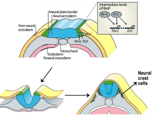
Subsequently the signalling molecules of the Wnt, FGF and BMP families cooperate to activate a distinct combination of transcription factors at the neural plate border including Snail2 (previously known as Slug, the earliest marker of specified neural crest) Sox9/10, Pax3, Id3, Foxd3, Zic and Msx1 which constitute the neural crest specification network (reviewed in Meulemans and Bronner-Fraser, 2004; Steventon et al., 2005; Fig. 1.2). For example, Pax genes are expressed initially in the neural plate border and dorsal neural tube and are targets of Wnt signalling pathway (Sato et al., 2005). Another example is the group of Snail transcription factors, which are among the earliest known markers of neural crest formation and their expression has been used as a read out of neural crest induction in response to different signals. Based on expression analyses, it has been suggested that, in the mouse, Snail1 performs the function of Snail2 in the chicken neural crest (Sefton et al., 1998). However, contrary to observations in frog and avian embryos neither Snail1 nor Snail2 seem to be involved in neural crest production in mice (Murray and Gridley, 2006), suggesting that some of the mechanisms of neural crest induction are not conserved among vertebrates.
Figure 1.2 Neural crest induction, specification and delamination.
Induction initiates at the neural plate border and is mediated by signals including fibroblast growth factor (FGF) from the underlying mesoderm as well as Wnts from mesoderm and adjacent non-neural ectoderm. These signals induce the expression of individual neural plate border specifiers, such as Pax3 and Zic1, in a manner that is dependent on intermediate levels of bone morphogenetic protein (BMP). Expression of these early neural crest specifiers, segregates neural crest from the dorsal neuroepithelium and control its delamination from the neural tube. Finally these cells diversify into a wide variety of cell types (Le Douarin and Kalcheim, 1999), including neurons and glial cells of the peripheral nervous system, melanocytes, smooth muscle and many of the skeletal and connective tissue of the head (adapted from Sauka-Spengler and Bronner-Fraser, 2008).
A.1.2 Delamination
Once the neural crest cells have been induced they migrate out of the neural tube. Neural crest cells lose cell-cell adhesions by undergoing a transition from epithelial to mesenchymal type, which allows them to delaminate and emigrate from the neuroepithelium. More specifically, this epithelial to mesenchymal transition (EMT) requires a switch in the repertoire of cell adhesion molecules expressed by these cells. Thus the crest cells down-regulate N-CAM, N-Cadherin and Cadherin-6B, which are generally expressed by cells of the neural tube and
upregulate Cadherin-7 and Cadherin-11 (Akitaya and Bronner-Fraser, 1992; Nakagawa and Takeichi, 1998; Cano et al., 2000). The transcriptional repressor Snail2 is thought to be crucial for the EMT and recent experiments in chick embryos show that the avian Snail2 directly represses Cadherin-6B during this process (Taneyhill et al., 2007).
The delamination process is also controlled by BMP signalling. Most prominently Bmp4, which comes to be expressed at the dorsal midline of the neural tube. If Bmp4 is added alone to neural tube explants, it stimulates production of neural crest cells. Interestingly, inhibition of BMP4 signalling by Noggin, a BMP4-specific antagonist, in the chick spinal cord prevents crest delamination; the neural crest cells cannot escape from the neural tube and accumulate within it, demonstrating that BMP4 and Noggin act antagonistically to control neural crest delamination (Sela-Donenfeld and Kalcheim, 1999; Smith and Graham, 2001).
Studies in chicken implicate Wnt signalling in the delamination process. It has been shown that BMP signalling controls the cell cycle by modulating cyclin levels and that this effect was mediated by Wnt1 (Burstyn-Cohen et al., 2004).
A.1.3 Migration
The neural crest can be divided into cranial, trunk and vagal neural crest (the cranial more anterior and the vagal the most posterior). Each population migrates along unique pathways contributing to specific cell and tissue types (Le Douarin and Kalcheim, 1999). In this thesis I will mainly focus on the cranial neural crest, that play an important role in sculpting the craniofacial structures by contributing to pigment cells, the neurons and glia of the sensory ganglia, cartilage, bone, smooth muscle and connective tissue of the face (Noden, 1983b; Kontges and Lumsden, 1996). Cranial NCCs migrate into the head periphery in distinct streams that correlate with their places of origin in the different brain regions: forebrain (telencephalon and diencephalon), midbrain (mesencephalon) or hindbrain (metencephalon or rhombencephalon). The hindbrain contains seven transient segments, called rhombomeres (r1-r7) (Lumsden et al., 1991; Birgbauer et al.,
1995; Kontges and Lumsden, 1996; Fig. 1.3). These rhombomeres dictate the axial levels where patterned neural crest cells exit the hindbrain to populate the pharyngeal arches. Neural crest cells that emanate from diencephalon and anterior mesencephalon migrate to the fronto-nasal and periocular regions of the head. These neural crest cells form the head mesenchyme generating some of the bones of the face (Le Douarin and Kalcheim, 1999). Neural crest cells from posterior mesencephalon and rhombomeres r1 and r2 migrate to the first pharyngeal arch originating some of the skeletal components of the face and ear (Table 1.1). The cells arising from r4 populate the second pharyngeal arch forming the lesser horn of the hyoid cartilage as well the stapes bone of the middle ear and the styloid process. The most caudal stream of cranial NCCs migrates from r6, 7 and 8 to populate the third and more caudal pharyngeal arches giving rise to some of the cartilages and connective tissue of the neck (Fig. 1.3; Le Lievre and Le Douarin, 1975; D'Amico-Martel and Noden, 1983).
Figure 1.3 Migratory streams of neural crest
Neural crest cells from diencephalon and mesencephalon migrate to the nasofrontal and periocular area. Migratory streams from posterior mesencephalon also populate the first pharyngeal arch (PA1), which is also populated by neural crest derived from rhombomeres 1 and 2 (r1/r2) together with a small contribution from r3 crest cells. The major contribution to the second pharyngeal arch (PA2) comes from rhombomere 4 crest cells. Neural crest cells arising from r3 and r5 split into strains participating, in two adjacent arches. Neural crest from r6 and r7 migrate to PA3 and PA4. r1-r7, rhombomeres 1-7, PA1-4, pharyngeal arches 1-4 (adapted from Couly et al., 2002).
reports showed that, in fact, these two rhombomeres produce a small amount of neural crest cells that do not exit the hindbrain lateral to these rhombomeres but move anteriorly or posteriorly to join the streams of crest migrating from the adjacent even-number rhombomeres (Sechrist et al., 1993; Birgbauer et al., 1995). The absence of neural crest cell streams associated to r3 and r5 has a key role in reinforcing the segregation of the migratory crest cells from their adjacent rhombomeres. Previous studies in chick showed that the majority of neural crest from r3 and r5 is lost by apoptosis, and this effect is mediated by Bmp4 expression (Graham et al., 1993; Smith and Graham, 2001). However studies in other species such as zebrafish (Schilling and Kimmel, 1994) or Xenopus (Hensey and Gautier, 1998; Hensey and Gautier, 1999) did not observe focal cell death in r3 and r5. Accordingly, mouse embryo studies have also suggested that the segregation of the neural crest cells into streams is not only due to induction of apoptosis in r3 and r5 as shown in chick, but also to the action of cues in the periphery, which act to reinforce and stabilize this region (Trainor et al., 2002). Genetic engineering in mice has shown several mouse embryo phenotypes with defects in cranial crest streams. ErbB4 and Twist act cell non-autonomously to regulate proper neural crest migration (Golding et al., 2000; Soo et al., 2002; Ota et al., 2004). In addition, other molecules such as Sema3A expressed in both r3 and r5 act cell autonomously to regulate cell migration (Eickholt et al., 1999). Studies in frog embryos suggest that Ephrin cell signalling plays an intrinsic role in keeping the neural crest streams segregated (Smith et al., 1997). EphA4 and EphB1 receptor tyrosine kinases are expressed in neural crest cells that populate the third and fourth arches, whereas second arch cells express EphrinB2 ligands. Inhibitory and repelling interactions between these r3/5 neural crest cells and second arch cells function to prevent intermingling between cell populations and keep the neural crest in their respective arch streams (Smith et al., 1997). Interestingly, in mouse it was shown that EphrinB1 acts cell autonomously in cranial neural crest cells and controls their migration. EphrinB1 null neural crest displays abnormal migration by invading regions normally devoid of this cell type.
This led to defects in middle ear, nerve fasciculation and branching (Davy et al., 2004). These studies showed that the segregation of the neural crest is a central step for the correct organization of the pharyngeal system.
The cardiac neural crest is a subpopulation of cranial neural crest originated in the caudal region of the cranial crest. It populates the third, fourth and sixth caudal arches and produces the muscular connective tissue wall of the large arteries as they arise from the heart, as well contributing to the septum that separates the pulmonary circulation from the aorta (Le Lievre and Le Douarin, 1975; Jiang et al., 2000). They can also develop into melanocytes, neurons or cartilage. This cell population will be emphasized in the second section of this introduction, where I will review some of the aspects regulating the development of the embryonic arch arteries.
A.2 The role of neural crest in pharyngeal arch development
There has been a general interest in understanding how the different embryonic populations pattern the pharyngeal arches. In the past, several studies have suggested that the neural crest played a crucial role in coordinating the development of the pharyngeal arches and in giving specific identity to individual arches (Noden, 1983b). More specifically, it was found that if the presumptive first arch neural crest was grafted and forced to populate the second arch, the second arch in the host embryo developed derivatives resembling the skeleton characteristic of the dorsal first arch (Noden, 1983b). These classic transplantation experiments suggested that the neural crest has positional information for organizing the pharyngeal arches already in the neural primordium, before emigration. Experiments by other groups suggested that the fate determination of cranial neural crest was due in part to differential Hox gene expression along the hindbrain, where these cells are originated (Trainor et al., 2002). Hox genes are involved in establishing anterior-posterior (cranial-caudal) positional identities (Hunt et al., 1991); unique combinations of these genes are
expressed among different hindbrain rhombomeres and their associated neural crest cells (reviewed in LaBonne and Bronner-Fraser, 1998). These studies indicated that these genes were also involved in specifying the anterior-posterior identity of the neural crest cells populating each of the arches. Mutational analysis of Hox genes in mice, have shown that these genes are required for the normal morphogenesis of arch-derived skeletal elements (Gendron-Maguire et al., 1993; Rijli et al., 1993; Kanzler et al., 1998; Gavalas et al., 2001). This is exemplified by inactivation studies of Hoxa2 gene, which is specifically expressed in the neural crest that originates from rhombomere 4 and populates the second pharyngeal arch. In mice mutant for Hoxa2 neither first nor second arches express this gene and consequently multiple cranial skeletal defects were evident, in which the second arch skeletal derivatives assume first arch characteristics. These results indicate that Hoxa2 expressed in rhombomere 4 crest cells is essential for the identity of the second arch (Gendron-Maguire et al., 1993; Rijli et al., 1993; Kanzler et al., 1998) by inhibiting the formation of skeletal elements in this arch. Hox3 group genes are expressed in the third pharyngeal arch (Hunt et al., 1991; Prince and Lumsden, 1994; Hunter and Prince, 2002). Compound mutants of Hoxa3 and Hoxd3, exhibit defects in the laryngeal cartilages (Condie and Capecchi, 1994).
Other transcription factors, in particular the Dlx family can also modulate the development of the derivatives of the pharyngeal arches, more specifically the skeletal tissues derived from neural crest. Dlx genes are expressed in the pharyngeal arches, with Dlx1/Dlx2 being expressed broadly and Dlx5/6 expressed in the more distal mesenchyme ( Qiu et al., 1997; Depew et al., 1999; Depew et al., 2002; Depew and Simpson, 2006). In Dlx5/6 mutant mice some of the elements of the first pharyngeal arch fail to form, such as the lower jaw. In their place an upper jaw is formed. The authors concluded that loss of Dlx5 and Dlx6 resulted in a homeotic transformation of the lower jaw into an upper jaw, indicating that Dlx5 and Dlx6 are major determinants of the cellular identity within the pharyngeal arches (Depew et al., 2002). Although these studies support a
major role for the neural crest and their transcription factors in patterning the pharyngeal arches, recent work shows that the fate determination of cranial neural crest is strongly influenced by instructive signals provided by adjacent tissues. For example, studies in mouse embryos showed that expression of Dlx transcription factors is regulated by ET-1 signals from the epithelia of the pharyngeal arches. Endothelins (ETs 1, 2 and 3) are a family of small peptides that play important roles in neural crest cell differentiation and survival (Yanagisawa et al., 1998). More specifically, endothelin-1 (ET-1) is proteolytically generated from its inactive precursor by endothelin-converting enzyme–1 (ECE-1) and acts on the endothelin-A (ET-A) receptor. ET-1 is expressed in the epithelium and endothelium of the pharyngeal arches (Kurihara et al., 1995), while the receptor ET-A is expressed in migrating neural crest populating the pharyngeal arches (Clouthier et al., 1998). In mouse studies, genetic disruption of this ET-1/ECE-1/ET-A pathway resulted in several developmental defects including craniofacial anomalies. More specifically, ET-1 null embryos display homeotic transformation of the mandibular arch with downregulation of Dlx5/Dlx6. These observations implicate the Endothelin pathway in the patterning of the pharyngeal arch system as a positive regulator of Dlx5/Dlx6 expression (Kurihara et al., 1994; Ozeki et al., 2004).
Even though neural crest is critical for the correct development of the pharyngeal arches, this role is more responsive than instructive. Cranial neural crest cells interact with and consequently respond to signals from endoderm (Couly et al., 2002; Cerny et al., 2004; Le Douarin et al., 2004), paraxial mesoderm (Trainor and Krumlauf, 2000) and ectoderm ( Lumsden, 1988; Bobola et al., 2003) giving rise to various types of tissues. These interactions modulate prior specifications and are necessary for patterning the arches as well the correct morphogenesis of the pharyngeal arch tissues.
Below I discuss some of the interactions neural crest cells have with the other cell players as well some of the genetic factors involved.
A.3 The role of endoderm
Ablation and transplantation experiments in birds revealed that the endoderm instructs neural crest cells to form the different skeletal derivatives of the arches, having an effect on the size, shape and position of some components of the facial skeleton (Couly et al., 2002). This work strongly suggests a central role for the pharyngeal endoderm during arch morphogenesis. Several studies also propose that this role comes from the ability of the endoderm to segment the pharyngeal apparatus by forming endodermal pouches, which provide specific signals to surrounding tissues (Piotrowski and Nusslein-Volhard, 2000; Piotrowski et al., 2003; Graham et al., 2004). Endodermal pouches form in an anterior to posterior sequence, separating the mesoderm and neural crest of the arches and defining the anterior and posterior limits of each arch. The formation of the pouches indicates the correct segmentation of the pharyngeal endoderm (Crump et al., 2004; Quinlan et al., 2004). These structures are polarized and have characteristic expression of several genes. For instance, the anterior half of each pouch expresses Bmp7, the posterior half expresses Fgf8 and Pax1 is expressed in the dorsal domain of each pouch (Veitch et al., 1999). In this study, early ablation of cranial neural crest cells in chick embryos did not affect the development and function of endoderm cells and the pharyngeal arches are normally patterned and regionalized. This implies that the patterning of endoderm and the pharyngeal segmentation is not dependent on neural crest (Veitch et al., 1999). In zebrafish mutants bonnie and clyde (bon) and casanova (cas), which do not produce endoderm, the pharyngeal cartilages fail to form (David et al., 2002). Moreover, the reintroduction of endoderm in these zebrafish embryos showed that the endoderm is required for the development of neural crest cells, including their identity, survival and differentiation into arch cartilages (David et al., 2002).
There are many genetic factors expressed in the endodermal pouches known to be key players for the correct morphogenesis of the arches. Several studies have shown a major role of FGF signalling for the formation of pharyngeal pouches.
Fgf8 is expressed within the developing pharyngeal arch ectoderm and endoderm during neural crest cells migration through the arches. Mouse embryos hypomorphic for Fgf8 have impaired development of the posterior pharyngeal pouches (Abu-Issa et al., 2002). In the zebrafish doubly reduced for Fgf8 and Fgf3, the pouches fail to form (Crump et al., 2004) and it has also been shown that the neural crest cells are induced to form cartilage via the action of FGF-3 and FGF8 emanating from the endoderm (David et al., 2002; Walshe and Mason, 2003). In these studies it was evident a correlation between alterations in pouch morphology and consequent defects in cartilage derived from neural crest.
Studies perfomed in early chicken embryos showed that ablation of the endoderm before neural crest migration eliminates the facial structures (Couly et al., 2002). This study showed that the most cranial endodermal regions were required for nasal capsule morphogenesis, while the more caudal regions were required for the development of Meckel’s cartilage and the mandibular joint (Couly et al., 2002).
Together, these zebrafish and chicken studies determine the causality between alterations in pharyngeal endoderm and consequent defects in cartilage derived from neural crest.
In addition, mutations of the Sonic hedgehog (Shh) gene, encoding for the morphogen Shh, have been associated to cases of holoprosencephaly in humans, a syndrome that includes a variety of malformations, such as absence of the lower jaw, cyclopia, and cleft palate (Belloni et al., 1996; Roessler et al., 1997). Recent studies, where Hedgehog signalling was blocked specifically in mouse NCCs, showed cell death of neural crest followed by severe head skeleton abnormalities (Jeong et al., 2004) suggesting that Shh is important for survival of the neural crest cells that populate the first pharyngeal arch. Furthermore, the absence of Shh in mutant mice results in strong abnormalities in the patterning of first branchial arch during development (Moore-Scott and Manley, 2005). Chicken studies showed Shh expression throughout the pharyngeal and gut endoderm, especially in the endoderm of the first pharyngeal arch (Brito et al., 2006). The
elimination of the anterior foregut endoderm, that expresses Shh, prevents upper and lower jaw development; however this phenotype can be rescued by applying a bead carrying Shh protein (Brito et al., 2006).
Therefore, all these studies show that the pharyngeal endoderm plays a crucial role in regulating the morphogenesis of pharyngeal arch derivatives.
A.4 The role of the ectoderm
The ectoderm that surrounds the neural crest mesenchyme also plays a role in regulating neural crest derivatives. Previous studies in birds showed that a zone of frontonasal ectoderm stimulated the proliferation and differentiation of underlying neural crest cells (Hu et al., 2003). A molecular boundary in the frontonasal ectoderm, defined by Fgf8 and Shh expression, indicates the initial site of outgrowth of the upper beak. When transplanted to an ectopic location, the frontonasal ectodermal zone activated a cascade of molecular events that ultimately re-programmed the neural crest-derived mesenchyme, producing a duplication in the upper beak structures. This indicates the ability of the ectodermal epithelium to re-pattern skeletal elements derived from the neural crest (Hu et al., 2003). The signals with the ability to re-specify the fates of neural crest are being discovered. Although Shh and Fgf8 are expressed in adjacent domains in this region it is not clear if they are the molecules that instruct the beak formation or simply molecular markers of this important boundary.
FGF signalling in the ectoderm plays critical functions in the development of the pharyngeal arch derivatives. Various studies have shown that conditional inactivation of the FGF8 signalling in the ectoderm resulted in the disappearance of the most proximal portion of the mandible (Trumpp et al., 1999) as well as cardiovascular defects derived from impaired development of the embryonic arteries (Macatee et al., 2003). Furthermore, analysis of mouse embryos hypomorphic for Fgfr1 demonstrate that ectoderm FGF signalling patterns the pharyngeal region to create a permissive environment for the entry of neural crest
cells (Trokovic et al., 2003; Trokovic et al., 2005). Moreover, pharyngeal arch explant experiments show that Fgf8 expression in the ectoderm of the second arch interacts with Hoxa2 positive crest cells to modulate the development of second arch skeletal derivatives (Bobola et al., 2003).
Taken together, it is clear that ectoderm mediated signalling plays an important role in determining the patterning of pharyngeal arch structures.
A.5 The role of cranial mesoderm
The cranial mesoderm generates the branchiomeric skeletal muscles of the head and neck which include muscles of mastication, derived from the first arch; muscles of facial expression, derived from the second arch; and muscles of the pharynx and larynx, derived from more caudal arches (Noden, 1983a; Couly et al., 1992; Trainor et al., 1994). It also provides sources for cardiovascular tissues such as the endothelia of the blood vessels and heart (Couly et al., 1995). Experiments in chick embryos have suggested that cranial mesoderm is able to direct cranial neural crest cell movement (Noden, 1986; Noden, 1988; Ferguson and Graham, 2004). Both cell populations have been examined in detail in the context of pharyngeal apparatus and it was observed that neural crest cells initially populate the superficial regions of each pharyngeal arch, and afterwards cover the mesodermal core tissue of the arches (Trainor and Tam, 1995). This study suggests that the consequent tissue-tissue interactions between neural crest and mesoderm could be important to organize the skeletal, myogenic and endothelial derivatives within the arches (Trainor and Tam, 1995).
Gene expression analysis of some transcription factors, such as Paraxis, Tbx1 and Hoxb1 suggest some degree of regionalization in the cranial mesoderm (Noden and Trainor, 2005). Some techniques were developed to allow the transposition of hindbrain cells in cultured mouse embryos (Trainor and Krumlauf, 2000). These experiments showed that, if neural crest from the second pharyngeal arch is transplanted into the first arch, it downregulates its expression
of Hoxb1. But, if it is transplanted along with second arch mesoderm, then, Hoxb1 expression is maintained. Although the cranial mesoderm doesn´t seem to have a primary role in pharyngeal arch patterning, it is responsible to maintain the normal expected Hox gene expression patterns (Trainor and Krumlauf, 2000).
Overall it is clear that pharyngeal arch development is a complex process, which involves co-ordinated interactions between different cell populations in order to generate a wide range of derivatives with correct morphology. Some signalling molecules that could also be included in this section (such as Tgfβ or Retinoic acid signalling molecules) will be referred in the next section in the context of vascular development.
B. EMBRYONIC ARTERIES AND HEART OUTFLOW TRACT
B.1 Formation of blood vessels
The cardiovascular system is the first functional organ to develop in the vertebrate embryo being essential for its survival, also assuring oxygen and nutrient supply and efficiently removal of waste products. The vasculature of the mouse embryo is composed of small and large blood vessels. The smaller vessels consist only of endothelial cells (ECs) whereas, the larger vessels, are surrounded by mural cells (perycites in medium-sized and smooth muscle cells in large vessels). In the embryo the vessels can develop by two distinct processes: vasculogenesis and angiogenesis.
Vasculogenesis assures the creation of the first embryonic vasculature. It starts in the yolk sac with the specification of endothelial cell precursors (angioblasts) from mesoderm. Subsequently the angioblasts proliferate and connect to construct a primitive tubular network composed of capillaries, arteries and veins (Risau and Flamme, 1995).
There are several factors essential for vasculogenesis. Experiments in chick and quail embryos have suggested that FGF signalling is important for the initiation of