The beginning of this adventure started when at university I had to look for an article for a presentation about molecular regulation in prokaryotes for a university project. I found in PubMed by chance a very interesting paper: Mani, N. & B. Dupuy, (2001) Regulation of toxin synthesis in Clostridium difficile by an alternative RNA polymerase sigma factor. PNAS 98:5844‐49. At the time I would never though that my life would change so much and that I would go to live in a new country, learn a new language, and have a chance to collaborate with the people that worked in this paper that I found fascinating. First of all I would like to say “Muito Obrigada” to my family, that have always encourage me in my pursuits and that have been at my side all the time in all my adventures, including this one. I would like to say also “Muito Obrigada” to my microbiology teacher Prof. Mario Santos that opened my eyes to this incredible big world of microbiology that is not at our sight, but it is so interesting, so complex and at the same time so small. I would like also to thank him for his assistance in finding my training course and accepting to be my supervisor of my PhD work.
The work of this PhD was done in two laboratories in the Institut Pasteur: Unité de Génétique Moléculaire Bactérienne and later in Unité des Toxines et Pathogénie Bacteriénne. I would like to thank Prof. Stewart Cole and Prof. Michele Mock for receiving me in their laboratories. A very special “Remerciment” goes to my mentor, Dr. Bruno Dupuy. I must say that it is very difficult to put in words how much grateful I am for all his support and sharing during these five years of work. Merci de m’avoir donné l’opportunité de travailler avec toi. J’ai beaucoup appris, pas seulement au niveau technique, mais aussi à penser, et à savoir dépasser les obstacles sans se décourager. Tous ces conseils resteront non seulement pour le travaille mais aussi pour la vie. Merci pour ta présence constante, même dans des situations ou le temps te manquait, ta patience pour toutes mes questions, ton encouragement qui m’a toujours fait aller plus loin, même dans des moments difficiles. C’est un plaisir travailler avec toi.
I am very grateful to Susana Matamouros who was my “Big Sister” in the lab and outside of it. Always interested in my work, very patient and available for my questions and also a very good friend.
I am also very grateful to Isabelle Martin‐Verstraete that showed me how to work with Bacillus subtilis and closely followed the advances in my thesis project and always found a little time for me in her busy agenda. Thank you very much for your time, interest and helpful discussions.
Emilie and Marc my labmates I am grateful for their good humour, optimism, helpful discussions and their friendship. Many thanks to the new arrivals in the lab, Marie and Fréderic, who are always available and keeping a good humour in the lab.
I am also grateful to Prof. Linc Sonenshein for helpful discussion and his support on this project.
Many thanks to the members of the Unité Bactéries anaérobies et Toxines directed by Michel Popoff. Especially to Philippe Bouvet, Maria Manich and Maryse Gibert, for helpful discussions about C. difficile and some techniques that I have learned with them. I would also like to thank the members of the Unité des Toxines et Pathogénie Bacteriénne. In particular, I am grateful to Agnès Fouet for helpful discussion and encouraging ideas, Patricia Sylvestre for the CcpA antibodies, and Evelyne Couture‐Tosi for contributing with her expertise on electron microscopy to my work. I am also thankful to the team of Claire Janoir with whom we collaborated on the use of the mouse model, with a special thanks to Cecile and Diana. Many thanks to the StaPa association where I had opportunity to meet people working in many different areas of research and discover new talents in me. Thanks to my friends from the microbiology course that have passed or will be passing their thesis soon. Many thanks also to the Portuguese community in the IP campus that brought some energy and laughs when I needed it. Thank you all that participated in the preparation of this manuscript, in particular Tarn Duong for correcting my English mistakes. I am also thankful to the Jury members that accepted to be part of my thesis defence and carefully read my work presented here. Last, but not the least...
In these past five years I got to know a lot of people. Some already left other just arrived. Having the opportunity to meet you all has contributed to who I am now and also to my way of working. All scientific discussions, ideas and helpful assistance have somehow contributed to this work. To YOU all I would like to say “Muito Obrigada “. This work was supported with the funding from the Fundação para a Ciência e a Tecnologia (PhD Fellowship SFRH/BD/16399/2004) for the first four years and from ERA‐NET PathoGenoMics/CDIFFGEN for the last year.
Everything has a beginning and an end
with its excitements,
but the journey is always the best part.
Main Articles
Antunes A, Martin-Verstraete I, Dupuy B. (2010) CcpA-mediated repression of Clostridium
difficile toxin expression, submitted in Molecular Microbiology.
Antunes A, Monot M, Deneve C, Janoir C, Martin-Verstraete I, Dupuy B. (2010) Role of the carbon catabolite repression in the C.difficile physiopathology, in preparation.
Collaborations
Carter GP, Douce GR, Howarth PM, Poon R, Antunes A, Govind R, Dupuy B, Rood JI, Lyras D. (2010) Clostridium difficile NAP1/027 hypervirulence is not associated with mutation of TcdC, submitted in Infection Immunity.
Camiade E, Peltier J, Bourgeois I, Couture-Tosi E, Courtin P, Antunes A, Chapot-Chartier MP, Dupuy B, Pons JL. (2009) Acp, a N-acetylglucosaminidase of Clostridium perfringens implicated in cell separation and stress-induced autolysis, Journal of Bacteriology. In
revision.
Dupuy B, Govind R, Antunes A, Matamouros S. (2008) Clostridium difficile toxin synthesis is negatively regulated by TcdC, (review) Journal Medical Microbiology, 57(Pt 6): 685-9.
Books
Ana Antunes and Bruno Dupuy. (2010) Chapter: Molecular Methods to study transcriptional regulation of Clostridium difficile toxin genes. Methods in Molecular Biology: Clostridium
difficile. Mullany P, Roberts, AP (Eds) in press.
Para os efeitos do disposto nº1 do Artigo 40º do Regulamento de Estudos Pós-Graduados da Universidade de Lisboa, publicado no Diário da República – nº153, II Série de 5 de Julho de 2003, declaro que participei integralmente na concepção e execução do trabalho experimental, na interpretação de resultados e na redacção dos manuscritos.
Resumo
Clostridium difficile é uma bactéria Gram-positiva, em forma de bastonete, anaeróbia estrita
e esporulante. Esta é reconhecida como uma das principais causas de enterocolites em pacientes hospitalizados e como a primeira causa de diarreias infecciosas nosocomiais em adultos. Os sintomas clínicos de infecções digestivas de C. difficile são diversos e variam desde simples diarreias até colites pseudomembranosas, podendo ser fatais para o paciente.
A patogenicidade de C. difficile inclui três etapas importantes: germinação dos esporos, colonização do hospedeiro e produção de toxinas. Após contaminação com C. difficile, ocorre germinação dos esporos e a multiplicação de formas vegetativas. C. difficile adere à mucosa intestinal e coloniza o intestino do hospedeiro. As estirpes patogénicas produzem duas toxinas (TcdA e TcdB) que são consideradas os principais factores de virulência desta bactéria. Apesar dos sintomas clínicos associados à infecção com C. difficile variarem, as formas severas da doença estão directamente correlacionadas com o nível de síntese destas duas toxinas. Assim sendo, a regulação da sua expressão e síntese constitui uma etapa importante na patogenicidade de C. difficile. Por isso, o estudo das condições de expressão e regulação das toxinas de C. difficile, contribuirá de maneira significativa para o estabelecimento de estratégias preventivas à doença e desenvolvimento de novos alvos terapêuticos.
A expressão génica das toxinas é regulada por vários factores ambientais, como a temperatura e a fase de crescimento. Em particular, fontes de carbono que são rapidamente metabolizadas através do sistema fosfotransferase dependente do fosfoenolpiruvico (PTS), tal como a glucose, reprimem a expressão dos genes das toxinas, sugerindo uma regulação através do mecanismo de repressão catabólica de carbono (CCR). O CCR é um mecanismo de regulação, que em presença de açúcares PTS no meio de cultura, reprime a expressão de certos genes e operões, cujos produtos estão geralmente envolvidos na utilização de fontes de carbono alternativas. O mecanismo global do CCR é mediado pela CcpA (Proteína A de Controlo Catabólico), um regulador global membro da família de reguladores transcripcionais LacI/GalR. De forma a exercer o controlo sobre os genes alvo, CcpA liga-se a uma região de ADN especifica no gene-alvo, designado elemento de resposta catabólica (cre). Dependendo da localização do local cre, a ligação de CcpA vai induzir uma repressão ou estimular a transcrição do gene-alvo. A proteína HPr-Ser46-P, uma forma fosforilada de HPr, elemento fundamental do sistema PTS, é capaz de interagir com CcpA e, assim,
aumentar a afinidade de CcpA ao ADN-alvo. Apesar do CCR estar muito bem caracterizado no organismo modelo Gram-positivo, B. subtilis, pouco foi descrito sobre o mecanismo global do CCR no grupo dos Clostridia e nada é conhecido em C. difficile.
No presente trabalho investigámos o papel do CCR na expressão génica das toxinas de C.
difficile utilizando abordagens in vivo e in vitro. Para tal construimos, usando a técnica do
ClosTron, estirpes de C. difficile mutantes nos elementos principais do mecanismo CCR, como os componentes do PTS e CcpA na estirpe JIR8094. A inactivação dos genes ptsI,
ptsH e ccpA resultarou na ausência de repressão da expressão génica das toxinas em meio
de cultura com glucose. No entanto, a inactivação de hprK não provocou qualquer alteração na repressão dos genes das toxinas em meio de cultura com glucose. Adicionalmente, demonstrámos que CcpA se liga a regiões reguladoras dos genes tcdA e tcdB mesmo na ausência de motivos cre evidentes. Para além disso, a proteína HPr e a sua forma fosforilada, HPr-Ser-P, não aumentam a capacidade de fixação de CcpA aos genes das toxinas. No entanto, a fructose-1,6-bifosfato (FBP), um cofactor do complexo CcpA-HPr-Ser-P, é capaz de aumentar a afinidade de fixação de CcpA na ausência de HPr-Ser-P. No seu todo, estes resultados sugerem que a regulação da expressão génica das toxinas em resposta a presença de fontes de carbono mediado por CcpA envolve um novo modo de regulação dependente de CcpA.
A regulação coordenada de genes de virulência dependentes de CcpA em resposta a disponibilidade de fontes de carbono, poderá constituir uma etapa importante na sobrevivência de C. difficile quando este compete com outros micróbios na colonização do hospedeiro. Para além disso, CcpA também foi demonstrado estar envolvido no controlo de várias funções importantes das bactérias patogénicas sem ser as toxinas, tal como o biofilme, pilus de tipo-IV, e a colonização. Embora os factores principais de virulência sejam estudados há muitos anos, os processos de virulência específicos à infecção com C. difficile ainda estão por descrever.
Para perceber a importância da glucose, como fonte de carbono e compreender o envolvimento de CcpA na fisiologia de C. difficile, executámos uma análise do transcriptoma da estirpe parental JIR8094 e da mutante ccpA cultivadas em meio sem ou com glucose. Em paralelo ao estudo do transcriptoma in vitro, usámos o modelo animal rato para determinar o regulão de CcpA que é especificamente expresso durante a infecção in vivo e que poderá ter um papel importante durante a colonização do intestino do hospedeiro pela bactéria C. difficile.
Os resultados obtidos mostram que a glucose afecta cerca de 18% dos genes de C. difficile, e que CcpA regula cerca de metade destes genes. Para além disso, CcpA regula genes cujas proteínas estão envolvidas no metabolismo do carbono, como a glicólise, a via das pentoses-fosfato e a fermentação. CcpA regula também várias vias de fermentação a partir de aminoácidos como a redução da glicina e da leucina, sugerindo que CcpA poderá agir como um elo de ligação entre o metabolismo do carbono e o metabolismo do azoto. CcpA também tem um papel importante em algumas funções celulares como a esporulação e a resposta ao stress.
Adicionalmente, investigámos também o envolvimento de CcpA na regulação de mecanismos de virulência que para além da expressão génica das toxinas poderão ser importantes no processo de infecção de C. difficile. Ao todo observámos que 183 genes são regulados por CcpA unicamente no transcriptoma in vivo, incluindo genes cujas proteínas estão envolvidas na utilização da etanolamina. A etanolamina é um elemento abundante no intestino do hospedeiro e que C. difficile provavelmente utiliza como fonte de carbono, azoto e energia.
Palavras-Chave:
Clostridium difficile, regulação de factores de virulência, repressão catabólica do carbono,
Abstract
Clostridium difficile, a low G+C Gram-positive, spore-forming anaerobic bacterium is the
major causative agent of nosocomial diseases associated to antibiotic therapy in adults. C.
difficile infection (CDI) can produce a wide spectrum of clinical manifestations that may range
from asymptomatic carriage to mild self-limiting antibiotic-associated diarrhoea (AAD) and in some cases to life-threatening pseudomembranous colitis (PMC). The pathology process of
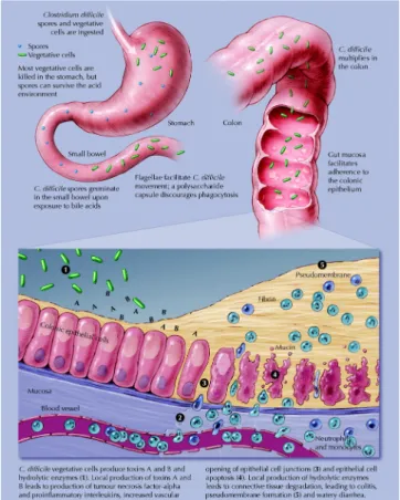
C. difficile is though to be divided in three main steps: spore germination, host colonization
and toxin production. After contamination by C. difficile, spores germinate and the vegetative forms multiply. C. difficile adhere to the mucus layer and colonize the gut. Then pathogenic strains produce two toxins (TcdA and TcdB), considered as the major virulence factors. The spectrum of diseases caused by C. difficile is highly variable and depends for the severe forms on the level of toxin produced. Thus, regulation of toxin synthesis is a critical determinant of the C. difficile pathogenicity.
Several environmental factors regulate toxin expression. In particularly, rapidly metabolizable sugars that enter by the phosphoenolpyruvate-dependent phosphotransferase system (PTS), such as glucose, repress toxin expression suggesting that this regulation involves the carbon catabolite repression (CCR) mechanism. In presence of PTS sugars in the medium, the CCR represses expression of genes and operons, whose genes products are generally involved in the utilization of alternative carbon sources(Stulke & Hillen, 1999). The pleiotropic regulator CcpA is the major global transcriptional regulator of CCR. CcpA-regulated genes involves the binding of CcpA to cis-acting catabolite responsive elements (cre site). The DNA-binding activity of CcpA is enhanced by its interaction with HPr-Ser-P a phosphorylated form of HPr, a component of the PTS. Although the CCR is well-known in the Gram-positive organism model B. subtilis, little is known about this global regulatory mechanism in C. difficile.
We investigated the role of CCR in C. difficile toxin expression by in vivo and in vitro approaches. We constructed, using the ClosTron technique, mutants of the major components of the CCR signal transduction pathway including PTS elements and CcpA in strain JIR8094. Inactivation of the ptsI, ptsH and ccpA genes resulted in derepression of toxin gene expression in glucose-medium, whereas repression of toxin expression was still observed in the hprK mutant.CcpA was found to bind to regulatory regions of tcdA and tcdB genes but not through consensus cre site motif. Interestingly, neither HPr nor HPr-Ser46-P stimulate CcpA binding affinity but fructose-1,6-biphosphate (FBP) alone enhanced CcpA binding in absence of HPr-Ser46-P.
Coordinate CcpA-dependent regulation of virulence and carbon utilization genes could be critical for fitness when C. diificile compete with other microbes for niche colonization. Although the main virulence factors have been studied since many years, in vivo specific virulence processes of C. difficile are still poorly understood.
We performed whole-transcriptome analysis during the growth of JIR98094 (wild-type) and its isogenic ccpA mutant strains in media without and with glucose, to understand the impact of glucose in C. difficile physiology and the role of CcpA in this process. In parallel, we used the axenic mouse model to determine the CcpA regulon specifically expressed during the in
vivo infection, which could be critical during the C. difficile establishment in the gut.
We showed that glucose affected about 18% of all C. difficile genes, and almost half of them were mediated by a CcpA-dependent mechanism. CcpA plays a central role in the carbon metabolism, regulating central pathways like glycolysis, pentose-phosphate shunt and fermentation. Moreover, CcpA regulates several amino acids fermentation pathways such as glycine and leucine reduction, suggesting that CcpA could act as a link between the carbon and the nitrogen metabolism in this bacterium. In addition, we showed evidence that CcpA is also involved in some important cellular functions like stress response and sporulation.
Finally, we also investigated the role of CcpA in the regulation of virulence mechanisms other than the toxin expression that are important for C. difficile infection process. Interestingly, 183 genes were regulated by CcpA only in the in vivo transcriptome. As an example, CcpA stimulated the expression of genes involved in the ethanolamine utilization, an abundant compound of the intestinal tract that can be utilized as a carbon and nitrogen source during the C. difficile gastrointestinal lifestyle.
Keywords: Clostridium difficile, regulation of virulence factors, carbon catabolite repression, transcriptome analysis, CcpA.
Table of contents
Abbreviations ………...……….………... 19
1. INTRODUCTION …...………..….... 21
1.1 The gut microbiota ………...……….……... 23
1.2 The Clostridium genus ……….…….. 28
1.2.1 Non pathogenic Clostridia
……….……... 31
Clostridium acetobutylicum: a solvent producer
………….….……….. 32
Clostridium butyricum: a hydrogen producer
……….……….………...…… 34
Clostridium thermocellum: a cellulose degradation organism
……….…….. 34
Clostridium pasteurianum: a nitrogen fixing organism
……..…….…...…… 35
Clostridia as cancer therapy agents
………...……….….. 35
1.2.2 Pathogenic Clostridia
...…... 37
Clostridium septicum
... 37
Clostridium tetani and Clostridium botulinum
... 38
Clostridium sordellii
... 40
Clostridium novyi... 40
Clostridium perfringens... 41
Clostridium difficile... 43
1.3 Clostridium difficile ... 43
1.3.1 C. difficile Epidemiology... 43
C. difficile Transmission... 45
Incidence of C. difficile infection
... 45
Risk Factors
... 46
Diagnosis
... 46
Treatment
... 47
Relapses
... 48
Costs of C. difficile infections
... 49
Prevention of CDI
... 49
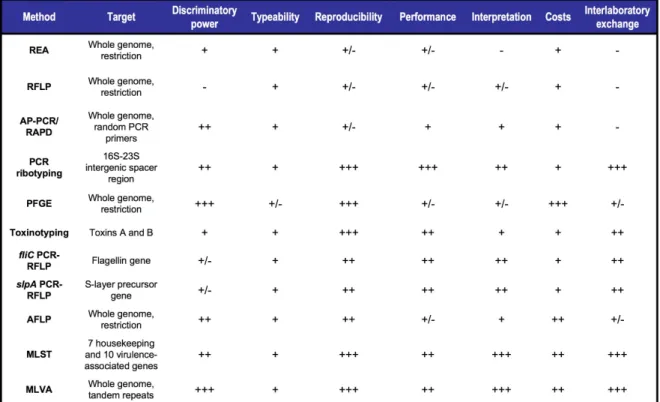
Typing
... 50
C. difficile Epidemic Strains
... 52
Animal Models
... 54
1.3.2 C. difficile Physiopathology
... 56
Spore Germination
... 58
Colonization and Adhesion
... 59
Toxins
... 61
1.3.3 Regulation of C. difficile virulence factors
... 68
1.3.3.1 Regulation of Colonization factors
... 68
1.3.3.2 Regulation of Toxins
... 69
1.3.3.2.1 Influence of environmental factors
... 76
Growth Phase
... 76
Temperature... 76
Antibiotics... 77
Biotin... 78
Amino Acids... 78
Carbon Sources... 80
1.4 Carbon Catabolite Repression (CCR) in Gram-positive bacteria ... 82
1.4.1 The PTS
... 82
1.4.2 Operon-specific CCR mechanisms
... 83
Inducer exclusion and Inducer expulsion
... 84
Regulation mediated by HPr-His-P and EIIB-P
... 85
1.4.3 Global mechanism of CCR
... 87
1.4.4 Impact of CCR in bacteria physiology
... 92
1.4.5 Impact of CCR in bacteria pathogenicity
... 94
1.4.6 CCR in Clostridia
... 96
2. RESULTS ... 99
ARTICLE 1
... 105
CcpA-mediated repression of Clostridium difficile toxin expression. Antunes A, Martin-Verstraete I, Dupuy B. (2010) submitted in Molecular Microbiology. ARTICLE 2
... 163
Role of Carbon Catabolite Repression (CCR) in the C. difficile physiopathology. Antunes A, Monot M, Deneve C, Janoir C. Martin-Verstraete I, Dupuy B. (2010) manuscript in preparation.
3. CONCLUSIONS AND PERSPECTIVES ... 243
4. REFERENCES ... 249
19
Abbreviations
BCAAs branched-chain amino acids BSA Bovine serum albumin CCA Carbon Catabolite Activation CcpA Catabolite Control protein A CCR Carbon Catabolite Repression CDI Clostridium difficile infection
CFU colony-forming unit
cre catabolite responsive element Cwp Clostridial wall protein
DMSO Dimethylsulfoxide
dNTP Desoxyribonucleotide triphosphate DTT Dithiothreitol
EDTA Ethylenediamine tetra-acetic acid FBP Fructose-1,6-bisphosphate GDH Glutamate dehydrogenase GTP guanosine triphosphate IPTG Isopropylthio-ß-D-galactoside
kDa KiloDalton
MIC Minimum inhibitory concentration MOPS 3-(N-morpholino)-propanesulfonic acid ORF Open Reading Frame
PBS phosphate buffer saline
PolydI-dC Polydeoxyinosinic-polydeoxycytidylic acid PEP phosphoenolpyruvate
PPi Pyrophosphate
PTS phosphoenolpyruvate:carbohydrate phosphotransferase system qRT-PCR quantitative reverse transcriptase polymerase chain reaction RNase Ribonuclease
SCFAS Short-chain fatty acids
SDS-PAGE Sodium dodecyl sulfate polyacrylamide gel electrophoresis TCA tricarboxylic acid
23
1.1 The gut microbiota
The adult human gut represents the biggest body surface in contact with the environment (300-400m2), in which the intestinal microflora forms a complex dynamic ecosystem with the “host” human cells. The homeostasis of this ecosystem is maintained by mutual crosstalk between the immune system, the resident prokaryotes and the epithelium cells. However when the gut-microbe relationships are disturbed, it results in a spectrum of intestinal disorders (Neish, 2009).
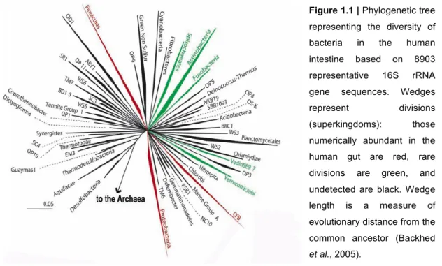
The gut is used for nutrients absorption and defence against potential pathogens. The number of microorganisms present is higher than the number of cells of our entire body. The normal human flora, or microbiota, is vast both in its quantitative mass and its qualitative diversity (Neish, 2009). It contains three types of organisms - bacteria, archaea and eukarya (Backhed et al., 2005). Bacteria achieve the highest cell densities in the normal human gut with approximately 1014 prokaryotic organisms constituted of 30 known genera and more than 500 species (Fig. 1.1) (Neish, 2002).
The population composition is quite stable along the different gut locations. However the number of cells varies greatly, ranging from 1011 cells/g content in the ascending colon to 107-8 in the distal ileum and 102-3 in the proximal ileum and jejunum (Fig. 1.2) (Neish, 2009).
Figure 1.1 | Phylogenetic tree
representing the diversity of bacteria in the human intestine based on 8903 representative 16S rRNA gene sequences. Wedges represent divisions (superkingdoms): those numerically abundant in the human gut are red, rare divisions are green, and undetected are black. Wedge length is a measure of evolutionary distance from the common ancestor (Backhed
475
Anaerobes are more abundant than aerobes in the bacterial community of the gut (more than 98 % of organisms are strict anaerobes (Neish, 2002). The majority of the bacterial population (60 %- 90 %) are representatives of two divisions: the Bacteroidetes and
Firmicutes (Neish, 2009). Interestingly, eukaryotic fungal species have also been identified
as components of the microbiota (Neish, 2009). Most of the bacterial community is indigenous, stable and long-term resident (autochthonous) although transient members (allochthonous) can be also found, such as enteric pathogens (Neish, 2009).
The foetal gut is sterile and colonization begins immediately after birth, with Enterobacteria and bifidobacteria being early colonizers (O'Hara & Shanahan, 2006). Moreover this colonization will lead to a stable community although with a microbial composition specific to each individual (Neish, 2009). Diet plays a major role on the ratio between the microbial species diversity and the number of strains in the intestinal flora. Furthermore, the intestinal physiology and the host defence mechanisms also influence the microbiota by preventing
Figure 1.2 | Distribution of microbiota in the gut and the main anaerobic and aerobic genera found.
Cecum/ascending colon is a “bioreactor” characterized by the greatest amount of bacteria, the most metabolic activities, and fermentation of the short-chain fatty acids (SCFA). The distal ileum is enriched in GALT (gut-associated lymphoid tissue) and is the dominant site of luminal sampling and mucosal adaptative immune activity (adapted from (Neish, 2009; O'Hara & Shanahan, 2006).
25 of the flora throughout the gastro-intestinal tract. The major factors influencing the composition of the microflora are summarized in Table 1.1 (Holzapfel et al., 1998).
These factors might be related to several changes such as the host physiological conditions (aging, stress, health status, cultural environment), the composition of the diet and the environmental circumstances (e.g. contamination with pathogens, use of medicaments). In this way the conditions underlying digestion (e.g. pH, substrate availability, redox potential, transit time, flow of enteric fluid, IgA secretion, etc) may be modulated. This could result in a decline of the beneficial bacteria and in an increase in potentially harmful bacteria. Changes in diet or climate, aging, medication, illness, stress or infection generally lead to an increase in anaerobes and E. coli in the small intestine and to an increase of Enterobacteriaceae and streptococci in the colon concomitantly with a decrease of bifidobacteria. Implicit interactions of typical intestinal bacteria may also contribute to stabilization or destabilization, e.g. by the production of H2O2, acids and bacteriocins (Holzapfel et al., 1998).
The gut microbiota can be viewed as a metabolic “organ” tuned to essential physiological functions that our organism lacks, such as processing of indigestible components of the diet like plant polysaccharides (Backhed et al., 2005), vitamin synthesis, bile salt metabolism and xenobiotic degradation. Furthermore it plays a major metabolic role by generating its own energy through the fermentation of dietary complex carbohydrates whose end products correspond to a range of organic acids, including short-chain fatty acids (SCFAs), like butyrate, succinate and propionate. The SCFAs constitute an important energy source for the colonic epithelium and the host, providing 5 % to 15 % of human energy requirements.
Firmicutes such as Clostridium and Bifidobacterium species are the most efficient organisms
which produce SCFAs (Neish, 2009).
The gut microbiota has also protective function against pathogen colonization. Studies done with germ-free mice revealed that mice lacking a microbiota had increased susceptibility to a variety of enteric pathogens (Fig. 1.3) (O'Hara & Shanahan, 2006).
Deficiency of microbiota (due for example to antibiotic use) can disturb the normal mechanisms of bacterial community regulation and pathogen colonization resistance, allowing the emergence of autochthonous bacteria that blur the distinction between symbiont and pathogen. As an example, Clostridium difficile proliferation following antibiotic treatment can result in pseudomembranous colitis (see below) (Neish, 2009). Actually, C. difficile is comensal in infancy and childhood, but may act as a frank pathogen under certain conditions in adults. Thus, disruption of host floral ecology by antibiotics or debilitating illness can allow
27 studies have shown that after an antibiotic treatment some taxa fail to recover within 6 months (Dethlefsen et al., 2008), preventing the microbiota ability to act as a protective barrier against pathogens.
In recent years, there has been interest in the use of living organisms as therapeutic agents or “probiotics” (Neish, 2002). Probiotics offer a dietary means to maintain the balance of intestinal flora. They can be used to counteract local immunological dysfunctions, to stabilize the gut mucosal barrier function, to prevent relapse of pathogenic microorganisms or to influence intestinal metabolism (Holzapfel et al., 1998). Moreover probiotics can also be used to provide supplementary bacteria to restore the deficient cell state following treatments with antibiotics (Neish, 2009).
The gut constitutes a privileged space for eukaryotic-prokaryotic interactions in humans (Neish, 2009). The intestinal microbiota is essential for human health, playing a major role in nutrition, development, metabolism, pathogen resistance, and regulation of immune responses. Disruption of these coevolved interactions can lead to acute or chronic disease (Fig. 1.4) (Dethlefsen et al., 2008).
Figure 1.4 | Mechanism of crosstalk between
the microbiota and the gut. Both parties have means to alter and shape each other, resulting in intestinal homeostasis. A breakdown on this crosstalk may result in clinical disorders (Neish, 2009).
1.2 The Clostridium genus
The first report on clostridia infections comes from the Greek physician Hippocrates (about 460-370 BC) that describes a disease similar to a gas gangrene caused by Clostridium
histolyticum. However, clostridia were first recognized as bacteria when Louis Pasteur, in
1861, described a microbe capable of growing without air, designating the term “anaerobic” to indicate life without free oxygen (Bahl & P., 2001).
The genus Clostridium (from the Greek word “klostridion” meaning “small spindle”) (Bahl & P., 2001), was described in 1880 by Prazmowski (Durre, 2005). It belongs to the Firmicutes phylum and it is one of the largest bacterial genera with more than 120 species (Bruggemann & Gottschalk, 2008).
Clostridia represent a heterogeneous taxonomic group sharing a combination of phenotypic characteristics, i.e., rod-shaped morphology, endospore formation, Gram-positive staining, anaerobic and fermentation metabolism in which sulfate is not reduced dissimilatorily (Bahl & P., 2001), and genomic DNA with a variable % G+C content (ranging from 22 mol% to 55 mol%, (Johnson & Francis, 1975)). Its members would be the descendants of a common ancestor that emerged early in the evolution of Gram-positive bacteria (Rood et al., 1997). However this genus is not a phylogenetically coherent and homogenous taxon (Durre, 2005).
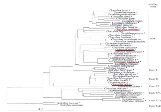
Phylogenetic studies using the 16S rDNA sequence analysis allowed the formation of 19 clusters inside the Clostridium genus (Fig. 1.5) (Rood et al., 1997).
29 Cluster I is the largest of the clostridial rRNA clusters and equivalent to the group I of Johnson and Francis (Johnson & Francis, 1975). This group is also designated the “Clostridium sensu stricto” and it includes psychrophilic, mesophilic, and thermophilic species, as well as cellulolytic, saccharolytic, and proteolytic representatives in addition to pathogenic and non-pathogenic species (Bahl & P., 2001). Cluster II contains highly proteolytic and acetate-producing species like C. hystolyticum and Clostridium proteolyticum (Bahl & P., 2001).
Cluster XIV is the second largest Clostridium species cluster, which however is significantly intermixed with representatives of other genera, such as Acetitomaculum, Eubacterium and Roseburia (Bahl & P., 2001). This cluster presents two subgroups: subclusters XIVa (e.g.
Clostridium oroticum and Clostridium coccoides) and XIVb (e.g. Clostridium lentocellum).
The C. coccoides subgroup Clusters XIVa, the C. leptum subgroup Cluster IV, and the
Clostridium subgroup Cluster XIX make up 70 % of the normal faecal flora of humans
(Finegold et al., 2002). This evidence accounts for the important role that clostridial species play in the microbiota of the human gut.
Figure 1.5 | Phylogenetic
dendrogram summarizing the 19 clusters of the genus
Clostridium as defined by
Collins et al. (Collins et al., 1994) and related taxa, using available 16S rRNA/DNA sequence data. The scale bar represents 5% sequence divergence (Rood et al., 1997).
The genus Clostridium contains 35 species, which are considered to have pathogenic potential. Clusters I and II together contain about half of the pathogenic species in the
Clostridium genus, including the major pathogenic clostridia, such as C. barati, C. botulinum, C. novyi and C. tetani (cluster I); as well as C. septicum, C. chauvoei and C. histolyticum
(cluster II) (Rood et al., 1997). Other major pathogens like C. difficile and C. sordellii are included in cluster XI, and are also able to infect human and animals hosts. This cluster is phylogenetically and phenotypically heterogeneous, including also non-pathogenic
Clostridium species such as Clostridium sticklandii. Several pathogens are also included in
clusters XII, XIV and XVIII (Fig. 1.6) (Rood et al., 1997).
Members of the Clostridium genus can be found in several sorts of environments, including soils, aquatic sediments, plants and gastrointestinal tract of animals and humans as mentioned above. They ferment a wide variety of organic compounds. They produce end products such as butyric acid, acetic acid, butanol and acetone, and large amounts of gas (CO2 and H2) during fermentation of sugars. They can produce a wide variety of extracellular
enzymes, which degrade numerous biological molecules (e.g. proteins, lipids, collagen,
Figure 1.6 | Phylogenetic dendogram indicating the relationships of the pathogenic Clostridium species*.
Clusters as defined by Collins et al. (Collins et al., 1994). The scale bar represents 10 nucleotide substitutions per 100 nucleotides (Rood et al., 1997).
31 cellulose, etc.) in the environment into fermentable components. Thus, the clostridia are considered to play an important role in nature in the biodegradation and the carbon cycle (Todar).
Among the Clostridium genus, the pathogenic species have also the capacity to produce the largest deadly toxins. Actually, they produce more toxins than any other bacterial genus (Rood et al., 1997). These toxins are usually the major virulence factors of the bacterium and are implicated in the disease caused by the major Clostridium pathogens, like botulism, tetanus, gas gangrene and pseudomembranous colitis (Hatheway, 1990).
1.2.1 Non-pathogenic Clostridia
Many clostridia species can degrade and utilize a wide range of plant polysaccharides and sugars (Bahl & P., 2001) to produce organic acids, alcohols or other neutral solvents providing a practical route for the conversion of renewable biomass and agricultural wastes to bulk chemicals and fuels. As a result, several non-pathogenic clostridia species have been used for biotechnological purposes, such as the solvent producer C. acetobutylicum (Bruggemann & Gottschalk, 2008). Other species are currently under extensive research because they employ metabolic pathways and enzymes of interest, e.g. C. pasteurianum (nitrogenase, hydrogenase, and ferredoxins), C. thermocellum (cellulases), C.
tetanomorphum (coenzyme B12-dependent reactions), C. kluyveri (energy metabolism and
reduction of enoates), C. formicoaceticum (tungsten-containing formate dehydrogenase), and C. ljungdahlii (growth with carbon monoxide) (Bruggemann & Gottschalk, 2008). Some psychrophilic, alkaliphilic, and thermophilic strains have been isolated and examined for the use of their proteases in washing detergents, or xylanase, in the pulp and paper industry (Bahl & P., 2001). Thermophilic species, whose fermentation process operates at high temperatures, and species capable of degrading hemicellulose and crystalline cellulose have been selected for their potential applications in biotechnology (Lee et al., 2008). In addition, clostridia can produce chiral products, which are difficult to make by chemical synthesis and degrade a number of toxic chemicals (Lee et al., 2008). Their diversity has allowed them to be exploited in different fields.
Here are some examples of non-pathogenic clostridia species involved in biotechnology and medical applications:
Clostridium acetobutylicum: a solvent producer
The only successful industrial fermentation process utilizing Clostridium species has been the acetone-butanol (AB) fermentation (Bahl & P., 2001). After Pasteur discovered bacterial butanol production from his landmark anaerobic cultivation in 1861, AB fermentative production prospered during the early 20th century and became after ethanol the second largest industrial fermentation process used in the world (Lee et al., 2008), playing a key role in the development of the chemical industry in the USA and Britain (Bahl & P., 2001). However, in the 1960s, the AB fermentation process had lost competitiveness due to the increase of feedstock costs and advancement of the petrochemical industry (Lee et al., 2008).
In spite of this, in the recent years, high crude oil prices and increasing concerns over global warming have renewed interest in biotechnological production of butanol, not only as, a chemical but also as an alternative fuel (Lee et al., 2008). In fact, acetone, butanol, and also isopropanol are now used as bulk chemicals as well as chemical feedstock for the synthesis of a wide range of other products. Moreover, these solvents are used for mixing in diesel liquid fuel and petrol, or as diesel supplements (Bahl & P., 2001).
The first industrial production of butanol was based on the fermentation of the bacterium C.
acetobutylicum, which ferments carbohydrates and produces mainly butanol and acetone
(Lee et al., 2008) from starch and molasses (Woods & Reid, 1995). C. acetobutylicum ATCC
824 is one of the most well-studied solventogenic clostridia and is closely related to the
historical Weizmann strain, which was used to develop an industrial starch-based acetone and butanol fermentation process (Bahl & P., 2001). Many other solventogenic clostridia were used at the time, although originally designated as C. acetobutylicum, such as C.
beijerinckii NCIMB 8052, Clostridium saccharobutylicum and Clostridium saccharoperbutylacetonicum that are also considered primary solvent producers (Lee et al.,
2008).
Solventogenic clostridia can utilize a large variety of substrates from monosaccharides (like xylose and arabinose) including many pentoses and hexoses to polysaccharides (Lee et al., 2008). Complex nitrogen sources such as yeast extract are generally required for good growth and solvent production, but otherwise the nutrient requirements for the growth of clostridia are rather simple (Lee et al., 2008). A typical feature of the clostridial solvent production is its biphasic fermentation. The first phase is the acidogenic phase, that usually occurs during the exponential phase, and from which the major products are acetate, butyrate, hydrogen, and carbon dioxide (Lee et al., 2008). The second phase is the
33 solventogenic phase, that occurs during the stationary phase, where production of butanol, ethanol, and acetone (or isopropanol in some C. beijerinckii strains) occurs (Lee et al., 2008).
The genetics of solvent-producing clostridia were initiated during the 1980s, allowing the first development of mutagenic techniques, gene transfer systems, and the use of plasmids for the construction of cloning vectors in a Clostridia species (Bahl & P., 2001).
The C. acetobutylicum ATCC 824 genome sequence was published in 2001 (Nolling et al., 2001). It possesses a 3.9 Mb chromosome and harbours pSOL1, a megaplasmid of 192 kb encoding for the genes involved in solvent formation (Desvaux et al., 2005). With the complete genome sequence of this strain and others already done or underway, it will be possible to carry out a more systematic strain improvement process by using genome-wide transcriptome and proteome studies, and the re-construction of the genomic-scale metabolic model, which will assist metabolic engineering of clostridia (Lee et al., 2008) (Table 1.2).
Table 1.2 | Summary of desirable metabolic engineering alterations of solventogenic clostridia and the
Clostridium butyricum: a hydrogen producer
The increasing burning of fossil fuels and their consequent changes on global climate, suggested the use of H2 as an alternative fuel, as its end product is only water (Nandi &
Sengupta, 1998). Anaerobic clostridia, such as C. pasteurianum, C. butyricum, and C.
beijerinkii, are potential strong hydrogen producers (Nath & Das, 2004). For instance
immobilized C. butyricum produces 2 mol H2/mol glucose at 50% efficiency (Nandi &
Sengupta, 1998). Several attempts have been made to produce H2 in fermentor reactors as
well as the production of biochemical fuel cell by the use of H2 producing C. butyricum (Nandi
& Sengupta, 1998). These results are in preliminary development (Nath & Das, 2004), and further research is still needed in order to develop biological systems to commercially feasibly produce H2 (Nath & Das, 2004).
Clostridium thermocellum: a cellulose degradation organism
About half of the carbonaceous compounds in terrestrial biomass are cellulose, which is the single most abundant organic compound on Earth. Moreover, the polysaccharide hydrolysis is one of the most important enzymatic processes since almost all of the biomass produced is again mineralized by enzymes from microorganisms (Schwarz, 2001).
Some Clostridia have developed a unique extracellular multi-enzyme complex, called cellulosome in order to degrade cellulose (Papoutsakis, 2008). This enzymatic complex is generally bound to the cell surface and contains binding-motifs to insoluble cellulose, and is made up of various cellulases that cleave oligosaccharides from insoluble cellulose (Papoutsakis, 2008). Cellulosomes present many advantages to be developed in the cellulosic biomass hydrolysis biotechnology.
The most well-investigated cellulosome is that of the thermophilic bacterium C.
thermocellum, usually isolated from hot springs and wet, rotting biomass (Schwarz, 2001).
The efficiency of its cellulosome makes it a good candidate for commercial bioconversion (Schwarz, 2001).
The majority of cellulolytic clostridia belong to the cluster III (the C. thermocellum subgroup) with the exception of C. stercorarium and C. cellulovorans (Schwarz, 2001). Several of these organisms are sequenced and all contain a complete and functional cellulosome, including
C. phytofermentans and C. cellulolyticum (Papoutsakis, 2008). C. cellulovorans, include in
cluster I, is a very active cellulose degrader, and was isolated with the intention of directly converting cellulosic wastes into industrial substrates, especially solvents (Schwarz, 2001). Unfortunately the cellulosome gene cluster found in C. acetobutylicum genome is not
35 expressed (Schwarz, 2001) and this bacterium is not able to grow on cellulose effectively (Papoutsakis, 2008).
Clostridium pasteurianum: a nitrogen fixing organism
Nitrogen fixation is the process by which atmospheric nitrogen is converted into nitrogen compounds (like ammonia or nitrate) useful for several chemical processes required for the biosynthesis of basic building blocks of life (Woods, 1993). C. pasteurianum was the first free-living nitrogen-fixing organism isolated, and where the first consistent cell-free nitrogen fixation was demonstrated (Kasap, 2006). As a result, C. pasteurianum has been extensively used in the study of the biochemistry and the physiology of nitrogen fixation (Woods, 1993).
Clostridia as cancer therapy agents
There have long been reports of tumour regression in presence of clostridia species. In 1813 Vautier reported that tumours regressed in his patients who were suffering from gas gangrene (C. perfringens associated disease) (Wei et al., 2008a). In 1935, Connell used sterile filtrates from C. histolyticum to treat advanced cancers and observed tumour regression (Wei et al., 2008a).
Clostridia bacteria present several advantages compared to classical approaches using virus and liposomes to treat cancer. They have been shown to specifically and preferentially target solid tumours where they are able to colonize and cause significant tumour lyses (Wei et al., 2008a). Furthermore, clostridial spores are easy to produce, stable to store and safe to use as well as having extensive oncolytic ability (Wei et al., 2008b) which is a major advantage when compare to other techniques. Therefore there has been continuous research on clostridial strains in the development of new cancer therapies tested in animal model and also in human trails (Wei et al., 2008a)
The first experiment performed in 1947 showed that direct injection of C. histolyticum spores into a mouse sarcoma caused oncolysis (liquefaction) and tumour regression. When spores of C. tetani, the causative agent of tetanus, was used, the tumour bearing mice died within 48 h because of C. tetani colonization and tetanus production. Thus, it was proven that C.
tetani was able to germinate/replicate selectively within the anaerobic environment of
tumours (Wei et al., 2008b). This result indicated that only non-pathogenic clostridia could be used in such therapeutic approach. The first example tested was the C. sporogenes strain (ATCC13732), a proteolytic species causing liquefaction of colonized tumours. Other strains were also tested, such as C. beijerinckii NCIMB8052, C. beijerinckii ATCC17778, C.
acetobutylicum ATCC824 and C. saccharoperbutylacetonicum that showed tumour
colonization although the tumour lysis was not complete (Wei et al., 2008b).
Spore treatment using wild-type clostridia were not sufficient to eradicate solid tumours and another approaches were developed. Clostridial spores were given in conjunction with other anticancer therapy (combination therapy) such as radio frequency and local X-irradiation. C.
sporogenes expressing the E. coli colicin E3 gene, which encodes a bacteriocin with
canceriostatic properties was also used. However, the overall anti-tumour efficacy of this bacteriocin was limited (Wei et al., 2008a). Some saccharolytic clostridia strains used in industry were also used to express therapeutic genes like the cytokine tumour necrosis factor alpha (TNF-α), the E.coli cytosine deaminase (CD) or the nitroreductase (NTR). No significant tumour regression was observed when these recombinant strains were used in solid tumour models (Wei et al., 2008b). Recent studies reported the development of new vectors using super tumour colonizer clostridial strains C. sporogenes or C. novyi-NT (lacking the lethal toxin, apathogenic). The recombinant C. sporogenes and C. novyi-NT strains overexpressing NTR have showed significant in vivo antitumor effects (Wei et al., 2008b). A combination therapy including C. novyi-NT with microtubule-interacting chemotherapeutic agents has demonstrated promising results and a phase 1 clinical trial of the combined approach is currently underway (Wei et al., 2008a). Other examples of studies using clostridia against cancer are presented in Table 1.3.
37 Currently, new genetic manipulation tools of clostridia are in progress to allow production of clostridia spores harbouring genes encoding for cancerstatic factors, or proteins or cytokines with additional tumour-killing capabilities (Wei et al., 2008b).
Therefore the use of Clostridium in cancer therapy holds considerable promise in the fight against cancer (Bahl & P., 2001).
1.2.2 Pathogenic Clostridia
On the basis of the main lesions or clinical signs observed, the major pathogens are classified as neurotoxic (C. botulinum and C. tetani), histotoxic (C. chauvoei, C.
haemolyticum, C. histolyticum, C. novyi, C. perfringens, C. septicum and C. sordellii) or
enterotoxic (C. colinum, C. difficile, C. perfringens, C. piliforme, C. septicum, C. sordellii and
C. spiroforme) (Dupuy & Matamouros, 2006). Some species, including isolates of C. perfringens, C. septicum and C. sordellii can be involved in more than one clinical entity
according to the combination of the toxins produced (Dupuy & Matamouros, 2006).
Some pathogenic clostridia infect animals, such as C. chauvoei which is responsible for the “Blackleg” (an emphysematous, necrotizing myositis) in cattle, sheep and other ruminants; C.
haemolyticum, the causative agent of bacillary hemoglobinuria in cattle and sheep and C. colinum which is responsible for ulcerative enteritis often lethal in domestic fowl and other
birds (Rood et al., 1997).
Clostridium septicum
C. septicum is found in soil and the faeces of domestic animals and humans (Neumann &
Rehberger, 2009). This anaerobic clostridium is highly virulent, and is responsible for spontaneous gas gangrene and atraumatic myonecrosis, and its infections are usually fulminant and lethal (Neumann & Rehberger, 2009). C. septicum produces four major toxins designated as alpha, beta, gamma and delta toxins. The alpha-toxin is a lethal pore-forming cytolysin and considered as a critical toxin in the disease establishment. The beta toxin is a DNase and has been shown to be the major extracellular protein produced by this organism. The delta toxin is an oxygen-labile hemolysin, antigenically related to the theta toxin of C.
perfringens, while the gamma toxin corresponds to a hyaluronidase. This last toxin belongs
to a class of enzymes generally known to aid in the spreading of the bacteria or toxins through tissues. They are produced by a number of pathogenic bacteria that initiate infections at the skin or mucosal surfaces (Neumann & Rehberger, 2009). C. septicum is a major cause of poultry infections in the United States and is considered as a primary concern
for both broiler and turkey producers (Neumann & Rehberger, 2009). C. septicum is also associated with “braxy”, a sheep disease in which the organism penetrates the lining of the abomasums (fourth stomach, where digestion takes place), resulting in a fatal bacteraemia. It is also responsible for malignant oedema in broiler hens (Rood et al., 1997).
Clostridium tetani and Clostridium botulinum
Tetanus was initially described by Hippocrates (Hatheway, 1990), while the first well-documented cases of botulinum intoxication occurred in Württemberg during the Napoleonic wars between 1795 and 1813. During this period, Justinus Kerner published the first case studies of botulinum poisoning linked to the consumption of blood sausages (Chaddock & Marks, 2006). Tetanus is an acute, spastic paralytic illness (Rood et al., 1997). Localized tetanus involves muscle rigidity and spasms near the site of the infected wound (Hatheway, 1990). Neonatal tetanus, which is quite prevalent in certain areas of the world, is due to infection of the umbilical stump, and often very severe and highly fatal (Hatheway, 1990). Botulism is a neuroparalytic disease that can weaken or paralyse all skeletal muscle in the body. Three forms of human botulism can be found: infant botulism, foodborne botulism and wound botulism (Rood et al., 1997).
As pathogens of tetanus and food-borne botulism, their virulence is due almost entirely to their toxigenicity (Todar). C. tetani and C. botulinum produce the most potent biological toxins known to affect humans. The potency of tetanus toxin (human lethal dose estimated at ~1 ng/kg) is exceeded only by the potency of botulinum toxin (human lethal dose estimated at about ≤0.1 ng/kg) (Rood et al., 1997).
Both neurotoxins are produced in the bacterial cell as a single polypeptide chain with a molecular weight of ~150kDa (Rood et al., 1997;Simpson, 2004). After release from the cell, the molecule is cleaved by a protease into two polypeptide fragments: a heavy chain (~100kDa) and a light chain (~50kDa) that remain joined by a disulfide bond (Hatheway, 1990; Simpson, 2004). The light chain of the tetanus toxin (TeNT) is a zinc-containing endoprotease whose substrate is synaptobrevin, one of the protein constituents of the docking complex that enables a synaptic vesicle to fuse with the terminal cell membrane (Rood et al., 1997). TeNT acts by blocking inhibitory synapses of the spinal cord motoneurons (Hatheway, 1990), which in turn induces muscle contraction (Schmitt et al., 1999). In the case of the botulinum toxin (BoNT), it utilizes the endocytosis and translocation pathways to enter into cytosol of cholinergic nerve endings where it acts like TeNT as a zinc-dependent endoprotease. In contrast to TeNT, BoNT cleaves polypeptides that are essential for exocytosis (Simpson, 2004), therefore blocking the transmitter release leading to the flaccid paralysis and autonomic dysfunction (Simpson, 2004) (Fig. 1.7).
39 Several clostridial species produce BoNT, including C. argentinese, C. butyricum and C.
baratti (Chaddock & Marks, 2006). According to their antigenic properties, seven antigenic
toxin types, labelled A-G, have been established. Neurotoxigenic C. butyricum strains produce type E-like toxin, while neurotoxigenic C. baratii strains produce type F-like toxin. Toxin types A, B, E and F are responsible for human botulism, while types C and D causes illness in animals. Type G was discovered in an Argentinean cornfield in 1970 and has not been established as a cause of either human or animal disease (Rood et al., 1997). Botulinum toxin is used to treat conditions such as migraine, headaches and cervical dystonia (Bruggemann & Gottschalk, 2008) but is also used for cosmetic purposes to smoothen facial lines, commercialized under the brand Botox.
Although the structure and the mode of action of the toxins are well understood, the regulation mechanisms of production of these toxins are still under investigation. It is known that the tetanus toxin encoding gene, tetX, is located on a large plasmid, and its expression is controlled by the regulator TetR, whose gene is located immediately upstream (Johnson & Bradshaw, 2001). In the case of C. botulinum toxin genes (ntnh-bont), they are located whether in the chromosome (ntnh-bont A, B, E, F) or in phages (ntnh-bont C, D) (Dupuy &
Figure 1.7 | Schematic models of the
neurotransmitter release and the actions of botulinum and tetanus toxins.
A- Synaptic vesicles containing neurotransmitters dock with plasma membrane through SNARE proteins (synaptobrevin, syntaxin and SNAP-25). Neurotransmitters are released through a Ca2+-triggered fusion process. SNARE proteins remain in random coil conformations until associated in the SNARE complex at docking, where they form a helical bundle.
B- Botulinum or tetanus toxin binds to the
presynaptic membrane through gangliosides and a protein receptor (step 1); it is internalized through endocytosis (step 2a), and its L chain is translocated across the membrane (step 2b). The L chain acts as specific endopeptidase against synaptobrevin (on synaptic vesicles), syntaxin (on the plasma membrane), or SNAP-25 (on the plasma membrane). BoNTs (or TeNT) cleave their substrates before the SNARE complex is formed (Singh, 2000).
Matamouros, 2006). Their expression is controlled by BotR, a regulator factor closely related to TetR (Dupuy & Matamouros, 2006).
Clostridium sordellii
C. sordellii is commonly found in the soil, in the intestines of animals and in 0.5 % of all
humans (Aldape et al., 2006). Many of C. sordellii strains are non-pathogenic, however, the virulent strains cause lethal infections in several animal species, such as enteritis and enterotoxaemia in sheep and cattle, and myonecrosis and gas gangrene in humans (Aldape et al., 2006). Usually, C. sordellii infections develop after childbirth or after gynaecologic procedures, although some cases involve sites of minor trauma such as lacerations (Rood et al., 1997). The absence of fever and a paucity of signs and symptoms of local infection make early diagnostics difficult (Rood et al., 1997)
Pathogenic strains of C. sordellii produce up to seven identified exotoxins. The lethal and hemorrhagic toxins, respectively TcsL and TcsH are regarded as the major virulent factors of
C. sordellii. Other exotoxins include an oxygen-labile hemolysin, a neuraminidase, a DNase,
a collagenase, and a lysolecithinase. TcsL and TcsH belong to the large clostridial cytotoxin (LCC) family (Just & Gerhard, 2004). TcsL and C. difficile toxin B are highly homologous (76 % identical) (Aldape et al., 2006). In the late 1970s, C. sordellii antitoxin was found to neutralize the cytotoxic effect of stool specimens collected from patients with C. difficile-associated pseudomembranous colitis (Aldape et al., 2006).
Clostridium novyi
C. novyi was first described by Novy in 1894 (Hatheway, 1990). This clostridia species is
responsible for gas gangrene in humans and is also the cause of infections in domestic animals, such as necrotic hepatitis in sheep (Hatheway, 1990).
C. novyi possess three toxigenic types: A, B and C. Types A and B are common in soil, but
type B can also be found in the intestine and in the liver of herbivorous animals. Type C isolates are nontoxigenic and do not cause disease (Rood et al., 1997). Type A strains are the causative agent of gas gangrene in humans and wound infections in animals (Rood et al., 1997). Type B strains causes infectious necrotic hepatitis of sheep and cattle, and occasionally infects horses and swine (Rood et al., 1997).
The major toxin mediating pathogenesis of C. novyi infections is the alpha-toxin (TcnA). It was shown that TcnA has cardio-, neuro-, histo-, and hepatotoxicity (Rood et al., 1997). Genetically, the alpha-toxin encoding gene is phage mediated (Rood et al., 1997). As already mentioned (see 1.2.1 Clostridia as cancer therapy agents), C. novyi is a promising candidate in cancer therapy (Bruggemann & Gottschalk, 2008). Tumours with their large
41 hypoxic regions provide an ideal growth environment for C. novyi, as the vegetative form is extremely sensitive to oxygen (Bruggemann & Gottschalk, 2008). Since C. novyi is a pathogenic strain, a new C. novyi strain lacking TcnA was produced (C. novyi-NT) that is currently being tested for cancer therapy.
Clostridium perfringens
C. perfringens is found in soil samples and in intestinal contents of animals and humans
(Hatheway, 1990). This organism was first described in 1892 by Welch and Nutall and at that time was named Bacillus aerogenes capsulatus (Hatheway, 1990).
C. perfringens has been shown to be the causative agent of several human diseases such as
gas gangrene and food poisoning as well as enterotoxemic disease in domestic animals (Rood et al., 1997). This organism produces four major lethal toxins (alpha, beta, epsilon, and iota) on which the toxin types of C. perfringens species are based (types A-E, Table 1.4). Moreover nine minor toxins (enzymes or soluble antigens) are expressed by most C.
perfringens strains and that can play some role in pathogenicity, and an enterotoxin which is
responsible for C. perfringens foodborne illness (Hatheway, 1990).
C. perfringens type A is commonly found in soil and also in intestinal contents of humans and
animals in absence of disease. Types B, C, D and E strains do not survive in soils. They are found in the intestinal tract of domestic animals and occasionally in humans usually associated with disease (Hatheway, 1990).
Toxin Type Major toxins present Diseases
A Alpha, Neuraminidase, Enterotoxin
Gas gangrene (myonecrosis), foodborne illness, and infectious diarrhea in humans; enterotoxemia of lambs, cattle, goats, horses, dogs, alpacas, and others; necrotic enteritis in fowl; equine intestinal clostridiosis; acute gastric dilation in nonhuman primates, various animal species, and humans
B Alpha, Beta, Epsilon, Theta, Kappa, Lambda, Mu, Nu, Neuraminidase
Lamb dysentery; sheep and goat enterotoxemia (Europe, Middle East); guinea pig enterotoxemia
C Alpha, Beta,Delta, Theta, Kappa, Neurominidase, Enterotoxin, Nu
Darmbrand (Germany) and pig-bel (New Guinea) in humans; "struck" in sheep; enterotoxemia in lambs and pigs; necrotic enteritis in fowls
D Alpha, Epsilon, Theta, Kappa, Neurominidae, Enterotoxin, Lambda
Enterotoxemia of sheep; pulpy kidney disease of lambs
E Alpha, Iota, Theta, Kappa, Lambda, Neurominidase
Enterotoxemia in calves; lamb dysentery; guinea pig enterotoxemia; rabbit "iota" enterotoxemia
Table 1.4 Toxin types of C. perfringens with the major toxins they produce and diseases (adapted from