Universidade de Lisboa
F
ACULDADE
D
E
C
IÊNCIAS
Departamento de Biologia Vegetal
R
RO
O
L
L
E
E
OF
O
F
NO
N
O
V
V
E
E
L
L
N
NU
U
C
C
L
L
E
E
A
A
R
R
EN
E
N
V
V
E
E
L
L
O
O
P
P
E
E
PR
P
R
O
O
T
T
E
E
I
I
N
N
S
S
I
IN
N
V
V
O
O
L
L
V
V
E
E
D
D
IN
I
N
NU
N
U
C
C
L
L
E
E
A
A
R
R
PO
P
O
S
S
I
I
T
T
I
I
O
O
N
N
I
I
N
N
G
G
D
DU
U
R
R
I
I
N
N
G
G
CE
C
E
L
L
L
L
M
M
I
I
G
G
R
R
A
A
T
T
I
I
O
O
N
N
J
OANAB
ORREGOP
INTOMESTRADO EM BIOLOGIA MOLECULAR EGENÉTICA
Universidade de Lisboa
F
ACULDADE
D
E
C
IÊNCIAS
Departamento de Biologia Vegetal
R
RO
O
L
L
E
E
OF
O
F
NO
N
O
V
V
E
E
L
L
N
NU
U
C
C
L
L
E
E
A
A
R
R
EN
E
N
V
V
E
E
L
L
O
O
P
P
E
E
PR
P
R
O
O
T
T
E
E
I
I
N
N
S
S
I
IN
N
V
V
O
O
L
L
V
V
E
E
D
D
IN
I
N
NU
N
U
C
C
L
L
E
E
A
A
R
R
PO
P
O
S
S
I
I
T
T
I
I
O
O
N
N
I
I
N
N
G
G
D
DU
U
R
R
I
I
N
N
G
G
CE
C
E
L
L
L
L
M
M
I
I
G
G
R
R
A
A
T
T
I
I
O
O
N
N
J
OANAB
ORREGOP
INTODissertação de mestrado orientada por:
Professora Doutora Rita Zilhão
Faculdade de Ciências da Universidade de Lisboa
Doutor Edgar Gomes
Myology Group UMR-S 787 Inserm, Université Paris 06, Institut Myologie
MESTRADO EM BIOLOGIA MOLECULAR EGENÉTICA
"Se eu não morresse nunca E eternamente buscasse E conseguisse (!)
A perfeição das coisas"
Cesário Verde
T
TAABBLLEEOOFFCOCONNTTEENNTTSS
Table of Contents ... i
List of Figures ... iii
List Of Abbreviations ... iv Acknowledgments/Agradecimentos ... v Abstract ... i Resumo alargado ... ii Keywords/Palavras-Chave ... vi Introduction ... 1
1) The Nuclear Envelope ... 1
a) What is already known ... 1
b) New studies, new proteins, new functionalities emerging ... 2
2) Cell migration ... 5
a) Centrosome reorientation ... 6
b) Moving forward ... 8
Objectives ... 10
Material and Methods ... 11
1) General ... 11
a) Chemicals and antibodies ... 11
b) Microscopy ... 12
2) NIH3T3 Fibroblasts procedure ... 12
a) Cell culture ... 12
b) siRNA transfection and wound-healing assay ... 12
c) Cell transfection ... 13
d) Immunolabeling procedures ... 13
e) Determination of centrosome reorientation quantification ... 13
f) Determination of nuclear and centrosome positioning ... 14
g) DNA microinjection ... 14
h) Live-imaging of Tmem201 depleted cells ... 15
i) Antibody production ... 15
Results ... 17
1) Tmem201 is a nuclear envelope protein ... 17
2) Tmem201 is involved in nuclear movement ... 18
a) Tmem201 depletion impairs centrosome reorientation through inhibition of the nuclear movement ... 18
b) Can Tmem201-dependent nuclear movement be rescued? ... 21
c) The functional domains of the protein – dominant negative approach ... 22
3) How is Tmem201 involved in nuclear movement? ... 23
a) How is actin organized in Tmem201-depleted cells? ... 23
b) Structural role ... 24
Discussion ... 26
1) Tmem201 is a nuclear envelope protein ... 26
2) Tmem201 is involved in nuclear movement ... 27
3) looking for Tmem201 role in nuclear movement ... 29
Conclusions ... 30
Appendix ... vii
a) Validate Tmem201 depletion ... vii
References ... viii
L
LIISSTTOOFFFIFIGGUURREESS
Figure 1. The LINC complex connects the NE to actin filaments. ... 4
Figure 2. Tmem201 conformational structure (predicted) (A) and different isoforms (B). ... 9
Figure 3. Tmem201 constructions that were injected. ... 12
Figure 4. Diagram showing criteria to consider a centrosome reorientated. ... 14
Figure 5. Tmem201 B-GFP localizes at nuclear envelope. ... 17
Figure 6. Tmem201 antibody efficiently recognized the Tmem201 isoforms, localized in NE. ... 17
Figure 7. Tmem201 form linear arrays at the nuclear envelope that colocalize with actin dorsal cables. ... 18
Figure 8. Tmem201 depletion inhibits centrosome reorientation. ... 19
Figure 9. Tmem201 depletion inhibits nuclear movement. ... 20
Figure 10. Tmem201 B GFP rescues centrosome reorientation and rearward nuclear movement. ... 21
Figure 11. Tmem201 628-GFP inhibits centrosome reorientation but promotes the rearward nuclear and centrosome movement. ... 23
Figure 12. Actin cytoskeleton is not affected by Tmem201 depletion. ... 24
Figure 13. Tmem201 depletion does not affect the localization of other known NE proteins. 25 Figure 14. siRNA transfection efficiently deplete endogenous Tmem201. ... vii
L
LIISSTTOFOFABABBBRREEVVIIAATTIIOONNSS
aPKC: atypical protein kinase C CTD: C-terminal domain
DTT: Dithiothreitol
ER: endoplasmic reticulum IF: Immunofluorescence INM: Inner Nuclear Membrane LPA: lysophosphatidic acid
MRCK: myotonic dystrophy kinase-related CDC42-binding kinase MT: microtubule
NE: Nuclear Envelope NPC: Nuclear Pore Complex NTD: N-terminal domain
ONM: Outer Nuclear Membrane PNS: perinuclear space
RT: room temperature SF: serum free
SPB: spindle pole body
TAN lines: transmembrance actin-associated nuclear lines TBST: Tris-buffered saline with 0.1% tween
TM: transmembrane domain
MTOC: microtubule organizing center
A
ACCKKNNOOWWLLEEDDGGMMEENNTTSS/A/AGGRRAADDEECCIIMMEENNTTOOSS
Ao longo deste ano conheci muitas pessoas, vindas de diversos países, falando várias línguas. Ao longo deste ano, mantive igualmente contacto com aqueles que eram especiais para mim e que o continuam a ser. Todos eles tornaram este ano muito especial para mim e um dos melhores anos da minha vida. Obrigada.
During this year, I met a lot of new people, coming from different countries, talking different languages. During this year, I kept some good relationships, which were and still are special for me. All of them turn this year into one of the most special years and one of the best years of my life. Thank you.
Pendant cette année, j’ai connu beaucoup de nouvelles personnes, qui venaient de différents pays, qui parlaient plusieurs langages. Pendant cette année, j’ai aussi gardé contact avec tous qui étaient déjà spéciales pour moi et qui le sont encore. J’ai passé une très bonne année, une des meilleurs de ma vie, grâce a vous tous. Merci.
Obrigada ao meu orientador externo: Edgar Gomes por me ter deixado fazer a tese de mestrado na sua equipa. Pela paciência que teve comigo (sobretudo durante a escrita da tese, ou quando tinha perguntas “parvas” para lhe fazer ou quando tinha algum papel para lhe pedir), por ter “travado” um pouco a minha dispersão e o meu “excesso” de energia, quando queria fazer tudo ao mesmo tempo. Por me ter “obrigado” a acabar coisas! Por me ter deixado fazer muita microscopia! Pelo incentivo! Por ter aprendido imenso com ele!
Obrigada à minha orientadora interna e Professora, Rita Zilhão. Pelo entusiasmo e boa disposição, simpatia como Professora. Por ter sempre tempo para ouvir. Por me ter ajudado durante este ano com a burocracia, com opiniões, com correcções, com paciência, com simpatia.
Obrigada ao Daniel, por ter sido o meu irmão “mais velho” do laboratório. Por me ter explicado o caminho para o lab no primeiro dia. Por falar português e por me ter “aturado”, mais do que os outros, porque simplesmente era mais fácil falar com ele. Por ser português! Pela paciência! Por me ter dado música quando precisava de escrever a tese! E por ter aprendido imenso com ele.
Merci à Thibaud pour être toujours disponible pour m’aider si j’avais besoins. Pour m’avoir donnés les plasmids pour microinjecter! Pour les figures jolies! Pour l’aide avec v
Illustrator! Pour m’avoir donné quelques bons conseils sur trucs à visiter à Paris, moins touristique.
Merci à tout le reste de l’équipe pour être une super équipe: Bruno, Vincent, Sestina. Pour m’avoir toujours aidé si j’avais besoins de quelque chose! Á Bruno pour sa calme, à Vincent pour ses “chansons”, à Sestina pour être l’autre fille du groupe!
Merci à toute l’unité U787, pour la bonne ambiance au labo, pour touts les petits pots, pour les biscuits, chocolats, gâteaux, soirées! Pour toute l’animation que donnais envie d’aller travailler.
Obrigada à Mónica Dias por ter sido ela que me falou do Edgar! E por me ter contagiado com o seu gosto pela ciência! Por se continuar a preocupar comigo e com o meu percurso pela ciência! Por me ter recebido de braços abertos na sua equipa, pelo muito que aprendi com todos eles!
Et “ça m’étonne“, je ne me suis pas oublier des Kikis! Merci pour être les Kikis! Pour tous les bons moments qu’on a passé ensemble, pour tous les moments moins bons qu’on a surmonté (?) ensemble. Pour tout les diners, les petites soirées, les grandes soirées! Pour toutes les promenades et visites! Pour la campagne (parce que c’est beau la campagne)! Pour la guerre! Pour la patience à toute mes “c’est quoi ça?”, “ça veut dire quoi ça?”. Pour m’avoir montré un peu mieux la vie et culture française. Pour toutes les chansons être dans Grease! Pour toutes les “papotes”! Pour les nounours! Pour “c’est un bon romans, c’est une belle histoire” chanter dans le metro! Pour savoir que je pourrais retourner en France avec une petite place oú dormir!
Thanks to all the Erasmus students that I met in Paris, especially the ones from the Collège Neerlandais. Thanks a lot for the amazing year, it was incredible! And a special special thanks to “the crew”, my “Erasmus family”: Ana, Lana, Benno, Yvonne, Patrick, Nienke, Patrick, Phillip, Laurentiu, Federico, Alessandro, Fanny, Skárlet, Samuele, Leonor, Neuza, Bouki, Pol, Annabel, Armel, Jean, Yann. Thanks for all the dinners (Italian, Portuguese, Hungarian, Dutch, Brazilian breakfast, German cocktails), all the tea breaks, all the parties, all the talks, all the laughs, all the crazy silent dances, all the times that we went out, all the Big Bang Theories and all the Bazingas, all the nights at the Paris-Tchéque Center, all the “happle pies”, the book with all the you-look-like-a-Big-Bang-character. Thanks to turn the Collège Neerlandais in the best residence to live! Thanks for everything!
Obrigada a todos os amigos que ficaram em Portugal. E que continuam lá, mas “cá”: Obrigada à Andreia, Ana Sofia, Sara, Diana, Carina Men., Mariana, André, Melissa, Ana vi
vii Margarida, Bruno Afonso, Ana Raquel, Tomás, Irina, Inês Tenente, Ana Luísa, Inês Talves, Carina e Tiago por serem os amigos de sempre. Por poder contar com vocês sempre. Obrigada a todos por nada ter mudado, mesmo que cada um tenha seguido o seu caminho. Por poder falar com vocês como se nos tivéssemos separado na semana anterior! Obrigada pelas notícias, por todas as vezes que vieram ter comigo na net para saberem de mim, por continuarem a partilhar a vossa vida comigo, apesar de tão longe.
E um especial obrigado à Andreia por todas as vezes que me andou a tratar de papeis por causa do Erasmus, e à Carina Men. por se ter “voluntariado” a pedido para imprimir a tese e para entregá-la.
Grazie mille Carlo. Pour tout. Merci pour être plus pratique que moi, et pour m’aider a ne me préoccupée “pas trop”. Parce que tu me fais toujours rire! Pour tout le support pendant toute cette année. Pour ta préoccupation. Pour sentir que je peux toujours compter avec toi. Pour tout les bons moments passés ensemble. Parce que tu continues ici.
Às primas e ao primo da França que me receberam de braços abertos quando passei a SDF. Por se preocuparem comigo, por toda a ajuda que me deram. Por me relembrarem que não estou sozinha.
À tia, às primas e ao primo mais lindos de Portugal, por me receberem sempre de braços abertos, por compreenderem que ando sempre feita barata-tonta quando vou a Portugal, por saber que estão sempre lá. Por estarem sempre cá.
Ao meu irmão, por se preocupar, sempre tentando mostrar que não se preocupa.
Obrigada à minha mãe. Por estar sempre lá, mesmo estando longe. Por mandar vir comigo quando me esqueço de carregar o telemóvel. Por me perdoar sempre. Por me ouvir sem refilar, quando tenho simplesmente de mandar vir com alguém. Por me ter ajudado a procurar casa em Paris, estando ela em Portugal. Por me ter apoiado nesta viagem. Por me ter ajudado tanto a chegar até aqui. Por estar, incondicionalmente, aqui.
A
ABBSSTTRRAACCTT
Centrosome reorientation is defined as the positioning of the centrosome in a region between the nucleus and the leading edge and is important for cell polarization and directional cell migration. Cdc42 is the key regulator on this process and two main pathways are involved in centrosome reorientation; on one side, centrosome centration by a mechanism dependent of Par complex and dynein/dynactin, and for other side, a rearward nuclear movement dependent on Cdc42-effector MRCK and actin-myosin retrograde flow.
Recently, the LINC complex was found to be involved in the rearward nuclear movement pathway. This complex spans the nuclear envelope and involves a SUN domain-containing protein which interacts with a KASH domain-containing protein, localized in the inner and in the outer nuclear membrane, respectively. SUN proteins bind to lamins and KASH proteins to actin filaments; in this way, the LINC complex makes the connection between actin retrograde flow and the nucleus. In fibroblasts, Sun2 and Nesprin-2 co-localize with dorsal actin cables on TAN lines; actin dorsal cables move back by actin retrograde flow and the nucleus moves with them.
Other proteins can be involved in the nuclear movement. In a siRNA screen for nuclear envelope proteins, a putative role for Tmem201 in nuclear movement was identified. In
S.pombe, Tmem201 homolog connects the heterochromatin with the LINC complex. A
connection with LINC complex in mammalians was never reported so far.
Tmem201is a nuclear envelope protein and is localized in TAN lines in fibroblasts. The TMEM201depletion by RNA interference inhibited centrosome reorientation. Tmem201 is involved in nuclear movement, probably by the stabilization of the LINC complex in the nuclear membrane. However, Tmem201 might also be involved in centrosome positioning and could act as a key regulator of both pathways.
R
REESSUUMMOOAALLAARRGGAADDOO
Durante muito tempo, o papel do invólucro nuclear foi desvalorizado, sendo visto como um mero compartimento de armazenamento de cromossomas. Hoje em dia, após a descoberta de uma nova categoria de proteínas que permitiram criar o elo entre os componentes nucleares e o citosqueleto, o seu papel como organizador essencial é lhe amplamente reconhecido. De facto, a dinâmica entre citosqueleto e invólucro nuclear é fundamental para um correcto posicionamento do núcleo, que depende, por sua vez, de uma correcta migração e ancoragem do núcleo. O posicionamento nuclear é importante em fenómenos tão diversificados como a fertilização, a formação de fibras musculares, a oogénese e a migração celular.
A reorientação do centrossoma é um processo que ocorre em fibroblastos, células endoteliais, células epiteliais, astrócitos, células T e neurónios e que leva à polarização celular. Considera-se que o centrossoma está orientado quando este se encontra posicionado entre o núcleo e a frente condutora da célula na migração. Pensa-se que a polarização do centrossoma é um fenómeno prévio à migração celular, importante para a orientação do Golgi, possibilitando a secreção polarizada de precursores membranares e de factores importantes para a frente da célula, possibilitando a migração.
Durante muito tempo, pensou-se que seria o centrossoma a mover-se para uma posição anterior ao núcleo. Hoje em dia, sabe-se que é o núcleo que se move, adquirindo uma posição posterior ao centrossoma. A proteína Cdc42 tem um papel fundamental neste processo, conduzindo à activação dos dois mecanismos necessários à reorientação do centrossoma: por um lado, o centrossoma é mantido no centro da célula. Este mecanismo é dependente do complexo Par (Par6, Par3 e aPKC), da dineína e de dinactina. Por outro lado, MRCK, um efector de Cdc42, activa o movimento retrógrado do núcleo. Este movimento aparece aliado ao movimento retrógrado da actina, dependendo da contracção da miosina.
Recentemente, um complexo de proteínas transmembranares do invólucro nuclear foi identificado como fundamental para este movimento nuclear. Este complexo é constituído por duas proteínas, uma com um domínio SUN e a outra com um domínio KASH, ambos localizados na extremidade C-terminal da proteína. Este domínio C-terminal é altamente conservado em todos os Metazoa e em leveduras. A proteína com domínio SUN localiza-se na membrana interna e interage com a lamina nuclear pela sua extremidade N-terminal. A proteína com domínio KASH localiza-se na membrana externa; algumas proteínas que contêm este domínio podem atingir dimensões de mais de 800kDa e podem interagir com a actina pelo seu domínio N-terminal. A localização da proteína com o domínio KASH na ii
membrana externa é absolutamente dependente da proteína com o domínio SUN. Uma grande especulação existe ainda em torno da retenção da proteína com o domínio SUN na membrana nuclear. Em mamíferos, por exemplo, a localização de Sun2 parece ser parcialmente dependente da lamina nuclear, enquanto que Sun1 não depende da lamina para se localizar na membrana nuclear interna.
Este complexo atravessa assim todo a membrana nuclear e estabelece assim a ligação entre o núcleo e a actina. Este par proteico é assim chamado de Complexo “LINC” – LIgação entre o Núcleo e o Citosqueleto (LInkers of Nucleoskeleton and Cytoskeleton). Um estudo recente reporta a implicação directa do complexo “LINC” na reorientação do centrossoma em fibroblastos de ratinho. Neste caso, o par de proteínas que está envolvido no movimento nuclear é Sun2/Nesprin-2. Este estudo descreve pela primeira vez a existência de cabos actínicos, organizados numa posição dorsal em relação ao núcleo, que se movem retrogradamente ao mesmo tempo do que o núcleo. Estes cabos, e logo também o movimento retrógrado de actina a eles associados, parecem ser o motor para o movimento nuclear. O envolvimento de Sun2 e Nesprin-2 é reportado, quando se observa que ambas as proteínas se co-localizam com estes cabos de actina e que a depleção destas proteínas inibe o movimento nuclear.
Esta associação das proteínas do complexo LINC com os cabos dorsais de actina define uma nova estrutura nuclear denominada de “linhas TAN” – linhas Nucleares Transmembranares associadas a Actina (Transmembrane Actin-associated Nuclear). A força gerada pelo movimento retrógrado de actina é assim transmitida ao núcleo através destas estruturas, conduzindo ao movimento.
Sabe-se que Sun2 e Nesprin-2 interagem no espaço perinuclear, mas pouco ainda se sabe sobre esta interacção. Para além do mais, a localização de Sun2 no invólucro nuclear continua mal elucidada. Para além disso, as forças aplicadas sobre estas proteínas durante o movimento nuclear, sugerem o envolvimento de outras proteína na estabilização, organização e interacção deste complexo.
Num screen de siRNA para proteínas do invólucro nuclear, algumas proteínas revelaram um potencial papel no movimento nuclear, entre estas a proteína Tmem201.
Esta proteína é conservada evolutivamente, estando presente em todos os Metazoa e ainda em S.pombe. Pouco se sabe ainda sobre esta proteína, havendo apenas dois estudos realizados até ao momento, um em S.pombe e outro em células humanas.
Em S.pombe, o homólogo de Tmem201, Ima1, participa na associação da heterocromatina com o complexo LINC. Ima1 é importante para a estabilização do complexo, e de facto, iii
quando é eliminada do sistema, observam-se deformações do invólucro nuclear e quebras no complexo LINC. Ima1 funcionará assim como estabilizador do complexo LINC à membrana, função ainda não atribuída a nenhuma outra proteína em mamíferos.
Em células humanas, a proteína homóloga Samp1 aparece associada com estruturas membranares que se sobrepõem ao fuso mitótico. É também sugerido um possível papel desta proteína na ligação entre centrossoma e núcleo.
Este trabalho propõe-se elucidar o envolvimento da proteína Tmem201 no movimento nuclear e na reorientação do centrossoma.
Infelizmente, um anticorpo eficaz para a marcação de Tmem201 não está disponível no mercado e assim, um dos passos fundamentais deste trabalho passou pela produção de um anticorpo capaz de reconhecer eficazmente as três isoformas da proteína. Tal foi conseguido com sucesso, sendo possível visualizar perfeitamente uma marcação nuclear da proteína. Uma marcação a nível dos centrossomas, provavelmente não específica, foi também observada.
Esta proteína também foi observada em associação aos cabos dorsais de actina que fazem parte das linhas TAN. Este fenómeno é particularmente interessante, se considerarmos que Sun2 e Nesprin-2 são as duas únicas proteínas que foram identificadas até ao momento com localização nestas estruturas. Tendo em conta que as laminas não se encontram nas linhas TAN, Tmem201 poderia funcionar como estabilizador do complexo LINC a este nível, num papel evolutivamente conservado ao papel de Ima1 em S.pombe.
O movimento nuclear e da reorientação do centrossoma pode ser estudado facilmente através de um ensaio experimental em fibroblastos em cultura. Neste ensaio, é efectuado uma lesão linear (através de uma ponta de pipeta) numa monocamda confluente de células aderentes (em meio desprovido de soro). A reorientação do centrossoma é depois estimulada por adição de um factor específico (LPA). Através deste ensaio, procurou-se estudar os efeitos da depleção da proteína, por siRNA, na reorientação do centrossoma. Verificou-se uma inibição da reorientação do centrossoma quando a proteína não está presente, o que corrobora o resultado inicial do screen efectuado. Analisando a posição do centrossoma e do núcleo, uma forte inibição do movimento nuclear é visualizada, enquanto que a posição do centrossoma não é afectada. Os efeitos de depleção podem ser parcialmente recuperados aquando da microinjecção de um plasmídeo que codifica para a mais pequena isoforma de Tmem201 (Tmem201 B-GFP).
Os resultados obtidos implicam o envolvimento de Tmem201 no movimento nuclear. Contudo, um resultado inesperado foi obtido, aquando da microinjecção do primeiro domínio
da proteína: uma deslocalização do centrossoma. Este resultado sugere um possível envolvimento também no posicionamento do centrossoma.
Através dos ensaios de microinjecção, foi também possível concluir que o primeiro domínio da proteína parece estar envolvido na localização nuclear da proteína (visto que Tmem201 628-GFP tem uma localização nuclear), enquanto que o segundo domínio da proteína deverá ter um papel no movimento do núcleo (visto que apenas a Tmem201 B-GFP é capaz de recuperar a reorientação do centrossoma).
A inibição do movimento nuclear pode dever-se a diversos factores: se Tmem201 for importante na localização nuclear de proteínas envolvidas no movimento do núcleo, ou se por acaso levar à inibição do movimento retrógrado de actina. Verificou-se que a depleção de Tmem201 não afecta a retenção de Sun2, Nesprin-2, lamin A/C, lamin B e Emerin. Quanto à actina, não parece haver uma alteração do citosqueleto actínico.
Em suma, Tmem201 aparece implicada na reorientação do centrossoma, mais precisamente no movimento nuclear. Tendo em conta que não afecta a localização de outras proteínas na membrana nuclear e não altera o citosqueleto actínico, Tmem201 poderá ter um papel como estabilizador do complexo LINC na membrana nuclear, sendo importante para o movimento nuclear.
Contudo, poderá também estar envolvida no posicionamento do centrossoma. Tmem201 poderá assim actuar como proteína reguladora das duas vias.
vi K KEEYYWWOORRDDSS/P/PAALLAAVVRRAASS-C-CHHAAVVEE KEYWORDS: Cell migration LINC complex TAN lines
Actin retrograde flow Centrosome reorientation Nuclear movement Centrosome centration PALAVRAS-CHAVE Migração celular Complexo LINC Linhas Tan
Movimento retrógrado de actina Reorientação do centrossoma Movimento nuclear
I
INNTTRROODDUUCCTTIIOONN
1) THE NUCLEAR ENVELOPE
a) What is already known i) Double membrane
For a long time, the nuclear envelope (NE) was only seen as a mere container for the chromosomes, separating them from the cytoplasm. Today, this vision is changing.
The NE is composed of a double lipid bilayer, the inner and outer nuclear membranes (INM and ONM), separated by the perinuclear space (PNS) (reviewed in (Stewart et al., 2007)). The two membranes are joined at the sites of nuclear pore complexes (NPCs). These complexes are large protein assemblies that are responsible for the transport between the cytoplasm and the nucleus (reviewed in (D'Angelo and Hetzer, 2006)).
Although the INM and ONM are contiguous with the ER, they contain distinct membrane proteins composition, thereby requiring specific targeting mechanisms. The “diffusion-retention” model proposes that INM proteins diffuse laterally along the membrane after being inserted in the ER membrane upon translation (reviewed in (Schirmer and Foisner, 2007; Worman and Courvalin, 2000)). After reaching their destination, INM proteins decrease their diffusional mobilities and bind to chromatin, lamins or other proteins, in order to be retained and do not return to the ER (reviewed in (Worman and Courvalin, 2000; Burke and Stewart, 2002)). For example, the lamin B receptor (LBR) binds to B-type lamins and chromatin, via HP1, a chromatin-associated-protein (Worman and Courvalin, 2000).
This model was challenged by the concept of a receptor-mediated translocation through NPCs of certain yeast INM proteins, with classical nuclear localization signals (cNLSs), normally directing the import of soluble nuclear transport (King et al., 2006; Kutay and Mühlhäusser, 2006). Recently, this was also reported for the first time in mammalians (Turgay et al., 2010).
ii) Lamins
The nuclear lamina constitutes a thin protein meshwork that underlies the INM. In mammals, it is constituted by A- and B-type lamins, members of the intermediate filament family. Lamins share the overall organization of all the intermediate filaments: globular N- and C- terminal domains flanking a central α-helical segment. There are two A-type lamins, A and C, which correspond to two different alternative splicing variants of LMNA gene.
Expression of A-type lamin is restricted to differentiated cell types. The two B-type lamin isoforms, B1 and B2, are encoded by two distinct genes. These proteins seem to be essential and are present in all nucleated cells (reviewed in (Crisp and Burke, 2008)).
b) New studies, new proteins, new functionalities emerging i) SUN domain containing proteins
The SUN domain containing proteins (or shortly referred as SUN proteins) share a highly conserved C-terminal luminal domain (CTD) that derives its name from the homologous regions in S.pombe Sad1 and C.elegans Unc-84 (SUN (Sad1 and UNC)) (Malone et al., 1999). The SUN domain is evolutionary conserved, and can also be found in D. melanogaster Klaroid and Giacomo, in S. cerevisiae Msp3 and in OsSAD1 in plants. C.elegans also has another SUN protein, more similar to Sun1, Matefin1 (MTF1). Four SUN proteins have been characterized in mammalians (reviewed in (Razafsky and Hodzic, 2009): Sun1, Sun2, Sun3 and SPAG4. Both Sun1 and Sun2 are widely expressed in a large variety of mouse tissues and in early developmental stages (Wang et al., 2006) and seem to be essential for development (Han et al., 2009). Sun3 and SPAG4 appear to have a more restricted expression patterns, mostly in testis (Crisp et al., 2006; Shao et al., 1999).
Sun1 and Sun2 are localized in the INM (Hodzic et al., 2004; Padmakumar et al., 2005). The N-terminal domain seems to be responsible for its INM-localization (Hodzic et al., 2004; Wang et al., 2006; Haque et al., 2006); actually, the presence of a cNLS in the Sun2 N-terminal domain, important for its localization in the INM, was recently described (Turgay et al., 2010). However, some studies also consider the CTD important for NE localization of both proteins (Wang et al., 2006; Haque et al., 2006; Turgay et al., 2010).
The N-terminal domain of Sun1 and Sun2 interact with A-type lamins (Haque et al., 2006; Padmakumar et al., 2005; Crisp et al., 2006). In C.elegans, UNC-84 depends on Ce-lamin binding to be stably retained at the NE (Lee et al., 2002). Retention of the mammalians SUN proteins seems to be more complex than in C.elegans. In Lmna-null mouse embryonic fibroblasts, Sun2 was displaced from the NE in the majority of the cells. Lamins can possibly contribute to the Sun2 localization, but they are not enough for full retention (Crisp et al., 2006).
Lamin A/C depletion does not affect Sun1 localization and probably, an additional binding partner is required for Sun1 NE localization (Crisp et al., 2006; Haque et al., 2006). D.
discoideum Sun-1 localization is mediated by direct binding of its N-terminal domain to
chromatin (Xiong et al., 2008). Therefore, multiple retention mechanisms at the NE can take place for different organisms and for different SUN proteins.
ii) KASH domain containing proteins
Proteins capable to make the connection between the cytoskeleton and the nucleoskeleton have become object of a great interest in recent years.
The identification of new NE proteins that were exclusively located at the ONM in the end of the XX century brought new insights into the importance of the NE. Studies in C.elegans revealed new NE proteins that were important for proper nuclear anchoring and migration: ANC-1, UNC-83, together with the Sun protein UNC-84 (Hedgecock, 1982; Starr et al., 2001; Malone et al., 1999).
ANC-1, together with the Drosophila MSP-300 and the mammalian Nesprin-1 and -2 are very large proteins (>800 kDa) that share interesting features: an N-terminal α-actinin-type actin binding domain, a central long rod containing spectrin repeats and a unique and highly conserved CTD (Starr and Han, 2002; Zhang et al., 2002, 2001; Apel et al., 2000; Zhen et al., 2002). C.elegans UNC-83, Drosophila Klarsicht and S.pombe Kms2 and two other mammalian Nesprin-3 and -4 also contain this CTD, (McGee et al., 2006; Mosley-Bishop et al., 1999; Starr and Fischer, 2005; Wilhelmsen et al., 2005; Roux et al., 2009) named KASH domain (for Klarsicht/Anc1/Nesprin homology (Starr and Han, 2002).
The actin binding KASH proteins in mammals, Nesprin-1 and -2 are encoded by two separate genes in two different chromosomes (Zhang et al., 2001), that generate multiple isoforms, by alternative splicing or alternative transcription initiation. Deletion of both genes in a mouse model caused neonatal lethality by respiratory failure, due to brain malformation, revealing the importance of these proteins for the organism (Zhang et al., 2007).
The CTD has been showed as sufficient to properly direct the Nesprin protein to the ONM (reviewed in (Starr and Fischer, 2005)). Starr and Hann, in C.elegans, made the first proposal for how Anc-1 KASH domain-containing protein could be retained and anchored to the ONM, (Starr and Han, 2002). Anc-1 retention would require the INM protein, UNC-84.
Interactions between SUN and KASH domain were broadly demonstrated (reviewed in (Razafsky and Hodzic, 2009; Crisp and Burke, 2008; Starr and Fischer, 2005), and also in mammals, the roles of Sun1 and Sun2 in Nesprin-2 and Nesprin-1 localization were shown (Crisp et al., 2006; Padmakumar et al., 2005; Han et al., 2009).
iii) LINC complex
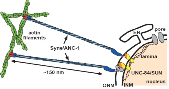
Both KASH and SUN proteins are type-II membrane proteins with a periplasmic CTD and a N-terminal domain (NTD) facing or the cytoplasm (in the case of KASH protein) or the nucleoplasm (in the case of SUN protein). As a result, both SUN and KASH domains are facing each other in the PNS (Hodzic et al., 2004; Haque et al., 2006; Padmakumar et al., 2005; Crisp et al., 2006) and form a complex, termed LINC complex (LInkers of Nucleoskeleton and Cytoskeleton) (Crisp et al., 2006)(Fig.1).
Figure 1. The LINC complex connects the NE to actin filaments.
Mammalians SUN proteins binds to nuclear lamins through their NTDs (yellow). Nesprin and Sun proteins interact in the PNS through their CTDs (KASH (light blue) and SUN (orange) respectively). The calponin NTD of Nesprin (red) attach to actin filaments (green). Picture adapted from (Starr and Han, 2003).
This complex is structurally important to keep the ONM and INM evenly spaced – the disruption of the LINC complex causes an expansion of PNS (Crisp et al., 2006). Functionally, it is important in several processes involving the attachment of the nucleus to the cytoskeletal networks:
(1) Centrosome-nucleus attachment. C.elegans ZYG-12 protein interacts with dynein, which will function to keep centrosome and nucleus together (Malone et al., 2003). In
S.pombe, the interaction between Sad1 and Kms2 provides a physical connection between the
spindle pole body (SPB) and centromeric chromatin (Razafsky and Hodzic, 2009; King et al., 2008). In D. melanogaster the Klarsicht protein, another KASH-domain protein, seems to be involved as well in the centrosome-nucleus attachment (Patterson et al., 2004).
(2) Nuclear positioning within the cell. In general, two related processes are required: nuclear migration to its appropriate place and nucleus anchorage to keep it in the right
position. This is necessary during different cellular and developmental events such as oogenesis, fertilization, muscle formation, and cell migration.
During Drosopila oogenesis, in the egg chamber, the nurse cells feed cytoplasmic components to the oocyte, through narrow ring canals, and then undergo apoptosis. The nuclei of the nurse cells are anchored by actin bundles in order not to block the ring canals. The LINC complex is involved in this process through the KASH protein Msp-300/Nesprin, binding to actin (reviewed in (Starr and Han, 2003; Wilhelmsen et al., 2006).
Nuclear migration is extremely important for fertilization - male and female pronuclei migrate towards each other in order to fuse. In C.elegans, the centrosome-nucleus attachment through ZYG-12 is important during pronuclear migration. The male pronucleus is attached to the centrosomes and theirs associated growing microtubules (MT) asters, and is pushed towards the center of the cell; by interaction with the male MT asters, the female pronucleus is also pushed towards the center (Starr and Fischer, 2005; Wilhelmsen et al., 2006). In this case, nuclear movement is MT-dependent.
Muscle cells are giant syncytial cells. Within these cells, nuclei are evenly spaced through the cytoplasm and some clustered, beneath the neuromuscular junction (synaptic nuclei). The KASH proteins, Nesprin-1 and -2 and the SUN proteins, Sun1 and Sun2, are involved in this nuclear positioning and the deletion of the KASH domain of Nesprin-1 affects synaptic and nonsynaptic nuclear anchorage (Han et al., 2009; Zhang et al., 2007).
The nuclear positioning is also important during cell migration, as described below.
2) CELL MIGRATION
The cell polarization is the triggering step for directed cell migration. In order to migrate, cells need an asymmetric morphology that will define leading and trailing edges. Cell migration occurs by a cyclical process of protusion in the cell front (leading edge) and retraction in the back (Ridley et al., 2003). Protrusion starts the migration cycle, and in the leading edge, two actin organizations can be found: lamellipodia and filopodia. These structures are stabilized by the binding to extracellular matrix or adjacent cells, via integrins. These transmembrane receptors provide traction sites for migration in the front and are detached in the cell rear, allowing the forward movement (Ridley et al., 2003; Ananthakrishnan and Ehrlicher, 2007).
a) Centrosome reorientation
Centrosome reorientation takes place in fibroblasts, endothelial cells, epithelial cells, astrocytes, T cells and neurons (Kupfer et al., 1982, 1983; Gundersen and Bulinski, 1988; Palazzo et al., 2001; Gotlieb et al., 1981; Etienne-Manneville and Hall, 2001; Gregory et al., 1988). Centrosome reorientation is important to define cell polarization and to direct migration. It is defined as the positioning of the centrosome in a region between the leading edge and the nucleus. As a result of centrosome reorientation, the Golgi apparatus is also reoriented. This pre-event to migration is thought to be important for the polarized delivery of vesicles traffics and new material to the leading edge (Gotlieb et al., 1981; Kupfer et al., 1982; Gundersen and Bulinski, 1988; Palazzo et al., 2001; Prigozhina and Waterman-Storer, 2004; Bergmann et al., 1983).
For a long time, the general idea was that the centrosome moved to the front of the nucleus during reorientation. However, recent data showed that for centrosome reorientation to occur, the nucleus has to move backwards, while the centrosome remains mostly stationary by an active process (Gomes et al., 2005). CDC42 is the key regulator of this pathway; alone is sufficient to induce centrosome reorientation (Palazzo et al., 2001). On one side, CDC42 recruits Par complex (PAR3, PAR6) and atypical protein kinase C (aPKC). This complex together with dynein and MTs are important for centrosome centration. When one of these components is disrupted, the centrosome is displaced from cell centroid, while the nucleus moves backwards (Gomes et al., 2005). A recent study implicates Par3 and dynein in the anchoring of MTs to the cell-to-cell contacts. Dynein could then generate tension (or pulling) on MT ends, or the simple anchoring of MTs to cell membrane would turn them more resistant to forces that could displace the centrosome from the cell center (Schmoranzer et al., 2009).
On the other side, CDC42 also activates nuclear movement by its effector, the myotonic dystrophy kinase-related CDC42-binding kinase (MRCK). MRCK activates Myosin II, by phosphorylation. This protein is essential for centrosome centration and rearward nuclear movement. MRCK induces centrosome centration by regulating the Par complex or dynein/dynactin. Gomes et al. also shown that nuclear movement is dependent on actin retrograde flow. Actin retrograde flow is defined as the movement of actin filaments away from the leading edge, in the opposite direction to the movement of the cell (Alexandrova et al., 2008; Ananthakrishnan and Ehrlicher, 2007). In the lamella, situated between the lamellipodium and the rest of the cell, the actin is depolymerized from the dendritic network and reorganized into actin bundles. Here the actin cytoskeleton is characterized by a slow 6
turnover and retrograde flow (reviewed in (Vicente-Manzanares et al., 2009)), and its organization in bundles could provide a strong support necessary for nuclear movement. Myosin II is a signature component, specific from this region, and it could generate the retrograde flow of actin in the lamella (reviewed in (Vicente-Manzanares et al., 2009)).
Summarizing, CDC42and CDC42-effector MRCK are the major regulators of centrosome reorientation. They act on the Par complex to center the centrosome by a dynein/dynactin dependent-mechanism, while the nucleus moves by a MRCK-regulated actin-myosin retrograde flow.
i) An in vitro assay to study the centrosome reorientation
A cell-based in vitro assay to study centrosome reorientation has been previously established and characterized in NIH 3T3 fibroblasts (Gundersen et al., 1994). When a confluent monolayer of fibroblasts is wounded, and in presence of medium with serum, cell polarization is triggered, and cells located at the wound edge start to migrate in the direction of the wound. But, if cells are serum starved before wounding, cell polarization only occurs after the addition of serum. Lysophosphatidic acid (LPA) is a major serum factor that is capable of induce cell polarization and centrosome reorientation, without stimulating cell migration (Palazzo et al., 2001; Cook et al., 1998). Mechanisms specific of centrosome reorientation can therefore be studied without the interference cell migration related processes and events (Gomes and Gundersen, 2006).
ii) The “LINC” between actin retrograde flow and nuclear movement is established through TAN lines
Actin retrograde flow can move nucleus backward during centrosome reorientation. If the KASH proteins can connect nucleus to actin filaments, and actin retrograde flow is involved in moving the nucleus backward, the obvious question would be: can the LINC complex couple the nuclear movement to the retrograde actin flow? This question was first elucidated by a recent study (Luxton and Gomes et al., 2010), in which the involvement of Sun2 and Nesprin-2 in nuclear movement was demonstrated. After LPA stimulation, and by the time nuclear movement began, actin cables are observed around the nucleus. Probably, those are cables that are proposed to be formed in the lamella region.
Luxton and Gomes distinguished two types of actin cables: ventral cables, with an orthogonal disposition relatively to the leading edge, and dorsal cables, parallel to the leading edge. The nucleus moves backward at the same rate as these dorsal cables, suggesting that nuclear movement could be driven by these backward moving cables.
Nesprin-2, Sun2 and these actin dorsal cables co-localized on the dorsal surface of the nucleus and move together with the nucleus, defining a new nuclear structure: TAN (Transmembrane Actin-associated Nuclear) lines. TAN lines move rearward at the same speed as nucleus, therefore strongly implicating them in nuclear movement. Actin dorsal cables would organize TAN lines, coupling actin to the LINC complex, and then the force generated by actin retrograde flow would move the nucleus backward.
b) Moving forward
There is still a lot of discussion about how SUN-KASH interactions are established, and other proteins should be involved to help the LINC complex in order to support the forces exerted by the cytoskeleton during nuclear movement. Furthermore the targeting of NE proteins is barely understood and many more players should be involved in the NE organization. Based on this hypothesis, Daniel Osório (member of the E. Gomes research team) performed a siRNA screen to identify potential NE proteins that could have a role on the nuclear movement in mammalian cells. Initially about 2000 potential candidates as NE proteins were selected from previously published organellar mass-spectrometry studies (Hallett et al., 2006; Foster et al., 2006; Schirmer et al., 2003). From these targets, proteins with potential transmembrane (TM) domains as predicted by bioinformatics algorithms were selected. Other relevant information like gene onthology (GO) and gene orthologs (NCBI homologene) were then checked and the results obtained were cross-referenced with other databases. This analysis identified 273 targets which fulfilled the imposed criteria and were used for the siRNA screen. From this set, 28 proteins were identified as involved on nuclear movement, by time-lapse microscopy and automated nuclear movement tracking.
The theme of this thesis is focused on the study of Tmem201, one of the proteins identified in the screen (together with Thibaud Jegou, a post-doc in the lab). This protein is evolutionary conserved and has a single gene homolog in all metazoan. It is also present in the fission yeast
S.pombe (designated Ima1), but is absent in S. cerevisiae (Buch et al., 2009). Until now, only
two studies have been published on this protein, one in fission yeast and other in human cells, which will be both briefly summarized here.
In S.pombe, nuclei are actively positioned at the cell center by MTs. The LINC complex is formed by the KASH proteins Kms1 and 2 and the SUN protein Sad1. It was already known that there was a coupling between the LINC complex and the SPB; King et al. demonstrated that Ima1 together with the Ndc80 complex contribute to the coupling between centromere and LINC complex, and consequently between SPB and cytoskeleton. Upon Ima1 depletion, 8
the LINC complex appeared fragmented and deformations of the NE were observed. MT-driven nuclear forces induced the loss of the spherical shape of the nucleus, since the Ndc80 complex alone was not sufficient to keep the connection between the centromere and the LINC complex (King et al., 2008). Ima1 could has a role in the stabilization of the LINC complex.
In a more recent study, in human cells, Samp1 (Tmem201 human homolog, which corresponds to the shorter isoform T201 2B – or simply, T201B), was found at the polar regions of the mitotic spindle. Samp1 seems to be associated with a new membranous structure which overlaps with the mitotic spindle, and its localization seems to be dependent of MTs. A functional connection between Samp1 and centrosomes is also proposed since Samp1 silencing seems to increase the distance between centrosomes and NE in interphase. As it is localized in INM, any interaction with the centrosome would have to be indirect (Buch et al., 2009).
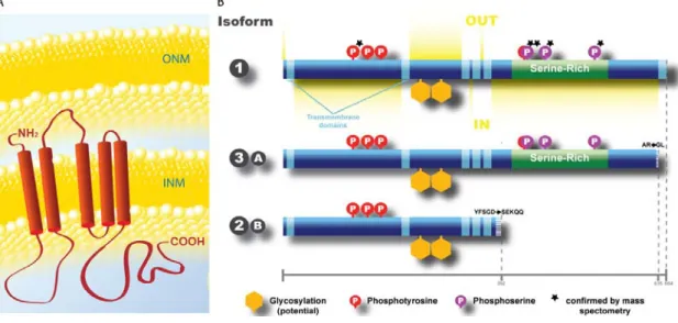
Some information about Tmem201 is already available in databases, and was compiled by Thibaud Jegou into a simplified picture (Fig. 2):
Figure 2. Tmem201 conformational structure (predicted) (A) and different isoforms (B).
Tmem201 is an INM with 5 TM domains. Three different sized isoforms are present in the mouse genome. The two larger isoforms have a serine-rich domain, absent in the smaller isoform. Picture adapted from Thibaud Jegou.
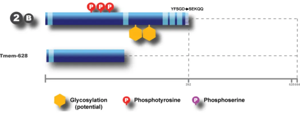
Three isoforms are described and all of them share the N-terminal domain. The isoform 1 (full-length) and the 3A (denominated as isoform 3 or isoform A) share almost the complete sequence, except the last amino acids. The isoform 2B (denominated as isoform 2 or B) lacks the serine-rich domain. All of them have potential sites for glycosylation and phosphorilation (some of them already confirmed by mass spectrometry), as well as five TM domains (see Fig. 2).
O
OBBJJEECCTTIIVVEESS
The recent implication of the LINC complex in nuclear movement raised even more questions namely: How is the LINC complex stabilized? How is the connection between SUN and KASH proteins established?
The answer to these questions is far from being understood and the identification of potential NE proteins involved in nuclear movement, including Tmem201, is extremely important to increase our knowledge about this.
Using a fibroblast wounding assay as model system, the principal aims of this work were: i. Confirm Tmem201 as a NE protein in mouse fibroblasts and determine if it also
localizes at TAN lines;
ii. Understand Tmem201´s role in centrosome reorientation, and determine if it is involved in centrosome positioning or in nuclear movement;
iii. Study the function of particular domains of the protein;
iv. Determine the role of Tmem201 on the retention of other NE proteins;
v. Study the involvement of Tmem201 in mediating cytoskeleton-nuclear interactions.
M
MAATTEERRIIAALLAANNDDMEMETTHHOODDSS
1) GENERAL
a) Chemicals and antibodies
For siRNA transfection: Lipofectamine RNAiMAX (Invitrogen); OPTI-MEM ® I (Invitrogen). The following siRNA sequences were used: Silencer® GAPDH siRNA mouse (Ambion) http://www.ambion.com/techlib/spec/sp_4624.pdf, Nesprin-2G siRNA mouse, 5´- CCAUCA-UCCUGCACUUUCATT-3´ (Genecust Europe); LBR siRNA mouse, 5´- GAGU AACAGCACUAUUUAATT-3´ (Genecust Europe); Emerin siRNA mouse, 5´- AUC AUUAUAGUCCUUGCUCTG-3´ (Genecust Europe); Silencer Select® Tmem201 s106693 siRNA mouse (Ambion), 5´- ACGAUACACUGGUGCCCUAtt-3´; Silencer Select® Tmem201 s106694 siRNA mouse (Ambion), 5´- CUAUGGGAAUCGCAACUGUtt-3´.
For plasmid transfection: Lipofectamine LTX (Invitrogen); PLUS Reagent (Invitrogen); OPTIMEM I Reduced Serum Medium (Invitrogen).
For western blots: NuPage 4x LDS sample buffer (Invitrogen), Dithiothreitol (DTT) (Sigma-Aldrich), 4-12% Bis-tris or Tris-glycine Gradient Precast gel (Invitrogen).
Antibodies used for Immunofluorescence (IF): Mouse anti-Emerin (Novacastra), Mouse anti-γ-tubulin (Sigma),, Mouse monoclonal anti-Lamin A/C (gift from Glen Morris), Mouse anti-Lamin B2 (gift from Harrald Herrman), Guinea Pig anti-LBR (gift from Harald Herrmann), Rabbit anti-Nesprin-2 (gift from Gregg Gundersen), Rabbit anti-Pericentrin (Eurogentec), Rabbit anti-Sun2 (Atlas Antibodies), Rat monoclonal anti-Tyrosinated α-tubulin- (YL1/2) (ECACC, UK).
Secondary antibodies conjugated with Cyanine or Alexa dyes (Jackson or Invitrogen). Plasmids: Tmem201 B-GFP and Tmem201 628-GFP (20 ng/μl) (Fig. 3). These plasmids were cloned using the Gateway® system (Invitrogen) by Thibaud Jegou.
Figure 3.Tmem201 constructions that were injected.
All the constructions were tagged with GFP in the C-terminal end.
b) Microscopy
All the images were acquired in a Nikon TE2000 or Nikon Ti microscope equipped with a Coolsnap HQ2 (Roper) CCD camera controlled by MetaMorph software (Universal Imaging), using an oil immersion 40x (0.6 NA) plan fluor objective.
For time-lapse imaging, a heated (37°C) chamber (Oko-lab, Italy) and an oil immersion 60x plan apo objective (Nikon) were used, together with the perfect focus system (Nikon). 2) NIH3T3FIBROBLASTS PROCEDURE
a) Cell culture
NIH3T3 fibroblasts were cultured in Dulbecco's Modified Eagle Medium 41965 (Invitrogen) with 10% bovine calf serum (Hyclone) and 10mM HEPES (pH = 7.5). Cells were split at a maximum confluence of 60%, using 0.05% Trypsin-EDTA (Invitrogen). Cells were kept in an incubator a 37ºC with 5% CO2 saturation.
b) siRNA transfection and wound-healing assay
All the siRNA experiments were performed by reverse transfection with Lipofectamine RNAiMAX according to the protocol provided by the manufacturer (Invitrogen). Briefly, the siRNA was diluted in OPTI-MEM® I medium and lipofectamine was then added. After 15-20min incubation (for complex formation), a cell suspension was added to give approximately 30% of confluence. siRNAs were used at the final concentration of 50uM, except for Silencer Select® siRNAs that were used at a final concentration of 20uM. After 24h of incubation, the cells were split in two coverslips (22 x 22 mm). At 48h post-transfection, the coverslips were washed 3 times in serum free (SF) culture medium and then starved. After 24h of starving, cells were wounded with a pipette tip and LPA-stimulated for 2h as previously described (Gundersen et al., 1994; Cook et al., 1998; Gomes et al., 2005).
Coverslips were then used for microinjection or directly fixed and stained (for centrosome reorientation analysis or nuclear and centrosome positioning in fixed cells).
c) Cell transfection
NIH 3T3 cells transfection with Tmem201B-GFP was performed using Lipofectamine LTX in 12 wells format according to the protocol provided by the manufacturer (Invitrogen). Briefly, for each well, 1μg of DNA was diluted in Opti-MEM I Reduced Serum Medium without serum, and PLUS Reagent was added (a 1:1 ratio to DNA). After 10min incubation, Lipofectamine LTX was added to the above diluted DNA solution and the mix incubated for 25mins for complex formation.
DNA-Lipofectamine LTX complexes were then added and cells were incubated for 16-24h at 37ºC in a CO2 incubator.
d) Immunolabeling procedures
Cell fixation: Cells were fixed in two different ways. In the first method, cells were fixed by Paraformaldehyde (3.7%), for 10 min at room temperature (RT), with 2 previous washes in PBS solution (1x, Invitrogen). Afterwards, cells were washed in PBS solution and then permeabilized with 0.5 % triton/PBS. Alternatively, cells were fixed in pure methanol (at -20ºC), for 10 min, and then washed in PBS.
Cells were kept in PBS buffer (1x) at 4ºC until immunolabeling. Blocking was performed simultaneously with primary antibody incubation using 10% Goat Serum-PBS (blocking buffer). Primary antibodies were incubated for 1h, and secondary antibodies, DAPI and phalloidin were incubated in blocking buffer for 30 min, always in a humid chamber, at RT. Secondary antibodies were used at a 1:400 dilution (except for DAPI, phallodin and Cy5 antibodies - 1:200).
Between each antibody incubation, coverslips were washed 3times, for 10 min, with PBS 1x. Coverslips were mounted in Fluoromount-G (Southernbiotech) and dried at RT.
e) Determination of centrosome reorientation quantification
Cells were considered centrosome-reoriented accordingly to what was already described (Palazzo et al., 2001; Gomes et al., 2005; Gomes and Gundersen, 2006). A putative sector between the center of the nucleus and the wound edge, which corresponds to one third of the cell, was considered (Fig. 4). If the centrosome localizes in this sector, between the wound edge and the nucleus, is considered reoriented.
The cells with neighboring gaps in the monolayer (that could lead to a centrosome reorientation in a wrong direction) were ignored. The size of the leading edge is also important, and normally should be around 30% of the total cell surface.
Centrosomes have to be in the putative sector between the wound edge and the nucleus center to be considered reoriented. If the centrosomes are separated in two centrioles, the mother centriole, which normally nucleates more MTs, is used as reference.
Figure 4. Diagram showing criteria to consider a centrosome reorientated
f) Determination of nuclear and centrosome positioning
Pictures were taken from wounded-monolayer of fibroblasts, stained for a centrosomal marker (pericentrin or γ-tubulin), microtubules (Tyr-tybulin) and DAPI.
The analysis of the nuclear movement in fixed cells was performed regarding the nucleus and centrosome position relative to the cell centroid, as previously described (Gomes et al., 2005). Using ImageJ software, pictures were rotated to place the wound-edge in a horizontal position, with the wound in the top and the cells in the bottom (to measure always the same sense of the movement). The cell perimeter from cells in the wound edge was traced and the area calculated. Cell centroid (given by the y-position of the cell) can be extracted and the equivalent radius calculated. Centrosome position and the center of the nucleus were determined manually using the “point selection” function. Only the y-coordinate – position of the nuclear centroid or centrosome along the y-axis – was used (the x-coordinate did not change significantly). A vector based on the y-coordinates distances of the centrosome and nucleus relative to the cell centroid was calculated and measurements were normalized to cell size. All the calculations were performed in Microsoft Excel.
g) DNA microinjection
Microneedles were prepared by pulling glass capillary tubing in a flaming/brown micropipette puller (Model P-97, Sutter Instrument Co.). Cells were kept, before and after injection, at 37ºC in a humidified CO2 environment. The wound was made 10-15 min before the microinjection, and only the cells on the wound were injected. The microinjection was performed in a Nikon TE2000 microscope equipped with a phase-contrast 40x (0.6 NA) plan fluor objective and a XenoWorks Micromanipulator (Sutter).
Microinjection was done in 24h-starved cells. Cells were subsequently incubated for a period of 1-2h for plasmid expression and then LPA-stimulated during 2h. Coverslips were then fixed and stained.
a) Tmem201 628-GFP construct injection: in mock-treated cells (dominant negative assay).
b) Tmem201 B-GFP construct injection: in siRNA-treated cells (rescue of the phenotype).
h) Live-imaging of Tmem201 depleted cells
Time-lapse video microscopy was performed in Tmem201 depleted cells in order to monitor nuclear movement by phase contrast. Nuclear movement was followed for 1h after LPA stimulation, by acquiring one image each 5min.
i) Antibody production
No Tmem201 antibody is commercially available in the market therefore we decided to raise one. Animals (rabbits and guiney pigs) were immunized with the first loop of Tmem201 and sera corresponding to different bleeds performed after immunization, was collected by a company (Eurogentec). Three bleeds from 2 different rabbits (sera 90 and 91) were tested, by IF and Western blot and for different concentrations. As a positive control, cells were transfected with Tmem201-B-GFP, which has an expected size of 68.5 kDa. As a negative control, pre-immune sera were used.
i) Immunofluorescence:
The protocol used was very similar to what was previously described. Each antisera was always tested in cells fixed in PFA and methanol. A previous blocking-step was made with Blocking Solution (2% BSA, 0.1% tween/PBS) for 30 min. Primary and secondary antibodies were diluted in the same solution. Multiple dilutions of each antisera were used (1:50, 1:100 or 1:400).
ii) Western blots:
Extracts from NIH 3T3 and Hela cells (transfected with Tmem201 full-length constructs) were used. Cells were lysed using 600ul (per 10cm plate) of the lysis solution (2x NuPage LDS sample buffer with 100mM DTT). Cells were scrapped off the dish and then collected and boiled at 98ºC. After 5min, the extract was sonicated for 10sec.
The Western-Blotting was then performed according to the protocol provided by the manufacturer – iBlot® Transfer Stacks, nitrocellulose (Invitrogen). Cell extracts (15ul per 15
lane) were loaded in a 4-12% Tris-glicine gradient pre-cast gel with protein molecular weight markers (Invitrogen). Proteins were then transferred to nitrocellulose membrane using the iBlot system (Invitrogen) program 3 for 6min. To improve the transfer of high molecular weight proteins gels were pre-incubated in tris-glycine buffer with 20% methanol. Transfer efficiency was assessed by Ponceau red staining and membranes were blocked with 5% milk in Tris-buffered saline with 0.1% tween (TBST).
The membrane was usually incubated overnight with the primary antibody and 30 min with the secondary antibody. The antisera were used in a dilution of 1:500, 1:1000 or 1:2000, the secondary antibody (HRP-conjugated antibody) was used 1:10 000. Both antibodies were diluted in Blocking solution. After each antibody, 3 washes of 10 min were made with TBST. Before the incubation with the Blocking Solution, a 10min wash was made with TBS.
Western-Blotting development was performed by Enhanced Chemiluminesce using the SuperSignal ® West Pico kit (Thermo Scientific). CL-XPosure ™ Film (Thermo Scientific) were used and developed manually in a dark room.
Same western protocol was performed to test siRNA-efficiency in Tmem201 depletion.
.
R
REESSUULLTTSS
1) TMEM201 IS A NUCLEAR ENVELOPE PROTEIN
Tmem201 had already been described as a nuclear envelope protein in previous studies (King et al., 2008; Buch et al., 2009; Schirmer and Gerace, 2005). It was necessary to confirm this localization in NIH3T3 fibroblasts. NIH3T3 cells transfected with Tmem201 B-GFP clearly show that the GFP-tagged protein localizes to the NE (Fig. 5).
Figure 5. Tmem201 B-GFP localizes at nuclear envelope.
Cells were transfected with Tmem201 B- GFP construct and fixed in PFA and then stained for DAPI. Scale bar: 10 μm.
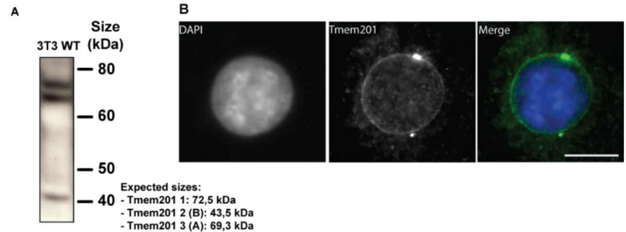
To determine the localization of the endogenous protein and since no commercially antibody against Tmem201 was available, an antibody was raised against the first loop of Tmem201. Two rabbit antisera (90 and 91) were tested by western blotting and IFs. Only the third bleed of serum 91 detected bands at all the expected sizes: 72.5 KDa (isoform 1), 43.5 KDa (isoform 2B) and 69.3 KDa (isoform 3A) (Fig. 6A).
All the bleeds of each serum were also tested by IFs. Once again, only the third bleed of sera 91 was able to stain nuclear rim efficiently (Fig. 6B). Nuclear envelope staining was detected more efficiently in methanol-fixed cells. In addition, a centrosome staining was also observed, which was not observed when GFP-tagged constructs were expressed.
Figure 6. Tmem201 antibody efficiently recognized the Tmem201 isoforms, localized in NE.
(A) Cells-extracts were analyzed by western-blot using α-Tmem201 as primary antibody. The three isoforms (72.5, 43.5 and 69.3 kDa) of the protein can be detected. (Figure provided by Thibaud Jegou) (B) Methanol-fixed cell were stained with α-Tmem201 and DAPI. Scale bar: 10μm.
Sera 91 (third bleed) was named α-Tmem201.
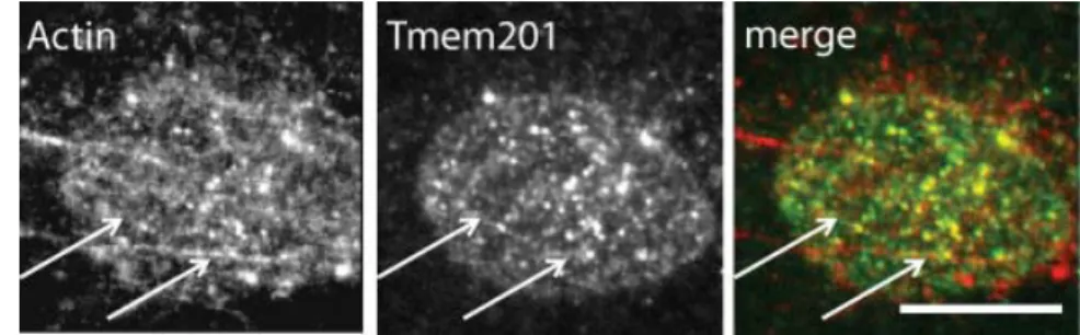
TAN lines are a new NE structure recently described and are involved in nuclear positioning (Luxton and Gomes et al., 2010). In cells stained with anti-Tmem201 antibody some lines were visualized on the dorsal surface of the nucleus. To see if Tmem201 was also a TAN lines component, α-Tmem201 was co-stained with Phalloidin (which labels F-actin), in some cells a co-localization of Tmem201 lines and actin dorsal cables can be observed (Fig. 7).
Figure 7.Tmem201 form linear arrays at the nuclear envelope that co-localize with actin dorsal cables.
Fluorescent pictures of a nucleus stained with rhodamine-phalloidin (F-actin) and α-Tmem201. Arrows: co-localized dorsal actin cables and Tmem201. Scale bar: 10μm.
Summarizing, Tmem201 is expressed in NIH3T3 fibroblasts. An antibody against Tmem201, α-Tmem201, was efficiently raised, and it is able to detect the three Tmem201 isoforms and their NE localization. In addition, Tmem201 is probably a new component of the TAN lines.
2) TMEM201 IS INVOLVED IN NUCLEAR MOVEMENT
a) Tmem201 depletion impairs centrosome reorientation through inhibition of the nuclear movement
To evaluate if Tmem201 was involved in nuclear movement, a centrosome reorientation assay (see MMAATTEERRIIAALLAANNDDMMEETTHHOODDSS for details) was performed – representative pictures for
mock and Tmem201 siRNA are shown in Fig. 8A.
The centrosome is oriented towards the leading edge if it is located into a specific area between the wound edge and the nucleus, which corresponds to 1/3 of the cell (Fig. 4 –
M
MAATTEERRIIAALLAANNDDMMEETTHHOODDSS). In starved cells, with no LPA stimulation, ~33% of the cells will
have the centrosome positioned in the front 1/3 of the cell, just out of random probability. In 18
all the conditions analyzed (mock- or siRNA-treated), around 30% of cells had their centrosome oriented, which corresponds to what we considered as a “basal centrosome reorientation state”, the “zero”. After LPA stimulation, the maximal centrosome reorientation was around 60% (in mock- and GAPDH siRNA-treated cells) (Fig. 8B) (Gomes et al., 2005; Gomes and Gundersen, 2006). So we can assume that a protein is involved in nuclear movement if the centrosome reorientation, after depletion of this protein, is closer to the levels of the non stimulated cells – for example, after Nesprin-2 depletion, centrosome reorientation levels were similar to starved cells (~38%) (Luxton and Gomes et al., 2010).
Three siRNAs for Tmem201 were tested, but only two were used in the successive assays (Tmem201 93- and siRNAs), since the other induced cell death. For Tmem201 93- and 94-siRNAs, Tmem201 was efficiently depleted (see Fig. 13 – SSUUPPPPLLEEMMEENNTTAARRYYDDAATTAA), without
affecting cell viability. After Tmem201 siRNA transfection, the level of centrosome reorientation after LPA stimulation was similar to Nesprin-2 siRNA (Fig. 8B). Tmem201 depletion inhibits centrosome reorientation.
Figure 8. Tmem201 depletion inhibits centrosome reorientation.
(A) Representative image of centrosome reorientation scoring in treated cells in SF (no LPA stimulation), and mock-treated and Tmem201 siRNA after LPA stimulation (green, MTs and blue, DAPI). Scale bar: 10 μm. (B) The percentage of centrosome reorientation was determined and plotted (green, mock-treated cells without LPA stimulation; orange, LPA stimulation). Only the cells at the leading edge were scored. Error bars in b) are SEM of at least three independent experiments. For each condition, at least 500 cells were counted.
To determine if the inhibition of centrosome reorientation was due to inhibition of nuclear movement, the position of the nucleus and the centrosome was measured, relatively to the cell centroid. The plotted positions can be visualized considering the 0% (yy axis) as cell centroid and the negative values as positions away from the leading edge. In starved cells, with no addition of LPA, nucleus and centrosome were close to the cell centroid, both in non-treated and siRNA treated cells (Fig. 9A). However, in LPA treated cells, the distances of the nucleus to the cell centroid increase in both mock- and GAPDH siRNA treated-cells (to an average position of -25.2% ± 2.7% and -23.9% ± 2.7%, respectively) (Fig. 9A). These distances express a clearly rearward movement of the nucleus. On the other hand, the centrosome stays, as expected, close to the cell centroid (Gomes et al., 2005).
Figure 9. Tmem201 depletion inhibits nuclear movement.
(A) The average position of the centrosome and the nucleus was determined, according to their distance to the cell centroid. A representative case of starved cells without LPA stimulation is shown (mock, -LPA), all the others correspond to cells stimulated by LPA for 2h. All the cells were rotated in order to keep their leading edge perpendicular to the xx axis. Error