dressed by measuring fibrinogen-erythrocyte unbinding forces by atomic force microsco-py (AFM) based force spectroscomicrosco-py.For the sake of comparison the measurements of the fibrinogen-erythrocyte interaction were always conducted in parallel with measure-ments of the well known interaction of fi-brinogen with platelets. It is worth of notice that the later interaction has been exten-sively studied, but not by the methodology described here.
AFM is mainly an imaging technique, in which the surface of a sample is scanned, line by line, by the movement of a thin tip, mounted on a flexible cantilever (for a review see, [6]). The tip-sample repulsion at the atomic level, transduced by the cantilever deflection and by an optical lever mechanism, permits the association of a height value to each position on the x,y plan and, therefore, the reconsti-tution of a pseudo-3D image of the sample surface. In addition to being used for imag-ing, the AFM can also be used to quantify the interaction between the tip and a specific spot of the sample, taking advantage of the pico-Newton sensitivity of the method. This
1. FIBRINOGEN-ERYTHROCYTE MEMBRANE INTERACTION
Upon bleeding due to the opening of a wound on a blood vessel, the coagulation process is triggered. The plasma protein fibrinogen builds up the scaffold for the blood clot formation, by polymerizing in a fibrin network and entrapping the blood cells. The main physiological receptor for fibrinogen is the platelet membrane in-tegrin αIIbβ3. As fibrinogen has more than one integrin-binding site, it is able to simul-taneously bind two platelets, bridging them and regulating blood hemostasis.
Beside the relevance of the fibrinogen-platelet binding, it is also known that high plasma fibrinogen levels induce erythro-cyte hyperaggregation [1-3], and that this increased aggregation constitutes a seri-ous cardiovascular risk factor [4, 5]. This prompted us to study: (i) how does fibrino-gen increase erythrocyte aggregation? (ii) Is there a specific receptor for fibrinogen on the erythrocyte membrane? (iii) Can this in-teraction be modulated to reduce hyperag-gregation? These three questions were
ad-ABSTRACT
Biological membranes are dynamic struc-tures essential for several cellular phenom-ena. The scope of the work of the Institute of Molecular Medicine (IMM) Biomembranes Unit is the study of biochemical and bio-physical processes occurring at the mem-brane level on human cells and on their viral and bacterial pathogens. On the viral context, we are primarily interested on HIV and dengue virus, and particularly on the two steps of their life cycle involving their interaction with host cell membranes: the viral entry into target cells and the assembly of new viral particles. A special focus will be given to the study of the role of biologi-cal membranes on the mechanism of action of the HIV entry (membrane fusion) inhibi-tors enfuvirtide and T-1249. We are also in-volved in assessing the molecular basis of the activity of microbicides, such as rBPI21,
that bind to specific components of bacte-rial membranes. Additionally, our line of work on the binding of fibrinogen to erythro-cytes, and its relevance as a cardiovascular risk factor will be presented. An approach to the latter problem by single-molecule force spectroscopy, using an atomic force micro-scope (AFM), allowed the molecular recog-nition, characterization and partial identifi-cation of the human erythrocyte receptor for fibrinogen.
Protein-Biomembrane
interactions
as therapeutic targets
Marco M. Domingues, Pedro M. Matos, Filomena A. Carvalho, Nuno C. Santos*
Instituto de Medicina Molecular, Faculdade de Medicina da Universidade de Lisboa, Av. Prof. Egas Moniz, 1649-028 Lisboa, Portugal.
approach is usually termed “force spectros-copy” (despite not being in fact a “spectros-copy”, as it is not based on the interaction of radiation with matter!).
On the context of this study, we used iso-lated human erythrocytes or platelets, in buffer, lightly adherent by electrostatic interactions to poly-L-lysine-treated glass slides. The force spectroscopy approach is based on the covalent attachment of fibrinogen molecules to the AFM silicon nitride tips, following methodologies pre-viously applied to other proteins [7, 8]. Af-ter this, we use the AFM to tap with the fi-brinogen-functionalized tip on the surface of the blood cell (Figure 1) [9, 10]. In the situations in which the binding occurs, the additional force necessary to detach the tip (and the fibrinogen molecule) from the cell can be calculated, based on force vs. distance curves [11]. After performing and analyzing hundreds or thousands of these binding / unbinding curves, a frequency
vs. rupture force histogram can be built up. The fitting using Gaussian functions of the histogram peaks allowed the deter-mination of the force necessary to break a single fibrinogen-cell interaction, with ad-ditional peaks corresponding to multiples of this value (Figure 2). The comparison of the results obtained by this methodology led us to a first surprise: The average force necessary to break the fibrinogen-eryth-rocyte interaction (79 ± 3 pN) is lower but quite comparable with the force necessary to break the fibrinogen-platelet interaction (97 ± 2 pN) [9], pointing out to the existence of a receptor for fibrinogen on the erythro-cyte membrane, instead of the non-specific interaction previously proposed by some authors [3].
By performing identical measurements for different loading rates and analyzing the data with the Bell, Evans and Ritchie for-malism [12-14], it was possible to obtain other parameters characterizing this inter-action, namely, the energy and width of the energy barrier that must be overcome for the unbinding to occur, and the dissociation rate, or its inverse, the unperturbed bond lifetime. The results obtained for this last parameter allowed us to conclude that one of the reasons why the fibrinogen-erythro-cyte binding is so difficult to study by more conventional methodologies, such as flow cytometry, is its reduced lifetime [9]. As it is common for integrins, the αIIbβ3 re-ceptor for fibrinogen in platelets requires
calcium to be functional. After the initial ex-periments, described above, being conduct-ed in the presence of a physiological con-centration of Ca2+, new experiments were carried out in its absence, additionally add-ing EDTA to chelate any trace amount that could be present. As a first control or proof of concept to demonstrate that the interac-tion measured by force spectroscopy was in fact between fibrinogen and the blood cells membrane receptor, results obtained in the absence of Ca2+ demonstrate, as expected, the reduction of the fibrinogen-platelet binding, both in terms of force necessary to break the bond (decreasing to values characteristic of non-specific interactions) and of binding frequency [9]. The binding of fibrinogen to erythrocytes is also impaired by the absence of calcium. This observation suggested that the receptor for fibrinogen in erythrocytes is also an integrin.
As a second control, or proof of concept, we tested the effect of eptifibatide on the fibrinogen binding to either of the blood cells. This is a cyclic peptide derived from barbourin, a component of the venom of the southeastern pigmy rattlesnake [15]. It Figure 1: Force Spectroscopy technique at an Atomic Force
Micros-cope. (A) Schematic representation of the erythrocytes deposited on a poly-L-lysine-coated glass slide and an AFM tip chemically functionalized with fibrinogen molecule(s). The arrows represent the approach and retraction cycles during the force spectrosco-py measurements. When approaching the tip to the sample, the fibrinogen molecule may contact with cell receptor(s) and the binding between them can occur. By retracting the tip away from the sample, the binding force necessary to break this bond, at the single-molecule level, can be measured. (B) Air tapping-mode AFM images of typical circular, biconcave human erythrocytes, from he-althy blood donors, deposited on poly-L-lysine coated glass slides (height 3D image). Reprinted from [10].
Figure 2: Force spectroscopy results from healthy human blood donors in the presence of Ca2+ 1 mM. Rupture-force histograms from about
8,000 single event-force measurements for the fibrinogen–erythrocyte (red) and fibrinogen-platelet (green) systems. Measurements were done with an applied force of 1 nN, pulling speed of 2 μm/s and at a loading rate of 4 nN/s. Reprinted with permission from [9] (© 2010, American Chemical Society).
pathogenic mutation was identified on the ITGB3 gene, we could demonstrate that one of the two sub-units of the receptor for fi-brinogen on the erythrocyte membrane is a product of the expression of this gene. New experiments are being conducted to identify the other sub-unit of the receptor.
In parallel, we also evaluated the effect of erythrocyte aging on the ability to bind fi-brinogen [10]. The results indicated that upon erythrocytes aging, there was a signif-icant decrease on the fibrinogen binding, by decreasing the frequency of its occurrence but not its binding strength. For the bind-ing between fibrinogen and erythrocytes to occur, a lower fibrinogen concentration was needed on young erythrocytes than for the older ones. We could conclude that the fibrinogen-erythrocyte receptor binding can be lost, masked or progressively turned to non-functional with the in vivo erythrocyte senescence process. Knowing that younger erythrocytes bind more to fibrinogen, we could presume that this population is the main responsible for some cardiovascular diseases associated with an increase on the fibrinogen content in blood, which could disturb its normal flow.
2. HIV FUSION INHIBITORS
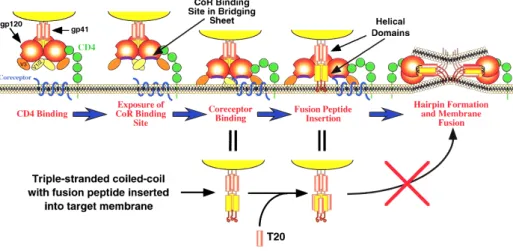
The infection of a target cell by the human immunodeficiency virus type 1 (HIV-1) is me-diated by the gp120-gp41 viral glycoproteins
complex (a homotrimer of heterodimers). gp120 binds to the cell receptor CD4 and to a co-receptor, usually CCR5 or CXCR4 [18]. After these bindings, gp41 is exposed, acquiring an extended conformation and enabling the insertion of its fusion peptide in the target cell membrane. Subsequent-ly, another conformational change takes place, in which two heptad repeat domains of each of the three gp41 monomers come together, forming a structure named six-helix bundle. It brings the viral and cell membranes together, enabling the forma-tion of the fusion pore and the entrance of the viral content into the cell [19] (Figure 3). HIV fusion inhibitor peptides were initially designed with a sequence corresponding to one of the heptad repeat domains, in order to inhibit the binding to the opposite heptad repeat domain of gp41, impairing the six-helix bundle formation and, there-fore, the entry of the viral content into the target cell [20]. Experimental evidences showed that this is, at least, a partial ex-planation, as inhibitors with a higher bind-ing to the gp41 heptad repeat domain were shown to be ineffective in vivo, while some peptides with a lower binding to gp41 are efficient [21].
The peptides enfuvirtide (T-20) and T-1249 are two of these HIV fusion inhibitors. Enfu-virtide is the only fusion inhibitor approved so far for clinical use. Its sequence corre-sponds to part of the HIV gp41 C-terminal is considered to be a specific inhibitor of the
αIIbβ3 integrin, used in hospital settings to treat platelet hyperaggregation. As expect-ed, eptifibatide inhibited fibrinogen-platelet binding at the extension and concentra-tions range expected based on the clinical studies [15, 16]. Regarding the binding to erythrocytes, eptifibatide also inhibited the binding of fibrinogen. However, the same threshold level of inhibition observed for platelets was reached only at higher epti-fibatide concentrations [9]. This indicates that the receptor for fibrinogen on eryth-rocytes is not the glycoprotein αIIbβ3, but an αIIbβ3-related integrin.
All the results described so far were obtained using blood samples from healthy volunteer blood donors. As a third and final control or proof of concept, identical measurements were carried out with cells isolated from a blood sample of a patient with Glanzmann thrombasthenia. This is a rare hereditary disease which patients have impaired co-agulation due to the absence of the αIIbβ3 receptor or its presence on a non-functional mutated form [17]. Expectedly, tapping with the fibrinogen-derivatized tip on platelets from this patient almost yielded no binding. On the few situations in which the binding occurred, the force necessary to break the bond was rather low, characteristic of non-specific interactions. Performing identical measurements with erythrocytes, we could demonstrate for the first time that Glanz-mann thrombasthenia patients can also have impaired fibrinogen-erythrocyte binding [9]. Genetic studies were conducted to identify if the mutation responsible for the Glanzmann thrombasthenia in this patient was on the IT-GA2B or on the ITGB3 gene (responsible for the expression of the αIIb and β3 sub-units of the platelet receptor, respectively). As the
Figure 3: Steps required for HIV-1 glycoprotein mediated membrane fusion with the host cells, as explained in the main text. A fusion inhibitor peptide (e.g., enfuvirtide and T-1249) may block the formation of the six-helix bundle, preventing the fusion of the viral and cell membranes. Reprinted with permission from [45] (© 2000, Cold Spring Harbor Laboratory Press).
heptad repeat domain. T-1249 is a second generation drug, with successful prelimi-nary clinical results, which development is presently on hold. Its sequence includes elements from fusion proteins of HIV-1, HIV-2 and SIV (simian immunodeficiency virus). In a line of work developed together with Miguel Castanho’s group (formerly at the Faculty of Sciences of the University of Lisbon, and now also at the Institute of Molecular Medicine (IMM) / Faculty of Medicine of the University of Lisbon), we have used the intrinsic fluorescence of the tryptophan residues in both peptides to study their interaction with biomembrane model systems (lipid vesicles) [22, 23]. In these studies, we showed that enfuvirtide and T-1249 partition to fluid phase lipid membranes, and that T-1249 (but not enfu-virtide) additionally adsorbs to the surface of cholesterol rich membrane domains. Recently, we extend these observations to the interaction of enfuvirtide and T-1249 with biological membranes, using human PBMC (peripheral blood mononuclear cells) and erythrocytes [24]. PBMC were chosen because they include one of the main targets for HIV infection: CD4+ T-lym-phocytes. Erythrocytes are not infected by HIV. However, it is found in vivo bound to the erythrocyte membrane, constituting a significant reservoir of infective viruses, even during highly active antiretroviral therapy (HAART) [25, 26].
To study the interaction of the peptides with the cell membranes, we could not use the same methodology as on the pre-vious studies with biomembrane model systems (changes on peptide fluorescence properties upon membrane insertion [27]), as the fluorescence of the peptides would be masked by the fluorescence of the innumerous aromatic amino acid resi-dues of the different protein components of the cells. Therefore, a new strategy had to be used: Instead of looking at the fluorescence of the peptide itself, we fol-lowed the changes induced by the pep-tide on the fluorescence of a membrane probe sensitive to the membrane potential [28]. After testing different fluorescent probes, we choose to use di-8-ANEPPS (4-[2-[6-(dioctylamino)-2-naphthalenyl] ethenyl]-1-(3-sulfopropyl)-pyridinium) for that purpose. The fluorescence of di-8-ANEPPS is highly sensitive to the mem-brane dipole potential, with an increase on this potential leading to a blue-shift on the probe excitation spectra. Quantita-tively, the variations of the membrane di-pole potential are easier to assess using as experimental output the ratio between the values obtained at two wavelengths of the fluorescence difference spectrum (ob-tained subtracting the spectrum without peptide from the spectrum obtained for a given concentration of the peptide). di-8-ANEPPS was shown by confocal
mi-croscopy to properly label the cell mem-branes of PBMC and erythrocytes [24]. By adding enfuvirtide or T-1249 to the labeled cells, the shape of the difference spectra revealed concentration-dependent shifts to the red, indicative of a decrease in membrane dipole potential upon the bind-ing of these peptides. The quantitative val-ues allowed the calculation of the peptide-cell dissociation constants [24]. This way, both peptides were shown to interact with erythrocyte and lymphocyte membranes. There is a complete agreement between these results and the data previously ob-tained with biomembrane model systems [22, 23]. T-1249 has an affinity for these blood cells approximately eight-fold high-er than enfuvirtide (Figure 4). This can be related with its higher partition constant and with the ability to adsorb to choles-terol-rich membrane domains, probably contributing for its improved antiretroviral efficacy [22].
In order to close the gap between the re-sults obtained with biomembrane model systems, using the peptides intrinsic tryp-tophan fluorescence, and those obtained with blood cells, based on di-8-ANEPPS fluorescence, we also performed experi-ments using di-8-ANEPPS-labelled lipid vesicles, with different membrane com-positions. The results were in agreement with the expected, with the addition of cholesterol to fluid phase lipid vesicles leading to a decrease on the enfuvirtide-membrane binding and to an increase on the binding of T-1249 [24].
Based on our findings, we proposed a model in which the HIV-1 association with erythrocytes in vivo could constitute a route to deliver peptide to the viral membranes (Figure 5). This should be especially rel-Figure 4: Peptide affinity towards erythrocytes and PBMC cell membranes. The plots represent the dependence of the ratio on the enfuvirtide
(red) and T-1249 (blue) concentrations for erythrocytes (A) and PBMC (B), the dashed curves are fittings to a single binding site model. Ratio values were normalized for the initial value of zero peptide concentration. Plotted values represent the mean ± standard error of mean (error bars not visible for erythrocytes due to small errors). N = 6 for erythrocytes and N = 7 for PBMC. Reprinted from [24].
evant for T-1249, as the viral membrane is rich in cholesterol [29]. Regarding lym-phocytes, their membrane can concentrate and accelerate the drug interactions with its molecular target, gp41 in its exposed conformation. The local enrichment of the inhibitor at the membrane level would in-crease the probability of taking advantage of the narrow spatial and time window dur-ing which gp41 is in the extended confor-mation in order to bind to it.
Going against the “textbook dogma” that the membrane fusion step of HIV-1 infec-tion occurs at the surface of the cell, the group of Gregory Melikian published in 2009 a study with compelling evidences for an entry of HIV-1 via endocytosis [30]. Subsequent evidences additionally sup-ported this theory [31]. If this is true, the relevance of the membrane binding of HIV fusion inhibitors is further strengthened, as a possible endocytic entry pathway would protect HIV from a fusion inhibi-tor only present in solution. However, its membrane binding and enrichment would drastically increase the inhibitory efficacy, by assuring a significant concentration to be present at the endosome level, where membrane fusion would occur.
Recently, the same experimental approach was also used for sifuvirtide, another sec-ond generation HIV fusion inhibitor that successfully completed phase IIb clinical trials in 2010 [32].
3. rBPI21 MOLECULAR LEVEL MECHANISM OF ACTION
Section 1 was dedicated to an AFM-based force microscopy approach to a specific subject. Section 2 describes the use of fluo-rescence spectroscopy to address another
one. Finally, in Section 3 another line of work ongoing at the Biomembranes Unit of IMM will be described; namely, the study of the mechanism of action of the antimicro-bial protein rBPI21, addressed by a different methodology: light scattering spectroscopy. Gram-negative bacteria have two mem-branes, separated by a peptidoglycan layer. The outer leaflet of their outer mem-brane is constituted mainly of lipopolysac-charide (LPS). Both the inner membrane and the inner leaflet of the outer mem-brane do not present LPS, having in their content high proportions of negative phos-pholipids, such as phosphatidylglycerol (PG). LPS is also negatively charged. Upon infection by Gram-negative bacteria, it is the causative agent of sepsis. At high con-centrations, LPS may lead to septic shock and, eventually, to death [33].
Our organism has two structurally similar proteins that bind LPS: the LPS-binding protein (LBP) and the bactericidal/perme-ability-increasing protein (BPI). Despite the structural similarity, their binding to LPS leads to opposite outcomes: The bind-ing of LBP to LPS enables the activation of the inflammatory cascade, through the binding to CD14 [34]. On the contrary, the binding of BPI to LPS allows its
neutrali-zation and clearance, together with the blocking of the inflammatory cascade [34]. BPI is produced by neutrophils. It exerts its antibacterial activity both intracellu-larly (at the azurophil or primary granules of the neutrophil) and extracellularly (re-lease of antimicrobial peptides and pro-teins from the specific or secondary gran-ules during neutrophil activation). This protein has a boomerang-like shape, with a cationic N-terminal domain, responsible for the antimicrobial activity and LPS neu-tralization, and with the C-terminal being responsible for cell-recognition [35, 36]. rBPI21 was designed with the sequence of the 193 amino acid residues of the N-terminal of BPI, except for the change of a cysteine for an alanine [37].
To study the mechanism of action of rBPI21, we used mainly dynamic light scattering (DLS) and zeta-potential measurements [38, 39]. DLS spectroscopy is based on the evaluation of the Brownian motion of the scattering particle, in order to obtain its dif-fusion coefficient and, based on it, to cal-culate the hydrodynamic radius or diameter of the particle using the Stokes-Einstein equation. Zeta-potential is a parameter that can be used to evaluate the electro-static interaction between particles and Figure 5: Proposed mode of action of enfuvirtide and T-1249 in circulation with blood cells. Reprinted from [24].
to give information on the stability of col-loidal dispersions. Most of the particles in aqueous solution carry an electric charge and this charge influences the distribu-tion of the ions at the particle surface. The counterions attached to the particle cre-ate an electrical double layer, with an in-ner region (Stern layer), where the ions are firmly attached, and an outer layer, where the ions diffuse more freely. In this diffuse layer, there is a boundary where ions and particle inside it form a stable entity. Dur-ing the zeta-potential measurements, an electric field is superimposed to the ran-dom Brownian motion. When a scattering particle is moving, ions within the bound-ary region move with the particle. The zeta-potential is defined as the zeta-potential created at this boundary [38].
In this study, we started by following the changes on the size and zeta-potential of the LPS aggregates naturally formed in so-lution [40]. Upon their titration with rBPI21, there was a marked increase of the hydro-dynamic diameter of the aggregates, with the size distribution becoming bimodal above a threshold level of the [rBPI21]/[LPS] ratio. The zeta-potential of the LPS aggre-gates (in the absence of rBPI21) is highly negative. With the addition of rBPI21, it pro-gressively increases, leveling up at positive zeta-potential values.
The interaction of rBPI21 with biomembrane model systems (large unilamellar vesicles; LUV) with different lipid compositions was
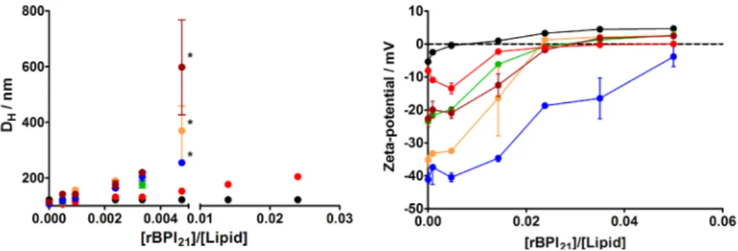
also addressed [40, 41], including systems containing LPS and/or POPG (palmitoy-loleoylphosphatidylglycerol), in order to mimetize Gram-negative bacteria mem-branes. On previous studies, we partici-pated on the development of spectroscopi-cal methodologies to quantify the partition constant (Kp) of a given molecule between the aqueous phase and membranes (for reviews see, [27, 42, 43]). Based on the changes on the intrinsic fluorescence of the tryptophan residues of rBPI21 upon mem-brane binding (followed by fluorescence spectroscopy), we measured its partition to membranes of different lipid compositions, and verified that high partitions to mem-branes occurred not only to LPS-containing systems, but also to membranes containing negative phospholipids, such as POPG [41]. By the opposite, no significant partition was observed for LUV made exclusively of zwit-terionic (neutral) phospholipids, such as POPC (palmitoyloleoylphosphatidylcholine). By using DLS to assess the lipid vesicles size variations induced by the presence of increasing concentrations of rBPI21, we realized that extensive aggregation was induced in the presence of POPG in the membranes, but not by the presence of LPS or POPC (Figure 6) [40]. Zeta-potential measurements shown that there is a higher affinity of rBPI21 for POPC:POPG mixtures, relative to pure POPG (Figure 7). Therefore, the rBPI21 interaction with membranes is not purely electrostatic.
To complement the DLS and zeta-potential studies, two other types of measurements were conducted, namely, membrane fusion and membrane leakage assays, in order to evaluate if these processes are promoted by rBPI21 [40]. For the membrane fusion (lipid mixing) assay, we used a Förster resonance energy transfer (FRET; see Fernandes et al. in this Canal BQ issue) methodology, with nitro-2-1,3-benzoxadiazol-4-yl-dipalmi-toylphosphatidylehtanolamine (NBD-PE) as donor and sulforhodamine B-dipalmi-toylphosphatidylehtanolamine (RhB-PE) as acceptor. One-fourth of the LUV used for the assay was labeled with both fluorescent membrane probes, while the remaining li-pid vesicles were unlabeled. The fusion ef-ficiency could be calculated based on the decrease of the energy transfer between both probes, as the fusion between labeled and unlabeled vesicles increases the aver-age distance between the two probes (sur-face dilution effect). With this assay, we ob-served that the rBPI21-induced membrane fusion (tested with increasing concentra-tions of the protein) is promoted by the presence of POPG, but not by the presence of LPS or POPC.
For the membrane leakage assay, we used LUV with carboxyfluorescein entrapped on their lumen. This fluorescent probe, when at high concentrations, undergoes a fluo-rescence self-quenching effect. Thus, the quantification is made by adding the protein and registering the eventual increase of the carboxyfluorescein fluorescence due to its dilution upon leakage. In order to obtain a reference value, total leakage was pro-moted afterwards by adding triton X-100. The results showed that rBPI21 promotes membrane leakage as long as the lipid membrane contains either POPG or LPS. Figure 6: Aggregation of LUV caused by rBPI21. Hydrodynamic
dia-meter variation of lipid vesicles with increasing rBPI21
concentra-tion. Bars represents the size range from three independent ex-periments. Larger ranges are intrinsically associated to vesicles flocculation/precipitation (marked *). POPC (black); POPG (blue); POPC:POPG 55:45 (orange); POPC:POPG 80:20 (green); POPC:LPS 80:20 (red); POPC:POPG:LPS 60:20:20 (brown). Reprinted from [40].
Figure 7: Zeta-potential for membrane model systems in the presence of rBPI21. Bars represent the zeta-potential range
from at least two independent experiments. POPC (black); POPG (blue); POPC:POPG 55:45 (orange); POPC:POPG 80:20 (green); POPC:LPS 80:20 (red); POPC:POPG:LPS 60:20:20 (brown). The lipid concentration was kept constant at 200 mM. Reprinted from [40].
ACKNOWLEDGMENTS
The study described in Section 1 of this article counted with the valuable collaborations of Simon Connell (School of Physics, University of Leeds, UK), Robert Ariëns (Unit of Molecular Vascular Medicine, University of Leeds, UK), Alice Tavares (Hospital de Santa Maria – Centro Hospitalar de Lisboa Norte) and Gabriel Miltenberger-Miltenyi (GenoMed, Lisbon). The projects of Sections 2 and 3 were developed together with Miguel Castanho (IMM / FMUL) and his group. These lines of work were supported by Fundação para a Ciência e a Tecnologia – Ministério da Ciência, Tecnologia e Ensino Superior (FCT-MCTES, Portugal; projects PTDC/SAU-OSM/73449/2006 and PTDC/ QUI-BIQ/104787/2008), by the FP7-PEOPLE IRSES (International Research Staff Exchange Scheme) project MEMPEPACROSS (EU), and by Fundação Calouste Gulbenkian (Portugal). MMD and PMM also thank FCT-MCTES for the PhD fellowships SFRH/ BD/41750/2007 and SFRH/BD/42205/2007, respec-tively.
REFERENCES
1. Kwaan HC (2010) Role of plasma proteins in whole
blood viscosity: a brief clinical review. Clin Hemorheol Microcirc 44, 167-176.
2. Lominadze D, Schuschke DA, Joshua IG & Dean WL
(2002) Increased ability of erythrocytes to aggregate in spontaneously hypertensive rats. Clin Exp Hypertens 24, 397-406.
3. Maeda N, Seike M, Kume S, Takaku T & Shiga T (1987)
Fibrinogen-induced erythrocyte aggregation: erythro-cyte-binding site in the fibrinogen molecule. Biochim Biophys Acta 904, 81-91.
4. Delamaire M & Durand F (1990) Erythrocyte
ag-gregation and vascular pathology. J Mal Vasc 15, 344-345.
5. Falco C, Vaya A, Simo M, Contreras T, Santaolaria M
& Aznar J (2005) Influence of fibrinogen levels on eryth-rocyte aggregation determined with the Myrenne ag-gregometer and the Sefam erythro-agag-gregometer. Clin Hemorheol Microcirc 33, 145-151.
6. Santos NC & Castanho MA (2004) An overview of the
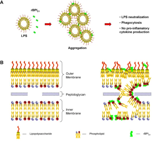
Based on these and on other comple-mentary results, we proposed a model to explain the molecular basis of the rBPI21 effects [40]: The interaction of this pro-tein with the LPS aggregates in solution causes their massive aggregation. This process may impair the recognition by LPS receptors, and facilitate the phagocytosis of the aggregates by macrophages. Re-garding the interaction with membranes, it is highly dependent on their composi-tion. rBPI21 can partition to the outer mem-brane of Gram-negative bacteria (rich in LPS and PG), attain an equilibrium with the intermembrane space, and reach the inner membrane. As both the inner leaf-let of the outer membrane and the inner membrane are rich in PG, rBPI21 may pro-mote the fusion (or hemifusion) of the two bacterial membranes (Figure 8). This can create a leakage or, at lower concentra-tions, an increased permeability across the membranes, leading to cell death. This increased permeability had already been previously reported [44]. Based on our findings, the disruption of mammalian membranes by rBPI21 is prevented by the absence of LPS and by the low content of POPG.
Calorimetry and atomic force microscopy experiments are being conducted in order to complement the previous findings.
OTHER PROJECTS
Adding to the lines of work addressed in the present article, the IMM Biomembranes Unit is also involved on the study of some poorly understood steps of the dengue virus life cycle, and on the identification of the mechanism of action at the molecular level of another family of antimicrobial peptides (AMP) and cell-pene-trating peptides (CPP).
Figure 8: Schematic representation of the proposed mechanism of action of rBPI21. (A) The protein interacts with LPS aggregates
pro-moting their aggregation, which allow LPS phagocytosis by innate immune cells and reduce LPS availability for its receptor. (B) rBPI21
interacts electrostatically with the LPS of the outer leaflet of outer membrane. This interaction disrupts the higher tight bond of the LPS, which allow the peptide insertion and translocation to the outer membrane inner leaflet and to the intermembrane space. rBPI21 induces
the fusion (or hemifusion) of the inner leaflet of the outer membrane and the inner membrane (both rich in PG). These membrane fusion events increase the membrane permeability, culminating with the leakage of the bacterial content. Reprinted from [40].
biophysical applications of atomic force microscopy. Biophys Chem 107, 133-149.
7. de Odrowaz Piramowicz M, Czuba P, Targosz M, Burda
K & Szymonski M (2006) Dynamic force measurements of avidin-biotin and streptavdin-biotin interactions using AFM. Acta Biochim Pol 53, 93-100.
8. Barattin R & Voyer N (2008) Chemical modifications of
AFM tips for the study of molecular recognition events. Chem Commun (Camb), 1513-1532.
9. Carvalho FA, Connell S, Miltenberger-Miltenyi G,
Pereira SV, Tavares A, Ariens RA & Santos NC (2010) Atomic force microscopy-based molecular recognition of a fibrinogen receptor on human erythrocytes. ACS Nano 4, 4609-4620.
10. Carvalho FA, de Oliveira S, Freitas T, Goncalves S &
Santos NC (2011) Variations on fibrinogen-erythrocyte interactions during cell aging. PLoS One 6, e18167.
11. Heinz WF & Hoh JH (1999) Spatially resolved force
spectroscopy of biological surfaces using the atomic force microscope. Trends Biotechnol 17, 143-150.
12. Bell GI (1978) Models for the specific adhesion of
cells to cells. Science 200, 618-627.
13. Evans E & Ritchie K (1997) Dynamic strength of
mo-lecular adhesion bonds. Biophys J 72, 1541-1555.
14. Lee CK, Wang YM, Huang LS & Lin S (2007) Atomic
force microscopy: determination of unbinding force, off rate and energy barrier for protein-ligand interaction. Micron 38, 446-461.
15. Holmes MB, Sobel BE & Schneider DJ (1999)
Varia-ble responses to inhibition of fibrinogen binding induced by tirofiban and eptifibatide in blood from healthy sub-jects. Am J Cardiol 84, 203-207.
16. Katori N, Szlam F, Levy JH & Tanaka KA (2004) A novel method to assess platelet inhibition by eptifibatide with thrombelastograph. Anesth Analg 99, 1794-1799.
17. Nurden AT (2006) Glanzmann thrombasthenia.
Or-phanet J Rare Dis 1, 10-17.
18. Wyatt R & Sodroski J (1998) The HIV-1 envelope
glycoproteins: fusogens, antigens, and immunogens. Science 280, 1884-1888.
19. Chan DC & Kim PS (1998) HIV entry and its inhibition.
Cell 93, 681-684.
20. Wild CT, Shugars DC, Greenwell TK, McDanal CB &
Matthews TJ (1994) Peptides corresponding to a predic-tive alpha-helical domain of human immunodeficiency virus type 1 gp41 are potent inhibitors of virus infection. Proc Natl Acad Sci U S A 91, 9770-9774.
21. Liu S, Lu H, Niu J, Xu Y, Wu S & Jiang S (2005)
Differ-ent from the HIV fusion inhibitor C34, the anti-HIV drug Fuzeon (T-20) inhibits HIV-1 entry by targeting multiple
sites in gp41 and gp120. J Biol Chem 280, 11259-11273.
22. Veiga AS, Santos NC, Loura LM, Fedorov A &
Castan-ho MA (2004) HIV fusion inhibitor peptide T-1249 is able to insert or adsorb to lipidic bilayers. Putative corre-lation with improved efficiency. J Am Chem Soc 126, 14758-14763.
23. Veiga S, Henriques S, Santos NC & Castanho M
(2004) Putative role of membranes in the HIV fusion in-hibitor enfuvirtide mode of action at the molecular level. Biochem J 377, 107-110.
24. Matos PM, Castanho MA & Santos NC (2010) HIV-1
fusion inhibitor peptides enfuvirtide and T-1249 interact with erythrocyte and lymphocyte membranes. PLoS One 5, e9830.
25. Hess C, Klimkait T, Schlapbach L, Del Zenero V,
Sadallah S, Horakova E, Balestra G, Werder V, Schaefer C, Battegay M & Schifferli JA (2002) Association of a pool of HIV-1 with erythrocytes in vivo: a cohort study. Lancet 359, 2230-2234.
26. Beck Z, Brown BK, Wieczorek L, Peachman KK,
Mat-yas GR, Polonis VR, Rao M & Alving CR (2009) Human erythrocytes selectively bind and enrich infectious HIV-1 virions. PLoS One 4, e8297.
27. Santos NC, Prieto M & Castanho MA (2003)
Quantify-ing molecular partition into model systems of biomem-branes: an emphasis on optical spectroscopic methods. Biochim Biophys Acta 1612, 123-135.
28. Matos PM, Goncalves S & Santos NC (2008)
Interac-tion of peptides with biomembranes assessed by poten-tial-sensitive fluorescent probes. J Pept Sci 14, 407-415.
29. Brugger B, Glass B, Haberkant P, Leibrecht I,
Wie-land FT & Krausslich HG (2006) The HIV lipidome: a raft with an unusual composition. Proc Natl Acad Sci U S A 103, 2641-2646.
30. Miyauchi K, Kim Y, Latinovic O, Morozov V & Melikyan
GB (2009) HIV enters cells via endocytosis and dynamin-dependent fusion with endosomes. Cell 137, 433-444.
31. Miyauchi K, Marin M & Melikyan GB (2011)
Visualiza-tion of retrovirus uptake and delivery into acidic endo-somes. Biochem J 434, 559-569.
32. Matos PM, Freitas T, Castanho MA & Santos NC
(2010) The role of blood cell membrane lipids on the mode of action of HIV-1 fusion inhibitor sifuvirtide. Bio-chem Biophys Res Commun 403, 270-274.
33. Raetz CR & Whitfield C (2002) Lipopolysaccharide
endotoxins. Annu Rev Biochem 71, 635-700.
34. Weiss J (2003) Bactericidal/permeability-increasing
protein (BPI) and lipopolysaccharide-binding protein (LBP): structure, function and regulation in host defence against Gram-negative bacteria. Biochem Soc Trans 31, 785-790.
35. Beamer LJ, Carroll SF & Eisenberg D (1997) Crystal
structure of human BPI and two bound phospholipids at 2.4 angstrom resolution. Science 276, 1861-1864.
36. Beamer LJ, Carroll SF & Eisenberg D (1998) The
BPI/LBP family of proteins: a structural analysis of con-served regions. Protein Sci 7, 906-914.
37. Horwitz AH, Carroll SF, Williams RE & Liu PS (2000)
Inclusion of S-sepharose beads in the culture medium significantly improves recovery of secreted rBPI(21) from transfected CHO-K1 cells. Protein Expr Purif 18, 77-85.
38. Domingues MM, Santiago PS, Castanho MA &
San-tos NC (2008) What can light scattering spectroscopy do for membrane-active peptide studies? J Pept Sci 14, 394-400.
39. Santos NC & Castanho MA (1996) Teaching light
scattering spectroscopy: the dimension and shape of tobacco mosaic virus. Biophys J 71, 1641-1650.
40. Domingues MM, Castanho MA & Santos NC (2009)
rBPI21 promotes lipopolysaccharide aggregation and
exerts its antimicrobial effects by (hemi)fusion of PG-containing membranes. PLoS One 4, e8385.
41. Domingues MM, Lopes SC, Santos NC, Quintas A
& Castanho MA (2009) Fold-unfold transitions in the selectivity and mechanism of action of the N-terminal fragment of the bactericidal/permeability-increasing
protein (rBPI21). Biophys J 96, 987-996.
42. Matos PM, Franquelim HG, Castanho MA & Santos
NC (2010) Quantitative assessment of peptide-lipid in-teractions. Ubiquitous fluorescence methodologies. Biochim Biophys Acta 1798, 1999-2012.
43. Ribeiro MM, Melo MN, Serrano ID, Santos NC &
Castanho MA (2010) Drug-lipid interaction evaluation: why a 19th century solution? Trends Pharmacol Sci 31, 449-454.
44. Wiese A, Brandenburg K, Carroll SF, Rietschel ET &
Seydel U (1997) Mechanisms of action of bactericidal/per-meability-increasing protein BPI on reconstituted outer membranes of gram-negative bacteria. Biochemistry 36, 10311-10319.
45. Doms RW & Trono D (2000) The plasma membrane
as a combat zone in the HIV battlefield. Genes Dev 14, 2677-2688.