Outubro, 2016
Cindy Nunes Soares
[Nome completo do autor]
[Nome completo do autor]
[Nome completo do autor]
[Nome completo do autor]
[Nome completo do autor]
[Nome completo do autor]
[Nome completo do autor]
Licenciatura em Bioquímica [Habilitações Académicas] [Habilitações Académicas] [Habilitações Académicas] [Habilitações Académicas] [Habilitações Académicas] [Habilitações Académicas] [Habilitações Académicas]
Isolation and Characterization of Novel Iron-Sulphur
Proteins from DvH Involved in Cell Division
Dissertação para obtenção do Grau de Mestre em Bioquímica
Dissertação para obtenção do Grau de Mestre em
[Engenharia Informática]
Orientadora:
Doutora Sofia Rocha Pauleta, Investigadora Principal,
Faculdade de Ciências e Tecnologia da UNL
Júri:
Presidente: Doutor José Ricardo Ramos Franco Tavares, FCT/UNL
Arguente: Doutora Maria Cristina Oliveira Costa, FCT/UNL
iii N o v e l I r o n -S u l f u r P r o t e i n s f r o m D v H
LOMBADA
2016v
Cindy Nunes Soares
Licenciatura em Bioquímica
Isolation and Characterization of Novel Iron-Sulphur
Proteins from DvH Involved in Cell Division
Dissertação de Mestre em Bioquímica
Orientadora: Doutora Sofia Rocha Pauleta, Investigadora
Principal, Faculdade de Ciências e Tecnologia da UNL
Júri:
Presidente: Doutor José Ricardo Ramos Franco Tavares, FCT/UNL
Arguente: Doutora Maria Cristina Oliveira Costa, FCT/UNL
Vogal: Doutora Sofia Rocha Pauleta, FCT/UNL
Outubro 2016
vii
Isolation and Characterization of Novel Iron-Sulphur Proteins from DvH Involved in Cell Division?
Copyright © Cindy Nunes Soares
Faculdade de Ciências e Tecnologia, Universidade Nova de Lisboa.
A Faculdade de Ciências e Tecnologia e a Universidade Nova de Lisboa têm o direito, perpétuo e sem limites geográficos, de arquivar e publicar esta dissertação através de exemplares impressos reproduzidos em papel ou de forma digital, ou por qualquer outro meio conhecido ou que venha a ser inventado, e de a divulgar através de repositórios científicos e de admitir a sua cópia e distribuição com objetivos educacionais ou de investigação, não comerciais, desde que seja dado crédito ao autor e editor.
ix
Acknowledgments
O trabalho desenvolvido nesta dissertação de mestrado não teria sido possível sem a participação, direta ou indireta, de várias pessoas a quem gostaria de expressar os meus agradecimentos.
Em primeiro lugar gostaria de agradecer à Doutora Sofia Rocha Pauleta, a minha orientadora, por todo o tempo que me dispensou, pelas orientações, pela disponibilidade que manifestou e pela transmissão de conhecimentos que contribuíram para aumentar o meu gosto por esta área cientifica.
Gostaria também de agradecer à Prof. Isabel Moura e ao Prof. José G. Moura por me terem acolhido nos seus laboratórios e grupos de investigação e por me proporcionarem todas as condições necessárias para a realização deste trabalho.
Queria agradecer a todos os membros do grupo Bioin e Bioprot pela boa disposição sempre presente, pelos momentos de descontração e conversas diárias, pelo companheirismo e pelo bom ambiente vivido no local de trabalho que me proporcionaram. Um agradecimento muito muito especial à Cíntia Carreira por toda a ajuda que me deu no laboratório, por todos os conselhos, por todas as ajudas (que ainda foram imensas) em situações complicadas, pelo exemplo de dedicação à investigação científica, pela preocupação, pelos telefonemas a meio da noite para saber se não tinha incendiado o laboratório, pela boa e má disposição que mostrou que calhava sempre bem a qualquer momento do dia e até por me fazer chorar e de cada vez me tornar mais segura de mim mesma. Também queria agradecer à Rute Nunes por toda a ajuda que me deu, pela alegria que transmitiu a cada momento, pelo companheirismo, pelas conversas divertidas e por ser a melhor companheira de gabinete que podia ter. Quero agradecer à Cláudia Nóbrega por toda a motivação, pelas conversas que me animavam, pela ajuda em coisas simples, mas importantes, pela boa disposição e estado de espirito zen demonstrados todos os dias que me acalmavam quando estava nervosa. Não poderia esquecer de agradecer ao Rui Almeida pelos almoços divertidos que ele me proporcionou com as suas conversas nem sempre próprias e pela boa disposição que sempre demonstrava. Acima de tudo quero agradecer a todos pela paciência em me aturarem e por me divertirem em momentos de maior tristeza, mesmo com o bullying que sofri vocês sabem que vos adoro. Esta tese nunca teria sido possível sem vocês, um obrigado gigantesco do fundo do coração.
Não poderia deixar de agradecer também às pessoas que estiveram comigo durante 5 anos e que me acompanharam durante este percurso que foi Bioquímica, sem vocês não teria
x
conseguido. Obrigada Cindy, Carina, Marisa, Inês e Filipa pelos almoços divertidos, pelas conversas que me descontraíram, pelo bullying sempre positivo, pelas piadas, pela compreensão, pelas conversas em momentos difíceis, pela ajuda a resolver situações boas e más, pela amizade acima de tudo. Nunca conseguirei expressar de forma 100 % verdadeira tudo o que vocês significam para mim, mas eu sei que vocês sabem. Rita não podias ficar de fora, tenho de te agradecer todo o apoio, todas as conversas, tudo o que fizeste por mim todos os dias quando chegava a casa exausta e mesmo assim conseguias animar-me, és e serás sempre a melhor colega de casa que poderei ter na vida e uma amiga que guardarei para sempre no coração, adoro-te imenso.
Tenho de agradecer também aos meus pais sem os quais a minha vinda para a faculdade não seria possível e que sempre me apoiaram nesta minha jornada. Muito obrigada mãe por toda a paciência que demonstraste comigo sempre e principalmente nas horas que mais precisei, pelas palavras de carinho que sempre me disseste, por ouvires as minhas explicações e reclamações apesar de nem sempre saberes muito bem do que eu estava a falar, mas concordavas na mesma. Pai muito obrigada pelo apoio, pelas saudades e por todos os momentos em que falar contigo me animava. A ti Julie que sempre ouviste os meus desabafos (maioritariamente maus), me deste na cabeça quando precisava, me fizeste rir e apoiaste de força incondicional em todas as fases que tive ao longo destes últimos anos, sem uma irmã como tu não me teria tornado a pessoa que sou hoje.
Muitas outras pessoas foram importantes para mim durante esta tese e estes anos e queria agradecer a todos do fundo do coração. Não nomeie os nomes todos senão nunca entregaria esta dissertação dentro do prazo, mas vocês sabem que são importantes para mim e não se preocupem que quando precisar vou chorar para ao pé de vocês. Muito obrigada!
xi
Abstract
Firstly, isolated from Desulfovibrio gigas, the Orange Protein presents a unique type of mixed metal sulphur cluster composed by two different metals, molybdenum and copper. This protein forms a complex with other proteins, that has recently been proposed to be involved in anaerobic cell division of D. vulgaris Hildenborough.
In this Master thesis, DVU2103 ATPase from D. vulgaris Hildenborough was purified and biochemically characterized. Since the metal cluster present in this protein has been proven to be oxygen sensitive, the purification was performed under anoxic environment. The protein was co-purified with other proteins of the orp operon, DVU2108 and possibly DVU2104, as a protein complex (heterotrimer) confirming that this complex has physiological meaning. The protein complex was purified with an average yield of 58 ± 28 µg, and presents 5.3 ± 0.3 Fe atoms/Total protein. DVU2103 complex presents a broad absorption band at 400 nm in its visible spectrum characteristic of [4Fe-4S] clusters and ICP-AES analysis confirm the presence of either one or two [4Fe4S] clusters. As-isolated protein is mainly EPR active presenting a rhombic signal, with g values of 2.06, 1.89 and 1.85, that switches to an axial signal when reduced with dithionite. Dithionite, unlike ascorbate, is responsible for the full reduction of the metallic centre. Studies to determine the apparent molecular mass of the complex revealed that ATP affects its conformational structure, making it more compact, and oxic conditions lead to the destruction of the [Fe-S] cluster, with concomitant formation of a larger oligomer. ATPase activity of “DVU2103” complex was tested under anoxic and oxic conditions, showing that the protein has a higher activity under the former. Considering that the exposure to oxygen leads to the destruction of the [Fe-S] cluster, it can be concluded that it is essential for higher activity.
In this thesis the homologous expression and purification of DVU2108 in D. vulgaris Hildenborough was also performed. No Mo/Cu heterometallic cluster was detected in this protein after purification under anoxic conditions, nevertheless Fe/protein ratio of 1 was estimated for this sample.
KeyWords
Desulfovibrio vulgaris Hildenborough – ORP complex - ATPase – [4Fe-4S] cluster - Orange Protein - DVU2108 - DVU2103
xiii
Resumo
Isolado pela primeira vez de Desulfovibrio gigas, esta proteína laranja possui um centro metálico único composto por dois metais diferentes, molibdénio e cobre, misturado com enxofre. Esta proteína forma um complexo com outras proteínas recentemente proposto como estando envolvido na divisão celular anaeróbia de bactérias sulfato-redutoras D. vulgaris Hildenborough.
Neste trabalho, a proteína ATPase DVU2103 de D. vulgaris Hildenborough foi purificada e caracterizada bioquimicamente. Uma vez que o centro metálico presente nesta proteína se revelou ser sensível ao oxigénio toda a purificação foi realizada em ambiente anóxico. A proteína foi co-purificada junto com outras proteínas do operão orp, DVU2108 e possivelmente a DVU2104 num complexo proteico (heterotrimero), o que indica a formação de um complexo com significado fisiológico entre estas proteínas. O complexo proteico foi purificado com um rendimento médio de 58 ± 28 µg, e apresenta 5,3 ± 0,3 átomos de ferro/total de proteína. O complexo “DVU2103” apresenta uma banda alargada de absorção a 400 nm no seu espectro de visível característica de centros [4Fe-4S] e a análise de ICP-AES confirma a presença de um ou dois centros [4Fe-4S]. A proteína nativa apresenta um sinal rômbico de EPR com valores de g a 2,06, 1,89 e 1,85, que muda para um sinal axial quando reduzida com ditionito. O ditionito, ao contrário do ascorbato, é responsável pela complexa redução do centro metálico. Estudos para determinar a massa molecular aparente do complexo revelaram que a presença de ATP afeta a sua estrutura conformacional, tornando-a mais compacta, e condições oxidantes levam à destruição do centro [Fe-S], com consequente formação de um oligómero de maiores dimensões. A atividade ATPase do complexo “DVU2103” foi testada em condições anóxicas e oxidantes, mostrando que a proteína tem uma maior atividade em condições anóxicas. Considerando que a exposição ao oxigénio leva à destruição do centro de [Fe-S], pode concluir-se que este é esconcluir-sencial para atividades mais elevadas.
Neste trabalho realizou-se também a expressão homóloga e purificação da DVU2108 em D. vulgaris Hildenborough. Nenhum centro heterometálico Mo/Cu foi detetado nesta proteína após purificação em condições anóxicas, no entanto um rácio de 1 Fe/proteína foi estimado para esta amostra.
xiv Termos Chave
Desulfovibrio vulgaris Hildenborough – ORP Complex - ATPase – Centro [4Fe-4S] - ORange Protein – DVU2108 – DVU2103
xv
Contents
1 INTRODUCTION ... 1
1.1 SULPHATE-REDUCING BACTERIA ... 1
1.2 DESULFOVIBRIO VULGARIS GENERA ... 5
1.3 IRON-SULPHUR PROTEINS ... 6
1.3.1 Types of [Fe-S] Clusters ... 7
1.3.2 Biosynthesis of Iron-Sulphur Clusters ...11
1.4 MOTIVES COMMONLY FOUND IN ATPASES... 14
1.5 THE ORANGE PROTEIN –NOVEL MO-CU CLUSTER ... 16
1.5.1 Homologous Proteins of the orp operon ...22
1.6 AIMS ... 24
2 MATERIALS AND METHODS ... 27
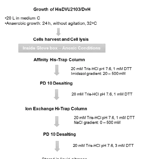
2.1 HOMOLOGOUS PRODUCTION OF DVU2103 IN DESULFOVIBRIO VULGARIS HILDENBOROUGH ... 27
2.1.1 Purification of HisDVU2103 ...28
2.2 HOMOLOGOUS PRODUCTION OF STREPDVU2108 FROM DESULFOVIBRIO VULGARIS HILDENBOROUGH ... 30
2.2.1 StrepDVU2108: Normal Bacterial Growth ...30
2.2.2 StrepDVU2108: Bacterial Growth Supplemented with Mo ...32
2.3 SPECTROSCOPIC CHARACTERIZATION ... 33
2.3.2 Electron paramagnetic resonance (EPR) ...34
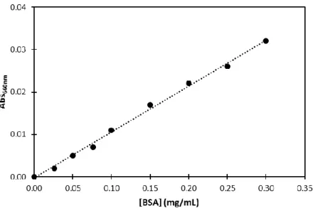
2.3.3 Protein quantification-660 nm Pierce Protein Quantification Method ..34
xvi
2.3.5 Determination of the apparent molecular mass of DVU2103 ...36
2.3.6 ATPase activity of DVU2103 - Malachite Green Phosphate Assay ...39
3 RESULTS AND DISCUSSION ... 43
3.1 DVU2103 FROM DESULFOVIBRIO VULGARIS HILDENBOROUGH ... 43
3.1.1 Homologue expression of protein DVU2103 in D. vulgaris Hildenborough 46 3.1.2 Purification of DVU2103 ...47
3.2 BIOCHEMICAL CHARACTERIZATION ... 56
3.2.1 The Apparent molecular mass ...56
3.2.2 Malachite Green Phosphate Assay – Determination of ATP activity of “DVU2103” 61 3.3 SPECTROSCOPIC CHARACTERIZATION ... 63
3.3.1 UV-visible Spectroscopy ...63
3.3.2 EPR Spectroscopy ...65
3.4 HOMOLOGOUS EXPRESSION OF DVU2108 IN D. VULGARIS HILDENBOROUGH ... 71
3.4.1 Purification of DVU2108 ...71
4 CONCLUSIONS AND FUTURE PERSPECTIVES ... 81
5 REFERENCES ... 85
xvii
Figure Index
FIGURE 1.1-THE SULPHUR CYCLE.THE ARROWS REPRESENT SULPHUR TRANSFORMATION THROUGH OXIDATION AND REDUCTION REACTIONS.IMAGE FROM [2] ... 2 FIGURE 1.2-SCHEMATIC REPRESENTATION OF DIFFERENT TYPES OF [FE-S] CLUSTERS.A–[1FE]
CLUSTER.B–[2FE-2S] CLUSTER.C–[3FE-4S] CLUSTER.D–[4FE-4S] CLUSTER. IMAGE FROM [19] ... 8 FIGURE 1.3–BIOCHEMICAL MODEL OF THE NIF SYSTEM SPECIFICALLY FEMO-CO BIOSYNTHESIS IN
NITROGENASE MATURATION.IMAGE FROM [31]. ... 12 FIGURE 1.4–SCHEMATIC MODEL FOR THE BACTERIAL IRON-SULPHUR PROTEIN BIOSYNTHESIS BY THE
ISC MACHINERY.IMAGE FROM [31]. ... 13 FIGURE 1.5–STRUCTURE OF HETEROMETALIC CLUSTER OF ORP[S2MOS2CUS2MOS2] PROPOSED BY
EXAFS ANALYSIS.IMAGE FROM [42]. ... 17 FIGURE 1.6–UV-VISIBLE SPECTRUM OF NATIVE ORP FROM D. GIGAS IN 50 MMTRIS-HCL PH7.6 AND
60 MMNACL BUFFER.ADAPTED FROM [43]. ... 18 FIGURE 1.7-SCHEMATIC REPRESENTATION OF GENES BELONGING TO THE ORP GENE CLUSTER IN D.
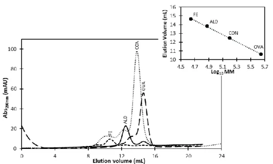
VULGARIS HILDENBOROUGH.ADAPTED FROM [49]. ... 20 FIGURE 1.8-SCHEMATIC REPRESENTATION OF ORP INTERACTION NETWORK.ADAPTED FROM [49]. 21 FIGURE 2.1-SCHEME OF THE PURIFICATION PROCESS OF HISDVU2103 FROM DVH ... 29 FIGURE 2.2-SCHEME OF PURIFICATION PROCESS OF DVU2108 FROM DVH. ... 32 FIGURE 2.3-A TYPICAL CALIBRATION CURVE FOR PROTEIN QUANTIFICATION USING 660 NM PIERCE
PROTEIN QUANTIFICATION METHOD.Y=0.1083X-0.0003, WITH A R=0.9982 ... 35 FIGURE 2.4-CHROMATOGRAM OBTAINED FOR THE CALIBRATION CURVE OF SIZE-EXCLUSION
xviii
PROTEINS OF THE CALIBRATION KIT OF SIZE-EXCLUSION CHROMATOGRAPHY (LMWGE
HEALTHCARE):FE–FERRITIN,ALD–ALDOLASE,CON–CONALBUMIN AND OVA–OVALBUMIN. A TYPICAL CALIBRATION CURVE FOR SUPERDEX 200 SIZE-EXCLUSION CHROMATOGRAPHY IN 50 MMTRIS-HCL BUFFER PH7.6,150 MMNACL AND 1 MMDTT.Y=-4.0559X +33.5363, WITH A R=0.9990(Y CORRESPOND TO ELUTION VOLUME AND X TO THE LOG10MM). ... 37 FIGURE 2.5-CHROMATOGRAM OBTAINED FOR THE CALIBRATION CURVE OF SIZE-EXCLUSION
CHROMATOGRAPHY IN A SUPERDEX7510/300GL COLUMN. THE FIGURE REPRESENTS THE PROTEINS OF THE CALIBRATION KIT OF SIZE-EXCLUSION CHROMATOGRAPHY (LMWGE
HEALTHCARE):CON–CONALBUMIN,OVA–OVALBUMIN,CA–CARBONIC ANHYDRASE,RIB– RIBONUCLEASE,APO–APOPROTEIN.A TYPICAL CALIBRATION CURVE FOR SUPERDEX 75 SIZE -EXCLUSION CHROMATOGRAPHY IN 50 MMTRIS-HCL BUFFER PH7.6,150 MMNACL AND 1 MM DTT.Y=-5.7026X +37.0710, WITH A R=0.9963(Y CORRESPOND TO ELUTION VOLUME AND X TO THE LOG10MM). ... 38 FIGURE 2.6–SCHEMATIC MODEL FOR THE MALACHITE GREEN PHOSPHATE ASSAY REACTION.
MALACHITE GREEN AND MOLYBDATE REACT WITH FREE ORTHOPHOSPHATE RELEASED FROM THE ATP HYDROLYSIS REACTION TO FORM A GREEN COMPLEX. ... 39 FIGURE 2.7-CALIBRATION CURVE FOR MALACHITE GREEN PHOSPHATE ASSAY UNDER OXIC
CONDITIONS.THE EQUATION OF CURVE FOR THE CALIBRATION CURVE OBTAINED,ABS620NM =
0.0052[PHOSPHATE](µM)+0.0080, WITH A R=0.9977, WAS USED TO DETERMINE THE AMOUNT OF PHOSPHATE PRODUCED IN THE REACTION. ... 40 FIGURE 2.8-CALIBRATION CURVE FOR MALACHITE GREEN PHOSPHATE ASSAY IN ANAEROBIOSIS.THE
EQUATION OF THE CALIBRATION CURVE OBTAINED,ABS620NM =0.0076[PHOSPHATE](µM)
-0.0008, WITH A R=0.9985, WAS USED TO DETERMINE THE AMOUNT OF PHOSPHATE PRODUCED IN THE REACTION. ... 41 FIGURE 3.1-SEQUENCE ALIGNMENT OF DVU2103 FROM D. VULGARIS HILDENBOROUGH (NCBI CODE:
WP_010939380.1) WITH THE SIMILAR PROTEINS FROM D. TERMITIDIS (NCBI CODE:
WP_035068143.1),D. AMINOPHILUS (NCBI CODE:WP_051202990.1),D. AFRICANUS (NCBI CODE:WP_027367473.1),D. OXYCLINAE (NCBI CODE:WP_018123934.1)D. BASTINII (NCBI CODE:WP_027178178.1),D. ALASKENSIS (NCBI CODE:WP_011368948.1) AND D. GIGAS (NCBI CODE:WP_051286230.1).THE SEQUENCES WERE OBTAINED FROM THE
INTERFACE BLASTP, AND BIOINFORMATICS CLUSTALX[61]2.1 AND JALVIEW2.9 WAS USED FOR MULTIPLE SEQUENCE ALIGNMENT.HIGHLIGHT THE CONSERVED AMINO ACID RESIDUES.THE WALKER A MOTIFS,CYS-X-X-CYS MOTIFS AND LOCALS OF [FE-S] CLUSTER BINDING ARE
xix
FIGURE 3.2-PROTEINS EXPRESSION PROFILE IN 12.5%TRIS-TRICINE SDS-PAGE.LANES:1.LOW MOLECULAR MASS MARKER (LMW–NZYTECH),2.SOLUBLE FRACTION,3.CELL DEBRIS,4. MEMBRANE FRACTION. ... 47 FIGURE 3.3–A)ANALYSIS OF THE FRACTIONS DURING DVU2103 PURIFICATION.LANES:1.LOW
MOLECULAR MASS MARKER (LMW–NZYTECH),2.SOLUBLE FRACTION,3.DVU2103
FRACTION AFTER AFFINITY CHROMATOGRAPHY,4.DVU2103 FRACTION AFTER IONIC EXCHANGE CHROMATOGRAPHY.12.5%SDS-PAGE.B)PAGE OF THE PURIFIED DVU2103 AFTER IONIC EXCHANGE CHROMATOGRAPHY (10%PAGE).GELS WERE STAINED WITH COOMASSIE BLUE. ... 49 FIGURE 3.4-SEQUENCE ALIGNMENT OF DVU2103 AND DVU2104 OF D. VULGARIS HILDENBOROUGH
THE SEQUENCES WERE OBTAINED IN THE INTERFACE BLASTP AND BIOINFORMATICS CLUSTALX 2.1 AND JALVIEW2.9 WAS USED FOR MULTIPLE SEQUENCE ALIGNMENT.HIGHLIGHT THE
IDENTICAL RESIDUES. ... 51 FIGURE 3.5–UV-VISIBLE SPECTRUM OF AS-ISOLATED “DVU2103" COMPLEX OF D. VULGARIS
HILDENBOROUGH IN 20 MMTRIS-HCL BUFFER PH7.6 AND 3 MMDTT. ... 52 FIGURE 3.6-CHROMATOGRAPHIC PROFILE OF DVU2103 ELUTED IN A SUPERDEX7510/300GL
COLUMN.THE SAMPLE WAS ELUTED IN A RUNNING BUFFER CONTAINING 20 MMTRIS-HCL PH 7.6,150 MMNACL AND 1MMDTT.ABSORBANCE WAS MONITORED AT 280 NM AND 400NM. THE CHROMATOGRAM WAS OBTAINED RECURRING TO A NANODROP.THE CHROMATOGRAM REPRESENTS ABSORBANCE VALUES AT 280NM (FULL LINE) AND AT 400NM (DASHED LINE). ... 54 FIGURE 3.7-SDS-PAGE ACRYLAMIDE GEL AFTER SIZE-EXCLUSION CHROMATOGRAPHY PURIFICATION.
PROTEINS WERE ELUTED IN 20 MMTRIS-HCL PH7.6,150MMNACL AND 1MMDTT RUNNING BUFFER.M.MARKER LOW MOLECULAR MASS (LMW–NZYTECH) AND NUMBERS (9 TO 21). ELUTION VOLUME OF COLLECTED FRACTIONS.HIGHLIGHT THE FRACTIONS OF INTEREST. ... 54 FIGURE 3.8-UV–VISIBLE SPECTRA OF FRACTIONS OF INTEREST IDENTIFIED AFTER SIZE-EXCLUSION
CHROMATOGRAPHY IN A SUPERDEX7510/300GL IN 20 MMTRIS-HCL BUFFER PH7.6 AND 3 MMDTT.PANEL A.FRACTION 11,PANEL B.MIX OF FRACTIONS 14 TO 15’. ... 55 FIGURE 3.9-CHROMATOGRAM OBTAINED FOR THE CALIBRATION CURVE OF SIZE-EXCLUSION
CHROMATOGRAPHY IN A SUPERDEX20010/300GL COLUMN.THE FIGURE REPRESENTS THE PROTEINS OF THE CALIBRATION KIT OF SIZE-EXCLUSION CHROMATOGRAPHY (LMWGE
HEALTHCARE):FE–FERRITIN,ALD–ALDOLASE,CON–CONALBUMIN AND OVA-OVALBUMIN (IN GREY) AND “DVU2103” IN THE AS-ISOLATED FORM (BLUE).AT THE TOP RIGHT CORNER IS REPRESENTED THE CALIBRATION CURVE FOR THIS COLUMN, THE BLACK CIRCLES ARE
REPRESENTATIVE OF THE STANDARD PROTEINS AND THE BLUE CIRCLE IS REPRESENTATIVE OF “DVU2103” PROTEIN,ELUTION BUFFER:50 MMTRIS-HCL BUFFER PH7.6,150 MMNACL AND
xx
1 MMDTT.EQUATION:ELUTION VOLUME =-4.0559 X LOG10MM+33.536, WITH A R= 0.9990. ... 56 FIGURE 3.10-CHROMATOGRAM OBTAINED FOR THE CALIBRATION CURVE OF SIZE-EXCLUSION
CHROMATOGRAPHY IN A SUPERDEX7510/300GL COLUMN.THE FIGURE REPRESENTS THE PROTEINS OF THE CALIBRATION KIT OF SIZE-EXCLUSION CHROMATOGRAPHY (LMWGE
HEALTHCARE):CON–CONALBUMIN,OVA–OVALBUMIN,CA–CARBONIC ANHYDRASE,RIB– RIBONUCLEASE,APO-APROTININ (IN GREY) AND "DVU2103" IN THE AS-ISOLATED FORM (GREEN).AT THE TOP RIGHT CORNER IS REPRESENTED THE CALIBRATION CURVE FOR THE COLUMN, THE BLACK CIRCLES ARE REPRESENTATIVE OF THE STANDARD PROTEINS, THE GREEN CIRCLES ARE REPRESENTATIVE OF "DVU2103" AND DVU2108.ELUTION BUFFER:50 MMTRIS -HCL BUFFER PH7.6,150 MMNACL AND 1 MMDTTEQUATION:ELUTION VOLUME =-5.7026 X LOG10MW+37.071, WITH A R=0.9962. ... 58 FIGURE 3.11-COMPARISON OF CHROMATOGRAMS OBTAINED FOR SIZE-EXCLUSION CHROMATOGRAPHY
IN A SUPERDEX7510/300GL COLUMN OF “DVU2103” FROM D. VULGARIS HILDENBOROUGH ELUTED WITH A BUFFER CONTAINING ATP(0.5 MM)(BLUE) AND NORMAL BUFFER (ORANGE), WITH NO ATP. ... 59 FIGURE 3.12-COMPARISON OF CHROMATOGRAMS OBTAINED FOR SIZE-EXCLUSION CHROMATOGRAPHY
IN A SUPERDEX7510/300GL COLUMN OF “DVU2103” FROM DESULFOVIBRIO VULGARIS HILDENBOROUGH WHEN EXPOSED TO OXIC CONDITIONS (BLUE) AND ANOXIC CONDITIONS
(PRESENCE OF REDUCING AGENT)(ORANGE). ... 60 FIGURE 3.13-EVALUATION OF PHOSPHATE CONCENTRATION THROUGH TIME (MIN) FOR 10 µM
“DVU2103” AS ATPASE. THE ASSAY WAS PERFORMED IN 50 MMTRIS-HCL BUFFER PH8.1, 100 MMNACL,2 MMMGCL2AND 2 MMDTT AT ROOM TEMPERATURE.AT VARIOUS TIME INTERVALS ALIQUOTS OF THE ASSAY MIXTURE WERE STOPPED BY ADDITION OF A STOP REAGENT, AND INORGANIC PHOSPHATE WAS DETERMINED USING THE MALACHITE GREEN ASSAY.FIGURE A AND B REPRESENT TWO ASSAYS PERFORMED IN TWO DIFFERENT CONDITIONS: IN ANOXIC (FULL CIRCLES) AND OXIC CONDITIONS (OPEN CIRCLES).A.ASSAY PERFORMED WITH 0.25 MMATP, EQUATION (OXIC CONDITION):PHOSPHATE CONCENTRATION =0.0253 X TIME –0.0215,R= 0.9999;EQUATION (ANOXIC CONDITION):PHOSPHATE CONCENTRATION =0.0811 X TIME – 0.0201, WITH A R=0.9831.B.ASSAY PERFORMED WITH 2.5 MMATP,EQUATION (OXIC CONDITION):PHOSPHATE CONCENTRATION =0.4145 X TIME –0.8781,R=0.9858;EQUATION (ANOXIC CONDITION):PHOSPHATE CONCENTRATION =1.0482 X TIME +4.7324, WITH A R= 0.9870. ... 62
xxi
FIGURE 3.14-SPECTROSCOPIC PROPERTIES OF D. VULGARIS HILDENBOROUGH "DVU2103".UV– VISIBLE SPECTRA OF “DVU2103” IN 20 MMTRIS-HCL BUFFER PH7.6 AND 1 MMDTT.THE AS -ISOLATED SPECTRUM OF “DVU2103” IS SHOWN AS A CONTINUOUS BLUE LINE AND THE REDUCED SPECTRUM WITH INCREASING AMOUNT OF ASCORBATE IS SHOWN AS A DASHED LINE. ... 63 FIGURE 3.15-SPECTROSCOPIC PROPERTIES OF D. VULGARIS HILDENBOROUGH “DVU2103”.UV–
VISIBLE SPECTRA OF “DVU2103” IN 20 MMTRIS-HCL BUFFER PH7.6 AND 1 MMDTT.THE AS -ISOLATED SPECTRUM OF “DVU2103” IS SHOWN AS A CONTINUOUS BLUE LINE AND THE REDUCED SPECTRUM WITH INCREASING AMOUNT OF DITHIONITE IS SHOWN AS A DASHED LINE. ... 64 FIGURE 3.16-SPECTROSCOPIC PROPERTIES OF D. VULGARIS HILDENBOROUGH “DVU2103”.UV–
VISIBLE SPECTRA OF “DVU2103” IN 20 MMTRIS-HCL BUFFER PH7.6 AND 1 MMDTT.A) RECOVERY OF THE PROTEIN CLUSTER REDUCED BY DITHIONITE AFTER OXIDATION UNDER OXIC CONDITIONS.THE AS-ISOLATED SPECTRUM OF “DVU2103” IS SHOWN AS A CONTINUOUS BLUE LINE AND THE OXIDATION SPECTRUM WITH INCREASING INCUBATION TIME UNDER OXIC CONDITION IS SHOWN AS A DASHED LINE.B)UV-VISIBLE SPECTRA OF “DVU2103”24 H AFTER EXPOSURE TO OXIC CONDITIONS. ... 65 FIGURE 3.17-X-BAND EPR SPECTRUM OF 126 µM“DVU2103” IN 20 MMTRIS-HCL BUFFER PH7.6
AND 3 MMDTT, AT 9.65GHZ OF FREQUENCY,15 DB,5GPP OF MODULATION AND 1×10⁵GAIN. THE FIGURE REPRESENTS THE AS-ISOLATED SPECTRUM OF "DVU2103" AT 10K(BLUE) AND 20 K(ORANGE). ... 66 FIGURE 3.18-X-BAND EPR SPECTRUM OF 126 ΜM“DVU2103” IN 20 MMTRIS-HCL BUFFER PH7.6
AND 3 MMDTT, AT 10K,9.65GHZ OF FREQUENCY15 DB,5GPP OF MODULATION AND 1×10⁵ GAIN.THE FIGURE REPRESENTS THE AS-ISOLATED (BLUE), ASCORBATE ADDITION (ORANGE) AND DITHIONITE ADDITION (GREY) SPECTRA OF “DVU2103”. ... 68 FIGURE 3.19-X-BAND EPR SPECTRUM OF 126 ΜM“DVU2103” IN 20 MMTRIS-HCL BUFFER PH7.6
AND 3 MMDTT, AT 10K,9.65GHZ OF FREQUENCY 15 DB,5GPP OF MODULATION AND 1×10⁵ GAIN.A-REPRESENTATIVE SPECTRA OF AS-ISOLATED (BLUE) AND ASCORBATE REDUCTION (ORANGE) FORM.B–REPRESENTATIVE SPECTRA OF AS-ISOLATED (BLUE) AND DITHIONITE REDUCTION (GREY) FORM. ... 69 FIGURE 3.20–PROTEINS PROFILE IN 12.5%TRIS-TRICINE SDS-PAGE.A)NORMAL BACTERIAL
GROWTH.B)SUPPLEMENTED BACTERIAL GROWTH.LANES:1.LOW MOLECULAR MASS MARKER (LMW–NZYTECH),2.CELL DEBRIS,3.MEMBRANE FRACTION,4.SOLUBLE FRACTION.GELS WERE STAINED WITH COOMASSIE BLUE. ... 71 FIGURE 3.21-SDS-PAGE ACRYLAMIDE GEL AFTER DVU2108 PURIFICATION RECURRING TO AN
xxii
FRACTIONS CORRESPONDING TO PROTEIN ELUTION,8.WASHING STEP,9.SOLUBLE PROTEIN FRACTION.GELS WERE STAINED WITH COOMASSIE BLUE. ... 72 FIGURE 3.22-UV–VISIBLE SPECTRUM OF AS ISOLATED HYPOTHETICAL PROTEIN DVU2108 OF
DESULFOVIBRIO VULGARIS HILDENBOROUGH IN 100 MMTRIS-HCL BUFFER PH7.6,500 MM NACL AND 3 MMDTT. ... 73 FIGURE 3.23-CHROMATOGRAPHIC PROFILE OF DVU2108 SEPARATION IN A SUPERDEX7510/300GL
COLUMN.THE SAMPLE WAS ELUTED IN A RUNNING BUFFER CONTAINING 50 MMTRIS-HCL PH 8.1,150 MMNACL AND 1 MMDTT.ABSORBANCE WAS MONITORED AT 280 NM AND 400 NM. THE CHROMATOGRAM WAS OBTAINED RECURRING TO A NANODROP.THE CHROMATOGRAM REPRESENTS ABSORBANCE VALUES AT 280 NM (FULL LINE) AND AT 400 NM (DASHED LINE). .... 74 FIGURE 3.24- SDS-PAGE OF THE FRACTIONS COLLECTED FROM THE SIZE-EXCLUSION
CHROMATOGRAPHY DURING DVU2108 PURIFICATION.PROTEINS WERE ELUTED IN 50 MMTRIS -HCL PH8.1,150 MMNACL AND 1 MMDTT RUNNING BUFFER.LANES:INJ.INJECTION,M. MARKER LOW MOLECULAR MASS (LMW–NZYTECH) AND NUMBERS (8 TO 18). COLLECTED FRACTIONS DURING ELUTION.HIGHLIGHTED ARE THE FRACTIONS OF INTEREST. ... 74 FIGURE 3.25-UV–VISIBLE SPECTRA OF AS-ISOLATED HYPOTHETICAL PROTEIN DVU2108 OF D.
VULGARIS HILDENBOROUGH IN 100 MMTRIS-HCL BUFFER PH7.6,500 MMNACL AND 3 MM DTT AFTER THE SIZE-EXCLUSION CHROMATOGRAPHY. ... 75 FIGURE 3.26-ANALYSIS OF THE FRACTIONS DURING DVU2108 PURIFICATION.LANES:1.LOW
MOLECULAR MASS MARKER (LMW–NZYTECH),2.DVU2108 FRACTION BEFORE SIZE -EXCLUSION CHROMATOGRAPHY,3.DVU2108 FINAL FRACTION AFTER SIZE-EXCLUSION
CHROMATOGRAPHY.12.5%SDS-PAGE.GELS WERE STAINED WITH COOMASSIE BLUE. ... 76 FIGURE 3.27-SDS-PAGE ACRYLAMIDE GEL AFTER DVU2108 PURIFICATION RECURRING TO AN
AFFINITY COLUMN.LANES:1.MARKER LOW MOLECULAR MASS (LMW–NZYTECH),2-3. SOLUBLE FRACTION 4-8.FRACTIONS CORRESPONDING TO WASH STEPS.9-12.FRACTIONS
CORRESPONDING TO PROTEIN ELUTION.GELS WERE STAINED WITH COOMASSIE BLUE. ... 79 FIGURE 6.1–ELECTRON TRANSFER FROM THE L127ΔFE PROTEIN TO THE P-CLUSTERS OF THE MOFE
PROTEIN MONITORED BY PERPENDICULAR MODE EPR SPECTROSCOPY.MOFE PROTEIN, WITH EACH P-CLUSTER OXIDIZED BY TWO ELECTRONS (P2+ STATE), AND THE REDUCED BUT DITHIONITE-FREE L127ΔFE PROTEIN WERE PREPARED AS DESCRIBED IN EXPERIMENTAL PROCEDURES.ALL SAMPLES WERE INCUBATED FOR 2 MIN PRIOR TO FREEZING IN LIQUID
NITROGEN, AND THE BUFFER WAS 50 MMMOPS(PH7.0).PERPENDICULAR MODE EPR SPECTRA ARE SHOWN FOR THE OXIDIZED (P2+) STATE OF THE MOFE PROTEIN (54 µM)(TRACE 1), THE REDUCED STATE OF THE L127ΔFE PROTEIN (91 µM)(TRACE 2), THE MATHEMATICAL ADDITIVE
xxiii
SPECTRUM FOR TRACES 1 AND 2(TRACE 3), AND THE MIXTURE OF L127ΔFE PROTEIN (91ÍM) AND THE P2+ STATE OF THE MOFE PROTEIN (54 µM)(TRACE 4).ALL SPECTRA WERE RECORDED AT 12K WITH A MICROWAVE FREQUENCY OF 9.64GHZ, A MODULATION FREQUENCY OF 100 KHZ, A MODULATION AMPLITUDE OF 5.028G, A TIME CONSTANT OF 20.48 MS, AND A MICROWAVE POWER OF 10.1 MW.IMAGE FROM [74] ... 98 FIGURE 6.2-X-BAND EPR SPECTRUM OF 126 ΜM“DVU2103” IN 20 MMTRIS-HCL BUFFER, PH7.6
AND 3 MMDTT, AT 10K,9.65GHZ OF FREQUENCY,15 DB,5GPP OF MODULATION AND 1×10⁵GAIN.THE FIGURE REPRESENTS THE AS-ISOLATED SPECTRUM OF "DVU2103"(BLUE) AND DITHIONITE ADDITION: AFTER 30 MIN (GREY) AND 1H (DARK GREY). ... 99
xxv
Table Index
TABLE 1.1 - STRUCTURAL MOTIFS OF DIFFERENT [4FE-4S]. ... 10 TABLE 1.2 - MOLECULAR MASS AND PI OF PROTEINS ENCODED BY ORP OPERON OF D. VULGARIS
HILDENBOROUGH.DATA OBTAINED BY [56]–[58] ... 24 TABLE 3.1 - IDENTIFICATION OF THE THREE BANDS THAT FORM A COMPLEX THROUGH PEPTIDE MASS FINGERPRINT ANALYSIS BY MALDI-TOF-MS ... 50 TABLE 3.2 - QUANTIFICATION OF PROTEIN AND FE FOR THE DIFFERENT PURIFICATION PERFORMED DURING THIS WORK. ... 53 TABLE 3.3 - ASSIGNMENT OF THE MOLECULAR MASS CORRESPONDING TO THE ELUTION VOLUME OF THE MAIN PEAKS OBSERVED IN THE SIZE-EXCLUSION CHROMATOGRAPHY CHROMATOGRAM (FIGURE 3.11) TO DETERMINE THE EFFECT OF THE ATP. ... 59 TABLE 3.4 - ASSIGNMENT OF THE MOLECULAR MASS (KDA) CORRESPONDING TO ELUTION VOLUME (ML) OF MAIN PEAKS OF THE CHROMATOGRAM OF SIZE-EXCLUSION CHROMATOGRAPHY (FIGURE 3.12) ... 61 TABLE 3.5 - DATA OBTAINED FROM ICP-AES ANALYSIS FOR FE, MO AND CU ATOMS IN DVU2108
PROTEIN. QUANTIFICATION OF PERFORMED BY 660 NM PIERCE PROTEIN QUANTIFICATION METHOD. ... 77 TABLE 3.6 - GROWTH OF D. VULGARIS HILDENBOROUGH IN MEDIUM C SUPPLEMENTED WITH MO. .... 78 TABLE 3.7 - QUANTIFICATION OF PERFORMED BY 660 NM PIERCE PROTEIN QUANTIFICATION METHOD FOR ELUTED FRACTIONS OF STREPDVU2108 PURIFICATION. ... 80 TABLE 6.1 - GROWTH OF D. VULGARIS HILDENBOROUGH IN MEDIUM C. ... 93
xxvi
TABLE 6.2 - COMPOSITION OF MEDIUM C FOR DESULFOVIBRIO VULGARIS HILDENBOROUGH GROWTH. THE MEDIUM WAS STERILIZED IN AUTOCLAVE AT 120°C FOR 20 MIN AT 1 BAR. THE RECIPE PRESENTED IS FOR A TOTAL VOLUME OF 1L. ... 94 TABLE 6.3 - QUANTITATIVE COMPOSITION OF POLYACRYLAMIDE GELS 12.5%(M/V) AND 10%(M/V)
IN BUFFER SOLUTION TRIS-TRICINE.INDEPENDENTLY OF THE PERCENTAGE OF THE RUNNING GEL, THE STACKING GEL’S CONCENTRATION IS ALWAYS THE SAME. APS(AMMONIUM PERSULPHATE) AND TEMED(TETRAMETHYLETHYLENEDIAMINE,(CH3)2NCH2CH2N(CH3)2) ARE ONLY ADDED AT THE END OF GEL PREPARATION.ALL SOLUTIONS ARE STORED AT 4°C, EXCEPT APS THAT IS STORED AT -20°C. STOCK OF ACRYLAMIDE/BIS-ACRYLAMIDE AND GEL BUFFER SOLUTIONS IS 41.5%/1.5%(M/V) AND 3MTRIS-HCL PH8.3 AND 0.3%SDS(M/V), RESPECTIVELY.FOR PAGE GELS, GEL BUFFER COMPOSITION IS DIFFERENT (WITHOUT DENATURANT AGENT,SDS) IN THE SAME VOLUME. ... 95 TABLE 6.4 - QUANTITATIVE COMPOSITION OF STAINING SOLUTION FOR SDS-PAGE AND PAGE GELS.
... 95 TABLE 6.5 - QUANTITATIVE COMPOSITION OF DISTAINING SOLUTION FOR SDS-PAGE AND PAGE GELS.
... 96 TABLE 6.6 - PROTEINS OF THE CALIBRATION KIT OF SIZE-EXCLUSION CHROMATOGRAPHY (LMWGE HEALTHCARE) USED TO CONSTRUCT THE CALIBRATION CURVE FOR MM DETERMINATION FOR SUPERDEX 20010/300GL... 97 TABLE 6.7 - PROTEINS OF THE CALIBRATION KIT OF SIZE-EXCLUSION CHROMATOGRAPHY (LMWGE HEALTHCARE) USED TO DETERMINE THE MOLECULAR MASS FOR SUPERDEX 7510/300GL. ... 97
xxvii
Abbreviations
Ɛ Molar Extinction Coefficient
λ Wavelength
Abs Absorbance
ATP Adenosine Triphosphate
AU Arbitrary Unit
BLAST Basic Local Alignments Search Tool COG Cluster of groups of orthologous proteins
D. Desulfovibrio
EBP Enhancer binding activator protein
EPR Electron Paramagnetic Resonance
EXAFS Extended X-ray absorption fine structure
Fds Ferredoxines
[Fe-S] Iron-Sulphur cluster
G Gauss
HiPIP High Potential Iron-Sulphur Protein
ICP Induced Coupled Plasma
LMW Low Molecular Weight
MALDI-TOF-MS Matrix Assisted Laser Desorption Ionization – Time of Flight – Mass Spectrometry
Min Minute
MM Molecular Mass
NCBI National Center for Biotechnology Information
xxviii
ORP Orange Protein
PAGE Polyacrylamide Gel Electrophoresis
RNA Ribonucleic Acid
SDS Sodium Dodecyl Sulphate
SDS-PAGE Polyacrylamide Gel Electrophoresis in denaturant conditions SRB Sulphate Reducing Bacteria
TM Thiomolybdate
Tris Tris(hydroxylmethyl)aminomethane
1
1
Introduction
1.1 Sulphate-Reducing Bacteria
The biogeochemical sulphur cycle (Figure 1.1) is known due to the chemical and biological complexity of its main intervenient, sulphur [1]. Sulphur is one of the most abundant elements on Earth, being present mainly in rocks and sediments [1]. In the sulphur cycle, sulphur goes through a huge range of oxidation states, from - 2 (completely reduced) to + 6 (completely oxidized) [1]. Microorganisms have an important role in this cycle, being responsible for sulphur transformations [1].
Oxidation and reduction reactions of this element are very important in some microorganisms for the generation of metabolic energy [1]. One of the most studied classes of these organisms are the sulphate-reducing bacteria (SRB) [1]. SRB are anaerobic microorganisms that use sulphate as a terminal electron acceptor for the degradation of organic compound (commonly acetate or lactate), producing hydrogen sulphide (H2S) and carbon dioxide (CO2) as final products [1].
2
Figure 1.1 - The sulphur cycle. The arrows represent sulphur transformation through oxidation and
reduction reactions. Image from [2]
SRB were identified in more than 60 genera, making a total of about 220 species widely distributed around Earth, from terrestrial to marine ecosystems [3], [4]. It is believed that these microorganisms have inhabited our planet for 3.5 billion years, being considered ancestral microorganisms [4]. Regarding its cellular classification, SRB can be classified as gram-negative eubacteria, gram-positive eubacteria or archaea [3]. In terms of acquiring organic compounds to growth, SRB can be heterotrophic, autotrophic, lithoautotrophic or respiration-type, always under anaerobiosis [4]. For many years, scientists thought that lactate and pyruvate were the only two compounds used by SRB to support growth, a theory that was dropped after confirming that these microorganisms can use a variety of compounds [4]. Nowadays, SRB can also be divided in two main groups according to how they degrade organic compounds: incomplete or complete degradation. Incomplete oxidizers refer to a group of microorganisms that carry out an incomplete lactate oxidation leading to the formation of a mixture of acetate and CO2 [5]. Considered a group of oxidizers less effective than the group of SBR that possesses complete degradation, this group rarely oxidize fatty acids being more common the use of low-molecular-weight alcohols [5]. Various SRB are included in this category, such as Desulfomonas, Desulfobubus, Desulfolobus and Desulfovibrio genera [6]. In the latter group, sulphate reducers degrade organic compounds to carbon dioxide, using acetate as a carbon source [1]. Organisms present in this
3
group are usually metabolically more diverse and have slower growth rates. They can oxidize a variety of compounds, including fatty acids and aromatic compounds, to CO2 [6].
The capacity to couple anaerobic electron transport to ATP synthesis makes SRB a unique physiological group of prokaryotes, being mainly present in Bacteria and Archaea [1], [3]. Found in marine sediments, polluted environments, cyanobacterial microbial mats, rice fields and even responsible for human diseases, these bacteria have a functional importance in many ecosystems [4]. Able to grow in extreme environments, SRB can reach from high acidic (pH 4) to basic habitats (pH 9.5), without affecting its way of living [3]. Cultures of SRB obtained at extreme temperatures have already been documented, confirming that these microorganisms can adapt their metabolism from psychrophilic to hyperthermophilic conditions [7]. The same happens when SRB are exposed to variable ranges of salt concentrations [7].
Relative to the terminal electron acceptor, sulphate is not the only compound used by SRB, as these bacteria are able to use various other organic and inorganic compounds [4]. SRB are now considered the microorganisms that reduce the greatest number of different terminal electron acceptors [4].
More than 125 compounds are known to be used by pure cultures of SRB, most of them are fermentation products and intermediate break-down products, such as glycerol, amino acids and fatty acids [7]. Sulphate-reducing bacteria’s metabolism focuses on the oxidation of hydrogen or an organic compound with the reduction of sulphate to sulphide [8]. The chemistry involved in that process was discovered in 1984 by Postgate and is represented in the following chemical equation (Equation 1) [6]:
4 AH2 + SO4 2-+ H+ 4 A + HS- + 4 H20 (1)
Referred as a dissimilatory process, SRB oxidize an electron donor (AH2) with subsequent reduction of sulphate in a bioenergetic reaction [6]. Electrons are transferred from the interior of the cell to the exterior, with the help of proteins associated with the membrane [6]. This reaction is catalysed in a single step by a single enzyme, bisulphite reductase [6].
Metabolism of a particular substrate can involve modification or removal of a special group from the substrate, an incomplete oxidation to an important intermediate (such as acetate) or its complete oxidation to CO2 [7]. Several experiments have been conducted to understand
4
more about the biochemistry and physiology of SRB mainly on the genus Desulfovibrio (D.), specifically in D. vulgaris Hildenborough and D. alaskensis G20 [9].
Beyond their function in the sulphur cycle, SRBs also play important roles in other biogeochemical cycles, as for example in the carbon cycle [9]. In anaerobiosis, SRB are part of the conglomerates that mineralize organic carbon (mainly polymers like cellulose) along with other microbes, producing organic acid and reduced gas (CO and H2) as a result of fermentation or oxidative pathways [9].
The fact that these microorganisms possess the ability to inhabit deeply extreme environments reflects its metabolic and phylogenetic versatility [4]. Thus, SRB display significant roles in Nature by virtue of their potential for numerous interaction, not only in sulphur cycle [1]. In biogeochemistry, SRB contribute to the complete oxidation of organic matter, through sulphide production and/or metal reduction, which would be very difficult without this kind of organisms [4]. Many biotechnological processes are affected by the presence of SRB, both in a positive and in a negative way.
Many industrial processes are prejudiced due to the presence of SRB, since the use of sulphuric acid results in the production of sulphate in waste waters because of the presence of these microorganisms [1]. The reduction of sulphate then occurs and when this happens in anaerobic environments, like anaerobic treatment of agro-industrial waste waters, a lower amount of methane is produced [1]. The formation of hydrogen sulphide as product of this reaction is therefore undesirable due to its toxic, odorous and corrosive properties [1]. Biocorrosion of ferrous metals in anaerobic environments is another severe problem with which the industries have to deal [9]. Because SRB are abundant in oil fields, many negative consequences come for the petroleum industry [9]. The issues associated to corrosion of machinery, acidification of oil and sulphide precipitation due to their metabolism are some [9]. Due to increasing concern about the environment, scientists were forced to innovate these processes, finding new ways to resolve these issues without minimizing profits [4].
On the other hand, the advances made in the past few years in the field of biotechnologies are making SRB more economically interesting [4]. The unique electron transport components found in SRB give them the possibility to be used in important environmental activities [4]. Areas like bioremediation, fuel production and wastewater treatment implicate nowadays the use of SRB, in a more economical way than that used a few years ago [4]. The implementation of SRB in systems for the removal of sulphate and heavy metals from waste waters is becoming increasingly utilized, such as the removal of sulphur dioxide from flue gas [7]. Bioremediation of BTEX compounds (Benzene, Toluene,
5
Ethylbenzene and Xylene) in contaminated soils are today made resorting to SRB because of its capability of utilizing hydrocarbons [4]. The SRB also contribute to bioremediation of toxic metal ions from anaerobic sediments [9]. The increase of pH in the surrounding environment due to their metabolism cause the precipitation of toxic metal, such as copper, cadmium and nickel as metal sulphides in acidic aquatic environments [9]. Nowadays, microbiologists consider that SRB are one of the cleaner ways to minimize pollution caused by toxic compounds used in industrial activities [4]. For that reason the use of SRB in bioremediation and biocontrol fields has become very appealing, both economically and ecologically [4].
1.2 Desulfovibrio vulgaris genera
The extreme versatility presented by SRB has always intrigued researchers that have tried to understand the processes behind how some microorganisms are able to support conditions such as high pressure or temperature [1]. Desulfovibrio (D.) genus has been the most extensively studied of the SRB members, both at the biochemical and physiological level, due to its extreme versatility [10].
The first species of Desulfovibrio genera was identified in 1895 by W. M. Beyerink in contaminated sewage and since then various species and strains of Desulfovibrio have been described, between then D. vulgaris Hildenborough (in 1946) [11]. Belonging to the sulphate-reducing class of bacteria that are ubiquitous in Nature, D. vulgaris Hildenborough presents the major metabolic versatility and is actually an organism of great interest by researchers [12]. Present in places with high toxicity levels due to heavy metal contamination (as uranium and chromate) or high concentration of nitrates, Desulfovibrio genus is able to colonize almost all kind of environments [4].
D. vulgaris Hildenborough was initially envisage as a strict anaerobic organism, however, studies suggested that it is in fact an aero-tolerant microorganism [10]. D. vulgaris Hildenborough was the first SRB to have its genome completely sequenced, which allowed functional genomic studies in order to expand the understanding of its electron transport and energy transduction mechanisms, as well as other metabolic studies [12]. Several genomic studies have shown that D. vulgaris Hildenborough is involved in the activation of different response pathways that are specific in a wide range of stresses [13]. A high number of response regulators have been reported to be involved in signal transduction, since D. vulgaris Hildenborough genome encode 64 histidine kinase sensors and 74 response regulators involved in the detection and integration of environmental stimulus [13], [14]. This diversity is believed
6
to be related to the various stresses that SRB face in its growth environments [4]. The fact that this microorganism is easily and rapidly grown renders it a perfect genetic, biochemical and genomic tool to investigate and perform physiological studies, using it as a model for SRB [4], [11].
In terms of metabolic features, D. vulgaris Hildenborough belongs to a group of incompletely-oxidizing sulphate reducers, in which acetate is formed by enzymatic oxidation of organic acids and alcohols, subsequently operating as electron donor for sulphate reduction [12]. Fumarate, ethanol, malate, lactate or pyruvate can also act as electron donors to sulphide reduction [12]. This species generate energy by reducing sulphate, playing an important role in sulphur cycle and complete mineralization of organic matter [4]. Capable of chemolithotrophic growth, this microorganism can also use H2 as electron donor to sulphate reduction, using acetate and CO2 as carbon source [4].
Although its pejorative impacts as biocorrosion agent (in oils and gas pipelines in oceans), this organism also acts as a bioremediator agent, making it economically viable [1].
1.3 Iron-sulphur Proteins
Iron-sulphur clusters ([Fe-S] clusters) are considered the most ancient and versatile inorganic cofactors being widely distributed in all kingdoms life [18]. Chemical versatility of these clusters allied with the abundance of iron and sulphur in prebiotic Earth, facilitated their use in a large number of different proteins (referred to Fe-S proteins), such as simply structural cofactors, redox sensors or catalysts [12]. Composed of iron and inorganic sulphide, these [Fe-S] clusters are formed from attachment to the polypeptide chain primarily via cysteinate iron ligation [19]. Other metals apart from iron can be part of these clusters, such as nickel and molybdenum, however these are less common [20].
[Fe-S] clusters can participate in a wide range of biological and biochemical processes, such as biosynthesis of [Fe-S] clusters, substrate binding and catalysis, DNA repair and gene regulation, central metabolism or even respiration [15], [16]. Biological electron transfer is the main functional role of [Fe-S] clusters, since they are able to delocalize electron density over both Fe and S atoms [16]. Propensity of iron to formally switch between oxidative states + 2 and + 3, makes [Fe-S] clusters excellent donors and acceptors of electrons in many biological reactions [15]. [Fe-S] clusters are also used in enzyme catalysis because they are able to reach very low reduction potentials, being able to reduce redox resistant substrates [15], [17]. These
7
clusters also play an important role in sensing environmental or intracellular conditions for regulation of gene expression due to their reversible interconversion between clusters [15], [17]. [Fe-S] clusters may be distinguished by the number of iron and inorganic sulphur atoms present in their structure as [1Fe], [2Fe-2S], [3Fe-4S] and [4Fe-4S] [20]. In general, [Fe-S] clusters feature two main conformations, rhombic [2Fe-2S] and cubic [4Fe-4S] types [18]. Both clusters contain iron atoms however its oxidation state can differ depending on the type of cluster, number of ferrous (Fe2+) or ferric (Fe3+) iron, and sulphide (S2-) that present always the same oxidation state [21]. [Fe-S] clusters are usually integrated into proteins via coordination of the iron ions by cysteine or histidine residues, but in more complex clusters alternative ligands have also been observed (Asp, Ser, CO or CN-) [18], [22].
1.3.1 Types of [Fe-S] Clusters
As previously mentioned, [Fe-S] cluster can be classified based on the number of iron and sulphur atoms in the cluster [16]. In addition to the number of atoms, protein can also be classified due to certain structural motifs and spectroscopic and electrochemical properties [18]. Four main groups of Fe-S proteins can be identified based on the type of cluster that they possess ([1Fe], [2Fe-2S], [3Fe-4S] or [4Fe-4S] cluster) (Figure 1.2) [18].
8
Figure 1.2 - Schematic representation of different types of [Fe-S] clusters. A – [1Fe] cluster. B –
[2Fe-2S] cluster. C – [3Fe-4S] cluster. D – [4Fe-4S] cluster. Image from [19]
Rubredoxins (Rd) are the simplest group of Fe-S proteins mainly found in Bacteria, Archaea and anaerobes [20]. With approximately 55 residues, these proteins have a unique [1Fe] cluster and does not have inorganic sulphur [20]. The single iron atom is coordinated by four thiolate ligands provided by cysteine segments located around the cluster [17]. As other Fe-S proteins, rubredoxins are electron carriers in several redox chains, such as oxygenation reactions or protection against reactive oxygen species (ROS) [16]. Able to switch between two redox states, [1Fe]3+ and [1Fe]2+, rubredoxins can reach reduction potentials ranging from - 100 to + 200 mV [18]. The iron in this metal centre can also be replaced by other metals, such as cobalt, nickel and zinc, in vitro, which makes them an interesting group of proteins [18].
Several proteins, such as plant type ferredoxins (involved in electron transport between photosystem I and several enzymes), biothin synthase (involved in the conversion of desthiobiotin into biotin by sulphur donation) or MitoNEET (involved in redox-sensitive signalling in [Fe-S] cluster transfer) coordinate a [2Fe-2S] cluster [20]. This cluster can exist in two redox states, [2Fe-2S]2+ and [2Fe-2S]1+, possessing a high reducing power since their
9
reduction potentials can range from - 250 to - 420 mV [16]. EPR active, iron sites of [2Fe-2S] clusters in the reduced form present an S = 1/2 group state resulting in localized valences (S = 5/2 /(Fe3+) and S = 2 (Fe2+)) antiferromagnetially coupled [16]. A subclass of the [2Fe-2S] protein is composed by the Rieske proteins, where [Fe-S] cluster is coordinated by a nitrogen atom of a histidine residue instead of the usual cysteine sulphur [20]. These proteins were first discovered as subunits of respiratory and photosynthetic electron-transfer complexes, and later identified in small electron-transfer proteins, such as ferredoxins, bacterial and eukaryotic oxygenases [18]. With the same redox transition as other 2Fe-2S proteins, the Rieske protein have the highest reduction potential of these type of centres due to the presence of the histidine ligation, reaching positive redox potential from + 400 to + 100 mV [17], [18].
[3Fe-4S] clusters were firstly recognized in anaerobic nitrogen-fixing bacterium Azotobacter vinelandii, that beside a [3Fe-4S] cluster also holds a [4Fe-4S] cluster [16]. This cluster was later identified in two other proteins (D. gigas ferredoxin II and aconitase), as well as in other ferredoxins and several enzymes, such as succinate dehydrogenase, nitrate reductase and [NiFe] hydrogenases [18]. Coordinating four cysteine residues this type of cluster, like other Fe-S proteins, presents magnetic and electronic properties [18]. Two stabilized oxidation states were observed in these clusters, + 1 and 0. In the oxidized state + 1, the cluster exhibits an isotropic EPR signal resulting in spin coupled form (S = 1/2) of three high-spin ferric ions, the signal is lost when the cluster is reduced to the [3Fe-4S]0 state [20]. The redox potential of this redox pair varies between - 70 and - 460 mV, depending on the protein [20]. The interconversion of [3Fe-4S] into [4Fe-4S] clusters can occur (particularly in proteins where the fourth iron atom is not coordinated by a cysteinyl ligand) and have been extensively studied in order to better understand the presence of iron-sulphur in enzymes like aconitase [18]. This type of clusters is rare when compared with other [Fe-S] clusters, like [2Fe-2S] and [4Fe-4S], that are more ubiquitously distributed [17].
Several proteins present in bacteria, mammalian tissues and some plants are constituted by [4Fe-4S] clusters [21]. These are one of the most common [Fe-S] clusters found in Nature and function mainly as ubiquitous electron transfer members in anaerobic bacteria [20], [22]. In some organisms, such as Clostridium, these proteins also play a role as immediate electron donor to nitrogenase and hydrogenase [20].
First detected in Desulfovibrio gigas, this type of cluster acts as electron paramagnetic resonance signatures in mitochondrial membrane proteins and are also involved in electron transfer in small proteins, such as ferredoxins (Fds) [16]. Ferredoxins are one of the major classes of electron transfer proteins in Nature [18]. These are important in a series of processes
10
essentials to life as oxidative phosphorylation, nitrogen fixation and photosynthesis [18]. Ferredoxins are usually distinct based on their reduction potential in two major classes: low-potential Fe-S proteins (- 150 to - 700 mV) and high-low-potential Fe-S proteins (+ 100 to + 450 mV), HiPIPs [16]. Although the distinction made between these proteins, these are all classified as ferredoxins and have in common the presence of a [4Fe-4S] cluster [16].
Metalloproteins usually coordinate metals using amino acid residue as ligands to increase stability [20]. Concerning [Fe-S] clusters, the most commonly observed ligand is cysteine [22]. In most ferredoxins, the [4Fe-4S] cluster is bound to the polypeptide recurring to cysteines residues, that link to Fe provide four thiolate donors to the 1Fe, 2Fe or 4Fe cluster [16], [20]. However, there are also others amino acids residues that have been observed as histidine, glutamine, aspartate or serine (Table 1.1) [22].
Table 1.1 - Structural motifs of different [4Fe-4S].
Due to its disposition in the cluster, iron and sulphur atoms are positioned alternatively in the apexes causing the cluster to take the form of a distorted cube [18]. The cysteine ligands are arranged in a Cys-X2-Cys-X2-Cys, the so-called [4Fe-4S] motif [16]. In this particular motif, three iron atoms are bound to almost adjacent cysteines, while the last one is bound to a more distant cysteine residue in the polypeptide chain [16]. The fact that the cluster is bound by cysteine residues from different parts of the polypeptide chain confers an extra stabilization of the tertiary structure of the protein [16].
The [4Fe-4S] clusters can exist in three stable oxidation states in proteins, which allows the change between two oxidation states through a simple electron transfer [20]. For this structure, the three accessible states are [4Fe-4S]3+, [4Fe-4S]2+ and [4Fe-4S]1+ corresponding to
Protein Iron-Sulphur Ligation Mode Reference
4Fe Ferredoxin Cys8 Cys11 Cys14 Cys50 [23]
4Fe Ferredoxin (Pyrococcus furiosus) Cys12 Asp15(Nδ) Cys18 Cys57 [24]
8Fe Ferredoxin Cys8 Cys11 Cys14 Cys47 [25]
8Fe Ferredoxin (2x [4Fe-4S]) Cys18 Cys37 Cys40 Cys43 [25]
HiPIP Cys41 Cys46 Cys61 Cys75 [26]
Hybrid cluster protein Cys3 Cys6 Cys15 Cys21 [27] [NiFe] hydrogenase His185(N) Cys188 Cys213 Cys219 [28] [FeFe] hydrogenase His94(N) Cys98 Cys101 Cys107 [29]
11
[3Fe3+- Fe2+], [2Fe3+- 2Fe2+] and [Fe3+- 3Fe2+] valence-state combinations, respectively [20], [22].
Usually proteins can present one or more [Fe-S] clusters [18]. In the case of a protein with more than one cluster, it will usually present a brown colour and a broad absorption band in the 380-400 nm region [18]. Characteristic of proteins containing [4Fe-4S]2+ clusters, this absorption band arises from [Fe-S] charge transfer bands resulting from the interaction of [Fe-S] clusters [21]. Studies indicate that the absorption band observed is attributed to d-d transitions of Fe2+ atoms from the lowest layer d orbital into t
2g sets [18]. The overlapping of bands from S Fe3+ charge transfer results in a spectrum with a broad absorption band [18].
EPR spectroscopy is also used to characterize this type of metal clusters [21]. Weak spin-spin interactions between paramagnetic clusters are responsible for the appearance of the EPR signal [21]. [4Fe-4S] clusters exhibit EPR resonances, detectable at low temperatures (below 35 K), only when the cluster is in the Fe3+ - 3Fe2+ (S = 1/2) state, being EPR silent when it goes to a 2Fe3+ - 2Fe2+ state (S = 0) [18]. Depending on the polypeptide environment and number of [4Fe-4S] clusters present in the protein, the EPR spectrum can exhibit different forms [18]. In the case of a single [4Fe-4S]1+ cluster, the spectrum can present either an axial or rhombic signal, while a more complex EPR spectrum is observed in the presence of two [4Fe-4S]1+ clusters [21]. [4Fe-4S]3+ cluster spectrum is also more sensitive to peptide environment comparing to that of a [4Fe-4S]1+ cluster [18].
1.3.2 Biosynthesis of Iron-Sulphur Clusters
In the 1990s, scientists improved chemical assemble of [Fe-S] clusters into apoproteins, determining that this process in vitro was different from what occurring in vivo [15]. Fe-S assembly require sources of inorganic iron and sulphur, although in vitro this can be achieved by spontaneous processes, with FeCl and Na2S through metallic reconstitution, in living cells this process is more complex and needs to be catalysed [12]. Thus, organisms need to resort to multicomponent systems, so-called Fe-S biogenesis systems, to perform the production of Fe-S proteins [17]. Cellular surroundings are also protected by these systems, avoiding potentially harmful effects of free Fe2+ , Fe3+ and S2- ions [17]. Depending on the organism, the number and the type of Fe-S biogenesis systems varies, also as the enzymes and compounds involved [12]. Three different systems for the biogenesis of bacterial Fe-S proteins have been identified based on biochemical evidence and organization of genes in bacterial operons: NIF, ISC and SUF systems [15], [30].
12
NIF system (Figure 1.3) is specific for the maturation of nitrogenase in azotrophic bacteria, being involved in nitrogen fixation, ISC and SUF systems are more common in prokaryotes [12], [16]. ISC system have as main objective to promote the formation of [Fe-S] clusters, while SUF system clusters in mobilization of sulphur [16]. Conserved in eubacteria and eukaryotes (mitochondria), ISC system has a broad specificity targeting general Fe-S proteins [12]. SUF system represent an alternative pathway to the ICS system and operates under iron starvation and can be found in eubacteria, archaea and eukaryotes (plastids) [15]. However, both act in maturation of all Fe-S proteins in the cell and are important for their organization under normal and oxidative-stress conditions, respectively [12].
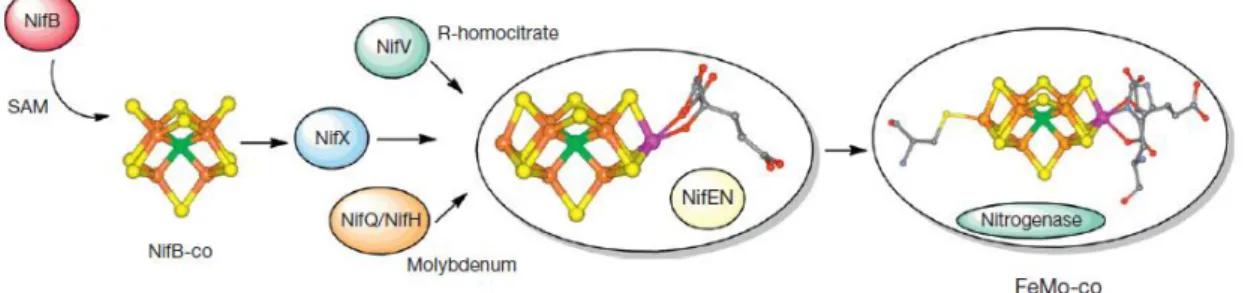
Figure 1.3 – Biochemical model of the NIF system specifically FeMo-co biosynthesis in nitrogenase
maturation. Image from [31].
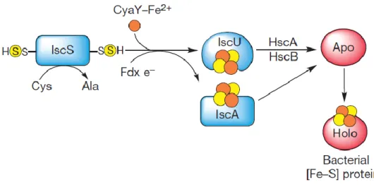
Fe-S biogenesis is a molecular process divided in two major steps: assembly and traffic [17]. The ISC system (Figure 1.4) consists in a set of genes that encodes several proteins necessary to its operation, such as a regulator (IscR), a cysteine desulphurase (IscS), a scaffold (IscU in bacteria and Isc1 in mitochondria), an A-type protein (IscA), a DnaJ-like co-chaperone (HscB), a DnaK-like chaperone (HscA) and a ferredoxin (Fdx) [17]. In the first step, sulphur is acquired from amino acid L-cysteine produced by the cysteine desulphurase (IscS) [17]. This reaction involves the conversion of L-cysteine into L-alanine through IscS and the release of a sulphur atom, in the form of a hydrogen disulphide [17]. Scaffold proteins contain three conserved cysteine residues (Cys37, Cys63 and Cys106) that helps in coordination of the [Fe-S] cluster [16]. Iron is provided to the system through CyaY protein, which is an iron donor [17]. A close interaction between it and the components of isc operon is necessary to provide iron atoms to the system, preventing any leakage of harmful free ions [17].
Since sulphur present in cysteine residues is in a S0 form, it is necessary that a reduction to sulphide (S2-) occurs in order for it to be used in the [Fe-S] cluster [15]. An electron transfer
13
mechanism generally provided by ferredoxin reductase and ferredoxin of the Isc assembly machineries is required for that step [15]. Then the [Fe-S] cluster is initially assembled in the scaffold proteins (IscU) [15]. IscU is necessary because it interacts with IscS and the iron source, allowing sulphur and iron to meet, receiving sulphur directly from IscS [12]. The IscU-IscS interaction is provided by the chemical and structural environment prepared, which facilitates the formation of the [Fe-S] cluster [12], [17]. The second step refers to the release of the [Fe-S] cluster from IscS-IscU, through interaction with an ATP-hydrolysing component [12], [15]. HscA and HscB, two members of DnaK/DnaJ chaperones/co-chaperones family, increase the cluster transfer rate [12]. HscA interacts with IscU while HscB regulates their interaction with the scaffold protein [12]. Then transfer to apo-protein targets occurs by coordination with specific amino acid residues and finally its assembly into the apo-protein [15]. The contribution of these components to [Fe-S] cluster biogenesis is crucial, as indicated by the gene knockout experiments performed [17].
Figure 1.4 – Schematic model for the bacterial iron-sulphur protein biosynthesis by the ISC machinery.
Image from [31].
In vitro experiments demonstrated that when FeCl3, L-cysteine and IscS are incubated with IscU, this last protein binds and forms a [Fe-S] cluster in a sequential way [17]. Starting with a dimeric form containing one [2Fe-2S] cluster, IscU then converts it to two [2Fe-S2] clusters and finally to one [4Fe-4S] cluster [17]. Exposure to oxic conditions reverses this process, converting a [4Fe-4S] cluster back into a [2Fe-2S] cluster [17]. Despite that this is not a reversible electrochemical process, spectroscopic studies support that theory [17].
14
SUF and ISC systems play the same role in [Fe-S] cluster biogenesis, however are triggered under different environmental conditions [30]. When organisms are under oxidative-stress or iron limitation conditions, SUF system is activated [30]. The suf operon encodes an A-type protein (SufA), a heterodimeric cysteine desulphurase (composed of SufS and SufE) and a pseudo-ABC (ATP-binding cassette)-transporter (composed of SufB, SufC and SufD) that might act as a scaffold, as binding and transfer of [4Fe-4S] clusters to apo-protein is mediated by SufBCD complex [12], [30]. Interaction of SufB and SufD is involved in iron entry into the complex, SufB acts as a scaffold promoting the formation of the cluster [18]. Role of SufC is not clear yet [12]. The heterodimeric complex SufSE acts as the sulphur donor for [Fe-S] cluster assembly. SufS is a homologue of IscS, a cysteine desulphurase, which mobilizes sulphur from L-cysteine, while SufE enhances SufS activity [12]. SufE exhibits a structure similar to that of IscU, but is unable to bind a cluster or interact with HscA/B [12]. Sulphur is then transferred from SufS to SufE and finally to SufB [12]. The assembly of [2Fe- 2S] to [4Fe-4S] occurs thanks to SufA, that acts as a scaffold protein [12].
[Fe-S] biogenesis regulation is important to maintain, rebuild and when necessary repair groups of [Fe-S] cluster-containing proteins in environments with variable conditions [12]. Regulation of ISC and SUF systems is made by [Fe-S] proteins present in each operon, namely IscR and SufR, respectively [15]. These proteins act as transcriptional repressors of their respective operons [15]. In case of iron deficiency or oxidative stress, regulator IscR in the apo form activates SUF operon [30]. The accumulation of apo IscR that occurs, leads to induction of the expression of both operons and consequently accumulation of both systems in the cell [12]. Efficiency of Fe-S proteins maturation depends in this way on the expression of the two operons [15]. IscR also acts as a sensor for S homeostasis [12]. Due to capacity to detect poorest Fe-S substrates in IFe-SC machinery, IscR is able to instruct the cell about the equilibrium between [Fe-S] cluster demand and its capacity to respond to it [12].
1.4 Motives commonly found in ATPases
Some structural elements (or motifs) can be conserved among different proteins representing a specific pattern of required or permitted amino acids and it may be associated with certain functions [32]. The study of sequences through database similarity search (performed by bioinformatics programs as FASTA or BLAST) allows the identification of common patterns along proteins or family of proteins [32]. The results associated with these
15
experiments enables the association of certain motifs to a particular function, making it easier to study related or analogous proteins [32].
In the past few years, computer-assisted comparative analysis of amino acid sequences has had significant impact on the identification and functional characterization of the nucleic acid-dependent ATPases [33]. These proteins can present a group of several conserved motifs that are normally found in the nucleotide binding domain (NBD): the Walker motif, Q-loop, signature, D-loop and H-loop [33], [34]. Three main conserved motifs can be found in ATPases of the ATP-binding cassette family: P-loop, the Walker A and the Walker B motifs [32], [35].
Regions of highest sequence homology in ATPases (the putative ATP-binding domains) are generally maintained in this type of proteins and appears in the form of the so-called Walker-type purine NTP binding pattern [33], [36]. Represented as the sequence GXXXXGKT (X, any residue) this motif has a common nucleotide binding fold in several ATP-requiring enzymes and was first recognized by Walker and colleagues in 1982 [37]. Found in many proteins that bind nucleotides, crystal structure data of this proteins indicated that this motif is present in a loop around nucleotides and uses its highly conserved residues of lysine and threonine to bind to the phosphate oxygen atoms [37]. Due to this structural characteristic, the consensus sequence of GXXXXGKT (S) (with serine substituting threonine in some cases) is also commonly known as Walker loop or P-loop (phosphate binding loop) [37]. P-loop NTPases represent a large protein family that are involved in a variety of cellular functions as signal transduction, protein transport, translation, membrane transport, among others [35].
The Walker ATPase domain consists of two separate motifs, the conserved Walker A and Walker B motifs, crucial components of the nucleotide binding site [36].
The Walker A ATP-binding motif (also called the P-loop or phosphate-binding loop) is a common feature of P-loop NTPase fold that bind nucleotide [33]. Known as one of the most common and highly conserved motifs found in genomes, this pattern is considered to be a fundamental and ancient functional motif in biological systems [38]. Several studies proved that the Walker sequence is widely distributed and that the P-loop structure is not restricted to nucleotide binding proteins [37]. Proteins that use and bind nucleotide phosphates also share this common loop structure being present, for example, in kinases, phosphatases, ATPases and heat shock proteins [37]. More recent studies have proved the existence of this type of motifs in ATPase protein and this pattern is believed to be essential for proteins’ ATPase activity [39].
Experiments have shown that the lysine residue in the GKT/S box of the Walker A motif is essential for the ATP/GTP-binding and contact the phosphate of the nucleotide [36].