INSTITUTO DE INVESTIGAÇÃO E FORMAÇÃO AVANÇADA ÉVORA, AGOSTO 2014 ORIENTADORES: Profª Doutora Rosa Maria Lino Neto Pereira
Prof. Doutor Carlos Eugénio Plancha dos Santos
Professor Doutor António Plancha Tese apresentada à Universidade de Évora para obtenção do Grau de Doutor em Ciências Veterinárias
Ricardo Jorge da Costa Trindade Palmeiro Romão
Cryopreservation of sheep embryos
produced in vitro: the effect of
protocols of lipid reduction
i
INDEX
Abstract ... iii Resumo ... iv Dedication ... v Funding acknowledgments ... viPersonal acknowledgments ... vii
Figure index ... ix
Table index ... xi
List of abbreviations ... xii
1 Preamble ... 1
2 Introduction ... 4
2.1 Application ... 5
2.2 In vitro embryo production ... 7
2.2.1 Collection of oocytes ... 8
2.2.2 In vitro maturation (IVM) ... 9
2.2.3 In vitro fertilization ... 10
2.2.4 In vitro culture ... 10
2.3 Cryopreservation of sheep embryos ... 11
2.3.1 Slow freezing ... 12
2.3.2 Vitrification ... 13
2.3.3 Vitrification in OPS ... 14
2.3.4 Cryoprotectants ... 15
2.3.5 Warming of thawed embryos ... 16
2.4 Methods for improving cryopreservation of sheep embryos ... 16
2.5 Evaluation of cryopreserved embryos ... 19
3 Published preliminary results ... 22
3.1 In vitro production of ovine embryos using fresh semen can improve blastocyst quality in Portuguese Merino breed ... 23
3.2 Cryopreservation of ovine in vitro produced embryos using centrifugation and cytocalasin D ... 24
4 Published papers ... 26
4.1 Evaluation of two methods of in vitro production of ovine embryos using fresh or cryopreserved semen... 27
4.1.1 Introduction ... 29
4.1.2 Materials and methods ... 30
4.1.3 Results ... 34
4.1.4 Discussion ... 36
4.1.5 Conclusion ... 38
4.1.6 References ... 38
5 Papers submitted to publication ... 41
5.1 Ultrastructural characterization of fresh and vitrified in vitro and in vivo produced sheep embryos ... 42
5.1.1 Introduction ... 44
5.1.2 Materials and methods ... 44
5.1.3 Results ... 47
5.1.4 Discussion ... 48
ii
5.2 Cryopreservation of in vitro produced sheep embryos: effect of different protocols of
lipid reduction ... 57
5.2.1 Introduction ... 59
5.2.2 Materials and methods ... 60
5.2.3 Results ... 63
5.2.4 Discussion ... 66
5.2.5 References ... 70
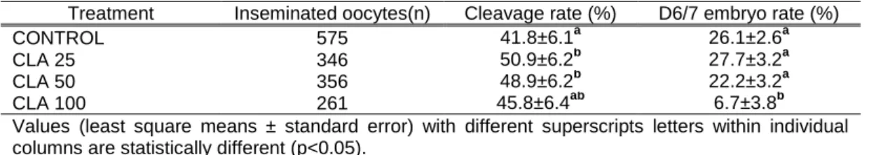
5.3 Ultrastructure of in vitro produced sheep embryos: effect of trans-10 cis-12 conjugated linoleic acid (CLA) in fresh and cryopreserved blastocysts ... 74
5.3.1 Introduction ... 76
5.3.2 Materials and methods ... 77
5.3.3 Results ... 80
5.3.4 Discussion ... 81
5.3.5 References ... 84
6 Discussion and conclusions ... 91
iii
Abstract
In vitro production of sheep embryos and cryopreservation are developing reproductive technologies that haven’t reached yet the expected results for routine application, mainly by the reduced cryotolerance when compared to in vivo derived ones. A series of experiments were developed searching for alternative methods of production and cryopreservation of this source of embryos. It was concluded that in vitro fertilization with fresh ram semen improves quality of in vitro produced sheep embryos when conjugated with a medium containing pyruvate and FSH. Also different protocols of lipid reduction can be successfully applied, when using a combination of cytochalasin D and centrifugation or by adding trans-10 cis-12 conjugated linoleic acid isomer (CLA) in culture, as they increased cryotolerance of embryos and also the production rates. In addition it was described the ultrastructural characteristics of in vitro produced sheep embryos and those cultured in the presence of CLA, either fresh or cryopreserved.
iv
Criopreservação de embriões de ovino obtidos in vitro: o efeito de
protocolos de redução lipídica
Resumo
A produção in vitro de embriões ovinos e respectiva criopreservação são biotecnologias reprodutivas em desenvolvimento que ainda não atingiram os resultados esperados para serem aplicados rotineiramente, sobretudo pela menor criotolerância destes embriões quando comparados com os produzidos in vivo. Realizaram-se uma série de experiências procurando métodos alternativos de produção e criopreservação deste tipo de embriões. Concluiu-se que a fertilização in vitro com sémen fresco de carneiro aumenta a qualidade dos embriões produzidos quando conjugada com meio contendo piruvato e FSH. Também se concluiu que é possível aplicar com sucesso protocolos de redução lipídica usando uma combinação de citocalasina D com centrifugação ou adicionando o isómero conjugado trans-10 cis-12 do ácido linoleico (CLA) ao meio de cultura, responsáveis pelo aumento da criotolerância dos embriões e maiores taxas de produção. Adicionalmente descreveram-se as características ultraestruturais dos embriões ovinos produzidos in vitro e dos produzidos em presença de CLA, quer frescos quer congelados.
v
Dedication
To Afonso, André and Ana Isabel,
who came to light together with this thesis, wishing they be the carriers of our ancestors’ best virtues through the future.
vi
Funding acknowledgments
This work was supported by Funds from the Portuguese Foundation for Science and Technology (PPTDC/CVT/98607/2008) and PhD Grant (SFRH/BD/37853/2007).
This work is funded by FEDER Funds through the Operational Programme for Competitiveness Factors - COMPETE and National Funds through FCT - Foundation for Science and Technology under the Strategic Project PEst-C/AGR/UI0115/2011.
The work of Mário Sousa and Elsa Oliveira were partially supported by UMIB-National Funds through FCT-Foundation for Science and Technology, under the Pest-OE/SAU/UI0215/2014.
Participation in the Conference ESDAR 2013 was supported by ICAAM – “Instituto de Ciências Agrárias e Ambientais Mediterrânicas”- Universidade de Évora – Núcleo da Mitra, Ap. 94, 7002-554, and funded by FEDER Funds through the Operational Programme for Competitiveness Factors - COMPETE and National Funds through FCT - Foundation for Science and Technology under the Strategic Project PEst-C/AGR/UI0115/2011.
vii
Personal acknowledgments
To Dr. Rosa Lino Neto, by accepting be my PhD advisor, for all the time spent with this work that provided me the necessary knowledge and other tools that I shall use in the future. I appreciated especially the patience, goodwill and accuracy applied during the last 6 years. Maybe because I know that attending my expectations, hesitations, and difficulties was not an easy task. Now I know we succeeded.
To Dr. Carla Marques, Dr. Maria Conceição Baptista, Dr. João Pedro Barbas, Dr. António Horta and all other colleagues and staff of Estação Zootécnica Nacional (EZN), who allowed the production of all this work, wishing them all continue to struggle for new research in Animal Science that is needed for Portugal and other countries. All this colleagues and friends welcomed me since the first day and are responsible that I see EZN also as one of my references through the future.
A special thanks to Dr. Nuno Carolino for putting the pieces of our work together whenever needed with his statistics that was essential in all this research. Also for the optimistic however realistic and professional way of acting that must be an example for all Animal Science students as me.
To Dr. Carlos Plancha, my co-adviser, and Patricia Rodrigues that opened me the possibility of sharing their experiences and knowledge in Reproductive Biology, and also allowed the use of the facilities of the Faculty of Medicine of University of Lisbon.
To Dr. Mário Sousa and lab technician Mrs. Elsa Oliveira for all the hard work in processing and preparing the material for electron microscopic evaluation of embryos. It was also patent the competence of all the team of the Department of Microscopy, Laboratory of Cell Biology, Multidisciplinary Unit for Biomedical Research-UMIB of the Institute of Biomedical Sciences Abel Salazar and also the possibility of cooperation with other research teams. This is very important in the multidisciplinary approach of making science, as I believe.
To Dr. Carlos Bettencourt and all the staff of Centro de Experimentação do Baixo Alentejo, for the possibility of using sheep flock and facilities that, once again, were put into service for research purposes.
viii
To all the staff and colleagues of the slaughterhouse where oocytes have been collected, MATREZE, Matadouro Regional do Zêzere, in Pedrogão Grande. In addition to the possibility of refreshing the view of meat inspection activity, it was a pleasure to review some of you again after the times of Veterinary Faculty.
To all my colleagues in Universidade de Évora that shared many of my difficulties, and cooperated also in my absence. My wish is to return with new strengths and skills to contribute, all together, for the future of our University.
To all my students, some of them already graduated during this long working period, knowing that much of this effort is also for them. Shall I have the opportunity and ability to contribute more and more for the expected challenges in education and research with students.
To all my other friends not menctioned, knowing that all friends are important, for being present in my life and because I know they are there whenever needed.
To Elisa Bettencourt, who is present since ever. In addition to our friendship (either in good and bad moments) that can positively influence decisions, she is a special inspiration as an example of dedication to work and duty, grounded in values as simplicity, honesty and determination.
To my family, headed by my parents, Guida and Joaquim, who provided me the values of life, the gift of education and the tools for building the same for their grandchildren.
To my wife, Alexandra, whose female side complements my existence and to whom I owe some of the time spent in this stage of my life. I know this was not a spent time but, instead, a tonic for new steps in our lives, with other challenges and hills to climb next.
ix
Figure index
Figure 1: sample of poster (100x70 cm) presented in the ESDAR conference 2013, 12-14 September
Bologna, Italy... 25
Figure 2: Percentual distribution of embryo (n=279) morphological quality [grade 1 embryos (G1); grade 2
embryos (G2); grade 3 embryos (G3)] in the four groups according to maturation method (M1: maturation medium containing EGF and M2: medium containing EGF, pyruvate and FSH) and type of semen used: fresh (F) or frozen–thawed (FT) (a≠b, P=0.007). ... 35
Figure 3: Ultrastructure of fresh in vitro ovine embryos at early blastocyst stage. Outer (A,C,E) and inner
cells (B,D,F), cells. Microvilli (Mv), perivitelline space (PvS), cell debris (*), intercellular junctions (black arrows), mitochondria (m), smooth endoplasmic reticulum (white arrowheads), lipid droplets (L), fusion (white open arrow) of light (LV) and dense vesicles (DV), Golgi complexes (G), nucleus (N), nucleolus (Nc). ... 53
Figure 4: Ultrastructure of fresh in vivo ovine embryos at early blastocyst stage. Outer (A,C) and inner
cells (B,D), cells. Microvilli (Mv),perivitelline space (PvS); fusion of light vesicles (white arrow), intercellular junctions (black arrows), mitochondria (m), smooth endoplasmic reticulum (white arrowheads), rough endoplasmic reticulum (black arrowheads), light vesicles (LV), dense vesicles (DV), Golgi complexes (G), nucleus (N), nucleolus (Nc), lipid droplets (L),secondary lysosomes (Ly). 54
Figure 5: Ultrastructure of OPS vitrified in vitro sheep embryos at early blastocyst stage fixed 1h after
thawing. Blastocysts were classified as grade 2 (A and B) and grade 3 (C and D) in semithin sections. Zona pellucida (ZP) perivitelline space (PvS), microvilli (Mv), blastocoelic cavity (b), nuclear membrane disruption (black open arrow), mitochondria (m), degenerated mitochondria (dm), smooth endoplasmic reticulum (white arrowheads), light vesicles (LV), fusion of light vesicles (white open arrow), dense vesicles (DV), Golgi complexes (G), nucleus (N), nucleolus (Nc), cell debris (*), cell lysis (**) ... 55
Figure 6: Ultrastructure of OPS vitrified in vivo ovine embryos at early blastocyst stage fixed 1h after
thawing. Blastocysts were classified as grade 1 (A and B) and grade 2 (C and D) in semithin sections. Perivitelline space (PvS), microvilli (Mv), blastocoelic cavity (b), intercellular junctions (black arrows), mitochondria (m), smooth endoplasmic reticulum (white arrowheads), light vesicles (LV), dense vesicles (DV), Golgi complexes (G), nucleus (N), nucleolus (Nc), lipid droplets (L). ... 56
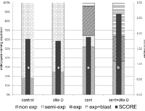
Figure 7: Evaluation of vitrified (OPS) sheep embryos after warming, previously submitted to different
protocols of lipid reduction. Embryos were scored (1-4) according to the post warming expansion: without expansion (non exp) = 1, semi-expansion (semi-exp) = 2, expansion (exp) = 3 and expansion with an excellent blastocoele (exp+blast) = 4. Values with different letter within individual columns are significantly different (p<0.05). Experiment 1: cytochalasin D (cyto-D, n=24), centrifugation (cent, n=25), centrifugation and cytochalasin D (cent+cyto-D, n=22), control (control, n= 27) groups. ... 64
Figure 8: Evaluation of vitrified (OPS) sheep embryos after warming, previously submitted to a protocol of
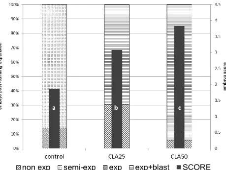
lipid reduction using different concentrations of trans-10 cis-12 conjugated linoleic acid isomer (CLA; 25 and 50 µM) supplemented to the culture medium. Embryos were scored (1-4) according to the post warming expansion: without expansion (non exp) = 1, semi-expansion (semi-exp) = 2, expansion (exp) = 3 and expansion with an excellent blastocoele (exp+blast) = 4. Values with different letter within individual columns are significantly different (p<0.05). Experiment 2: control (n= 14), CLA25 (n=23) and CLA50 (n=18) groups. ... 66
Figure 9: Evaluation of vitrified (OPS) embryos after warming, previously submitted to different protocol of
x
expansion (non exp) = 1, semi-expansion (semi-exp) = 2, expansion (exp) = 3 and expansion with an excellent blastocoele (exp+blast) = 4. Values with different letter within individual columns are significantly different (p<0.01). Experiment 3: control (control, n= 25), cytochalasin D (cyto-D, n=22), centrifugation and cytochalasin D (cent+cyto-D, n=20), 25 or 50 µM trans-10 cis-12 conjugated linoleic acid isomer (CLA) added in the culture medium (CLA25, n=33) and (CLA50, n=18) groups. ... 67
Figure 10: Ultrastructure of fresh sheep blastocysts produced in culture media supplemented with 25 µM
of trans-10 cis-12 linoleic acid isomer (CLA25F). Outer (A,C,E) and inner cells (B,D,F), cells. ZP pellucida (ZP), microvilli (Mv), perivitelline space (PvS), intercellular junctions (black arrows), mitochondria (m), smooth endoplasmic reticulum (white arrowheads), light (LV), dense vesicles (DV), lipid droplets (L), fusion of lipid droplets with dense vesicles (white open arrow), fusion of light vesicles (black arrow head), Golgi complexes (G), nucleus (N), nucleolus (Nc), blastocoel cavity (Bc) . ... 87
Figure 11: Ultrastructure of fresh in vitro produced sheep blastocysts (control fresh, CF). Outer (A,C) and
inner cells (B,D), cells. Microvilli (Mv),perivitelline space (PvS); mitochondria (m), smooth endoplasmic reticulum (white arrowheads), light vesicles (LV), dense vesicles (DV), Golgi complexes (G), nucleus (N), nucleolus (Nc), disrupted nuclear membrane (white open arrow), ** cell debris cytosolic areas devoid of organelles (Cy) blastocoel cavity (Bc). ... 88
Figure 12: Ultrastructure of OPS vitrified sheep blastocysts produced in culture media supplemented with
25 µM of trans-10 cis-12 linoleic acid isomer (CLA25V) and fixed 1h after warming. Perivitelline space (PvS), microvilli (Mv), blastocoel cavity (b), mitochondria (m), smooth endoplasmic reticulum (white arrowheads), light vesicles (LV), fusion of light vesicles (black arrow head), dense vesicles (DV), Golgi complexes (G), nucleus (N), nucleolus (Nc), cytosolic areas devoid of organelles (Cy). ... 89
Figure 13: Ultrastructure of OPS vitrified in vitro produced sheep blastocysts (control vitrified, CV), fixed 1h
after warming. Perivitelline space (PvS), microvilli (Mv), blastocoel cavity (b), intercellular junctions (black arrows), mitochondria (m), smooth endoplasmic reticulum (white arrowheads), light vesicles (LV), dense vesicles (DV), Golgi complexes (G), nucleus (N), nucleolus (Nc), *, cell lysis,** cell debris, disrupted nuclear membrane (black arrow). ... 90
xi
Table index
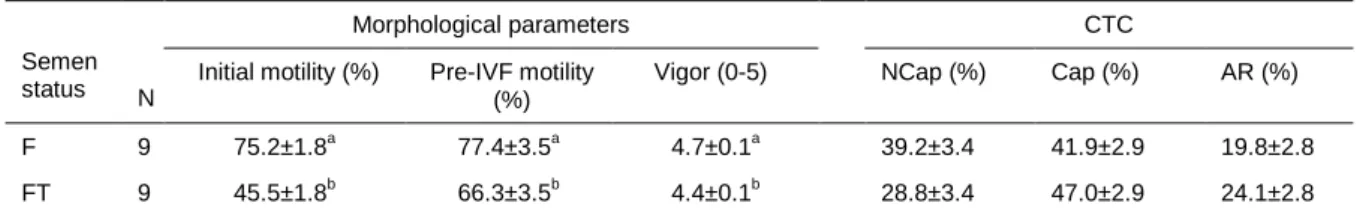
Table 1: Evaluation of morphological parameters and capacitation status (chlortetracycline staining-CTC)
of fresh (F) and frozen thawed (FT) ovine semen (least squares means ± standard error). ... 34
Table 2: Effect of different maturation media (M1 and M2) and oocyte fertilization by fresh (F) or frozen– thawed (FT) spermatozoa on embryo production rates (least squares means ± standard error and p-values). ... 35
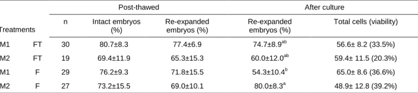
Table 3: Effect of different maturation media (M1 and M2) and oocyte fertilization by fresh (F) or frozen– thawed (FT) spermatozoa on post-thawed embryo (n=105) viability evaluated by intact and re-expanded blastocyst rates and embryo cell counts (least squares means ± standard error). ... 36
Table 4: Effect of different protocols of lipid reduction on sheep embryo post warming evaluation (4
sessions, experiment 1). ... 64
Table 5: Effect of a protocol of lipid reduction using different concentrations of trans-10 cis-12 conjugated
linoleic acid isomer (CLA) during sheep embryo culture on production rates (9 sessions, experiment 2). ... 65
Table 6: Percentual distribution of sheep embryo morphological quality after culture with different
concentrations of trans-10 cis-12 conjugated linoleic acid isomer (CLA; 9 sessions, experiment 2). .... 65
Table 7: Effect of different concentrations of trans-10 cis-12 conjugated linoleic acid isomer (CLA) during
sheep embryo culture on production rates (9 sessions, experiment 3). ... 66
Table 8: Production rates (6 sessions) of in vitro produced sheep embryos cultured with 25µM of trans-10
cis-12 linoleic acid isomer (CLA25 group) or without (control group). ... 80
Table 9: Percentual distribution of morphological quality (6 sessions) of in vitro produced sheep embryos
cultured with 25 µM of trans-10 cis-12 linoleic acid isomer (CLA25 group) or without (control group). . 80
Table 10: Post-warming survival (3 sessions) of in vitro produced sheep embryo cultured with 25 µM of
trans-10 cis-12 linoleic acid isomer (CLA25 group) or without (control group) and vitrified by open
xii
List of abbreviations
Abbreviation Unabbreviated Abbreviation Unabbreviated
µL microliter INIAV National Institute of Agrarian and Veterinarian Research
AI artificial insemination INRB Instituto Nacional dos Recursos Biológicos
BL blastocysts IVC in vitro culture
BME BME Amino Acids Solution IVF in vitro fertilization
BSA bovine serum albumin IVM in vitro maturation
Ca calcium IVP in vitro production
CLA trans-10 cis-12 conjugated linoleic
acid isomer LH luteinizing hormone CO2 carbon dioxide LN2 liquid nitrogen
COCs cumulus oocyte complexes LOPU laparoscopic ovum pick-up CTC chlortetracycline M199 tissue culture medium 199 D6 day 6 MEM MEM Amino Acids Solution
D8 day 8 min minutes
DMSO dimethylsulphoxide mm millimeters
E2 estradiol MOET multiple ovulation and embryo transfer
EG ethylene glycol N2 nitrogen
EGF epidermal growth factor mM miliMole
F fresh O2 oxygen
FCS fetal calf serum oFSH ovine FSH FF follicular fluid OPS open pulled straws fig. figure p.i. post-insemination FSH follicle stimulating hormone PBS phosphate buffer saline FT frozen-tawed PI propidium iodide
GSH glutathione PPARs peroxisome proliferator-activated receptors
h hour PUFA polyunsaturated fatty acid HA hyaluronic acid PVs perivitelline space HBP hexosamine biosynthetic pathway ROS reactive oxygen species HM holding medium SOF synthetic oviductal fluid hr hour SOFserum fetal calf serum ICBAS Institute of Biomedical Sciences
Abel Salazar TCM tissue culture medium
IETS International Embryo Transfer
Society TUNEL
terminal deoxynucleotidyl transferase dUTP nick end labeling
1
1 Preamble
Sheep production is one of the main livestock outputs in the Mediterranean area, being characterized by a large genetic diversity with several breeds of small ruminants (Gama, 2006). The importance of applying reproductive technologies is one of the cores of field action of the technicians working in Animal Science and Veterinary Medicine areas hereafter. In this context it is our opinion that progress in reproductive biotechnologies research is essential to guarantee the support of farmers, breeders associations and research centers.
Production and storage of sheep embryos is one of the new challenges that need to be appraised. This technology can be associated to other techniques as oestrus synchronization and superovulation, as well as semen technology, wich have been applied more often, and should also include in vitro production (IVP) of embryos. In Portugal IVP of sheep embryos started in 2000 (Baptista et al., 2002) and in vivo derived sheep embryos production has being routinely used with the foundation of the Portuguese Animal Germplasm Bank, established in 2005 (Pereira and Marques, 2008).
As will be discussed below, IVP of sheep embryo is still a demanding issue for researchers due to the inferior results when compared to other species and even more if cryopreservation is applied (Cognié et al., 2004; Dattena et al., 2004; Dalcin et al., 2013). Therefore special efforts to develop new methods that could overcome the low cryotolerance of IVP sheep embryos should be addressed. Challenged by these constraints and motivated by the believe in the future need of knowledge in this area, we developed several experiments focused in the characterization of the effects of cryopreservation in in vitro produced sheep embryos and we delineate a strategy to reduce lipid content of IVP sheep embryos pointed as one of the causes of the low success in their cryopreservation. To develop this research work we joined some research teams working in this area, based in the Unit of Genetic Resources, Reproduction and Animal Breeding, of the National Institute of Agrarian and Veterinarian Research (INIAV), in Estação Zootécnica Nacional, Santarém, where the main part of the works took place, namely the steps of in vitro embryo production, semen technology, embryo cryopreservation and storage as well as other essential research steps. The source of oocytes was a regional slaughterhouse, Matreze, in Pedrogão Grande, and in vivo embryos derived from Merino sheep collected in the facilities of the Regional Agriculture Direction of Alentejo located in Herdade da Abóbada in Vila Nova de S. Bento, Serpa.
The need of fluorescence microscopy facilities led us to the Institute of Molecular Medicine, in the Faculty of Medicine of the University of Lisbon, where embryo cell counts and viability were performed.
2
The processing of embryos for electron microscopy occurred in the laboratory of Pathology of University of Évora and in the Department of Microscopy, Laboratory of Cell Biology of the Multidisciplinary Unit for Biomedical Research-UMIB, Institute of Biomedical Sciences Abel Salazar (ICBAS), University of Porto, where images were obtained.
In a first approach we studied two methods of in vitro sheep embryo production using two different methods of in vitro maturation and also comparing fresh and frozen-thawed ram semen for in vitro fertilization. This contribution was published in the paper:
Romão R, Marques CC, Baptista MC, Vasques, MI, Barbas JP, Horta AEM, Carolino N, Bettencourt E, Plancha C, Rodrigues P, Pereira RM. Evaluation of two methods of in vitro production of ovine embryos using fresh or cryopreserved semen. Small Ruminant Research 2013;110:36-41.
Afterwards we studied the effects of cryopreservation in sheep embryo, by highlighting the alterations that occur at ultrastructural level in IVP and their in vivo counterparts, either fresh or cryopreserved. At our knowledge these can be the first published results showing ultrastructural changes in IVP sheep embryos. The results of this experiment were compiled in a submitted paper:
Romão R, Bettencourt E, Pereira RMLN, Marques CC, Baptista MC, Barbas JP, Horta AEM, Oliveira E, Bettencourt C, Sousa M. Ultrastructural characterization of fresh and vitrified in vitro and in vivo produced sheep embryos. Theriogenology (submitted).
Postulating that delipidation of IVP of sheep embryos is essential for increasing their cryotolerance several experiments were performed using physical or chemical delipidation. Some of these techniques were already tried in other species being adapted to sheep IVP embryos for the first time. These experiments were included in another submitted paper:
Romão R, Marques CC, Baptista MC, Barbas JP, Horta AEM, Carolino N, Bettencourt E, Pereira RM. Cryopreservation of in vitro produced sheep embryos: effect of different protocols of lipid reduction. Theriogenology (submitted).
Last but not least we studied the postulated positive effect of CLA in the ultrastructure of IVP sheep embryos enhancing cryotolerance. A paper was written with these results:
Romão R, Marques CC, Bettencourt E, Baptista MC, Barbas JP, Carolino N, Oliveira E, Sousa M, Pereira RMLN. Ultrastructure of in vitro produced sheep embryos: effect of trans-10 cis-12 conjugated linoleic acid (CLA) in fresh and cryopreserved blastocysts. Animal (submitted).
3 In the 6 years of this research, besides the published and submitted publications we had also the opportunity to present two communications in the scope of a National and an International Congresses, reporting some preliminary results of our data.
Knowing that much effort will still be needed to reach definitive methods to be used in cryopreservation of IVP sheep embryos, it is our believe that with the contributions of those that were involved in this work, we can add a small contribution to future fruitful discussion and research in small ruminant reproductive biotechnologies.
4
2 Introduction
1In the last decade, the production of sheep embryos did not get an improvement as researchers would like to announce and its application is still lower than in other species (Isachenko et al., 2003; Dalcin et al., 2013), being embryo cryopreservation one of the trouble spots in this area. In fact, since the first steps in embryo cryopreservation, attempts have been made to understand the effect of freezing in sheep embryos (Willadsen et al., 1976; Willadsen, 1977). It is expected that a successful cryopreservation of sheep embryos can push forward all other reproductive biotechnologies in this species as multiple ovulation and embryo transfer (MOET), artificial insemination (AI) or in vitro production of embryos. These associated techniques are still expensive and this fact limits its widespread.
In sheep, as in other species, there are two ways of obtaining embryos: in vivo derived or in vitro produced (IVP). In vivo embryo production consists in collecting them directly from the uterus either after natural service or after AI, while IVP involves oocyte collection from sheep ovaries followed by laboratorial maturation, fertilization and embryo culture. Advantages and uses of each technique have been previously discussed (Cognié et al., 2003; Cognié et al., 2004).
The highest lambing rates can be achieved after transfer of fresh in vivo derived embryos being reported results of 46.4-66.7% (Folch et al., 2000), 67.8% (Martinez et al., 2006), 73.3% (Bettencourt et al., 2009b), 75% (Papadopoulos et al. 2002) and 81.2% (Dattena et al., 2000). Conversely transference of fresh IVP embryos has lower results with lambing rates of 32.8% (Papadopoulos et al., 2002), 37.5% (Martinez et al., 2006) or 40% (Dattena et al., 2000). Despite this variation, some authors referred no differences between surviving rates after transfer of fresh in vivo derived and IVP embryos (Dobrinsky, 2002).
Cryopreservation tries to preserve embryos to be maintained as genetic reserves or for easier application later on. However, the results are below the desirable. For instance, lambing rates reported after transfer of in vivo derived cryopreserved sheep embryos are of 32-36% (Folch et al., 2000), 50% (Gibbons et al., 2011), 58.3% (Bettencourt et al., 2009b), 60% (Dattena et al., 2001), 60.1-75.1% (Dattena et al., 2004), 75% (Dattena et al., 2000), whereas for IVP embryos are of about 21.7% (Dattena et al., 2000), 19.4-23.8% (Dattena et al., 2004) and 23-26.6% (Martinez et al., 2006).
The analysis of these results highlights that the low cryotolerance is the main obstacle for using cryopreserved sheep embryos, especially the IVP embryos that present a much lower survival rate
1
5 compared to in vivo derived ones (Rizos et al., 2002b). It has been demonstrated that IVP sheep embryos have a slower developmental rate with detrimental effects in placentation and higher fetal loss, leading to a lower lambing rate (Tveden-Nyborg et al., 2005). An enhanced efficiency would increase the number of viable produced embryos per ewe making more feasible its use. For example, Folch et al. (2000) referred 4.5 lambs produced by ewe using MOET and AI followed by cryopreservation of embryos. Also the use of cryopreserved embryos can facilitate the work planning process, eliminating distance limitations and reducing sanitary risks (Folch et al., 2000). In fact, the sanitary quality of in vitro produced embryos can theoretically be better controlled than those obtained in vivo (Guerin et al., 2000).
In the last 15 years, researchers have tried to understand the reason of the low cryotolerance of IVP embryos, searching for new methods capable of improved results in this species. Sheep cryopreserved embryos present ultrastructural damage (Bettencourt et al., 2009) clearly expressing the cryodamage of cells during the process of freezing and thawing. Cryopreserved embryos show less microvilli and lower mitochondria activity than fresh ones (Cocero et al., 2002; Bettencourt et al., 2009). The lower cyotolerance of IVP blastocysts can be related to the excessive accumulation of lipids (Thompson et al., 1995; Dattena et al., 2000; Rizos et al., 2002b; Pereira et al., 2007; Pereira and Marques, 2008) and these accumulation can be favored by their culture in serum containing media (Cho et al., 2002; Abe and Hoshi, 2003; Pereira et al., 2007) but also by the type and concentration of cryoprotectant used as well as the freezing protocol (Cocero et al., 2002). No studies have been done that can explain the different alterations induced by cryopreservation in IVP embryos at ultrastructural level.
2.1 Application
On the behalf of developing new and improved reproductive assisted techniques, mainly by research teams, there has been for long an interest in sheep embryo model research as confirmed by the birth of the first IVP lambs in 1991 (Czlonkowska et al., 1991; Pugh et al., 1991) and the first cloned animal (a lamb) by nuclear transfer in 1997 (Wilmut et al., 1997). Beyond that, great interest exists in developing embryo reproductive technology to be used commercially or in routine programs for conservation of species and breeds all around the world.
Food and Agriculture Organization (FAO) has established the minimum number of individuals in each population so they can have a classification of endangered (Henson, 1992; FAO, 1998). This classification is the base for choosing the target breeds or species to be urgently preserved. Also following the guidelines of Rio de Janeiro convention, in 1992, it was established that all countries should have a plan for the
6
conservation of autochthon genetic resources, also recognizing their qualities in what concerns the adaptation to local conditions as well as their potential use in agricultural niches of production (Båge, 2003).
One of the best ways of preserving the animal genetic resources of endangered populations is through the creation of germplasm banks in which the biological material can be stored for decades, as cryopreservation, at actual knowledge, is a reliable way of long-term conservation of genetic resources (Fogarty et al., 2000; Yao et al., 2012). Besides, one of the current alternative strategies for maintenance of some breeds or strains is through embryo cryopreservation (Amstislavskya and Trukshinb, 2010), now routinely used in mouse strains for long-term colony maintenance, with the advantages of saving costs, readiness in distant transport, health guarantee and also for preventing genetic drift occurrence (Woods et al., 2004; Mochida et al., 2013).
In other perspective, germplasm exchanges are nowadays crucial (Thibier, 2011), being extremely useful in improving genetic upgrading (Gibbons and Cueto, 2011). Also embryo transfer can have a major role in reducing or eliminating some transmissible diseases in germplasm livestock changes (Stringfellow and Givens, 2000; Thibier and Guérin, 2000; Cognié and Baril, 2002; Pereira and Marques, 2008). For commercial proposes embryo transfer should be chosen for those reasons in which other less expensive techniques are not as advantageous. For example Cognié and Baril (2002) estimated a 10 times higher cost when comparing embryo transfer (either using in vivo or in vitro embryos) with artificial insemination and this represents an elevated cost attending the economic value of the animal species (Cognié and Baril, 2002).
Based on International Embryo Transfer Society (IETS) data, a decade ago, Thibier (2004) reported 6674 fresh and 2907 frozen/thawed sheep embryos transferred in the world in 2003 and, for the year of 2012, Perry (2013) referred 8124 fresh and 6134 frozen/thawed transferred embryos, mainly represented by Australia, South America and South Africa. This numbers, although considered underestimated because of difficulty in retrieving data, show an evolution of the use of this reproductive biotechnology and, in our opinion, reflects the demands of an emerging global market in this area.
In sheep, as in other species, several methods can be used for ex situ conservation of genetic resources and attention in reproductive cells has been focused in oocyte, spermatozoa, zygote or embryo cryopreservation (Mullen and Fahy, 2012; Mara et al., 2013). In parallel, in the last years, research has been conducted in other methods as ovary, testicle (Arav et al., 2005, Faustino et al., 2010; Merdassi et al., 2011; Mara et al., 2013), and somatic cells cryopreservation (McCreath et al., 2000; Liu et al., 2010) as well as in
7 new methods in embryo technology as somatic cell nucleus transfer and transgenesis (McCreath et al., 2000; Betteridge, 2006).
Having the background of other species we can mention that, for example, in the United States, in 2011, 79% of all bovine collected embryos were later cryopreserved (Hasler, 2012). In sheep, cryopreservation of embryos is a crucial developing technique for commercial application and it is our idea that, when better results after the cryopreservation of embryos have been attended, it will be the dominant target for sheep embryo production and also using IVP embryos because of the possibilities that can be provided.
2.2 In vitro embryo production
In vitro embryo production simulates the natural development of an embryo and comprises the phases of oocyte maturation, in vitro fertilization (IVF) and in vitro culture (IVC) of embryos. In these steps the last one is the critical in terms of determining the blastocyst yield (Lonergan and Fair, 2014). In terms of lambing rate, IVP embryo survival is 25% lower than for in vivo derived ones mainly due to an increased embryo loss at day 30-40 of gestation (Cognié et al., 2004). Betts and King (2001) have discussed the mechanisms of preimplantation and cell death referring the importance of knowing the tools and conditions that can promote better results in IVP. Moreover Betts and Madan (2008) referred the importance of permanent embryo demise that occurs in the first week of development in the outcomes of assisted reproductive technologies.
In parallel with other species, IVP technology reduces the rate of produced embryos and leads to inferior embryo survival when compared to in vivo derived embryos, as previously discussed (Cognié and Baril, 2002; Tominaga, 2004; Pereira and Marques, 2008; Blanco et al., 2011; Lonergan and Fair, 2014). Cocero et al. (2011) obtained 34.6% (day 7) or 40.4% (day 8, D8) blastocysts produced from sheep abattoir derived oocytes and Cognié et al. (2003) referred 25% D8 blastocyst yield.
In vitro sheep embryo production is not yet a controlled and totally defined technique once that has been associated with the “large offspring syndrome” (Thompson et al., 1995; Holm et al., 1996) and other not well understood problems (Shirazi et al., 2009), also reported in other species (Farin et al., 2001; Lazzari et al., 2002). These abnormalities seem to be promoted in very early developmental stages, such as IVM or IVF, irrespectively of the subsequent in vivo or in vitro culture treatment (Holm et al., 1996; Galli and Lazzari, 2008). Apparently the use of serum supplementation and co-culture can be pointed as a justification (Galli and Lazzari, 2008). Also these problems could be extended following birth as Çörekçi and Yilmaz (2004) found higher growing rates until weaning age in lambs born from IVP. It is well established that IVP embryos
8
have an altered morphology when compared with in vivo derived counterparts (Crosier et al., 2000). It has also been observed an increase in the lipid content of IVP compared to in vivo derived embryos regardless of the composition of the medium of culture. Blastocyst’s differences depending on culture medium used were identified (Thompson et al., 1995; Crosier et al., 2001; Pereira et al., 2007). These changes have also been correlated to the lower cryotolerance of IVP embryos as stated before.
Several studies have tried to establish the most successful laboratory method of IVP including IVM, IVF and IVC of embryos since all these steps are crucial for good results.
2.2.1 Collection of oocytes
The physiological constraints on sheep uterus access, mainly by the peculiar anatomy of the cervix, have reduced the chances of an easy collection of oocytes and embryos, although interest in new techniques (Gusmão, 2011), which also could improve welfare issues in future, exists. There are several techniques of oocyte collection namely by follicular aspiration or slicing in abattoir derived ovaries (Wani et al., 2000; Zeinoaldini et al., 2013). Better quality of cumulus oocyte complexes (COCs) and easy application have been associated with the later technique (Wani et al., 2000; Wani, 2002). Also the number of harvested oocytes collected after slicing is higher (Amiridis and Csehb, 2012). However slicing is a very time consuming technique with the associated loss of oocyte viability during the process (Machado et al., 2012). On the other hand, sheep ovum pick up-systems are applied in living animals using minimal invasive procedures such as laparoscopic ovum pick-up (LOPU) technique guided by laparoscopy (Baldassarre et al., 1996, 2002; Rodriguez et al., 2006; Cox and Alfaro, 2007; Gibbons et al., 2008). These procedures, when used in vivo, are usually associated with follicular stimulation treatments (Gibbons et al., 2007) in order to raise the number of collected COCs (Mermillod et al., 2006; Crocomo et al., 2012) and can allow better maturation, fertilization and in vitro development capacities (O’Brien et al., 1997; Cocero et al., 2011). According to Stangl et al. (1999) and Bari et al. (2001) it is possible to repeatedly collect oocytes by this approach without reducing subsequent recovery rates. Moreover, development of in vivo techniques of oocyte collection is important because, when derived from slaughtered animals, oocytes usually cannot be used in genetic programs due to the unknown sanitary and even genetic status, being applied mainly for research proposes (Gibbons el at., 2008). However a safe animal identification system may overcome these issues.
9 Oocyte quality seems to be the key limiting factor to achieve high embryo production ratios and quality (Dieleman et al., 2002; Cognié et al., 2003; Blanco et al., 2011). Embryo production depends also in oocyte competence to complete meiosis which is affected by follicle size and quality (Cognié and Baril, 2002). It is known that pre-pubertal oocytes have an inferior developmental potential than those collected from adults (O’Brien et al., 1996, 1997). Nevertheless the expected potential in future results using juvenile oocyte collection, namely the decreasing to half of the generation interval in genetic programs, must be considered (Amiridis and Csehb, 2012).
2.2.2 In vitro maturation (IVM)
Collected oocytes must undergo cytoplasmic and nuclear maturation (reaching metaphase II stage) prior to fertilization. Once outside the follicle, spontaneous meiotic resumption is the beginning of oocyte in vitro maturation (Mermillod et al., 2006). This is usually performed in an incubator at 38-39ºC in a humidified atmosphere with 5% CO2 concentration (Pereira et al., 2009; Amiridis and Csehb, 2012). The widely used
medium for IVM is Tissue Culture Medium (TCM) supplemented with bicarbonate, pyruvate, luteinizing hormone (LH), follicle stimulating hormone (FSH) and estradiol (E2) and with 10% fetal calf serum (Cognié et al., 2004; Cocero et al., 2011). Gonadotrophins seem to play a central role in the process as well as serum (Accardo et al., 2004; Wani et al., 2012).
Nevertheless other media and supplementation can be also used. For example, Shabankareh et al. (2011) found good results using human menopausal serum and Birler et al. (2001) showed that, in sheep, synthetic oviductal fluid (SOF) medium improves the rate of cleavage compared to TCM199 medium. Guler et al. (2000) presented higher results adding follicular fluid (FF) to maturation medium or using epidermal growth factor (EGF) conjugated with FSH and E2. Also Cotterill et al. (2012) and Zhou et al. (2008) showed the positive effect of EGF or other EGF-like ligands in COCs during IVM and Alabart et al. (2000), Cocero et al. (2011) and Wani et al. (2012) with EGF and cysteamine.
Despite atmosphere control of O2 concentration in IVM, the whole process of IVP is stressful because
of the formation of reactive oxygen species (ROS) that promote cellular and nuclear damage (Deleuze and Goudet, 2010). Cumulus cells are important to the success of in vitro fertilization (Van Soom et al., 2002; Shirazi et al., 2007; Bogliolo et al., 2007) but it is known by studies in bovine that these cells also undergo apoptosis during the IVM process (Ikeda et al., 2003). In sheep these damage can be reduced with supplementation with the previously referred antioxidants as cysteamine, combined with growth factors, or
10
using serum as font of albumin that balances osmolarity and acts as a free radical scavenger (Thompson, 2000) consequently improving maturation rates and subsequent IVP rates (Wani et al., 2012).
2.2.3 In vitro fertilization
The fertilization of matured oocytes can be performed with frozen or fresh semen. Although some authors argue that early embryo development is not influenced by the method of semen preservation (Hollinshead et al., 2004), fresh semen can improve embryo production rates and quality (Romão et al., 2013), eventually because freezing and thawing disrupts the stability of ram sperm chromatin, reducing its fertilization efficiency (Peris et al., 2004; Khalifa and Lymberopoulos, 2013).
Cryopreserved semen is advantageous since it can be easily available for routine use and can be stored in seasonal propitious period. However when a proven fertility ram semen is available it should be used in fresh either.
Fertilization is performed with previously capacitated spermatozoa usually using media containing heparin or sheep serum (Cognié et al., 2004), that causes Ca++ income into the acrosome and thus capacitation and acrosome reaction (Freitas et al., 2007). The best semen fraction is usually achieved by a Percoll gradient centrifugation (Cognié et al., 2004) or swim-up (Suttiyotin and Thwaites, 1993; Mermillod et al., 2006) prior to fertilization. It is stated that both sperm preparation methods allow to select a sperm population with a low percentage of apoptotic spermatozoa (Ricci et al., 2009).
2.2.4 In vitro culture
The goal of IVC is to simulate the events that occur in the oviduct so that putative zygotes obtained after fertilization undergo optimal development into blastocysts. For this reason culture can be performed in vivo using surrogate oviducts (Lazzari et al., 2010). Usually embryo IVC is achieved in controlled atmosphere, at 38.5-39ºC with 5%O2, 5% CO2 and 90% N2 (Cognié et al., 2004). The culture medium is
usually composed of SOF supplemented with amino acids, bovine serum albumin (BSA) and/or serum (Birler et al., 2001; Pereira et al., 2009; Amiridis and Csehb, 2012; Romão et al., 2013) although co-culture with oviduct cells or fetal fibroblasts was also reported (Bernardi, 2005). During IVC it is important to keep low concentration of oxygen preventing oxidation (see Harvey, 2007, for details) and the deleterious effects of ROS; this is also the reason of adding low molecular weight thiol compounds in IVM and IVC like cysteamine, β-mercaptoethanol, cystine and cysteine that increase the synthesis of glutathione (GSH), which in turn reduces the oxidative stress (De et al., 2011). At this stage of embryo development, the generation of
11 energy is provided manly by the Krebs cycle and oxidative phosphorylation. Therefore substrates as pyruvate, amino acids, lactate and fatty acids are important to embryo development and mitochondria plays a central role in metabolic events. Oxygen consumption is lower before the morula stage, having an increasing during compaction with the higher glucose consumption via glycolysis after blastocysts formation (Leese, 2012). Providing the best IVC conditions is of primordial importance for embryo competence acquisition to complete a term pregnancy after transfer.
2.3 Cryopreservation of sheep embryos
The science of cryobiology aims to preserve cells at low temperatures (-196ºC), intending to avoid the negative effects of the process which can preclude the main objective. The survival of cells depends on both the freezing and the warming processes. Cyopreservation of sheep embryos is a difficult task and practical results are not as encouraging as in cattle mainly due to the reduced embryo cryotolerance in this species. Two major methods have been used to preserve embryos: slow freezing and vitrification. The difference between them is the velocity rate of freezing. While slow freezing tries to differentiate the cooling rate in sequential steps of freezing, vitrification increases the freezing velocity by reducing volume, thus minimizing ice formation (Gao and Critser, 2000; Arav 2014). Slow freezing is currently practiced in programmable freezers and has the advantage of using low concentrations of cryoprotectants, but their ability to prevent ice-crystal formation at low concentrations is limited (Pereira and Marques, 2008). Vitrification is considered an important methodology for long-term storage of embryos in a glass-like amorphous vitreous state without the formation of ice crystals (Vajta and Nagy, 2006; Kim et al., 2012). Several methods using this technique are presently available: conventional vitrification or adaptations of the last one, such as vitrification in open pulled straws (OPS, Vajta et al., 1998) or more recently in other devices such as cryotop (Kuwayama, 2007) and paper containers (Kim et al., 2012) among others.
As previous described, in all the species in vitro produced embryos presented a higher sensitivity to cryopreservation than in vivo ones. Several reasons have been indicated to justify this IVP cryosensivity, mainly cellular (Pereira et al., 2007; Rizos et al., 2002b), metabolic (Khurana and Niemann, 2000; Farin et al., 2001) and biochemical (Massip et al., 1995) changes. At ultrastructural level, in vitro produced blastocysts exhibited less microvilli and a less extensive network of intercellular junctions, specifically an apparent lack of desmosomal junctions, a higher incidence of cellular debris and a higher number of lipid droplets than their in vivo counterparts (Farin et al., 2001; Rizos et al., 2002a). These differences could
12
certainly contribute to the observed lower cryotolerance of in vitro produced embryos (Massip et al., 1995; Ohboshi et al., 1998; Cognié and Baril, 2002).
Although it seems that very low temperatures will disrupt cell components, the most complicated freezing window is between -15ºC and -60ºC followed by intracellular ice formation, between -5ºC and -15ºC (Gao and Critser, 2000), whereas between -50ºC and -150ºC the risks are fracture of the zona pellucida or cytoplasm (Rall and Meyer, 1989). Below -150ºC the risks in embryo damage are lower (Vajta and Nagy, 2006). In embryo cells with high lipid content, as in sheep, the lipid drops can combine with parts of the cytoskeleton, as well as membranes, organelles and other structures of the cytoplasm. When these embryos are cooled from 15ºC until -5ºC this association can be related to irreversible and fatal structural damage occurence, mostly when the traditional freezing methods are used (Dinnyes et al., 2006; Vajta and Nagy, 2006). Most of these injuries seem to be related with cell lipid content and specifically to the membrane’s saturated-to unsaturated fatty-acid ratio (Arav et al., 2000; Arav and Zvi, 2008). During exposure to sub-physiological temperatures, the membrane goes through a lipid phase transition with solidification of lamellar lipids, such as saturated fatty acids. The low-temperature-fluid lipids, such as unsaturated fatty acids, which in this case are non-lamellar, then separate and form a new non-lamellar structure, such a II hexagonal structure which interferes with membrane function and leads to ion leakage and cell death (Arav and Zvi, 2008). Different strategies can be implemented to minimize cryodamage (Arav and Zvi, 2008; Pereira and Marques, 2008).
Both approaches for cryopreservation, slow freezing and vitrification, have been applied using in vivo derived or IVP embryos in sheep (Baril et al., 2001; Dattena et al., 2004; Bettencourt et al., 2009) or in cattle (Assumpção et al., 2008). In in vivo derived sheep embryos no differences were observed in lambing or embryo survival rates after transfer of embyos cryopreserved using slow freezing, conventional vitrification or vitrification in OPS (Bettencourt et al., 2009).
Although the achieved results by each method can be relevant, the cost and application in routine field is also of primordial importance to spread its use (Baril el at., 2001; Dattena et al., 2004).
2.3.1 Slow freezing
This method tries to control the descending cooling event in several steps, regulating extracellular and intracellular water exchange, with a balance between ice crystal formation and structural and osmotic damage, as well as cryoprotectant toxicity (Vajta and Nagy, 2006; Gajda and Smorag, 2009). The method is
13 possible using as referred programmable freezers with a rapid cooling until -6 to -7ºC, when ice formation is induced (seeding), and then a lower rate of cooling (0.3 to 1ºC/min) causing freezing of extracellular ice and increasing the concentration of extracellular solution and cellular dehydratation. At -30ºC, straws are then plunged in liquid nitrogen (Woods et al., 2004; Saragusty and Arav, 2011).
In spite of still being used by many teams in the world it is argued that slow freezing has a limited future in embryology and according to Vajta and Nagy (2006) “the rate of advancement in oocyte and embryo cryopreservation will depend on the rate by which embryologists and decision-makers adopt the new approaches”.
2.3.2 Vitrification
Vitrification was found to be interesting in embryo cryopreservation because it reduces cryoinjuries caused by ice formation (Rall and Fahy, 1985), In fact, vitrification is based on embryo manipulation into different carrier tools applied to minimise the volume and to submerge the sample quickly into liquid nitrogen, allowing an ultra-fast freezing speed, which avoids ice crystals formation, thus eliminating its deleterious effects in the cell (Dalcin and Lucci, 2010). This technique combines the use of small volumes with high concentration of two or more cryoprotectants (Liebermann et al., 2002) and it can be more adapted to IVP embryos (Vajta et al., 1998; Massip, 2001; Kasai and Mukaida, 2004).
According to several authors, the slow freezing techniques are time consuming and laborious (Assumpção et al., 2008) whereas vitrification is simple, rapid and inexpensive (Vajta et al., 1998; Gupta and Lee, 2010; Saragusty and Arav, 2011). Moreover, no publications have demonstrated that results obtained by vitrification were significantly worse than those obtained by slow freezing (Vajta and Nagy, 2006). Concerns in safety of vitrification related to transmissible diseases are partially justified but new methods have been developed to avoid these constraints (Yu et al., 2010).
Several authors described good results with this cryopreservation technique in sheep. For example, Baril et al. (2001) and Bettencourt et al. (2009b) achieved good field results (50-60% lambing rate) with vitrification of in vivo derived sheep embryos even with direct transfer of cryopreserved embryos and Folch et al. (2000) referred lambing rates of 32 to 36%, also in in vivo derived embryos.
In IVP embryos, Ptak et al. (1999) found no differences in pregnancy rates between fresh and vitrified transferred embryos (47 vs 42%). However they achieved significantly differences in lambing rates (41 vs 23%), showing that vitrification of IVP embryos is not yet an optimized technique.
14
The success of vitrification depends on the stage at which embryos are cryopreserved apart from the method used. In fact there are different levels of sensitivity to low temperatures, depending on the stage of embryo development (Balasubramanian and Rho, 2006). Garcia-Garcia et al. (2005, 2006) and Shirazi et al. (2010) showed that cryotolerance of IVP embryos to conventional slow freezing or vitrification increased as the developmental stage of embryos progressed perhaps due to the higher cryotolerance of blastocysts when compared with early developmental embryos. In fact, Garcia-Garcia et al. (2006), using slow freezing, described similar rates of viability between fresh or frozen-thawed embryos (92.5 vs 83.7%) frozen at the blastocyst stage.
2.3.3 Vitrification in OPS
In order to achieve a higher rate of temperature reduction the OPS method uses thinner superfine straws that have the end of the straw with half the diameter of a conventional one. This characteristic enables filling by capillary action with a volume of approximately 1µL, different from the conventional straw that contains 5µL (Vajta et al., 1998; Vajta and Nagy, 2006). So, it can allow freezing rates of 20000ºC/min (Massip, 2001), i.e., 10 times higher than in 0.25mL straws and overcome some problems of vitrification as toxicity of cryoprotectants, and difficulties in the permeability of membranes that can lead to intracellular ice formation and osmotic over-swelling (Kasai and Mukaida, 2004). Allowing higher speed of freezing, this type of vitrification is helpful in preventing embryo and zona pellucida fracture that occurs at low temperatures, especially with appropriate adjustments of warming parameters (Kuwayama, 2007). The method has been introduced by Vajta et al. (1998) and, since then, it has been successfully used in several animal species. It is a robust and feasible method for animal embryo vitrification, more than other new, but delicate, techniques (Vajta and Nagy, 2006).
Vitrification by OPS is so effective that results in lambing rate of in vivo derived sheep embryos vitrified by this method are persuasive. Green et al. (2009) reported higher pregnancy rates when using vitrification by OPS and direct transference, thus enhancing the field application of this technique. Nevertheless, these good results with OPS vitrification cannot avoid the reduced cryotolerance of IVP embryos. Dattena et al. (2001) achieved lambing rates of 60% for in vivo produced embryos vitrified in OPS while only 24% for IVP.
15 2.3.4 Cryoprotectants
The role of these substances is to reduce damage to cryopreserved embryos, minimizing ice formation. They usually are classified in permeable and non-permeable cryoprotectants, being also important in osmotic dehydration (Vajta and Nagy, 2006; Pereira and Marques, 2008).
The negative effects of cryoprotectants are related to their osmotic and toxic effects, closely dependents of the time of exposure and the concentration used (Massip, 2001; Dinnyes et al., 2006; Arav, 2014). The intent to reduce cryoprotectants to a minimum is a goal that has been tried in both slow freezing and vitrification procedures to minimize their deleterious effects. According to Liebermann et al. (2002) a balance between the maximization of cooling rate and the minimization of cryoprotectant concentration is the key point for a successful cryopreservation. Mainly in vitrification, where it is necessary to deal with higher concentrations of cryoprotectants, one of the strategies is to use more than one. This strategy reduces individual toxicity of cryoprotectants and also permits adding them in a stepwise equilibration (two or three steps), with increasing concentration or after cooling from 4ºC to subzero temperatures when their toxicity is lower (Massip, 2001; Woods et al., 2004; Vajta and Nagy, 2006).
Ethylene glycol (EG) is a frequent choice as a permeable cryoprotectant but others can be used like acetamide, glycerol, raffinose and dimethylsulphoxide (DMSO) in several combinations. For non-permeable cryoprotectants mono- and disaccharides including sucrose, trehalose, glucose and galactose can be cited, being sucrose the most used (Vajta and Nagy, 2006). The latter molecules, with large molecular weights, do not penetrate the cell membrane, but they can significantly reduce the amount of cryoprotectant required as well as the toxicity of EG by decreasing the concentration required to achieve a successful cryopreservation (Liebermann et al., 2002). Other substitute substances like polymers and proteins (e.g. polyvinylpyrrolidone, polyethylene glycol and Ficoll), not being essential, have been tried to replace the former indicated components by minimizing toxic effects (Liebermann et al., 2002; Kasai and Mukaida, 2004).
Different embryos cryosurvival rates were found among cryoprotectants with higher results for EG than glycerol (Martínez and Matkovic, 1998; Cocero et al., 1996, 2002). According to Cocero et al. (2002) EG protects embryos membranes and cytoplasmic structures from cryoinjury but Kartberg et al. (2008) have also reported good results of DMSO in protecting embryo membranes.
16
2.3.5 Warming of thawed embryos
Embryo thawing should be performed also in order to reduce cryoinjuries, when passing critical temperatures. In embryos cryopreserved by slow freezing the best method depends on the specific protocol and cryoprotectant used. Briefly, if the freezing method finished at temperatures of -30 to -40ºC, a moderately rapid warming (200 to 350ºC/min) is required to maximize the survival rate but if freezing reached -60ºC or less, warming rate should be slow, in the range of 25ºC/min (Gupta and Lee, 2010) although injuries were reported (Hochi et al., 1996).
Warming of vitrified embryos is usually performed directly into a solution at body temperature although it can be advisable to wait 1-3 seg in air to avoid fracture damage (Vajta and Nagy, 2006). Vitrified embryos need to be warmed ultra-rapidly in the presence of non-permeating cryoprotectants (usually sucrose) to dilute and remove the very high levels of intracellular permeating cryoprotectants (Gupta and Lee, 2010); this can lead to changes in dynamics of water in the cells that can cause embryo injury caused by osmotic over-shrinkage (Kasai and Mukaida, 2004; Woods et al., 2004).
Regardless of the method used for embryo warming, and transference after observation of re-expansion, Isachenko et al. (2003) Dattena et al. (2004) and Green et al., (2009) obtained promising results for field use after direct transfer of vitrified sheep embryos. These authors reported that the interval between thawing and transference has a great influence on embryo subsequent viability and valorize direct transfer use for field conditions.
2.4 Methods for improving cryopreservation of sheep embryos
Different strategies to increase cryotolerance of embryos in sheep and in other species were developed mainly by improving embryo cryopreservation procedures, changing the composition of IVC media (Dattena et al., 2007) or through the decrease of their lipid content (Arav, 2014; Prates et al., 2014). As referred, methods of ultra-rapid vitrification have been developed to further increase freezing velocity. However, the ideal device and combination of cryoprotectants has not yet been achieved. Therefore, this technique continues to be a challenge in embryo cryopreservation (Gajda and Smorag, 2009). Some examples of devices are glass micropipette, solid surface vitrification, cryoloop, microdrop, cryotop, cryo-tip, electron microscopy grids or nylon mesh (Kong et al., 2000; Kelly et al., 2005; Kuwayama, 2005, 2007; Gajda and Smorag, 2009; Gupta and Lee, 2010; Gibbons et al., 2011). Also attempts were experienced in IVC conditions as it was argued that the effect of changing culture conditions can be seen only after embryo
17 cryopreservation and warming. For example, Dattena et al. (2007) confirmed higher cryosurvival results when including BSA and Hyaluronan during IVC and Gad et al. (2012) realized that changing culture conditions lead to adaptations of embryos, induced by different gene expression, which was responsible for different developmental ability.
Difference in species’ cryosensitivity of embryos is responsible for different approaches in their cryopreservation. As indicated before, one of the major concerns in embryo cryopreservation is its lipid content that can hamper cryopreservation (Thompson et al., 1995; Dattena et al., 2000; Tsujii et al. 2001; Rizos et al., 2002b; Steel and Hasler, 2004; Seidel Jr., 2006; Pereira et al., 2007; Pereira and Marques, 2008) as it happens in species as sheep or pigs. This phenomenon is more pronounced in IVP embryos (Leibo et al., 1995, 1996) and in those produced in serum containing media (Thompson et al., 1995; Cho et al., 2002; Abe and Hoshi, 2003; Abe et al., 2004; Pereira et al., 2007). Serum is useful in oocyte and embryo culture as a source of albumin that balances the osmolarity, acting as a free radical scavenger with an additional important nutritive role (Thompson, 2000). However, the fatty acids and lipoproteins of the serum seem to be the source of embryos’ cytoplasmic lipids, hampering embryo quality (Abe et al., 2004; Lapa et al., 2005), albeit the perturbations induced by the presence of serum in sheep embryo culture are higher before rather than after compaction (Rooke et al., 2007).
On the other hand, embryo lipid content effect on chilling sensitivity is not totally elucidated at the moment. However it seems that lipid droplets interact directly with the intermediate filaments of the cytoskeleton and changes within these organelles during the cryopreservation process may lead to irreversible damages in the cytoskeleton (Pereira et al., 2008; Zehmer et al., 2009). Also it has been observed that cryopreserved embryos have ultrastructural changes that are visible as degenerated cells, disruption of cell membranes, and mitochondrial injuries, mainly in poor quality embryos (Dobrinsky, 1996; Cocero et al., 2002; Bettencourt et al., 2009). Damages in mitochondria are clear and reported by other authors before (Vajta et al., 1997; Cocero et al., 2002; Cuello et al., 2007; Dalcin et al., 2013) and may be used as reliable sign of cellular damage (Bettencourt et al., 2009, 2014). Mitochondrial changes have been observed in poor quality and IVP embryos and were associated with culture in serum medium (Shamsuddin et al., 1994; Abe et al., 1999). Several changes have been reported, namely a reduced total number and an increase in the proportion of immature mitochondria (Abe et al., 2002). These changes have been also associated with inefficient lipid metabolism and poor quality embryos that compromises ATP production (Doorland et al., 1997; Crosier et al., 2000; Abe et al., 2002, 2004; Gad et al., 2012). Also these