Bioactivity and chemical characterization in hydrophilic and lipophilic
compounds of
Chenopodium ambrosioides
L.
Lillian Barrosa#, Eliana Pereiraa#, Ricardo C. Calhelhaa,b, Montserrat Dueñasc, Ana
Maria Carvalhoa, Celestino Santos-Buelgac,*, Isabel C.F.R. Ferreiraa,*
a
Centro de Investigação de Montanha (CIMO), ESA, Instituto Politécnico de Bragança,
Campus de Santa Apolónia, 1172, 5301-855 Bragança, Portugal.
b
Centro de Química, Universidade do Minho, Campus de Gualtar 4710-057 Braga,
Portugal.
c
GIP-USAL, Facultad de Farmacia, Universidad de Salamanca, Campus Miguel de
Unamuno, 37007 Salamanca, Spain.
#
Both authors contributed equally.
*Authors to whom correspondence should be addressed (e-mail: [email protected],
telephone +351273303219, fax +351273325405; e-mail: [email protected]; telephone +34
Abstract
The bioactive properties (antioxidant and antitumour activities, and hepatotoxicity) of
the infusion and methanolic extracts of Chenopodium ambrosioides L., a plant
commonly used in Portuguese folk medicine, were compared. The chemical
composition in hydrophilic (sugars, organic acids and phenolic compounds) and
lipophilic (fatty acids and tocopherols) fractions were determined. In general, the
infusion revealed higher antioxidant activity, while the methanolic extract was the only
one showing antitumour effects against colon, cervical and hepatocellular carcinoma
cell lines. No toxicity in non-tumour cells was observed either for the infusion or the
extract. The studied plant proved to be a good source of natural antioxidants and other
bioactive compounds, which may have industrial use.As far as we know, this is the first
detailed chemical characterization and bioactivity evaluation of C. ambrosioides
methanolic extract and infusion.
Keywords: Chenopodium ambrosioides L.; Antioxidant activity; Antitumour activity;
1. Introduction
Oxidative stress is an imbalance between the generation of reactive oxygen species
(ROS; which include unstable oxygen radicals such as superoxide radical and hydroxyl
radical and non-radical molecules like hydrogen peroxide) and the body's antioxidant
defence capacity, having an important role in normal cell functioning. When produced
in excess ROS can have harmful effects, affecting cellular lipids, proteins and DNA,
leading to their modification, and often destruction, and inhibiting their normal function
(Valko et al., 2007; Rosenfeldt et al., 2013). Relevant diseases such as cancer, diabetes,
cirrhosis, heart disease or dementia disorders, as well as aging process have been
associated with the uncontrolled production of free radicals (Valko et al., 2007;
Halliwell, 2012).
Some plants traditionally used have medicinal properties with great potential for
therapeutic applications in the treatment of some of the aforementioned diseases, since
they are a natural source of bioactive compounds, including antioxidants, such as
polyphenols, vitamins, carotenoids, unsaturated fatty acids and sugars, which can be
useful for various applications, especially as food additives and in health promotion as
ingredients in formulations of functional foods and nutraceuticals (Ramarathnam,
Osawa, Ochi, & Kawakishi, 1995; Skerget et al., 2005).
Chenopodium ambrosioides L. (Amaranthaceae; syn: Dysphania ambrosioides (L.)
Mosyakin & Clemants) is an example of a plant formerly used in Portuguese traditional
medicine, normally consumed as infusion of its dried leaves and flowering stems. It is an exotic plant from Central and South America that in former times was introduced by
migrants from those countries. Nowadays the species has escaped to wild and can be
applications in the treatment of influenza, cold or gastrointestinal and respiratory
ailments, as well as vomiting, antihelmintic, healing of skin ulceration caused by
Leishmania species, anti-inflammatory and antitumor properties (Nascimento et al.,
2006; Cruz et al., 2007; Carvalho, 2010; Kamel, El-Emam, Mahmoud, Fouda, & Bayaumy, 2011).
Studies on chemical characterization and bioactivity evaluation of this plant,
particularly in the most consumed form (infusion) are scarce. The present work aims to
characterize the chemical composition of C. ambrosioides in hydrophilic (sugars,
organic acids and phenolic compounds) and lipophilic (fatty acids and tocopherols)
molecules, as also some bioactive properties (antioxidant and antitumour activities, and
hepatotoxicity) of its infusion and methanolic extract.
2. Materials and methods
2.1. Sample
Chenopodium ambrosioides L. (Amaranthaceae) (English names: Epazote, wormseed,
Jesuit's tea, Mexican tea; Local names: Té; chá-bravo; chá de Santa Marinha), also
known as Dysphania ambrosioides (L.) Mosyakin & Clemants, (Amaranthaceae), used
to be cultivated in homegardens in Bragança (Northeastern Portugal). Nowadays it is
less frequent in gardens and there are some specimens growing wild nearby the local
villages. However, if available, inflorescences and upper leaves are still wild gathered,
dried and used as herbal infusions. The material was collected in Varge (Bragança) from
different plants considering the species availability and local consumers’ criteria for
medicinal use. A sample was made putting together all the material from several
Voucher specimens are deposited at the Herbarium of the Escola Superior Agrária de
Bragança (BRESA). The samples were lyophilized (FreeZone 4.5, Labconco, Kansas
City, MO, USA), reduced to a fine dried powder (20 mesh) and mixed to obtain a
homogenate sample.
2.2. Standards and Reagents
Acetonitrile (99.9%), n-hexane (97%) and ethyl acetate (99.8%) were of HPLC grade
from Fisher Scientific (Lisbon, Portugal). The fatty acids methyl ester (FAME)
reference standard mixture 37 (standard 47885-U) was purchased from Sigma (St.
Louis, MO, USA), as also were other individual fatty acid isomers and standards:
L-ascorbic acid, tocopherols (α-, β-, γ-, and δ-isoforms), sugars (D(-)-fructose,
D(+)-melezitose, D(+)-sucrose, D(+)-glucose, D(+)-trehalose and D(+)-raffinose
pentahydrate), organic acids and trolox
(6-hydroxy-2,5,7,8-tetramethylchroman-2-carboxylic acid). Phenolic compounds were purchased from Extrasynthèse (Genay,
France). Racemic tocol, 50 mg/mL, was purchased from Matreya (Pleasant Gap, PA,
USA). 2,2-Diphenyl-1- picrylhydrazyl (DPPH) was obtained from Alfa Aesar (Ward
Hill, MA, USA).Foetal bovine serum (FBS), L-glutamine, hank’s balanced salt solution
(HBSS), trypsin-EDTA (ethylenediaminetetraacetic acid), penicillin/streptomycin
solution (100 U/mL and 100 mg/mL, respectively), RPMI-1640 and DMEM media
were from Hyclone (Logan, USA). Acetic acid, ellipticine, sulphorhodamine B (SRB),
trypan blue, trichloroacetic acid (TCA) and Tris were from Sigma Chemical Co. (St
Louis, MO, USA). Water was treated in a Milli-Q water purification system (TGI Pure
Water Systems, Greenville, SC, USA). All other chemicals and solvents were of
2.3. Evaluation of bioactive properties
2.3.1. Samples preparation. The methanolic extract was obtained from the lyophilized
plant material. The sample (1 g) was extracted by stirring with 25 mL of methanol (25
ºC at 150 rpm) for 1 h and subsequently filtered through Whatman No. 4 paper. The
residue was then extracted with 25 mL of methanol (25 ºC at 150 rpm) for 1 h. The
combined methanolic extracts were evaporated at 40 ºC (rotary evaporator Büchi R-210,
Flawil, Switzerland) to dryness.
The infusion was also obtained from the lyophilized plant material. The sample (1 g)
was added to 200 mL of boiling distilled water and left to stand at room temperature for
5 min, and then filtered under reduced pressure. The obtained infusion was frozen and
lyophilized.
Methanolic extract and infusion were redissolved in i) methanol and water, respectively
(final concentration 2.5 mg/mL) for antioxidant activity evaluation, or ii) water (final
concentration 8 mg/mL) for antitumour activity evaluation. The final solutions were
further diluted to different concentrations to be submitted to distinct bioactivity
evaluation in in vitro assays. The results were expressed in i) EC50 values (sample
concentration providing 50% of antioxidant activity or 0.5 of absorbance in the reducing
power assay) for antioxidant activity, or ii) GI50 values (sample concentration that
inhibited 50% of the net cell growth) for antitumour activity. Trolox and ellipticine
were used as positive controls in antioxidant and antitumour activity evaluation assays,
respectively.
2.3.2. Antioxidant activity. DPPH radical-scavenging activity was evaluated by using an
ELX800 microplate reader (Bio-Tek Instruments, Inc; Winooski, VT, USA), and
[(ADPPH-AS)/ADPPH] × 100, where AS is the absorbance of the solution containing the sample at
515 nm, and ADPPH is the absorbance of the DPPH solution. Reducing power was
evaluated by the capacity to convert Fe3+ into Fe2+, measuring the absorbance at 690 nm
in the microplate reader mentioned above. Inhibition of β-carotene bleaching was
evaluated though the β-carotene/linoleate assay; the neutralization of linoleate free
radicals avoids β-carotene bleaching, which is measured by the formula: β-carotene
absorbance after 2h of assay/initial absorbance) × 100. Lipid peroxidation inhibition in
porcine (Sus scrofa) brain homogenates was evaluated by the decrease in thiobarbituric
acid reactive substances (TBARS); the colour intensity of the
malondialdehyde-thiobarbituric acid (MDA-TBA) was measured by its absorbance at 532 nm; the
inhibition ratio (%) was calculated using the following formula: [(A - B)/A] × 100%,
where A and B were the absorbance of the control and the sample solution, respectively
(Pinela et al., 2012).
2.3.3. Antitumour activity. Five human tumour cell lines were used: MCF-7 (breast
adenocarcinoma), NCI-H460 (non-small cell lung cancer), HCT-15 (colon carcinoma),
HeLa (cervical carcinoma) and HepG2 (hepatocellular carcinoma). Cells were routinely
maintained as adherent cell cultures in RPMI-1640 medium containing 10%
heat-inactivated FBS (MCF-7, NCI-H460 and HCT-15) and 2 mM glutamine or in DMEM
supplemented with 10% FBS, 2 mM glutamine, 100 U/mL penicillin and 100 mg/mL
streptomycin (HeLa and HepG2 cells), at 37 ºC, in a humidified air incubator containing
5% CO2. Each cell line was plated at an appropriate density (7.5 × 10³ cells/well for
MCF-7, NCI-H460 and HCT-15 or 1.0 × 10⁴ cells/well for HeLa and HepG2) in
96-well plates and allowed to attach for 24 h. Cells were then treated for 48 h with various
by adding cold 10% trichloroacetic acid (TCA, 100 μL) and incubated for 60 min at 4
ºC. Plates were then washed with deionised water and dried; sulphorhodamine B
solution (0.1% in 1% acetic acid, 100 μL) was then added to each plate well and
incubated for 30 min at room temperature. Unbound SRB was removed by washing
with 1% acetic acid. Plates were air dried, the bound SRB was solubilised with 10 mM
Tris (200 μL) and the absorbance was measured at 540 nm in the microplate reader
mentioned above (Guimarães et al., 2013).
2.3.4. Hepatotoxicity. A cell culture was prepared from a freshly harvested porcine liver
obtained from a local slaughter house, and it was designed as PLP2. Briefly, the liver
tissues were rinsed in hank’s balanced salt solution containing 100 U/mL penicillin, 100
µg/mL streptomycin and divided into 1×1 mm3 explants. Some of these explants were
placed in 25 cm2 tissue flasks in DMEM medium supplemented with 10% fetal bovine
serum, 2 mM nonessential amino acids and 100 U/mL penicillin, 100 mg/mL
streptomycin and incubated at 37 ºC with a humidified atmosphere containing 5% CO2.
The medium was changed every two days. Cultivation of the cells was continued with
direct monitoring every two to three days using a phase contrast microscope. Before
confluence was reached, cells were subcultured and plated in 96-well plates at a density
of 1.0×10⁴ cells/well, and cultivated in DMEM medium with 10% FBS, 100 U/mL
penicillin and 100 µg/mL streptomycin (Abreu et al., 2011).
2.4. Chemical composition in hydrophilic compounds
2.4.1. Sugars. Freesugars were determined by high performance liquid chromatography
coupled to a refraction index detector (HPLC-RI). Dried sample powder (1.0 g) was
40 mL of 80% aqueous ethanol at 80 ºC for 30 min. The resulting suspension was
centrifuged (Centurion K24OR refrigerated centrifuge, West Sussex, UK) at 15,000g
for 10 min. The supernatant was concentrated at 60 ºC under reduced pressure and
defatted three times with 10 mL of ethyl ether, successively. After concentration at 40
ºC, the solid residues were dissolved in water to a final volume of 5 mL and filtered
through 0.2 µm nylon filters from Whatman (Pinela et al., 2012). The equipment of
analysis consisted of an integrated system with a pump (Knauer, Smartline system
1000, Brelin, Germany), degasser system (Smartline manager 5000), auto-sampler
(AS-2057 Jasco, Easton, MD) and an RI detector (Knauer Smartline 2300). Data were
analysed using Clarity 2.4 Software (DataApex). The chromatographic separation was
achieved with a Eurospher 100-5 NH2 column (4.6 × 250 mm, 5 mm, Knauer) operating
at 30 ºC (7971 R Grace oven). The mobile phase was acetonitrile/deionized water,
70:30 (v/v) at a flow rate of 1 mL/min. The compounds were identified by
chromatographic comparisons with authentic standards. Quantification was performed
using the internal standard method and sugar contents were further expressed in g per
100 g of dry weight (dw).
2.4.2. Organic acids extraction and analysis. Organic acids were determined using
ultra-fast liquid chromatography coupled to a photodiode array detector (UFLC-PDA).
Samples (~2 g) were extracted by stirring with 25 mL of meta-phosphoric acid (25ºC at
150 rpm) for 45 min and subsequently filtered through Whatman No. 4 paper. Before
analysis, the sample was filtered through 0.2 µm nylon filters (Barros, Pereira, Ferreira,
2013). The analysis was performed using a Shimadzu 20A series UFLC (Shimadzu
Corporation, Kyoto, Japan). Separation was achieved on a SphereClone (Phenomenex,
thermostatted at 35 ºC. The elution was performed with sulphuric acid (3.6 mM) using a
flow rate of 0.8 mL/min. Detection was carried out in a PDA, using 215 and 245 nm
(for ascorbic acid) as preferred wavelengths. The organic acids found were quantified
by comparison of the area of their peaks recorded at 215 and 245 nm with calibration
curves obtained from commercial standards of each compound: ascorbic acid
(y=8E+07x+55079; R2=1); citric (y=1E+06x+4170.6; R2=1); fumaric acid
(y=172760x+52193; R2=0.999); malic acid (y=952269x+17803; R2=1); oxalic acid
(y=1E+07x+96178; R2=0.999); quinic acid (y=601768x+8853.2; R2=1). The results
were expressed in g per 100 g of dry weight (dw).
2.4.3. Phenolic compounds extraction and analysis. The previously described
methanolic extract and infusion were dissolved in water:methanol (80:20, v/v) and
water, respectively (final concentration 1 mg/mL) and analysed using a
Hewlett-Packard 1100 chromatograph (Hewlett-Hewlett-Packard 1100, Agilent Technologies, Santa
Clara, CA, US) with a quaternary pump and a diode array detector (DAD) coupled to an
HP Chem Station (rev. A.05.04) data-processing station. A Waters Spherisorb S3
ODS-2 C18, 3 µm (4.6 mm × 150 mm) column thermostatted at 35 °C was used. The solvents
used were: (A) 0.1% formic acid in water, (B) acetonitrile. The elution gradient
established was isocratic 15% for 5 min, 15% B to 20% B over 5 min, 20-25% B over
10 min, 25-35% B over 10 min, 35-50% for 10 min, and re-equilibration of the column,
using a flow rate of 0.5 mL/min. Double online detection was carried out in the DAD
using 280 nm and 370 nm as preferred wavelengths and in a mass spectrometer (MS)
connected to HPLC system via the DAD cell outlet.
MS detection was performed in an API 3200 Qtrap (Applied Biosystems, Darmstadt,
that was controlled by the Analyst 5.1 software. Zero grade air served as the nebulizer
gas (30 psi) and turbo gas for solvent drying (400 ºC, 40 psi). Nitrogen served as the
curtain (20 psi) and collision gas (medium). The quadrupols were set at unit resolution.
The ion spray voltage was set at -4500V in the negative mode. The MS detector was
programmed for recording in two consecutive modes: Enhanced MS (EMS) and
enhanced product ion (EPI) analysis. EMS was employed to show full scan spectra, so
as to obtain an overview of all of the ions in sample. Settings used were: declustering
potential (DP) -450 V, entrance potential (EP) -6 V, collision energy (CE) -10V. EPI
mode was performed in order to obtain the fragmentation pattern of the parent ion(s) in
the previous scan using the following parameters: DP -50 V, EP -6 V, CE -25V, and
collision energy spread (CES) 0 V. Spectra were recorded in negative ion mode between
m/z 100 and 1000.
The phenolic compounds present in the samples were characterised according to their
UV and mass spectra and retention times compared with standards when available. For
the quantitative analysis of phenolic compounds, a 5-level calibration curve was
obtained by injection of known concentrations (2.5-100 µg/mL) of different standards
compounds: p-coumaric (y=884.6x+184.49; R2=0.999); ferulic acid (y=505.97x-64.578;
R2=0.999); isorahmetin-3-O-rutinoside (y=327.42x+313.78; R2=0.999); luteolin-6-C -glucoside (y=508.54x-152.82; R2=0.997); luteolin-7-O-glucoside (y=80.829x-21.291;
R2=0.999); kaempferol-3-O-glucoside (y=288.55x-4.05; R2=1); kaempferol-3-O -rutinoside (y=239.16x-10.587; R2=1); quercetin-3-O-glucoside (y=253.52x-11.615;
R2=0.999) and quercetin-3-O-rutinoside (y=281.98x-0.3459; R2=1). The results were expressed in mg per 100 g of dry weight (dw).
2.5.1. Fatty acids. Fatty acids were determined by gas-liquid chromatography with
flame ionization detection (GC-FID)/capillary column, after trans-esterification
procedure. Fatty acids (obtained after Soxhlet extraction) were methylated with 5 mL of
methanol:sulphuric acid:toluene 2:1:1 (v:v:v), during at least 12 h in a bath at 50 ºC and
160 rpm; then 3 mL of deionised water were added, to obtain phase separation; the
FAME were recovered with 3 ml of diethyl ether by shaking in vortex , and the upper
phase was passed through a micro-column of sodium sulphate anhydrous, in order to
eliminate the water; the sample was recovered in a vial with Teflon, and before injection
the sample was filtered with 0.2 µm nylon filter from Whatman (Pinela et al., 2012).
The analysis was carried out with a DANI model GC 1000 instrument equipped with a
split/splitless injector, a flame ionization detector (FID at 260 ºC) and a Macherey–
Nagel (Düren, Germany) column (50% cyanopropyl-methyl-50%
phenylmethylpolysiloxane, 30 m × 0.32 mm i.d. × 0.25 μm df). The oven temperature
program was as follows: the initial temperature of the column was 50 ºC, held for 2 min,
then a 30 ºC/min ramp to 125 ºC, 5 ºC/min ramp to 160 ºC, 20 ºC/ min ramp to 180 ºC,
3 ºC/min ramp to 200 ºC, 20 ºC/min ramp to 220 ºC and held for 15 min. The carrier gas
(hydrogen) flow-rate was 4.0 mL/min (0.61 bar), measured at 50 ºC. Split injection
(1:40) was carried out at 250 ºC. Fatty acid identification was made by comparing the
relative retention times of FAME peaks from samples with standards. The results were
recorded and processed using the CSW 1.7 Software (DataApex 1.7) and expressed in
relative percentage of each fatty acid.
2.5.2. Tocopherols. Tocopherols were determined by HPLC (equipment described
above), and a fluorescence detector (FP-2020; Jasco). BHT solution in hexane (10
sample prior to the extraction procedure. The samples (~500 mg) were homogenized
with methanol (4 mL) by vortex mixing (1 min). Subsequently, hexane (4 mL) was
added and again vortex mixed for 1 min. After that, saturated NaCl aqueous solution (2
mL) was added, the mixture was homogenized (1 min), centrifuged (5 min, 4000g) and
the clear upper layer was carefully transferred to a vial. The sample was re-extracted
twice with hexane. The combined extracts were taken to dryness under a nitrogen
stream, redissolved in 2 mL of n-hexane, dehydrated with anhydrous sodium sulphate,
filtered through 0.2 µm nylon filters from Whatman, transferred into a dark injection
vial prior to the analysis (Pinela et al., 2012). The fluorescence detector was programmed for excitation at 290 nm and emission at 330 nm. The chromatographic
separation was achieved with a Polyamide II (250 mm × 4.6 mm i.d.) normal-phase
column from YMC Waters operating at 30 ºC. The mobile phase used was a mixture of
n-hexane and ethyl acetate (70:30, v/v) at a flow rate of 1 mL/min, and the injection
volume was 20 µL. The compounds were identified by chromatographic comparisons
with authentic standards. Quantification was based on calibration curves obtained from
commercial standards of each compound using the IS methodology. The results were
expressed in mg per 100 g of dry weight (dw).
2.6. Statistical analysis
Three samples were used and all the assays were carried out in triplicate. The results are
expressed as mean values and standard deviation (SD). The results were analyzed using
one-way analysis of variance (ANOVA) followed by Tukey’s HSD Test with α = 0.05.
This treatment was carried out using SPSS v. 18.0 program.
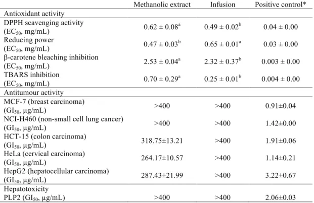
3.1. Evaluation of bioactive properties
The results obtained in the evaluation of the bioactive properties (antioxidant and
antitumour activities, and hepatotoxicity) of the infusion and the methanolic extract of
C. ambrosioides are given in Table 1. The infusion gave higher DPPH scavenging
activity and β-carotene bleaching and TBARS inhibitions than the methanolic extract.
The latter revealed higher reducing power.The essential oil extracted from the leaves of
C. ambrosioides (Kumar, Kumar, Dubey, & Tripathi, 2007) was also reported to show
powerful antioxidant activity. To the best of our knowledge, no reports are available on
the infusion or methanolic extract of the aforementioned plant.
The effects of C. ambrosioides methanolic extract and infusion on the growth of five
human tumour cell lines (MCF-7, NCI-H460, HCT-15, HeLa and HepG2), represented
as the concentrations that caused 50% of cell growth inhibition (GI50), are also
summarized in Table 1. The infusion of C. ambrosioides did not show any antitumour
potential; however, the methanolic extract presented some activity on HCT-15, HeLa
and HepG2 cell lines. It should be highlighted that no hepatotoxicity in non-tumour
cells was observed for any of the samples (GI50 > 400 µg/mL). Trolox and ellipticine
were used as positive controls of antioxidant and antitumour activities evaluation
assays, respectively, but comparison with the samples should be avoided, because they
are individual compounds and not mixtures.
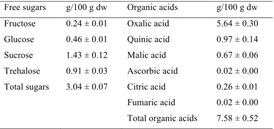
3.2. Chemical composition in hydrophilic compounds
The chemical composition of the samples in sugars and organic acids was also analyzed
and the results are shown in Table 2. The sugars found were fructose, glucose, sucrose
Oxalic, quinic, malic, ascorbic, citric and fumaric acids were also identified and
quantified (Table 2), being oxalic acid the most abundant organic acid. Some organic
acids (e.g., citric acid) have been reported as having antioxidant capacity (Hraš,
Halodin, Knez, & Bauman, 2000).
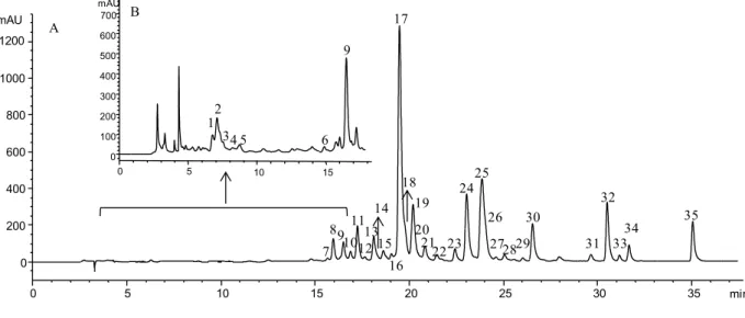
Phenolic compounds found in C. ambrosioides are presented in Table 3 and Figure 1.
Thirty-five compounds were detected, eight of which were phenolic acid derivatives
(hydroxycinnamic acid derivatives). Among them, five compounds (peaks 1-3, 5 and 9)
were p-coumaric acid derivatives identified according to their UV spectra and
pseudomolecular ion. Peak 9 was identified as trans p-coumaric acid by comparison of
its UV spectrum (λmax 312 nm) and retention time with a commercial standard. Peak 1
was identified as a p-coumaroyl pentoside acid according to its pseudomolecular [M-H]
-ion (m/z at 295) and the release of fragments at m/z 163 [p-coumaric acid-H]- (-132 mu,
pentose) and m/z 119 (loss of 132+44 mu, pentose + CO2). Peaks 2, 3 and 5 presented
pseudomolecular ions [M-H]- at m/z 287 and 387 releasing the same fragment ions at
m/z 163 and 119, which allowed assigning them to p-coumaroyl acid derivatives,
although their precise identities could not be established. The other three phenolic acid
derivatives were identified as ferulic acid derivatives based on the observation of the
ions at m/z 193 ([ferulic acid-H]-) and 149 ([ferulic acid-CO2-H]-). Peak 14 could be
identified as free ferulic acid by comparison of its UV spectrum (λmax 326 nm) and
retention time with a commercial standard. Peak 4 was associated to a feruloyl
pentoside acid based on its molecular ion fragmentation pattern similar to peak 1,
whereas no precise identity could be established for peak 6.
The remaining phenolic compounds corresponded to flavone and flavonol derivatives,
12 compounds) and kaempferol (λmax around 346 nm and MS2 fragment at m/z 285; 11
compounds) (Table 3). Quercetin 3-O-rutinoside (peak 17), quercetin 3-O-glucoside
(peak 21) and kaempferol 3-O-rutinoside (peak 24) were positively identified according
to their retention, mass and UV-vis characteristics by comparison with commercial
standards.
Peak 10 ([M-H]- at m/z 609) could be interpreted as a quercetin O-diglycosides in which
each of the sugar moieties are located at different positions on the aglycone, owing to
the observation of fragments derived from the loss of each sugar residue. However, it
might also be rationalised as a quercetin O-rhamnosyl-glucoside, in which the fragment
at m/z 447 would correspond to the loss of the terminal glucose of the dissacharide,
whereas that at m/z 463 might be rationalised as produced by an internal rearrangement
in the sugar moieties following the loss of the internal dehydrated glucose/pentose and
further linkage of the terminal rhamnose to the aglycone (Ma, Cuyckens, Heuvel, & Claeys, 2001). In that case, the greater abundance of the Y0 ion (m/z at 301; aglycone) than Y1 ion (m/z at 447; breakdown of the interglycosidic linkage) might support the
existence of a 1,2 interglycosidic linkage (Cuyckens, Rozenberg, Hoffmann, & Claeys, 2001), which allow the identification of peak 10 as quercetin 3-O-neohesperidose. Peaks 15 and 20, both with a pseudo molecular ion [M-H]- at m/z 579 releasing
fragments at m/z 447 (-132 mu; pentiosyl residue) and 301 (-132-146 mu; loss of
pentosyl+rhamnosyl residues), could be assigned as quercetin O-rhamnosyl-pentosides
in which the pentose is the terminal unit owing to the lack of a fragment at m/z 433,
which should result from the loss of the rhamnose residue if both sugars were located at
different positions on the aglycone. The observation that Y0 > Y1 ion in the case of peak
20 might point to a 1,2 interglycosidic linkage, whereas a 1,6 linkage might exist in
pseudo molecular ion [M-H]- at m/z 623 and releasing fragments at m/z 447 (-176 mu;
loss of a glucuronyl residue) and 301 (-176-146 mu; loss of glucuronyl+rhamnosyl
residues) should correspond to quercetin O-rhamnosyl-glucuronides. Furthermore, as
mentioned above, in peak 30 a 1,2 interglycosidic linkage could be observation (Y0 >
Y1 ), whereas a 1,6 linkage might exist in peak 29 (Y1 > Y0). Peak 34 can be assigned to
an acetyl derivative of peak 30 owing to its pseudomolecular ion ([M-H]- at m/z 665) 42
mu higher than that peak.
The pseudomolecular ion of peak 19 ([M-H]- at m/z 593) is coherent with a quercetin
derivative bearing two rhamnosyl residues. In principle, it can be supposed that each
sugar is located at different positions on the aglycone as suggested by the formation of a
fragment ion at m/z 447 from the loss of one of the rhamnosyl moieties, although the
possibility that they constituted a disaccharide cannot be disregarded, either.
Peak 11 ([M-H]- at m/z 741) can be assigned to a quercetin derivative bearing pentosyl,
rhamnosyl and hexosyl residues, based on the loss of 440 u (132+146+162 u) to yield
the corresponding aglycone (m/z at 301, quercetin). The fact that the three moieties were
lost simultaneously suggested that they might constitute a trisaccharide O-linked to the
aglycone. Similarly, peak 12 would be associated to a quercetin O-disaccharide
consisting of a pentose and a hexose.
Peak 8 ([M-H]- at m/z 755) would correspond to a quercetin derivative possessing two
rhamnosyl and one glucosyl moieties. The observation of a fragment at m/z 609 from
the lost of a rhamnosyl residue (-146 mu) points to this sugar is located on the aglycone
in a position different to the other two sugars that should constitute a disaccharide. The
presence of quercetin 3-O-rutinoside (peak 17) as majority flavonoid in the plant might
Similar reasoning as for the quercetin derivatives has been applied for assigning the
identities of kaempferol (peaks 13, 16, 18, 22, 23, 25, 26, 31, 32 and 35) and
isorhamnetin derivatives (peaks 27, 28 and 33), as indicated in Table 3.
Finally, peak 7 ([M-H]- at m/z 579) was assigned to a flavone, luteolin C-hexoside-O
-pentoside, based on its fragmentation. Thus, the ion at m/z 447 could be interpreted as
the loss of the pentosyl moiety (-132 mu) and a fragment of 120 mu characteristic of the
cleavage of pyran ring in the more strongly linked C-hexoses, whereas the ion at m/z
417 might correspond to the loss of the hexosyl moiety and a fragment of 30 mu
resulting from CH2O functional group of the hexose, also observed in the case of C
-hexoses (Abad-Garcia, Garmon-Lobato, Berrueta, Gallo, & Vicente, 2008). The fragments ions at m/z 447 and 285 would correspond to the respective losses of the
pentosyl and hexosyl moieties, respectively.
Flavonoids were the major phenolic compounds present in this sample (768 mg/100 g
dw), being quercetin (46.98%) and kaempferol derivatives (45.91%) the most abundant.
Quercetin 3-O-rutinoside was the compound found in the highest amount (205 mg/100
g dw, peak 17), followed by kaempferol dirhamnoside-O-pentoside (96 mg/100 g dw,
peak 25). Phenolic acids were 6.58% of the total phenolic compounds in this sample
and trans p-coumaric acid was the most abundant one (25.65 mg/100 g dw, peak 9).
Herbal infusions are frequently used in traditional medicine due to their beneficial
activities and among their constituents, special relevance has been given to phenolic
compounds, which often exhibit high antioxidant capacity being able to counteract
oxidative stress (Mejía, Songa, Hecka, Vinicio, & Ramírez-Mares, 2010; Pereira, Marcias, Perez, Marin & Cardoso, 2013). They act as antioxidants through various mechanisms, including hydrogen donating reactions, metal chelation, and up-regulation
2013). In particular, C. ambrosioides infusion is a rich source of diverse polyphenols that could contribute to the mentioned activity.
3.3. Chemical composition in lipophilic compounds
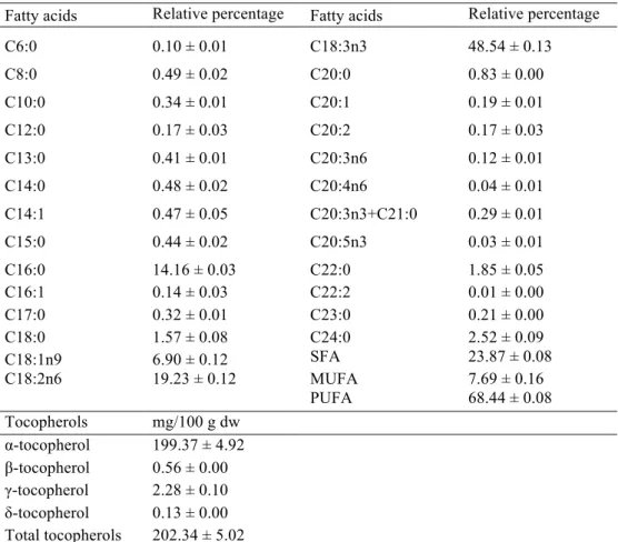
The results of lipophilic compounds (fatty acids and tocopherols) are shown in Table 4.
Up to 26 fatty acids were identified and quantified. Polyunsaturated fatty acids (PUFA)
predominated over saturated fatty acids (SFA) and monounsaturated fatty acids
(MUFA). α-Linolenic (C18:3n3; 48.54%) and linoleic (C18:2n6; 19.23%) acids
contribute to the high levels of PUFA observed (68.44%). Linoleic acid is the most
prominent PUFA in the Western diet and previous studies showed health benefits under
the prevention of cancer diseases(Whelan, 2008).
α-Tocopherol was, by far, the most abundant tocopherol in C. ambrosioides (199.37
mg/100 g dw from a total tocopherols amount of 202.34 mg/100 g dw; Table 4).
Tocopherols are very important natural antioxidants in plant foods, especially those that
are rich in PUFA. Their effectiveness as antioxidants depends not only on their
reactivity against harmful radicals, but also the relatively stable nature of his radical due
to relocation of the unpaired electron on the ring cromanol (Kagan et al., 2003).
Overall, C. ambrosioides infusion revealed, in general, higher antioxidant activity,
while the methanolic extract was the only one showing antitumour effects against colon,
cervical and hepatocellular carcinoma cell lines. Neither the infusion nor the extract
reveal toxicity for non-tumour cells. Bioactive compounds such as some sugars and
organic acids, phenolic compounds, unsaturated fatty acids and tocopherols were
chemical characterization of C. ambrosioides and bioactivity evaluation of its
methanolic extract and infusion.
Acknowledgements
The authors are grateful to Fundação para a Ciência e a Tecnologia (FCT, Portugal) for
financial support to CIMO (strategic project PEst-OE/AGR/UI0690/2011). R.C.
Calhelha and L. Barros also thank to FCT, POPH-QREN and FSE for their grants
(SFRH/BPD/ BPD/68344/2010 and SFRH/BPD/4609/2008, respectively). The
GIP-USAL is financially supported by the Spanish Government through the
Consolider-Ingenio 2010 Programme (FUN-C-FOOD, CSD2007-00063). M. Dueñas thanks to the
References
Abad-Garcia, B., Garmon-Lobato, S, Berrueta, L.A., Gallo B., & Vicente F. (2008)
New features on the fragmentation and differentiation of C-glycosidic flavone
isomers by positive electrospray ionization and triple quadrupole mass
spectrometry. Rapid Commun Mass Spectrom, 22, 1834–1842.
Abreu, R.M.V., Ferreira, I.C.F.R., Calhelha, R.C., Lima, R.T., Vasconcelos, M.H.,
Adega, F., Chaves, R., & Queiroz, M.J.R.P. (2011). Anti-hepatocellular carcinoma
activity using human HepG2 cells and hepatotoxicity of 6-substituted methyl
3-aminothieno[3,2-b]pyridine-2-carboxylate derivatives: In vitro evaluation, cell
cycle analysis and QSAR studies. European Journal of Medicinal Chemistry, 46,
5800-5806.
Barros, L., Pereira, C., & Ferreira, I.C.F.R. (2013). Optimized analysis of organic acids
in edible mushrooms from Portugal by ultra fast liquid chromatography and
photodiode array detection. Food Analytical Methods, 6, 309-316.
Carvalho, A.M. (2010). Plantas y sabiduría popular del Parque Natural de Montesinho.
Un estudio etnobotánico en Portugal. Biblioteca de Ciencias, vol. 35. Consejo
Superior de Investigaciones Científicas, Madrid.
Cruz, G.V.B., Pereira P.V.S., Patrício, F.J., Costa, G.C., Sousa, S.M., Frazão, J.B.,
Aragão-Filho, W.C., Maciel, M.C.G., Silva, L.A., Amaral, F.M.M., Barroqueiro,
E.S.B., Guerra, R.N.M., & Nascimento, F.R.F. (2007). Increase of cellular
recruitment, phagocytosis ability and nitric oxide production induced by
hydroalcoholic extract from Chenopodium ambrosioides leaves. Journal of
Cuyckens, F., Rozenberg, R., Hoffmann, E., & Claeys, M. (2001). Structure
characterization of flavonoid O-diglycosides by positive and negative
nano-electrospray ionization ion trap mass spectrometry. Journal of Mass Spectrometry,
36, 1203-1210.
Guimarães, R., Barros, L., Dueñas, M., Calhelha, R.C., Carvalho, A.M., Santos-Buelga,
C., Queiroz, M.J.R.P., & Ferreira, I.C.F.R. (2013). Nutrients, phytochemicals and
bioactivity of wild Roman chamomile: a comparison between the herb and its
preparations. Food Chemistry, 136, 718-725.
Halliwell, B. (2012).Free radicals and antioxidants: updating a personal view. Nutrition
Reviews, 70, 257–265
Hraš, A.R., Halodin, M., Knez, Z., & Bauman, D. (2000). Comparison of antioxidative
and synergistic effects of rosemary extract with α-tocopherol, ascorbyl palmitate
and citric acid in sunflower oil. Food Chemistry, 71, 229-233.
Kagan, V.E., Kuzmenko, A.I., Shvedova, A.A., Kisin, E.R., Li, R., Martin, I., Quinn,
P.J., Tyurin, V.A., Tyurina, Y.Y., & Yalowich, J.C. (2003). Direct evidence for
recycling of myeloperoxidase-catalyzed phenoxyl radicals of a vitamin E
homologue, 2,2,5,7,8-pentamethyl-6-hydroxy chromane, by
ascorbate/dihydrolipoate in living HL-60 cells. Biochimica et Biophysica Acta,
1620, 72-84.
Kamel, E.G., El-Emam, M.A., Mahmoud, S.S.M, Fouda, F.M., & Bayaumy, F.E.
(2011). Parasitological and biochemical parameters in Schistosoma
ambrosioides, Conyza dioscorides and Sesbania sesban. Parasitology
International, 60, 388–392.
Kumar, R., Kumar, M.A., Dubey, N.K., & Tripathi, Y.B. (2007) Evaluation of
Chenopodium ambroisioides oil as a potential source of antifungal,
antiaflatoxigenic and antioxidant activity. International Journal of Food
Microbiology, 115, 159-164.
Ma, Y.-L., Cuyckens, F., Heuvel, H.V., & Claeys, M. (2001). Mass Spectrometric
Methods for the Characterisation and Differentiation of Isomeric O-diglycosyl
Flavonoids. Phytochemical Analysis, 12, 159–165.
Mejía, E.G, Songa, Y.S., Hecka, C.I., & Ramírez-Mares, M.V. (2010). Yerba mate tea
(Ilex paraguariensis): Phenolics, antioxidant capacity and in vitro inhibition of
colon cancer cell proliferation. Journal of Functional Foods, 2, 23-34.
Nascimento, F.R.F., Cruz, G.V.B., Pereira, P.V.S., Maciel, M.C.G., Silva, L.A.,
Azevedo, A.P.S., Barroqueiro, E.S.B., & Guerra, R.N.M. (2006). Ascitic and solid
Ehrlich inhibition by Chenopodium ambrosioides L. treatment. Life science, 78,
2650-2653.
Pereira O.R., Marcias, R.I.R., Perez, M.J., Marin, J.J.G., & Cardoso, S.M. (2013).
Protective effects of phenolic constituents from Cytisus multiflorus, Lamium album
L. and Thymus citriodorus on liver cells. Journal of Functional Foods, in press,
doi.org/10.1016/j.jff.2013.03.014.
Pinela, J., Barros, L., Dueñas, M., Carvalho, A.M., Santos-Buelga, C., & Ferreira,
I.C.F.R. (2012). Antioxidant activity, ascorbic acid, phenolic compounds and
sugars of wild and commercial Tuberaria lignosa samples: effects of drying and
Ramarathnam, N., Osawa, T., Ochi, H., & Kawakishi, S. (1995). The contribution of
plant food antioxidants to human health. Trends in Food Science & Technology, 6,
75-82.
Rosenfeldt, F., Wilson, M., Lee, G., Kure, C., Ou, R., Braun,L., & Haan, J. (2013).
Oxidative stress in surgery in an ageing population: Pathophysiology and therapy.
Experimental Gerontology, 48, 45-54.
Skerget, M., Kotnik, P., Hadolin, M., Hras, A.R., Simonic, M., & Knez, Z. (2005).
Phenols, proanthocyanidins, flavones and flavonols in some plant materials and
their antioxidant activities. Food Chemistry, 89, 191-198.
Valko, M., Leibfritz D., Moncol, J., Cronin, M.T., Mazur, M., & Telser, J. (2007). Free
radicals and antioxidants in normal physiological functions and human disease.
International Journal of Biochemistry and Cell Biology, 39,44-84.
Whelan, J. (2008). The health implications of changing linoleic acid intakes.
Table 1. Bioactive properties of the methanolic extract and infusion of wild
Chenopodium ambrosioides.
Methanolic extract Infusion Positive control* Antioxidant activity
DPPH scavenging activity (EC50, mg/mL)
0.62 ± 0.08a 0.49 ± 0.02b 0.04 ± 0.00 Reducing power
(EC50, mg/mL)
0.47 ± 0.03b 0.65 ± 0.01a 0.03 ± 0.00 β-carotene bleaching inhibition
(EC50, mg/mL)
2.53 ± 0.04a 2.32 ± 0.37b 0.003 ± 0.00 TBARS inhibition
(EC50, mg/mL)
0.70 ± 0.29a 0.25 ± 0.01b 0.004 ± 0.00 Antitumour activity
MCF-7 (breast carcinoma) (GI50, µg/mL)
>400 >400 0.91±0.04 NCI-H460 (non-small cell lung cancer)
(GI50, µg/mL)
>400 >400 1.42±0.00 HCT-15 (colon carcinoma)
(GI50, µg/mL)
318.75±13.21 >400 1.91±0.06 HeLa (cervical carcinoma)
(GI50, µg/mL)
264.17±10.57 >400 1.14±0.21 HepG2 (hepatocellular carcinoma)
(GI50, µg/mL)
287.43±21.99 >400 3.22±0.67 Hepatotoxicity
PLP2 (GI50, µg/mL) >400 >400 2.06±0.03
Table 2. Chemical composition in hydrophilic compounds- sugars and organic acids- of wild Chenopodium ambrosioides.
Table 3. Retention time (Rt), wavelengths of maximum absorption in the UV-vis region (λmax), pseudomolecular and MS2 fragment ions (in
brackets, relative abundances), identification and quantification of phenolic compounds in wild C. ambrosiodes.
Peak Rt (min) λmax (nm)
Molecular ion
[M-H]- (m/z)
MS2
(m/z) Identification
Quantification
(mg/100 g dw)
1 6.8 310 295 163(100),119(60) p-Coumaroyl pentoside acid 3.53 ± 0.50
2 7.1 314 278 163(6),119(13) p-Coumaroyl acid derivative 9.75 ± 0.44
3 7.5 328 387 387(100)207(25),163(50),119(37) p-Coumaroyl acid derivative 1.41 ± 0.09
4 8.2 328 325 193(100),149(38) Feruloylpentoside acid 2.58 ± 0.27
5 8.8 308 278 163(6),119(13) p-Coumaroyl acid derivative 1.21 ± 0.15
6 14.8 326 473 267(27),193(100) Ferulicacid derivative 3.51 ± 0.14
7 15.7 332 579 447(15),417(7),327(7),285(100) Luteolin C-hexoside-O-pentoside 2.27 ± 0.09
8 15.9 354 755 609(2),301(100) Quercetin 3-O-rutinoside-(1→2)-O-rhamnoside 15.23 ± 0.41
9 16.4 312 163 119(100) transp-Coumaric acid 25.65 ± 0.77
10 16.9 356 609 463(30),447(33),301(36) Quercetin 3-O-neohesperide 7.19 ± 0.32
11 17.2 354 741 301(100) Quercetin O-pentosyl-rhamnosyl-hexoside 27.60 ± 0.31
12 17.6 356 595 301(100) Quercetin O-pentosyl-hexoside 3.55 ± 0.46
13 18.1 348 739 285(100) Kaempferol O-dirhamnosyl-hexoside 20.38 ± 0.74
14 18.3 326 193 149(17),135(100) Ferulic acid 6.43 ± 0.53
15 18.6 354 579 447(100),301(33) Quercetin O-rhamnosyl-pentoside 8.06 ± 0.87
16 19.0 348 739 593(83),431(17),285(67) Kaempferol dirhamnoside-O-hexoside 4.80 ± 0.49
17 19.4 354 609 301(100) Quercetin-3-O-rutinoside 204.95 ± 6.39
18 19.8 346 725 285(100) Kaempferol O-pentosyl-rhamnosyl-hexoside 31.42 ± 1.36
dw- dry weight; tr- traces
20 20.4 354 579 447(45),301(100) Quercetin O-rhamnosyl-pentoside 1.22 ± 0.03
21 20.8 350 463 301(100) Quercetin 3-O-glucoside 12.91 ± 0.80
22 21.5 348 563 431(53),285(100) Kaempferol O-rhamnosyl-pentoside 4.93 ± 0.08
23 22.4 344 739 593(24),431(24),285(100) Kaempferol dirhamnoside-O-hexoside 11.31 ± 0.44
24 23.1 348 593 285(100) Kaempferol 3-O-rutinoside 74.82 ± 2.29
25 23.5 342 709 563(25),431(63),285(100) Kaempferol dirhamnoside-O-pentoside 95.89 ± 1.64
26 23.9 344 563 431(47),285(100) Kaempferol O-rhamnosyl-pentoside 36.15 ± 1.40
27 24.6 350 607 461(50),315(100) Isorhamnetin dirhamnoside tr
28 25.1 352 593 461(80),315(100) Isorhamnetin O-rhamnosyl-pentoside 1.60 ± 0.09
29 26.0 352 623 447(50),301(43) Quercetin O-rhamnosyl-glucuronide 2.48 ± 0.04
30 26.6 352 623 447(33),301(51) Quercetin O-rhamnosyl-glucuronide 33.99 ± 0.28
31 29.6 344 607 459(30),431(20),285(50) Kaempferol O-rhamnosyl-glucuronide 6.54 ± 0.28
32 30.5 346 607 431(100),285(86) Kaempferol O-rhamnosyl-glucuronide 56.08 ± 0.35
33 31.2 350 637 461(100),315(87) Isorhamnetin O-rhamnosyl-glucuronide 0.50 ± 0.00
34 31.7 352 665 623(14),447(35),301(18) Quercetin (acyl)glucuronide-O-rhamnoside 12.53 ± 0.56
35 35.1 344 649 607(6),431(42),285(31) Kaempferol (acyl)glucuronide-O-rhamnoside 35.26 ± 1.43
Phenolic acids 54.07 ± 1.55
Flavonoids 768.27 ± 10.70
Table 4. Chemical composition in lipophilic compounds of wild Che ambrosioides.
dw- dry weight. SFA- Saturated fatty acids; MUFA- Monounsaturated fatty aci Polyunsaturated fatty acids.
Fatty acids Relative percentage Fatty acids Relative percentage
C6:0 0.10 ± 0.01 C18:3n3 48.54 ± 0.13
C8:0 0.49 ± 0.02 C20:0 0.83 ± 0.00
C10:0 0.34 ± 0.01 C20:1 0.19 ± 0.01
C12:0 0.17 ± 0.03 C20:2 0.17 ± 0.03
C13:0 0.41 ± 0.01 C20:3n6 0.12 ± 0.01
C14:0 0.48 ± 0.02 C20:4n6 0.04 ± 0.01
C14:1 0.47 ± 0.05 C20:3n3+C21:0 0.29 ± 0.01
C15:0 0.44 ± 0.02 C20:5n3 0.03 ± 0.01
C16:0 14.16 ± 0.03 C22:0 1.85 ± 0.05 C16:1 0.14 ± 0.03 C22:2 0.01 ± 0.00 C17:0 0.32 ± 0.01 C23:0 0.21 ± 0.00 C18:0 1.57 ± 0.08 C24:0 2.52 ± 0.09 C18:1n9 6.90 ± 0.12 SFA 23.87 ± 0.08 C18:2n6 19.23 ± 0.12 MUFA 7.69 ± 0.16
PUFA 68.44 ± 0.08 Tocopherols mg/100 g dw
Figure 1. HPLC phenolic profile of Chenopodium ambrosiodes,obtained at 370 nm (A) and 280 nm (B) for flavonoids and phenolic acids, respectively.
min
0 5 10 15 20 25 30 35
mAU 0 200 400 600 800 1000 1200 10 15 mAU 0 100 200 300 400 500 600 700 0 5 A B 1 2
34 5 6
7 8 9 10 11 9 12 13 14 15 16 17 18 19 20 21 2223 24 25 26