Braz. J. of Develop.,Curitiba, v. 6, n. 10, p. 75145-75151, oct. 2020. ISSN 2525-8761
Radiological Findings: a lamb skeleton deformities case report
Achados radiológicos: relato de caso de deformidades do esqueleto de Cordeiro
DOI:10.34117/bjdv6n10-078
Recebimento dos originais: 08/09/2020 Aceitação para publicação: 05/10/2020
Gabriela Döwich Pradella
Formação acadêmica mais alta: Médica veterinária especialista (residência na área de clínica e cirurgia de grandes animais)
Instituição: Universidade Federal do Pampa (Unipampa- campus Uruguaiana) Endereço: BR 472, km 585, caixa postal 118, CEP 97501-970, Uruguaiana/RS
E-mail:[email protected]
Patrícia Maurer Taschetto
Formação acadêmica mais alta: Médica veterinária especialista (residência na área de clínica e cirurgia de grandes animais)
Instituição: Universidade Federal do Pampa (Unipampa- campus Uruguaiana) Endereço: BR 472, km 585, caixa postal 118, CEP 97501-970, Uruguaiana/RS
E-mail: [email protected]
Susane Werle Dill
Formação acadêmica mais alta: Médica veterinária especialista (residência na área de diagnóstico por imagem)
Instituição: Universidade Federal do Pampa (Unipampa- campus Uruguaiana) Endereço: BR 472, km 585, caixa postal 118, CEP 97501-970, Uruguaiana/RS
E-mail: [email protected]
Claudia Acosta Duarte
Formação acadêmica mais alta: Doutora em Cirurgia Veterinária Instituição: Universidade Federal do Pampa (Unipampa- campus Uruguaiana) Endereço: BR 472, km 585, caixa postal 118, CEP 97501-970, Uruguaiana/RS
E-mail: [email protected]
Ingrid Rios Lima Machado
Formação acadêmica mais alta: Doutora em Ciência animal
Instituição: Universidade Federal do Pampa (Unipampa- campus Uruguaiana) Endereço: BR 472, km 585, caixa postal 118, CEP 97501-970, Uruguaiana/RS
E-mail: [email protected]
Geórgia Camargo Góss
Formação acadêmica mais alta: Mestre em Ciência animal
Instituição: Universidade Federal do Pampa (Unipampa- campus Uruguaiana) Endereço: BR 472, km 585, caixa postal 118, CEP 97501-970, Uruguaiana/RS
Braz. J. of Develop.,Curitiba, v. 6, n. 10, p. 75145-75151, oct. 2020. ISSN 2525-8761
ABSTRACT
Skeletal deformities are relatively common in animals. These abnormalities could be genetic or conditioned by environment factors. The radiographic exam is simple and allow us to identify a great number of congenital defects. Lambs with deformities are important economic problems in the sheep culture and there are few studies related to this. The objective is to describe the radiographic findings in one case of skeletal deformities in a lamb from the state of Rio Grande do Sul, Brazil. Radiological findings are compatible with paraxial hemimelia of the left tibia and polydactylies. General skeletal deformities were also observed in this study.
Keywords: Tibial hemimelia; sheep; supranumeric finger; perinatal mortality; congenital defects. RESUMO
As deformidades esqueléticas são relativamente comuns em animais. Essas anormalidades podem ser genéticas ou condicionadas por fatores ambientais. O exame radiográfico é simples e permite identificar muitos defeitos congênitos. Cordeiros com deformidades causam problemas econômicos importantes na ovinocultura e poucos são os estudos relacionados a isso. O objetivo é descrever os achados radiográficos em um caso de deformidades esqueléticas em um cordeiro do estado do Rio Grande do Sul, Brasil. Os achados radiológicos são compatíveis com hemimelia paraxial da tíbia esquerda e polidactilia. Deformidades esqueléticas gerais também foram observadas neste estudo.
Palavras-chave: Hemimelia tibial; ovelhas; dedo supra numérico; mortalidade perinatal; defeitos
congênitos.
1 INTRODUCTION
Congenital defects are featured by anomalies in structure or function of an organ or a system. They may be genetic or conditioned by the environment (Riet-Correa et al., 2001). Skeleton developmental errors may be primary abnormalities of bone, cartilage, or primitive mesenchyme. The processes by which the skeleton is formed are complex and these provides large opportunity for error. The conditions described as the cause of bone defects are presumed to have a genetic basis. However, abnormalities, like those of hereditary origin, can be caused by environmental factors such as chemicals, viruses, and nutritional deficiencies (Jubb et al., 1993).
Inside the skeleton defects there are hemimelia and polydactylies. The hemimelia is characterized by a defect of the midsection of the limb. There are two different classifications: transverse or paraxial. In transverse hemimelia the bone may be absent, whereas in paraxial hemimelia there is aplasia. When an increase in the number of digits appears, it is called polydactylies. This congenital defect is well known in cats, dogs, horses, and cattle (Jubb et al., 1993).
The objective is to describe the radiographic findings in one case of skeletal deformities in a lamb from the state of Rio Grande do Sul, Brazil.
Braz. J. of Develop.,Curitiba, v. 6, n. 10, p. 75145-75151, oct. 2020. ISSN 2525-8761
2 MATERIAL AND METHODS
An Ile de France lamb was received at the University hospital after it died in the property when it was three days old. The owner reported that the lamb was born with a limb defect. It remained in decubitus all the time until its dead. At the property there was no previous case about newborns with these changes. The radiographic exam of the pelvic limbs was made using craniocaudal and medium-lateral projections.
3 RESULTS
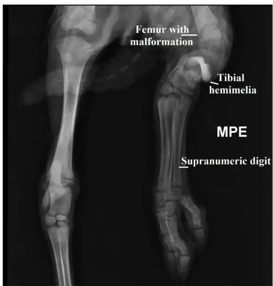
On the left limb a trace of tibia (paraxial hemimelia) was observed, and an increase in the number of digits in the first, second and third metatarsi and phalanges (Figure 1).
In addition to these defects, in the latero lateral projection of the spine, the lamb presented agenesis of the last thoracic vertebraes and ribs (11ª, 12ª e 13ª), mal formation of lumbar and sacral vertebrae as well, making anatomical identification of these structures impossible. The pelvis was also malformed with ileo hypoplasia in the left side (Figure 2). Changes in the femur bone with mal formation in the proximal epiphyses and metaphysis were also observed (Figure 3).
Radiological findings are compatible with paraxial hemimelia of the left tibia and polydactylies in the same limb. General skeletal deformities were also observed in this study.
4 DISCUSSION
Radiography was used because it is a simple but important tool in the diagnosis of
anatomical defects in animals. It is a practical method of examining a patient's skeletal system and may be the only procedure for the early detection of inherited defects in a given herd (Guffy and Leipold, 1977).
It was possible to evidence a skeletal deformity in the left hindlimb of the Ile de France lamb in this study. There were three reported cases of radial hemimelia in goatlings, two females and one male. The radiological presentation of these cases varies into agenesis, paraxial hemimelia and deformity of the radius (Corbera et al., 2002). A similar case was reported in a 6-year-old goat with complete absence of the radial bone (OVIAWE e colab., 2017). All these reports occurred in the forelimb and involved the radius. In our study skeletal deformity in the hindlimb with vestige of the tibia was observed. Some studies showed a tibial hemimelia syndrome in cattle (Apointe et al., 2000). In humans, tibial hemimelia may occur in isolation or may be associated with a variety of skeletal or extra-skeletal malformations, such us polydactyly (Salinas-Torres et al., 2015), as observed in the present case. There are reports of unilateral and / or bilateral tibial hemimelia in
Braz. J. of Develop.,Curitiba, v. 6, n. 10, p. 75145-75151, oct. 2020. ISSN 2525-8761 cattle (Lapointe et al., 2000). In goats, there are reports of hemimelia in the radio (Corbera et al., 2002). However, in the sheep species, there are no reports on the occurrence of tibial hemimelia, possibly due to the low number of occurrences and the failure to send these animals to be rigorously evaluated.
Two cases have also been reported of hemimelia affecting a 6-month-old goat with bilateral absence of metatarsal bones with shortening of hindlimbs and a newborn calf with abnormal hindlimbs (the tibia was rudimentary, and I don’t understand). No surgery was indicated for such conditions (Mosbah et al., 2012).
Besides the left hindlimb defects, general ossification deformities were observed in this study. Malformed bones are common in sheep (DANTAS et al., 2010). However, sheep or lambs born with congenital malformations do not get the laboratory diagnosis, especially if the percentage of perinatal mortality is within the expected limit, around 10% in the conditions of sheep in the southern region of the state (Méndez et al., 1982).
5 CONCLUSIONS
The present report describes a case of tibial hemimelia, polydactylies and general skeleton malformation in a newborn lamb and its radiological findings. The skeletal deformities are an important economic factor in the sheep farming. However, few cases are reported because of the low number of forward cases. The radiographic exam is a simple tool that can help in the diagnosis and farming management.
Braz. J. of Develop.,Curitiba, v. 6, n. 10, p. 75145-75151, oct. 2020. ISSN 2525-8761
REFERENCES
CORBERA, J. A. et al. Radiological Findings in Three Cases of Paraxial Radial Hemimelia in Goats. Journal of Veterinary Medical Science, v. 64, n. 9, p. 843–845, 2002.
DANTAS, A. F. M. et al. Malformações congênitas em ruminantes no semiárido do Nordeste Brasileiro. Pesquisa Veterinaria Brasileira, v. 30, n. 10, p. 807–815, 2010.
GUFFY, M. M.; LEIPOLD, H. W. Radiological Diagnosis of Economically Important Genetic Defects in Cattle. Veterinary Radiology, v. 18, n. 4, p. 109–115, 1977.
JUBB, K. V. F.; KENNEDY, P. C.; PALMER, N. Pathology of Domestic Animals. 4th ed. San Diego, California: 1993. v. 1.
LAPOINTE, J. M.; LACHANCE, S.; STEFFEN, D. J. Tibial hemimelia, meningocele, and abdominal hernia in shorthorn cattle. Veterinary Pathology, v. 37, n. 5, p. 508–511, 2000.
MOSBAH, E.; KARROUF, G.; RIZK, A. Congenital limb deformities in some farm animals. Fifth
animal wealth research conference in the Middle East and North Africa, v. 5, n. May 2014, p. 23–
38, 2012.
OVIAWE, E. I. et al. Radiographic Finding of Radial Hemimelia in a 6-Day- Old West African Dwarf Goat with a Fractured Ulna. Austin Journal of Radiology, v. 4, n. 1, p. 6–8, 2017.
RIET-CORREA, F. et al. Doenças dos Ruminantes e Equinos. 1th ed. São Paulo: 2001.
SALINAS-TORRES, V. M. et al. Bilateral tibial hemimelia type 1 (1a and 1b) with T9 and T10 hemivertebrae: a novel association. Sao Paulo Medical Journal, v. 131, n. 4, p. 275–278, 2015.
MÉNDEZ, M.C. et al. Mortalidade perinatal em ovinos nos municípios de Bagé, Pelotas e Santa Vitória do Palmar, Rio Grande do Sul. Pesquisa Veterinária Brasileira, v. 2, n. 2, p. 69-76, 1982.
Braz. J. of Develop.,Curitiba, v. 6, n. 10, p. 75145-75151, oct. 2020. ISSN 2525-8761
ANEXOS
Figure 1: Radiography in craniocaudal projection. It is possible to observe a polydactyly in the left hindlimb, with supranumeric first, second and third metatarsi and phalanges; a remnant of left tibia (tibial hypoplasia) and a malformed femur bone.
Braz. J. of Develop.,Curitiba, v. 6, n. 10, p. 75145-75151, oct. 2020. ISSN 2525-8761 Figure 2: Latero-lateral radiography.It showed agenesis of the last thoracic vertebraes and ribs (11ª, 12ª e 13ª); fusion of the vertebral bodies of the lumbar region; absence of sacral iliac joint and bilateral ileus malformation of an Ile de France lamb.
Figure 3: Craniocaudal projection of the left limb. It is possible to observe changes in the femur bone with mal formation in the proximal epiphyses and metaphysis.