Faculdade de Medicina de Lisboa
Deciphering the molecular role of DJ-1 in the etiology
of Parkinson’s and Huntington’s disease
Maria Leonor Lemos Pereira Miller Fleming
Tese orientada por:
Prof. Doutor Tiago Outeiro
Prof. Doutor Flaviano Giorgini
Ramo das Ciências Biomédicas Especialidade – Neurociências
Todas as afirmações efectuadas no presente documento são da exclusiva responsabilidade do seu autor, não cabendo qualquer responsabilidade à Faculdade de Medicina de Lisboa pelos conteúdos nele apresentados.
A impressão desta dissertação foi aprovada pelo Conselho
Cientifico da Faculdade de Medicina de Lisboa em reunião de
(15 de Outubro de 2013)
Acknowledgements ... iii
Abstract ... v
Resumo ... vii
Abbreviations ... xi
Chapter 1. Introduction ... 1
Protein misfolding diseases ... 1
Huntington’s disease ... 4
Parkinson’s disease ... 9
Baker’s yeast: a versatile “toolbox” for molecular biology studies ... 18
Modeling neurodegenerative diseases in yeast ... 19
Yeast models of HD ... 20
Yeast models of PD ... 21
References ... 23
Aims of the thesis ... 31
Chapter 2. Functional gene expression profiling in yeast implicates translational dysfunction in mutant huntingtin toxicity ... 35
Abstract ... 35
Introduction ... 35
Results ... 39
Yeast expressing a mutant Htt fragment differentially express genes involved in ribosome biogenesis and rRNA processing ... 39
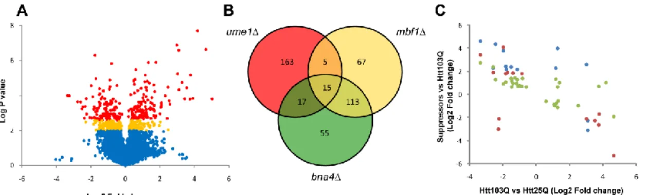
Common mechanisms underlie mutant Htt toxicity suppression in gene deletion suppressor strains ... 40
Cis-regulatory domain analysis of DEGs identifies enriched elements ... 44
A unique subset of differential expressed, highly interconnected genes modulate mutant Htt toxicity ... 45
Discussion ... 49
Materials and Methods ... 52
Acknowledgments ... 56
References ... 56
Chapter 3. Yeast DJ-1 family members are required for diauxic-shift reprogramming and cell survival in stationary phase ... 61
Abstract ... 61
Introduction ... 61
Results ... 66
The HSP31 mini-family is required for normal diauxic-shift and stationary phase . 66 Typical stationary phase characteristics are altered in HSP31 mini-family knockout strains ... 73
Autophagic response is impaired during carbon starvation in hsp31Δ cells ... 74
TORC1 signaling is perturbed in hsp31Δ cells ... 77
Discussion ... 80
Materials and Methods ... 83
Acknowledgements ... 89
References ... 89
Chapter 4. DJ-1 modulates huntingtin aggregation and toxicity in models of Huntington’s disease ... 97
Abstract ... 97
Introduction ... 97
Results ... 99
DJ-1 overexpression ameliorates mutant Htt toxicity in yeast and fruit flies ... 99
DJ-1 expression and its oxidation is increased in the HD brain and in cell and animal models of HD ... 100
DJ-1 directly modulates aggregation and toxicity of mutant Htt in an oxidation-sensitive manner ... 102
Discussion ... 108
Materials and Methods ... 109
Acknowledgements ... 114
References ... 114
Chapter 5. General discussion, future perspectives and conclusion ... 119
CONCLUSION ... 126
References ... 127
Esta tese é dedicada à minha querida avó Fátima. (This PhD thesis is dedicated to my sweet grandmother Fátima.)
“To be is to do – Socrates To do is to be – Sartre Do Be Do Be Do – Sinatra”
Kurt Vonnegut
A PhD is a long and extremely challenging journey, during which I had the pleasure to meet many people who I am thankful for, in one way or another, contributing to the knowledge and scientific skills I have acquired.
I thank my supervisor Tiago Outeiro for his guidance and support throughout my PhD and for teaching me how to think as a scientist, work hard and stay focused. He introduced me to the world of yeast and opened for me the doors to one of my biggest scientific passions: neuroscience research. He gave me the opportunity to grow as a researcher in his lab, where I spent most of my PhD time.
I started my PhD in the lab of Flaviano Giorgini (Flav) – my co-supervisor – and had the privilege to be one of his first PhD students. Besides being a very intelligent scientist, Flav is a fantastic person, with a big heart and great sense of humor. His mentorship was a permanent source of inspiration. I thank Flav for always being very supportive and for giving me motivation and positive thinking. I will never forget once that I realized I had cloned a gene upside down. I was afraid of his reaction, but instead of being angry at me, he showed me the positive side: “Most people don’t even realize they have made a mistake”. I also thank him for the scientific discussions and for his scientific guidance.
I owe my sincere gratitude to Teresa Pais, my friend and wise colleague, the person with whom I had the longest and funniest scientific discussions. Teresa was the person who was always by my side; listening, teaching, criticizing, bearing my frustrations and sharing the joys of success, helping me to think and create hypotheses. Her smile, good mood and enthusiasm were always an inspiration. I also thank her for allowing me to expose my ideas and contribute to her work, which was a great experience.
Pedro Antas, a very special person who unfortunately only entered my life at the end of my PhD. He helped me to finish some experiments. I believe the last months of the PhD may be a stressful moment, but for me these were the best. I had lots of fun
with Pedro’s jokes “night and day”. Thanks Pedro for the help and hard work, for the nice suggestions and for making our work so much fun.
I am thankful to Federico Herrera (Fede), for proofreading my work many times and having taught me several rules of scientific writing. I also thank him for all the support and constructive - and sometimes hard – criticism throughout my PhD, which I really appreciated. I also thank Fede for giving me the opportunity to work with him.
It was great to share ideas and long hours in the lab with Sandra Jacinto, Oldriska Marques (Oli), Elisa Basso (Elisinha) and Patricia Guerreiro (Patricinha). I will miss those times.
I would like to thank all of my colleagues from Tiago’s lab (UNCM) who in one way or another contributed to my work; especially Rita Oliveira and Hugo Miranda who were always very supportive and enriched my research with their questions and suggestions, to the present and former technicians of my lab, Tiago Mendes, Andreia Peixoto and Sónia Oliveira that always gave me help when they had time and to Sandra Tenreiro for always being helpful.
From Leicester, I am thankful to Rob Mason for teaching me the majority of the basic yeast molecular techniques I know, to Mahmut Ergoren, Rob Hardwick, Angelica Vittori, Raheleh Rahbari and Paul Ainsworth for receiving me in their homes during my short visits to Leicester and to all my friends in Leicester for always making me feel at home.
I would also like to thank Paula Ludovico for the fruitful discussions and constructive feedback, and Daniel Klionsky for spending some of his valuable time discussing some of my results and for kindly sharing material and protocols that were essential at a certain point of my PhD to move my work forward, and Marek Skoneczny for kindly providing several plasmids and strains - even more than I asked for.
I am grateful to all the collaborators, whose names are mentioned in the beginning of each chapter, for contributing to some of the experiments presented in this thesis.
Last but not least I want to thank my “Lisbon family”, in particular Carolina and Francisco for making my life outside the lab really great, and all my friends in Porto, especially Joaninha, Joana Maranhas, Ana Inês and my cousin Sofia who, although far away, were closer than ever, and to my parents for always believing in me and encourageing me to pursue my goals.
Neurodegenerative diseases are among the most complex and puzzling human disorders. These devastating disorders currently do not have any effective therapies or treatments, and thus are a social and economic burden for modern society. Intense efforts are being made to unravel the mechanisms underlying this group of diseases, however there is still much to be learnt and discovered. Therefore, it is of utmost importance to better understand the biological mechanisms involved in disease pathogenesis, and more importantly, to discover novel avenues for therapeutic intervention. Budding yeast have been successfully used to perform studies on these diseases that have resulted in the identification of several promising therapeutic drugs and targets. The ease of experimental manipulation, the high conservation in basic cellular mechanisms between yeast and humans, and the well defined genome are three of the numerous characteristics that make yeast so attractive to researchers studying neurodegenerative disorders. Here, we used the budding yeast to study the molecular basis of two neurodegenerative diseases: Huntington’s (HD) and Parkinson’s disease (PD).
HD is a fatal neurodegenerative disorder caused by a well defined genetic cause, which is an expansion of a polyglutamine tract in the huntingtin (Htt) protein. We have used a yeast HD model to find differentially expressed genes (DEGs) in wild-type yeast in response to mutant Htt toxicity, as well as in three Htt toxicity suppressor strains: bna4∆, mbf1∆, and ume1∆. We found common DEGs in the suppressor strains involved in stress response, translation elongation, and mitochondrial transport. We then tested whether overexpression of the DEGs suppress mutant Htt toxicity. We identified 12 novel suppressors, including genes that play a role in stress response and rRNA processing. We have integrated the microarray and the genetic screening data that allowed us to generate a robust network that showed enrichment in genes involved in rRNA processing and ribosome biogenesis. Altogether, our results suggest that dysfunctional translation is implicated in HD. Ultimately pharmacological manipulation of translation may have therapeutic value in HD.
PD is the most common progressive neurodegenerative movement disorder. One of the proteins that when mutated leads to autosomal recessive forms of PD is DJ-1. The precise function of this protein and how this protein leads to PD is unclear. Yeast has four proteins that belong to the conserved DJ-1 superfamily: Hsp31, Hsp32, Hsp33 and Hsp34. Very little is known about their function. Here, we show that the yeast DJ-1
homologs are required for diauxic-shift, an important metabolic reprogramming stage in yeast that is triggered by glucose limitation. We found that the Hsp31 genes are strongly induced in diauxic-shift and in stationary phase, and that deletion of these genes leads to reduced chronological lifespan, an inability to reprogram gene transcription at diauxic-shift, and failure to acquire several typical characteristics of stationary phase, including defective autophagy induction. Furthermore, we showed that these proteins contribute to TORC1 regulation, a central signaling pathway involved in diauxic-shift reprogramming and, importantly, in autophagy. As dysregulation of both autophagy and TORC1 are associated with several disorders – including PD – our work may have broad relevance to understanding these processes in health and disease. These novel insights into the function of the yeast DJ-1 homologs contribute to the understanding of the normal function of both human DJ-1 and the remaining DJ-1 superfamily members, and may therefore bear therapeutic implications in various human diseases.
The human DJ-1 is thought to act as a redox-dependent chaperone by preventing the aggregation of misfolded proteins in oxidative stress conditions. One of the potential targets of DJ-1 is α-synuclein, which is the major component of Lewy bodies in PD. Oxidative stress is linked to several neurodegenerative diseases including HD. Here, we show that DJ-1 plays a potential role in the pathogenesis of HD, by modulating Htt aggregation and toxicity likely, by acting as a redox-dependent chaperone. We found that DJ-1 is upregulated and abnormally oxidized in the brains of HD patients, which is how DJ-1 is found in brains of patients with PD and Alzheimer’s disease. Our results suggest a general role for DJ-1 as a redox-dependent chaperone that may have an impact in the pathogenesis of several protein misfolding diseases. Furthermore, it establishes DJ-1 as a potential therapeutic target for HD.
Overall, the work presented in this thesis opens novel avenues of research and therapeutics for HD and PD and proposes that DJ-1 has broad relevance in the pathogenesis of neurodegenerative diseases.
Keywords: Parkinson’s disease, Huntington’s disease, yeast, diauxic-shift, autophagy, translation, TORC1, protein misfolding
As doenças neurodegenerativas estão entre as doenças humanas mais complexas e enigmáticas. As terapias e tratamentos actualmente existentes não são eficazes, causando um impacto negativo na sociedade moderna, tanto a nível social como económico. Apesar do esforço empregue na compreensão dos mecanismos patogénicos associados a este grupo de doenças, ainda há muito por explorar e aprender. É urgente investigar os mecanismos moleculares que levam a essas patologias e descobrir novos alvos terapêuticos. A levedura Saccharomyces cerevisiae tem sido usada com sucesso no estudo de doenças neurodegenerativas e já resultou na identificação de vários alvos terapêuticos bem como de compostos com potencial terapêutico bastante promissor. As leveduras, como modelo biológico, apresentam várias vantagens que as tornam atractivas para estudar estas doenças, incluindo a sua fácil manipulação laboratorial, a conservação dos mecanismos celulares básicos até aos humanos, e extensa caracterização do seu genoma. Nesta tese, as leveduras foram o modelo escolhido para estudar os mecanismos moleculares associados a duas doenças neurodegenerativas: a doença de Huntington (DH) e a doença de Parkinson (DP).
A DH é uma doença neurodegenerativa fatal, geneticamente bem caracterizada, causada por uma mutação genética no gene IT15, que consiste na repetição anormal da sequência de nucleotídeos CAG, originando a expansão de uma região rica em glutaminas na proteína huntingtina (Htt). O modelo de levedura para a DH foi usado para detectar genes diferencialmente expressos (GDE) em células a expressar a proteína Htt mutada, assim como em três estirpes supressoras de toxicidade de Htt: bna4∆, mbf1∆, e ume1∆. Nestas estirpes supressoras da toxicidade foram encontrados GDE envolvidos na resposta ao stress, no alongamento durante a tradução do mRNA e no transporte mitocondrial. A sobre-expressão dos GDEs levou à identificação de 12 novos genes supressores da toxicidade provocada pela Htt, incluindo genes envolvidos na resposta ao stress e no processamento do rRNA. Pela integração dos resultados provenientes de microarrays e do screening genético, através de ferramentas informáticas, construiu-se uma rede de interacções que mostra um enriquecimento de genes envolvidos no processamento do rRNA e na biogénese de ribossomas associado à toxicidade da Htt. Estes resultados sugerem que a desregulação do processo de tradução está associado à DH e que novas terapias podem surgir pela modulação farmacológica deste processo.
A DP é a doença neurodenegerativa motora progressiva mais comum. Um dos genes geneticamente associados à DP, transmitida de uma forma autossómica recessiva, codifica a proteína DJ-1. A função normal desta proteína é ainda pouco clara e não se conhece ainda o papel da mutação desta proteína no desenvolvimento da DP. As leveduras têm quatro proteínas que pertencem à conservada superfamília da DJ-1: Hsp31, Hsp32, Hsp33 e a Hsp34, no entanto pouco se conhece sobre a sua função. Nesta tese, mostra-se que os homólogos da DJ-1 são necessários para a transição do metabolismo fermentativo para o respiratório, uma importante reprogramação metabólica nas leveduras originada pela falta de glucose. A deleção dos genes
HSP31-34 leva a um envelhecimento mais rápido de leveduras em fase estacionária e à
ausência de várias características desta fase, que incluem a ausência de indução do processo autofágico. Em particular, mostramos que estas proteínas contribuem para a regulação da TORC1, uma via de sinalização envolvida nesta reprogramação, e mais importante ainda, na autofagia. Tendo em conta, que tanto a desregulação da TORC1 como da autofagia estão associadas a várias doenças, incluindo a DP, este trabalho pode ser relevante para a compreensão destes processos não só em condições normais, como também patológicas. Estas novas descobertas associadas à função dos homólogos em leveduras da DJ-1 contribuem para a compreensão da função da proteína humana e dos outros membros da superfamília DJ-1, podendo trazer implicações terapêuticas em várias doenças humanas.
Uma das funções propostas para a DJ-1 humana é a de actuar como uma
chaperone dependente do estado redox, que previne a agregação de proteínas sem
conformação definida em condições de stress oxidativo. Um dos potenciais alvos da DJ-1 é a α-sinucleína, o componente mais importante dos corpos de Lewy, na DP.
O stress oxidativo é um mecanismo molecular associado a diversas doenças neurodegenerativas, incluindo a DH. Neste estudo, demonstramos que a DJ-1 poderá ter um papel importante na patogénese da DH, pois consegue modular a agregação e toxicidade da Htt, possivelmente através da sua função como chaperone. Neste trabalho demonstramos que a DJ-1 está mais expressa e anormalmente oxidada em cérebros de doentes com DH, à semelhança do que acontece nos doentes com DP e doença de Alzheimer. Os nossos resultados sugerem que a DJ-1 apresenta um papel geral como chaperone, dependente do estado redox, e cujo impacto na patogénese de diversas doenças relacionadas com a agregação de proteínas não deve ser desvalorizado. Para além disso, determina a DJ-1 como um potencial alvo terapêutico para a DH.
estratégias terapêuticas, sugerindo que a proteína DJ-1 está associada à patogénese de várias doenças neurodegenerativas como a DP e DH.
Palavras-chave: Doença de Parkinson, Doença de Huntington, leveduras, TORC1, tradução proteica
α-syn – α-synuclein
3-HK – 3-hydroxykynurenine AD - Alzheimer's disease ATG – autophagy-related ATP – adenosine triphosphate
BiFC - bimolecular fluorescence complementation CMA – chaperone mediated autophagy
Cys106 – cysteine residue on position 106 DEG – differentially expressed gene
eIF2α – eukaryotic translation initiation factor 2, subunit 1 α GFP – Green fluorescent protein
GPx – glutathione peroxidase GO – gene ontology
HD – Huntington’s disease HDAC – histone deacetylase Hsp – heat shock protein Htt – huntingtin
IB – inclusion bodies
KMO – kynurenine 3-monooxygenase KP – Kynurenine pathway
KYNA – kynurenic acid Log – logarithmic phase
LRRK2 – Leucine-rich repeat kinase 2 MPP+ – 1-methyl-4-phenylpyridinium
MPTP – 1-methyl-4-phenyl-1,2,3,6-tetrahydropyridine mRNA – messenger RNA
mtDNA – mitochondrial DNA NMDA – N-methyl-D-aspartate 6-OHDA – 6- hydroxydopamine ORF – open reading frame PD – Parkinson's disease
PINK1 – PTEN-induced kinase 1 polyQ – polyglutamine
QC – Quality control QUIN – quinolinic acid
RNS – reactive nitrogen species ROS – reactive oxygen species
RPL – ribosomal protein, large subunit RPS – ribosomal protein, small subunit rRNA – ribosomal RNA
SEM – standard error of the mean SNpc – substantia nigra pars compacta TBP – TATA-binding protein
TORC1 – target of rapamycin complex 1 tRNA – transfer RNA
UCHL1 – ubiquitin C-terminal hydrolases L1 UPS – ubiquitin-proteasome system
WT – wild type
YOK – yeast open reading frame collection YOC – yeast gene knockout collection
Chapter 1
Introduction
This chapter contains parts of the following publication:
Yeast as a model for studying human neurodegenerative disorders.
Miller-Fleming L, Giorgini F, Outeiro TF. Biotechnol J. 2008 Mar;3(3):325–38.
Chapter 1. Introduction
Protein misfolding diseases
Protein aggregation disorders are a group of diseases characterized by the misfolding and aggregation of one or more proteins, which includes Alzheimer’s disease (AD), Parkinson’s disease (PD) and Huntington’s disease (HD).
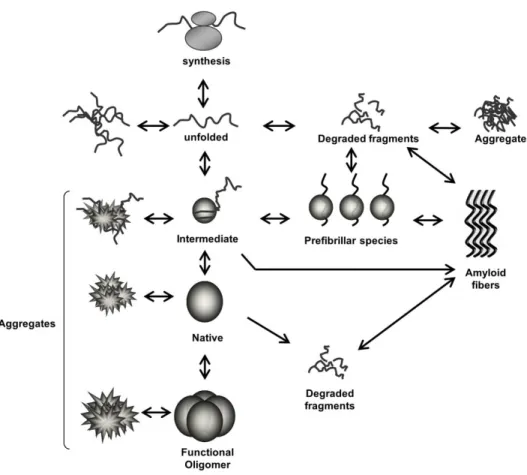
Proteins are constantly in risk of becoming unfolded, especially in stress conditions 1. Intermediate folding species typically expose hydrophobic residues which have higher tendency to lead to aggregation. Once certain proteins are misfolded they can act as a seed, leading to the misfolding of others and ultimately resulting in the formation of aggregates 2 (Fig. 1.1).
Figure 1.1. Schematic representation of protein folding/misfolding processes. Following synthesis, each protein needs to adopt a specific three dimensional structure (native conformation) in order to become biologically functional. Proteins often acquire intermediate states before being completely folded. The different species may be prone to misfolding, leading to their accumulation and, in certain cases, to the formation of aggregates, which can be amorphous or amyloid-like. Some of the intermediates and/or aggregates are likely to be cytotoxic, and therapeutic intervention might be possible at different levels along these pathways to prevent their accumulation.
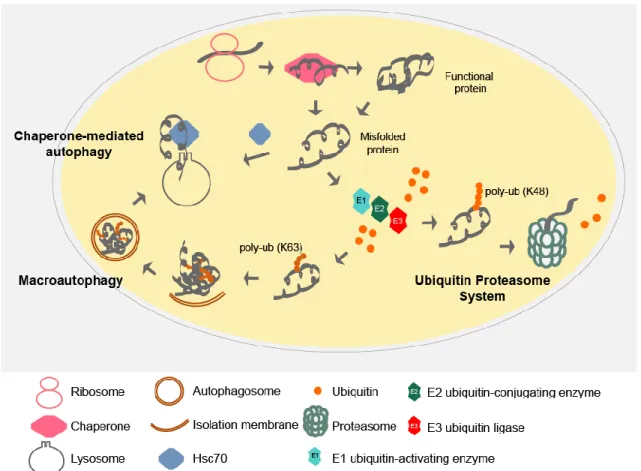
To deal with protein misfolding cells have developed a protein quality control (QC) system, which includes molecular chaperones, the ubiquitin proteasome system (UPS) and autophagy (Fig. 1.2). While chaperones help proteins to fold or refold, the UPS and autophagy are responsible for protein turnover. The degradation of proteins is critical not only to remove the damaged proteins and recycle amino acids but also to clear proteins that play essential roles in cellular processes such as signal transduction and cell division. Protein degradation by the UPS, the key mechanism to degrade short-lived proteins, consists in covalently linking the target proteins to multiple ubiquitin molecules (polyubiquitination) as a signal for degradation, which will subsequently be recognized and degraded by the 26S proteasome (Fig 1.2)3. Autophagy is a catabolic process essential to recycle cellular components that allows survival under nutrient-limited conditions. It is also required to eliminate dysfunctional organelles or other materials. Autophagy can be divided in three types: macroautophagy (hereafter autophagy), in which the cargo to be eliminated is engulfed by a double membrane vesicle, the autophagosome, and is transported to the vacuole, where it is degraded (Fig. 1.2); chaperone-mediated autophagy (CMA) which is characterized by selective translocation of the cargo across the lysosome through a specific receptor mediated by a chaperone (Fig. 1.2); and microautophagy, in which the material to be degraded directly enters the lysosome 4.
When the capacity of the protein QC system is exceeded or the system just fails, proteins may become toxic to the cell due to its accumulation and possible aggregation. Protein aggregation may be caused by additional factors, such as: mutations that increase the protein tendency to misfold and aggregate, errors in translation, environmental stresses such as oxidative stress and ageing (which is correlated with an increase in oxidation of proteins) 1. The accumulation of abnormally folded proteins results, on one hand, in the loss of the normal biological role of the protein and, on the other hand, in a putative gain of cytotoxic function (Fig. 1.1). The ‘amyloid hypothesis’ (developed originally for AD) states that the aggregation of proteins into an ordered fibrillar structure is causally related to aberrant protein interactions that culminate in neuronal dysfunction and ultimately neurodegeneration 5–7. However, it is still unclear which species correlate with toxicity, whether the aggregates or the intermediate species. It has been observed that cells can direct the aggregated proteins to specific cellular sites, which can be considered as the second line of protection against the potential toxic species upon failure of the QC system 1. Nevertheless, the presence of large aggregates can still contribute for cellular toxicity.
Since neurons are post-mitotic cells and highly metabolic active they are more sensitive to the accumulation of misfolded proteins and this may explain the high number of brain diseases associated with protein misfolding.
Figure 1.2. Protein quality control systems.
Following protein synthesis, chaperones help proteins to fold. If folding fails or the protein needs to be turned over, a chain of ubiquitin molecules is attached to the target protein by the action of a series of enzymes: ubiquitin activating (E1), conjugating (E2) and ligating (E3) enzymes. The ubiquitin molecules are usually linked between the terminal residue (G76) of one ubiquitin and a lysine of another ubiquitin molecule, usually lysine 48. The polyubiquitinated proteins are then recognized by the 26S proteasome and degraded. Ubiquitin molecules can instead be linked through the lysine 63, which promotes protein aggregation. Aggregates may be, engulfed by a double membrane – autophagosome – which will then fuse with the lysosome. Upon fusion, the cargo is degraded by the acidic lysosomal hydrolases (macroautophagy). Proteins can also be degraded by entering into the lysosome with the help of the chaperone Hsc70 through the receptor LAMP 2A (chaperone-mediated autophagy). poly-Ub – polyubiquitin chain.
Huntington’s disease
HD is a fatal adult onset neurodegenerative disorder that has a worldwide prevalence of ~1 in 10,000 people. The onset of the symptoms is typically around 45-50 years old but there are also juvenile forms of the disease. Death usually occurs 10-15 years after the onset of the disease. This disorder is inherited in an autosomal dominant manner and is characterized by abnormal motor movements, personality changes and cognitive decline 8. The neuropathology of HD is characterized by the loss of specific neuronal types in several brain regions, and can be related to specific symptoms and progression of the disease. However, neuronal loss can be detected before the clinical symptoms start to appear 9. The most striking pathology in HD is the cell loss observed in the striatal region of the basal ganglia, of which medium spiny neurons are the most affected.
HD is caused by a mutation in the IT15 gene (localized in exon 1) that consists in the expansion of CAG repeats beyond a critical length. This causes an expansion of the polyglutamine (polyQ) tract in the huntingtin (Htt) protein and consequently results in its misfolding. The CAG repeat in the IT15 is polymorphic in the general population and expanded in individuals affected by HD, which have repeat lengths greater than 35 10. HD genes carrying expansions in the range of 36-39 CAG repeats show an increasing risk of developing HD, while expansions of 40 CAG repeats and higher are fully penetrant 11. The length of the CAG repeat has also been found to be inversely correlated to the age of onset 12, with expansions of 70 CAG repeats or longer inevitably leading to juvenile onset HD 10,13. Though increased size of the triplet repeat expansion correlates to an earlier age of onset, there is great variability in the age of onset of HD, even when controlling for repeat length. Indeed, work by the U.S.-Venezuela Collaborative Research Project with HD kindreds containing over 18,000 individuals has found that approximately 40% of variation in age of onset in HD patients is due to several genetic modifiers 14. Although HD has a well-defined genetic cause, the molecular pathways that lead to neurodegeneration remain poorly understood and so far there is no therapy that prevents, delays or reverses this disease. However, the fact that several genetic modifiers can affect the HD age of onset suggest that many therapeutic targets may be available for treating progression of this devastating disorder.
Htt aggregation and inclusion bodies
The discovery of the polyQ expansion allowed the generation of several transgenic mice models expressing the first exon of the human Htt 15. The most studied HD model is the R6/2 model that exhibits some characteristics of HD, such as progressive neurodegeneration, motor abnormalities and reduced lifespan 15. The study of mouse models together with the analysis of HD brains led to discover the pathological hallmark of HD which consists in intraneuronal inclusion bodies (IB) containing aggregated Htt. IB are found in the nucleus, cytoplasm and neuronal processes 16–19. Although these aggregates have been causally related to HD pathology, there is great debate as to their role in this disorder. Mutant Htt could lead to neuronal loss through sequestration of crucial proteins into IB, such as transcription factors, which could explain why transcription is dysregulated in HD 20–23. On the other hand, sequestration into IB could also be biologically irrelevant 24,25, and soluble oligomeric species could be the actual cause of transcriptional dysregulation and mutant Htt neurotoxicity. Compelling evidence indicates that IB are not correlated with cell death. The cerebral cortex of HD patients has a high number of IB, whereas their striatum, which is where neuronal death is higher, shows only a few 16. Formation of aggregates could be a protective mechanism to scavenge more neurotoxic Htt oligomeric species 26–28.
The most important intrinsic factor affecting the aggregation of mutant Htt is the length of the polyQ tract, but there are other factors, such as the amino acids flanking the polyQ stretch 29–31. For instance, deleting the flanking proline rich region changes from non-toxic Htt to toxic one and it leads to changes in the aggregates shape and number 29,30. The proteolytic cleavage of Htt is also thought to promote aggregation 17,32 and influence the localization of IB to the cytosol or nucleus 33. Interestingly, transgenic mice expressing a caspase 6-resistant version of mutant Htt do not exhibit striatal degeneration 34, suggesting that proteolysis of Htt at the caspase-6 cleavage site is important for neurodegeneration in HD. Post translational modifications of Htt, such as ubiquitination, phosphorylation and sumoylation can influence Htt degradation and subsequently its aggregation 35.
Pathogenic mechanisms in HD
Since the cloning of the HD gene in 1993 10, a great deal has been learned about the cellular dysfunction caused by mutant Htt expression, and several mechanisms have been uncovered that likely contribute to the selective
neurodegeneration observed in the disease. The Htt protein is a large cytoplasmic protein that is ubiquitously expressed 36 and is required for embryogenesis and development 37–39. The precise function of this protein is still not clear, but it has been shown to be involved in several pathways and interact with multiple proteins that are involved in diverse mechanisms including intracellular transport, clathrin-mediated endocytosis, transcriptional regulation, RNA trafficking and cell survival 39–46. Therefore, the loss of Htt interactions with binding partners or anomalous interactions may contribute to Htt neurotoxicity.
Neurodegeneration in HD occurs not only by dysregulation of intrinsic mechanisms of the vulnerable cells (cell-autonomous) (Fig. 1.3), but also by contribution of other cell types (non-cell-autonomous) (Fig. 1.4). The cell-autonomous mechanisms affected in HD include transcriptional dysregulation, mitochondrial dysfunction, defective vesicle trafficking in axons and impairment of ubiquitination and proteasomal function, whereas the non-cell-autonomous mechanims include excitotoxicity, perturbations in the kynurenine pathway (Chapter 2), and inflammation.
Transcriptional dysregulation has long been implicated in the pathogenesis of HD. Early studies on post mortem HD brains showed that several mRNAs of neurotransmitter receptors and signaling neuropeptides are decreased in striatal neurons 47–49. The idea that transcription was affected by mutant Htt was further supported by the fact that wild-type Htt was mainly a cytosolic protein, while cleaved mutant Htt was strongly localized to the nucleus causing enhanced toxicity 32,50,51. Mutant Htt can interact with transcription factors, such as SP1 and members of the core transcription machinery, and by this way affects transcription 52–55. BDNF was one of the transcripts that was found reduced in the brains of HD patients 56. BDNF (Brain-derived neurotrophic factor) is important for the survival of striatal neurons and was shown to have beneficial effects on mouse models of HD when overexpressed 57,58. BDNF expression is regulated by the repressor element-1 transcription factor/neuron-restrictive silencer factor (REST/NRSF), which recognizes and binds to the neuron restrictive silencer element (NRSE) present in the BDNF promoter 59,60. Wild-type Htt regulates BDNF transcription by sequestering REST/NRSF in the cytosol, preventing its translocation into the nucleus and binding to NRSE. In the presence of mutant Htt, the REST/NRSF is not sequestered anymore and enters into the nucleus leading to the repression of NRSE regulated genes, including BDNF 59,60, explaining why this gene is decreased in striatal neurons, and suggesting a mechanism for the unique sensitivity of these neurons in HD.
Compelling evidence from patients and several models of HD suggest that mitochondrial dysfunction contributes to the pathogenesis of HD. The weight loss that patients suffer at early stages of the disease is indicative of mitochondrial impairment 61. The postmortem HD brain tissues show dysfunctional complex II, III, IV of the electron transport chain, mtDNA mutations 62–65, a reduced number of mitochondria and different morphology from controls 66. Defects in calcium homeostasis in mitochondria from HD patients and HD transgenic mice has also been observed 67. One hypothesis that explains how mutant Htt causes mitochondrial dysfunction is through its direct interaction with mitochondria67–70. Mutant Htt can also bind to the promoter of the Peroxisome proliferator-activated receptor gamma coactivator 1-alpha (PGC-1α) and repress its expression 71. PGC-1α is a transcriptional coactivator of mitochondrial genes involved in respiration and mitochondrial biogenesis. The repression of PGC-1α can cause a defect in the energy homeostasis of the cell that may lead to cell death 71. Interestingly, a correlation between the number of CAG repeats with the production of mitochondrial ATP has also been observed72.
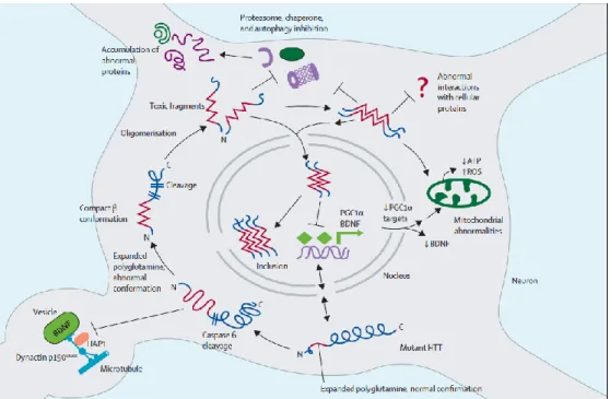
Figure 1.3. Cell autonomous mechanisms underlying HD.
Mutant huntingtin (HTT) can be cleaved by caspase-6 and interfere with essential neuronal processes, such as proteasomal function, autophagy and transcription. Moreover, it can lead to mitochondrial dysfunction and lead to an increase in oxidative stress.
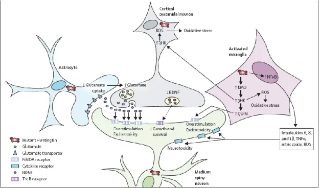
Excitotoxicity, one of the non-cell-autonomous mechanisms implicated in HD, is caused by overstimulation of glutamate receptors, especially the ionotropic N-methyl-D-aspartate (NMDA) receptors, which leads to an increase in calcium influx and ultimately neuronal death (Fig. 1.4) 74. Excitotoxicity is thought to be caused by reduced glutamate uptake by glutamate transporter 1 in astrocytes or increased glutamate release from cortical neurons 75,76. Overactivation of NMDA receptors may also result from mitochondrial dysfunction and the consequent deficient energy production. The entry of calcium ions by NMDA receptors is blocked by Mg2+, which is released by depolarization of the cell. Mitochondria are required to sustain the membrane potential. Upon its dysfunction, there is less energy to maintain the Na+/K+ pump working, leading to cell depolarization, loss of Mg2+ and consequently entry of calcium into the cell 77.
Figure 1.4. Non-cell-autonomous mechanisms underlying HD
Mutant huntingtin can lead to dysregulation of several mechanisms in different cell types, which will contribute for neuronal cell death. These mechanisms include excitotoxicity, perturbations in the kynurenine pathway and increase in neuroinflammation.
Parkinson’s disease
PD is the second most common progressive neurodegenerative disorder, after AD. It affects about 2% of people over 65 years old and 4–5% of people over 85 78. PD was originally characterized in the monograph “An Essay on the Shaking Palsy” by James Parkinson in 1817, in which he described the major symptoms of PD: muscle rigidity, bradykinesia (slowness of voluntary movement), resting tremor and postural instability. Non-motor symptoms are also characteristic of the disease, such as hyposmia (reduced ability to smell), depression and rapid eye movement sleep behavior disorder. At later stages the patients can eventually suffer from dementia 79.
The major pathological hallmarks of PD are the presence of intraneuronal concentric hyaline cytoplasmic inclusions called Lewy bodies (LB) and loss of dopaminergic neurons in the substantia nigra pars compacta (SNpc). LBs contain fibrillar forms of α-synuclein (α-syn), as well as proteasomal and lysosomal subunits and molecular chaperones 80,81. Curiously, LBs can also be found in patients with other neurodegenerative disorders, such as dementia with LBs and AD 82, and even in clinically normal people over 60 years old (10%), although in this case it is thought to be pre-symptomatic 83. Surprisingly, LBs are not present in the brains of some patients with autosomal recessive PD. There is a great debate regarding whether LBs are cytotoxic or a protective mechanism to preclude the accumulation of the pathogenic intermediates, which have been proposed to be α-syn oligomers 84
.
PD was first thought to affect essentially dopaminergic neurons, but nowadays is considered a multisystem disorder that affects several regions of the nervous system 79. In the majority of idiopathic cases, it is possible to predict the spreading of LBs and thus it has been divided in 6 stages 85. However, how exactly this pathology is spread is still unclear.
Despite the intense research on PD, there are still no effective therapies, neither a way to slow down the progression of the disease 86. Dopamine replacement therapy (with the drug levodopa) ameliorates the motor symptoms (especially at the beginning), but generally causes severe secondary effects 87. When the patients do not respond well to the drug treatment, they are candidates for an alternative procedure, deep brain stimulation, which consists in surgically inserting an electrode in the subthalamic nucleus or the internal global pallidus, both localized in basal ganglia. This electrode, by sending electric stimulus, can block the brain impulses that cause PD symptoms. However, it cannot slow the progression of the disease 87. It is therefore of extreme
importance to understand the molecular mechanisms that underlie PD so that effective therapies can be discovered in a near future.
ETIOLOGY OF PD
The etiology of PD is complex and unclear and in many cases the cause is completely unknown (idiopathic). The majority of PD cases arise sporadically 88. The major risk factor to develop this disease is clearly ageing, but there also some environmental factors associated to PD. For instance, subjects that continuously self-injected “synthetic heroin” with the contaminant MPTP (1-methyl-4-phenyl-1,2,3,6-tetrahydropyridine) developed typical symptoms of parkinsonism. Interestingly, although their brains showed nigrostriatal degeneration, LBs were not found 89. In addition, chronic exposure to agricultural chemicals, such us rotenone, manganese ethylenebis(dithiocarbamate) (maneb) and paraquat, is considered a potential risk factor of PD 90. Caffeine and nicotine have also been shown to be inversely correlated with the incidence of PD 91. Until 1977, PD was considered to be a non-genetic disease, but today it is known that approximately 10% of PD cases are familial 88, and that genetics may also affect susceptibility to disease onset 92.
GENETICS OF PD
The first gene to be associated with PD was SNCA, the gene that encodes α-syn. Since then 18 loci named PARK have been genetically linked to PD. However, only 6 genes have been clearly found to cause monogenic PD: SNCA (PARK1/4), LRRK2 (PARK8), Parkin (PARK2), PINK1 (PARK6), DJ-1 (PARK7), and ATP13A2 (PARK9) 93 (Table 1.1). Mutations in these genes only explain around 30% of familial cases and 3% of sporadic 93.
Mutations in SNCA and LRRK2 (Leucine-rich repeat kinase 2) are associated with autosomal dominant cases of PD. Although it is rare to find mutations in SNCA, five missense mutations (A53T, A30P, E46K, H50Q and G51D) have been identified in patients with familial PD or Dementia with LBs 94–98 as well as duplications and triplications of the genomic region of SNCA 99,100. In addition, two genome-wide association studies showed that several single nucleotide polymorphisms in SNCA are risk factors to develop sporadic PD 101,102. LRRK2 mutations are the most frequent cause of familial and sporadic PD 103. Patients with mutations in this gene present late-onset PD, clinically indistinguishable from sporadic cases 103.
Table 1.1. Loci associated with PD
Locus Gene Disorder Inheritance Status
PARK1 SNCA EOPD AD Confirmed
PARK2 Parkin EOPD AR Confirmed
PARK3 Unknown Classical PD AD Unconfirmed; may represent a risk factor PARK4 SNCA EOPD AD Identical to Park1 (error)
PARK5 UCHL1 Classical PD AD Unconfirmed
PARK6 PINK1 EOPD AR Confirmed
PARK7 DJ-1 EOPD AR Confirmed
PARK8 LRRK2 Classical PD AD Confirmed
PARK9 ATP13A2 Kufor-Rakeb syndrome;
atypical PD AR Confirmed
PARK10 Unknown Classical PD Risk factor Confirmed PARK11 Unknown LOPD AD Possibly is a risk factor PARK12 Unknown Classical PD Risk factor Possibly is a risk factor PARK13 HTRA2 Classical PD AD or Risk
factor Unconfirmed PARK14 PLA2G6 Early-onset
dystonia-parkinsonism AR Confirmed
PARK15 FBX07 Early-onset
parkinsonian-pyramidal syndrome AR Confirmed PARK16 Unknown Classical PD Risk factor Confirmed
PARK17 VPS35 Classical PD AD Confirmed
PARK18 EIF4G1 Classical PD AD Unconfirmed
Table adapted from Corti et al, Physiol Rev, 2011 86 and Klein and Westenberger, Cold Spring Harbor Persp in Med, 2012 93.
The remaining genes Parkin, PINK1, DJ-1 and ATP13A2 are associated with juvenile (≤ 20 years-old) and early-onset (≤ 45 years-old) autosomal recessive PD. Mutations in Parkin are the most frequent cause of autosomal recessive PD, followed by mutations in PINK1 104–106. An intriguing aspect of the autosomal recessive cases is that LBs are absent in the majority of cases with Parkin mutations and so far only one case with mutations in PINK1 was reported to show LBs 107. There are still no autopsy reports of patients with DJ-1 or ATP13A2 mutations. Additional genes have been linked to PD but need further confirmation: UCHL1 (PARK5), GYGYF2 (PARK11),
OMI/HTRA2 (PARK13), PLA2G6 (PARK14), and FBXO7 (PARK15) 93.
PATHOGENESIS OF PD
Although familial and sporadic forms of PD have certain differences, these likely share several pathogenic mechanisms. Therefore, intense research is focusing on investigating the proteins involved in familial PD, both in terms of their normal physiological role as well as their role in the disease. The discovery of PD genes and environmental factors such as MPTP have allowed the PD research community to
generate in vivo and in vitro models of the disease, which together with epidemiological studies and post-mortem analysis has already provided several insights into the molecular pathways and mechanisms that underlie PD. The fact that most genes linked to PD result in similar phenotypes, suggests that at least some of the proteins encoded by these may work in a single pathway (Fig. 1.5). Therefore, one of the goals of the PD research is to understand the relationship between these genes.
The key mechanisms that contribute for PD pathology are thought to be the loss of protein homeostasis and mitochondrial dysfunction (Fig. 1.5). These pathogenic factors do not act independently and it is not known exactly if their dysfunction is a cause or consequence of neurodegeneration.
α-synuclein and proteostasis imbalance
The fact that mutations in SNCA cause familial PD and that α-syn is the major component of LB strongly indicates that α-syn has a central role in the pathogenesis of PD. Therefore, much research attention is being devoted to this protein. α-syn is a small protein of 140 amino acids, enriched at presynaptic terminals 108. The role of α-syn is still unclear, but it is thought to play a role in synaptic vesicle recycling, neurotransmission, and synaptic plasticity 109.
α-syn is natively unfolded with very little secondary structure and once it interacts with phospholipid membranes, its N-terminal region acquires an amphipathic α-helical structure 110. The central region of α-syn contains a highly hydrophobic motif known as the non-amyloid-β component (NAC), which is highly aggregation prone 111. A recent study reported that α-syn exists as an α-helical tetramer, suggesting that the protein can be stabilized by interacting with the neighboring subunits 112. Nevertheless, it was further suggested that α-syn exists predominantly as an unfolded protein in the central nervous system 113. Aggregation of α-syn starts by the formation of assembly intermediates, followed by oligomers, soluble protofibrils and amyloid fibrils that are deposited in LBs. The propensity of α-syn to aggregate increases with several factors, such as: familial point mutations, excess of α-syn caused by higher expression (gene duplications, triplications or polymorphisms) or defective clearance, oxidative stress or post-translational modifications 114.
Figure 1.5. Pathogenic mechanisms that underlie Parkinson’s disease.
The study of the genes associated with the familial forms of Parkinson’s disease (PD) has been helping in elucidating the pathways that lead to neurodegeneration. Dysfunctional mitochondria, ubiquitin proteasome system or autophagy can promote the formation of intermediate α-synuclein species and aggregation, which in turn can impair the proteasome, mitochondria or autophagy. Dysfunctional mitochondria lead to higher generation of reactive oxygen species (ROS), which might cause further mitochondrial impairment and promote further aggregation.
Increasing evidence shows that the three protein QC systems – molecular chaperones, UPS and autophagy (especially macroautophagy and chaperone mediated autophagy) – are involved in α-syn homeostasis (Fig. 1.2) 115. However, there are conflicting results regarding the exact mechanism by which α-syn is degraded and it is still unknown how the cell decides the route of protein degradation. The different results obtained in different labs seem to be dependent on the model system used and also on which kind of α-syn species the system has to deal with 116. Nevertheless, several studies show that α-syn is degraded by both UPS and autophagy 116 and when one of these systems is inhibited causes α-syn accumulation and aggregation 116. Conversely, the increase in α-syn and the consequent generation of toxic species may affect the QC systems and disturb cell homeostasis.
Ebrahimi-Fakhari and colleagues have proposed a model for α-syn degradation that hypothesizes that in non-pathological conditions, in which the levels of α-syn are maintained homeostatic, α-syn is degraded by both UPS and CMA; if the levels of α-syn increase in a way that exceeds the capacity of the QC system, toxic species may appear, blocking UPS and CMA (at initial stages of the disease). At this point, a vicious cycle begins, in which the dysfunction of UPS and CMA further increases the levels of toxic α-syn species (Fig 1.5). In order to cope with this dysfunction, cells upregulate macroautophagy, however in later stages of the disease, macroautophagy may also become impaired 117.
A strong indication that UPS and autophagy are implicated in PD pathogenesis and are responsible for α-syn degradation is the fact that among the genes linked to PD, three are strongly associated with these mechanisms: parkin, ATP13A2 and UCHL1. Parkin is an E3 ubiquitin ligase that is involved in UPS and more recently it was shown that it plays a role in mitochondria (discussed below). The role of an E3 ubiquitin ligase is to covalently link ubiquitin to a specific target protein. Parkin is capable of mediating either polyubiquitination through the ubiquitin lysine 48 (targets to the proteasome) or lysine 63 (targets to non degradative processes) or monoubiquitination (targets to non degradative processes) 118.
Mitochondria and PD
Mitochondria are essential organelles for the function and survival of neurons. These are the major energy suppliers and play a role in other critical mechanisms, such as calcium buffering and apoptosis. Importantly, mitochondria are the major source of reactive oxygen species (ROS). These are very mobile and dynamic organelles, constantly undergoing fission and fusion. These two mechanisms are required to maintain the mitochondrial network functional. While fusion is responsible for redistributing the mitochondrial components (which is a strategy of functional mitochondria to complement the damaged ones), fission occurs to create new mitochondria or remove the damaged components. When the damage is irreparable, mitochondria are removed by mitophagy.
Mounting evidence suggests that mitochondrial dysfunction is implicated in the pathogenesis of PD. However, whether it is a cause or consequence of neurodegeneration is still unclear. The first finding that led researchers to investigate the role of mitochondria in PD was that MPTP led to progressive and irreversible parkinsonism in humans 89,119. Oxidation of MPTP in glial cells leads to formation of
MPP+ – an inhibitor of complex I – which is then released and selectively uptaken by dopaminergic neurons via the dopamine transporter. Within dopaminergic neurons this will inhibit complex I and thus impair the electron flux. As a consequence, ATP production decreases and both ROS and reactive nitrogen species (RNS) increase 119. This finding raised the question whether complex I was impaired in idiopathic PD patients and in fact post-mortem analysis of the brains of PD patients showed reduced complex I activity in the SNpc 120. The administration of MPTP and other complex I inhibitors, such as rotenone or paraquat, in rodents can also induce dopaminergic degeneration, further supporting that mitochondrial dysfunction has a central role in PD pathogenesis 121. Additionally, many of the proteins encoded by the genes associated to PD (α-syn, PINK1, parkin, DJ-1, LRRK2) play a role directly or indirectly in mitochondrial functions and at least PINK1 and parkin act in a common pathway.
PINK1 (PTEN-induced kinase 1) is a nuclear encoded kinase that is localized in the outer mitochondrial membrane, with the kinase domain turned to the cytosol 122,123. Mutations in PINK1 are thought to be loss of function; the majority of mutations are localized in the kinase domain and result in impairment of the kinase activity. So far, only three PINK1 substrates have been identified: the mitochondrial chaperone TNF-receptor-associated protein 1 (TRAP1), the mitochondrial serine protease Omi/HtrA2 and parkin 124–126. Both TRAP1 and Omi/HtrA2 are involved in protecting cells against cell death in stress conditions 126,127. Interestingly, the knockout mouse for Omi/HtrA2 develops a parkinsonian phenotype and dies after one month 127. PINK1 and parkin were elegantly shown to function in the same pathway using Drosophila as a model system 128,129. Drosophila deleted for PINK1 or Parkin exhibit similar phenotypes due to mitochondrial defects, including male sterility, apoptotic muscle degeneration, higher sensitivity to several stresses and locomotive impairment 128,129. The overexpression of parkin complements the phenotypes caused by deletion of PINK1, but not vice-versa, indicating that PINK1 is an upstream regulator of parkin. Compelling evidence shows that PINK1-parkin pathway is involved in maintaining the integrity of the mitochondrial network by modulating mitochondrial dynamics and promoting degradation of damaged mitochondria through mitophagy 130,131. More recently it was shown that it can also induce the selective degradation of respiratory subunits 132. It is not clear whether dysfunctional quality control of mitochondria is involved in the pathogenesis of sporadic PD, however parkin was found impaired even when it was not mutated 133,134, supporting this idea.
How DJ-1 is related to mitochondria is also unclear. DJ-1 is a cytosolic, nuclear and mitochondrial protein 135,136. Localization of DJ-1 to mitochondria increases upon oxidative stress and is neuroprotective 137,138. The precise cellular role of DJ-1 is unknown, but compelling evidence indicates that it is involved in cellular responses of oxidative stress (further discussed in Chapter 3). The link between mitochondria protection and DJ-1 may be in fact the protection against oxidative stress 139. Nevertheless, multiple studies have shown that loss of DJ-1 leads to several mitochondrial defects, including mitochondria fragmentation 140–144. Interestingly, fruit flies deleted for DJ-1 (both orthologs DJ-1a and DJ-1b) exhibited similar phenotypes to flies deleted for pink1 or parkin 145. Overexpression of DJ-1 was able to complement the
loss of PINK1, but not parkin 145. These results suggest that DJ-1 acts in a parallel pathway or downstream PINK1, but likely is independent of PINK1-parkin pathway.
Finally, overexpression of wild-type or familial mutant forms of α-syn in transgenic mouse and cell culture models results in mitochondrial dysfunction 146–148.
Taking all this into account, it is clear that dissecting further the role of these proteins in the mitochondrial function will contribute for a better understanding of PD pathogenesis.
Oxidative stress
Oxidative stress occurs when the generation of ROS and RNS exceeds the cell antioxidant defense mechanisms. These reactive species have the capacity to chemically modify cell constituents such as proteins, lipids and nucleic acids, which may result in their irreversible damage and ultimately trigger neuronal injury 149. High levels of ROS can even trigger apoptosis or autophagy leading to cell death 150. Apart from its toxic effect in the cell, ROS have also an important signaling role under normal conditions 151. Therefore, it is essential to control the levels of ROS and RNS in the cell. Mitochondria are the major source of ROS. Electrons leak from the electron transport chain and partially reduce oxygen resulting in the formation of superoxide, which subsequently can be converted to hydrogen peroxide. Hydrogen peroxide by reacting with iron (Fenton reaction) results in the formation of the hydroxyl radical 150. Superoxide can also be transformed into peroxynitrite by reacting with nitric oxide 152,153. The accumulation of ROS in mitochondria leads a vicious cycle in which dysfunctional mitochondria result in higher production of ROS that in turn enhance mitochondrial impairment.
The brain is thought to be more susceptible to oxidative stress since is highly metabolically active and has a low capacity to regenerate the post mitotic neurons compared to other organs. Oxidative stress has been implicated as one of the mechanisms that contribute to the pathogenesis of several neurodegenerative diseases including PD, as well as one of the causes of ageing, proposed as the “free radical theory of ageing” 149,154.
Several observations indicate that dopaminergic neurons in SNpc are particularly exposed to oxidative stress, making them more vulnerable for cell death. As mentioned already, post mortem PD brains show decreased complex I activity in SNpc when compared with healthy controls, 120 as well as increased lipid peroxidation, protein carbonylation, DNA and RNA oxidation and decreased antioxidant defenses, such as glutathione 155–158. Dopaminergic neurons in SNpc contain higher concentrations of iron, which can be toxic if iron reacts with hydrogen peroxide and the hydroxyl radical is generated 159–161. In addition, these neurons contain dopamine, which oxidation products are potentially toxic 162. An additional hypothesis that explains why nigral dopaminergic neurons have higher levels of oxidative stress is the presence of L-type calcium channels. These channels help to maintain the pacemaking activity which is characteristic of SNpc dopaminergic neurons and consists in maintaining neuronal activity autonomously, i.e. without any synaptic input 163. L-type calcium channels are open frequently, since they open at relatively hyperpolarized state. This comes with an energetic cost, since the calcium concentration has to be under tight control and this is maintained by ATP dependent pumps. Since this is an energy dependent mechanism, mitochondria activity increases and subsequently the basal ROS also increase, turning the dopaminergic neurons more vulnerable for stressors 163.
In addition to the intracellular sources of ROS, these species can also come from activated microglial cells. Microglia are generally on a resting state, but become activated upon several insults, such as factors released by damaged neurons. Microglial cells are found activated in the brains of PD patients 164,165. Activation of microglia may result in a vicious cycle, since these cells release neurotoxic factors (such as ROS) which will potentiate neuronal cell death 164. Several studies have shown that neuroinflammation also contributes to the pathogenesis of PD 164. Therefore, dissecting the mechanisms that control neuroinflammation may help in finding novel strategies to control PD. Excitingly, we have recently found that SIRT2 is a promising target to prevent sustained inflammation and neurotoxicity. We showed that reduction of SIRT2 expression in mouse models and microglial cell lines leads to enhanced
neuroinflammation and neurotoxicity induced by higher generation of ROS and RNS. Conversely, SIRT2 overexpression in microglial cell lines inhibits microglial activation166. The gene implicated in PD that predominantly supports the role of oxidative stress in the pathogenesis of PD is DJ-1 (see Chapter 3).
Baker’s yeast: a versatile “toolbox” for molecular biology
studies
Since ancient times the budding yeast Saccharomyces cerevisiae has been used in baking and brewing 167. This unicellular eukaryotic organism is extremely useful for molecular biologists and our knowledge about fundamental cellular mechanisms has greatly benefited from the use of yeast as a model 168. It has been extensively used in the study of numerous complex and devastating disorders, such as HD and PD and has provided insight into the molecular events underlying these disorders 169–171.
Multiple characteristics make this simple eukaryote a model system of choice. First of all, it is well defined genetically and genomically (S. cerevisiae was the first eukaryote organism to be fully sequenced). Of the 6000 genes predicted to be encoded by its 12,000-kilobase genome, ~80% are functionally characterized 172. Since the electronic release of its genomic sequence several highly annotated online databases have become available, which provide multitudes of detailed information on yeast genes and proteins, such as protein-protein interactions, genetic interactions, protein function, and predicted orthologs in other organisms. The remarkably high degree of conservation between yeast and higher eukaryotes, with respect to basic processes such as cell cycle, intracellular transport, and cellular QC systems is another feature that makes yeast so attractive. Therefore, yeast studies may shed light into the conserved mechanisms involved in human diseases. In fact, yeast has been extensively used as an informative organism for human gene function, since approximately half of the genes involved in human heritable diseases are predicted to have yeast homologues 173.
Genetic and biochemical manipulations in yeast are extremely simple, rather quick, and inexpensive when compared to manipulations in other eukaryotes. Yeast is easy to grow on appropriate media, with a doubling time of approximately 90 minutes on rich medium, and survive indefinitely in frozen glycerol stocks. Strains can be maintained as stable haploids or diploids, allowing the study of lethal mutations in heterozygous diploids and recessive mutations in haploids. It is easy to mate the haploid strains and to sporulate diploid strains, making classical genetics extremely
facile. Other versatile characteristics of this organism are the ease of transformation and the ability to integrate genes by homologous recombination 167,174,175. The experimental tractability of yeast has allowed the development of several sophisticated functional tools, such as yeast two-hybrid variants, affinity purification and mass-spectrometry identification providing substantial amounts of information about protein-protein interactions 176,177. In addition, several yeast strain collections have been created, permitting rapid genomic systematic screenings and the identification of genetic interactions. These libraries include the yeast open reading frame (ORF) collection (YOC) and the yeast gene knock-out collection (YKO), which is available in haploid, homozygous diploid and heterozygous diploid strains 175. So far, such screens have allowed the identification of several possible genetic modifiers (suppressors or enhancers) of amyloid diseases, the identification of new therapeutic targets, and the dissection of the pathogenic mechanisms and pathways involved in these diseases.
Although the simplicity of yeast is an advantage for experimental manipulation and interpretation of data, it does present a caveat of this model. For obvious reasons, several mechanisms and biological pathways present in higher eukaryotes are absent in yeast and, on the other hand, some pathways, such as cell wall biosynthesis, have no counterpart in mammals. Therefore, all findings in yeast related to human disease ultimately must be validated in more physiologically relevant systems prior to their trial in humans.
Modeling neurodegenerative diseases in yeast
Despite the obvious absence of a nervous system in yeast, basic mechanisms and pathways underlying neurodegenerative diseases, such as mitochondrial dysfunction, transcriptional dysregulation, trafficking defects and proteasomal dysfunction, are extremely well conserved between humans and yeast, enabling detailed studies of the molecular events involved in those conditions. In fact, several remarkable insights into the understanding of brain diseases have been recently achieved 171,178–180. In order to develop disease models, it is essential that some relevant aspects of the disease phenotype are recapitulated. If the gene implicated in the disease has a yeast homolog, it is possible to study its function directly. If, on the other hand, the gene underlying the disease is absent in yeast but causes disease by a toxic gain-of-function mechanism in humans, it can still be modeled via the heterologous expression of the human gene in yeast cells.
Yeast models of HD
Several yeast models of HD have been developed to study the folding and behavior of mutant Htt as well as to dissect the underlying conserved mechanisms of mutant Htt-dependent toxicity 81,181–183. Expression in yeast of an amino-terminal fragment of Htt fused to GFP results in polyQ length-dependent formation of cytoplasmic inclusions that can be visualized in cells via fluorescence microscopy 81,182. A simple filter-retention assay has also been used to isolate detergent-insoluble aggregates formed by expression in yeast of a mutant Htt fragment 181. One of these models exhibits polyQ length-dependent toxicity, such that expression of a Htt fragment with polyQ length in the pathogenic range (Htt103Q) produces cellular toxicity within yeast, while expression of the same construct containing a non-expanded polyQ tract (Htt25Q) shows no negative effect upon growth 182. Recent work suggests that several factors are contributing to toxicity in this yeast model of HD, including perturbations in the kynurenine pathway (discussed in Chapter 2) 171, defects in endocytosis 184,185, apoptotic-like events 186, increased levels of ROS 171,187, and mitochondrial dysfunction 186,187. Interestingly, the polyQ-length dependent toxicity observed in the Meriin et al. model requires the presence of the yeast prion Rnq1 in its prion conformation 182. Several other glutamine-rich proteins play an additional role in modulating toxicity 29,171. In addition, it was observed that the flanking amino acid sequences are required for modulating toxic conformations of these Htt fragments in yeast 29,30. This conversion of benign species to toxic species (and vice-versa) can operate in cis or in trans 29.
The availability of yeast models of HD has facilitated the identification of candidate therapeutic targets for HD in the form of gene deletions that either enhance or suppress toxicity of a mutant Htt fragment 170,171,188. Additionally, this model has allowed screening and individual testing of small molecules to identify novel candidate therapeutic compounds which have been validated in higher eukaryotes 189–191. The YKO was screened for loss-of-function suppressors of the toxic Htt103Q construct 171,182. Of the genes identified in this screen, 25% encode proteins that cluster into the functionally related categories of vesicular transport, vacuolar protein sorting, and vacuolar import; 25% encode proteins that are involved directly in transcription or in establishment/maintenance of chromatin architecture; and ~21% encode known yeast prions or proteins containing Q/N-rich regions that may mediate prion-like aggregation. Over half of the genes isolated are annotated as having one or more human orthologs. This suggests that there are conserved biological pathways associated to mutant Htt that ultimately may be relevant to HD. One of the identified suppressors was BNA4,