Contribution for the knowledge of
toxoplasmosis in Portugal
Dissertação para obtenção do Grau de Doutor em Biologia
Orientador: Doutor João Paulo dos Santos Gomes,
Investigador Auxiliar com Habilitação, Instituto
Nacional de Saúde Doutor Ricardo Jorge
Co-orientadora: Doutora Isabel Maria Godinho de Sá Nogueira,
Professora Associada com Agregação,
Faculdade de Ciências e Tecnologia,
Universidade Nova de Lisboa
Júri:
Presidente: Prof. Doutora Ana Isabel Nobre Martins Aguiar
de Oliveira
Ricardo
Arguentes: Prof. Doutor Luis Madeira de Carvalho
Prof. Doutor Jaime Manuel Simões Nina
Vogai: Doutor João Paulo dos Santos Gomes
Doutor Jacinto José Carneiro Gomes
Contribution for the knowledge of toxoplasmosis in Portugal
Copyright © Maria João Gargate
A Faculdade de Ciências e Tecnologia e a Universidade Nova de Lisboa têm o direito, perpétuo e sem limites geográficos, de arquivar e publicar esta dissertação através de exemplares impressos reproduzidos em papel ou de forma digital, ou por qualquer outro meio conhecido ou que venha a ser inventado, e de a divulgar através de repositórios científicos e de admitir a sua cópia e distribuição com objetivos educacionais ou de investigação, não comerciais, desde que seja dado crédito ao autor e editor.
As secções desta dissertação já publicadas por editores para os quais foram transferidos direitos de cópia pelos autores, encontram-se devidamente identificadas ao longo da dissertação e são reproduzidas sob permissão dos editores originais e sujeitas às restrições de cópia impostas pelos mesmos.
É o tempo da travessia:
E, se não ousarmos fazê – la,
Teremos ficado, para sempre, à margem de nós mesmos.
Fernando Pessoa
Ao meu orientador Doutor João Paulo Gomes, Responsável da Unidade Investigação do Departamento de Doenças Infecciosas do Instituto Nacional de Saúde Doutor Ricardo Jorge, pelo incentivo e motivação e pelo rigor científico, sapiência e experiência com que me orientou e enriqueceu este trabalho. Muito obrigada pela tua amizade. Admiro te muito pelo investigador que és!
À minha coorientadora Professora Isabel Sá Nogueira, Professora Associada com Agregação e Coordenadora do Programa Doutoral em Biologia da Faculdade de Ciências e Tecnologia da Universidade Nova de Lisboa, por ter reconhecido valor cientifico na minha tese e a ter aceite no Programa Doutoral que coordena e pelo seu profissionalismo e cordialidade.
Ao Doutor Jorge Machado, Coordenador do Departamento de Doenças Infecciosas do Instituto Nacional de Saúde Doutor Ricardo Jorge, pela iniciativa e reconhecimento da minha capacidade para levar a cabo esta tarefa, pelo encorajamento, pela liderança e pela amizade.
Aos membros da Comissão de Acompanhamento de Tese, Doutora Gabriela Rodrigues, Professora Auxiliar da Faculdade de Ciências da Universidade de Lisboa e Doutor Baltazar Nunes, Investigador do Departamento de Epidemiologia do Instituto Nacional de Saúde Doutor Ricardo Jorge, pela revisão do manuscrito e sugestões concedidas e pelo incentivo e afabilidade.
Ao Doutor Vitor Borges Investigador do Núcleo de Bioinformática do Departamento de Doenças Infeciosas do Instituto Nacional de Saúde Doutor Ricardo Jorge,uma alma antiga numa cabeça de vanguarda, pela sabedoria, amabilidade e maneira única de ser.
À Doutora Helena Ângelo pela mão de quem vim parar a esta ciência e de quem sou sucessora, espero ter estado e continuar a estar à sua altura.
Às minhas amigas e colegas do Laboratório Nacional de Referencia de Infeções Parasitárias e Fúngicas do Departamento de Doenças Infecciosas do Instituto Nacional de Saúde Doutor Ricardo Jorge, que trabalharam, me acompanharam, encorajaram e animaram em todo o período da tese bem como em todos os dias dos últimos 16 anos; Idalina Ferreira, com quem dei os primeiros passos no mundo da parasitologia, com quem fiz as primeiras incursões no biotério com os nossos ratinhos e de quem ouvi e oiço tantos conselhos sábios, obrigada de todo o coração por tudo isto e pela sua amizade, é insubstituível; Anabela Vilares uma das minhas grandes lutas, pela forma entusiasta, sabedoria e rigor com que trabalhas, pelo ânimo e paixão pelos parasitas, pela
disponibilidade sempre pronta, Cristina Verissimo, Helena Simões e Raquel Sabino pelas palavras de apoio e motivação que sempre me disseram e por fim mas não no fim, Assunção António pelos Bons dias mais afáveis do INSA e pelo optimismo que sempre me transmite.
Aos meus amigos Raquel Guiomar e Nuno Verdasca, companheiros, confidentes e conselheiros há quase 25 anos, muito obrigada pela ajuda sempre voluntária, pelo apoio incondicional, pela vossa solidariedade e amizade sincera. Raquel a tua opinião é muito importante para mim, Nuno não podia viver sem as tuas formatações e ajustes informáticos.
Aos meus familiares e amigos que me acompanham sempre.
À memória dos meus avós queridos.
Aos meus pais que gostam de mim mais do que eu própria e me mimam como se eu ainda tivesse cinco anos, adoro viver com a graça, o humor, as histórias, a cultura e o mimo de um; a sensatez, a retaguarda, a sabedoria, os auspícios e o mimo do outro. São o meu pilar.
Aos meus três filhos únicos Joana Maria, António Maria e Francisco Maria, gigantes amores da minha vida, a vossa existência, as vossas conversas, lógicas, ideias, piadas, gargalhadas e até o vosso mau feitio, atestam-me a alma e inspiram me todos os dias.
Ao cúmplice de uma vida, o meu marido, Miguel, que me conhece como só a minha mãe, que testemunha, acompanha e engrandece a minha vida desde há muitos anos.
toxoplasmose uma ameaça permanente, uma vez que os seres humanos permanecem infectados a vida toda. Esta tese de doutoramento teve como objetivo enriquecer o conhecimento sobre a toxoplasmose em Portugal e implementar abordagens optimizadas para a propagação de T. gondii no laboratório de referência.
Começámos por avaliar a seroprevalência da população portuguesa comparando três estudos transversais ao longo de três décadas (1979/80, 2001-2002, 2013), com especial foco nas mulheres em idade fértil. Observámos uma tendência decrescente da seroprevalência ao longo do tempo (de 47 % em 1979/80 para 22 % em 2013), aumentando esta com a idade. O cenário observado para as mulheres em idade fértil indica que mais de 80 % são suscetíveis à infecção primária encontrando-se assim em risco de contrair toxoplasmose.
Focámo-nos também no estudo do parasita e caracterizámos geneticamente 48 estirpes isoladas a partir de amostras biológicas de pacientes diagnosticados no Instituto Nacional de Saúde (INSA), para os quais efetuámos uma avaliação retrospectiva que estimou 1,6% de novos casos de toxoplasmose congénita nos últimos 10 anos. Este estudo revelou variações genéticas nas estirpes de T. gondii causadoras de infecção, mais especificamente, a existência de uma proporção considerável (21 %) de estirpes recombinantes, que se acredita estarem associadas a fenótipos específicos.
Finalmente, avaliámos diversas abordagens laboratoriais com o objectivo de reduzir a utilização do número de ratinhos sacrificados na actividade laboratorial de referência. Concluímos que a propagação de uma forma alternada do parasita numa uma linhagem celular e em ratinhos constitui um procedimento laboratorial promissor, pois, para além de reduzir o número de animais sacrificados em mais de 80 %, permite também que T. gondii não perca a sua virulência, mantendo potencialmente o seu genoma inalterado.
Globalmente, esta tese de doutoramento não só contribuiu para o conhecimento da toxoplasmose em Portugal, ao nível do individuo, elucidando a tendência cronológica do estado imunitário da população portuguesa, bem como ao nível do parasita, identificando o perfil genético das estirpes de T. gondii circulantes causadoras de infecção humana e a sua virulência no ratinho. Finalmente, esta dissertação de doutoramento estabeleceu procedimentos que modificaram o modus operandi do laboratório de referência que visam uma redução significativa do número de ratinhos sacrificados.
Palavras-chave Toxoplasma gondii; Genotipagem; Estirpes recombinantes; Toxoplasmose congénita; Seroprevalencia; Vigilância laboratorial.
constitutes a life-long threat since humans remain infected throughout life. This PhD dissertation aimed to enrich the knowledge on toxoplasmosis in Portugal and to implement updated approaches regarding T. gondii propagation in the reference laboratory.
We started by evaluating the seroprevalence in Portugal by comparing three cross-sectional studies spanning three decades (1979/80, 2001-2002, 2013), with focus on childbearing women. Seroprevalence showed a decreasing trend over time (from 47 % in 1979/80 to 22 % in 2013) and increased with age. The scenario observed for childbearing women indicates that more than 80 % of these are susceptible to primary infection and thus to congenital toxoplasmosis.
We also focused on the parasite and genetically characterized 48 strains isolated from biological samples from patients attending to the NIH, for which a retrospective evaluation estimated 1.6 % of new cases of congenital toxoplasmosis in the last 10 years. This study revealed genetic variations in T. gondii and more specifically the existence of a considerable proportion (21 %) of recombinant strains, which are believed to be associated with specific phenotypes.
Finally, we evaluated laboratory approaches towards the reduction of sacrificed mice in toxoplasmosis reference laboratories. We observed that the alternate passaging of the parasite in a cell line and in mice constitutes a promising laboratory procedure as, besides the reduction of sacrificed mice in more than 80 %, it enabled T. gondii to retain the virulence potential while keeping a putative stable genome.
Globally, this PhD dissertation not only increased the knowledge on toxoplasmosis in Portugal by elucidating the chronological trend of the immune status of the population and the general genetic profile of the T. gondii strains causing human infection, but it also modified the modus operandi of the reference laboratory towards the significant reduction of scarified mice.
Keywords Toxoplasma gondii; Genotyping; Recombinant strains; congenital toxoplasmosis;
Agradecimentos VII
Resumo IX
Abstract XI
Table of contents XIII
Figure index XV
Table index XVI
List of abbreviations XVII
Notes of the author: thesis organization, format and outline XIX
1. Chapter I: General Introduction 1
1.1. Toxoplasma gondii, the parasite 3
1.1.1. Historical background and taxonomy 3
1.1.2. Biology 5
1.1.3. Life Cycle 6
1.1.4. Mechanisms of cell invasion 8
1.1.5. The Genome 10
1.1.6. Genetic variation and disease severity 12
1.1.7. Assessment of strain virulence 15
1.2. Toxoplasmosis, the human disease 16
1.2.1. Pathophysiology 16
1.2.2. Transmission 16
1.2.3. Clinical Features 17
1.2.3.1. The immunocompetent patient 17
1.2.3.2. The immunosuppressed patient 18
1.2.3.3. Ocular disease 18
1.2.3.4. Congenital toxoplasmosis 19
1.2.4. Diagnosis 20
1.2.4.1. The immunocompetent patient 20
1.2.4.2. The immunosuppressed patient 21
1.2.4.3 Ocular disease 21
1.2.4.4. Congenital Toxoplasmosis 21
1.2.5. Prevention and Treatment 22
1.2.5.1. The immonocompetent patient 23
1.2.5.2. The immunosuppressed patient 23
1.2.5.3. Ocular disease 23
1.4. Aims of the thesis 26
2. Chapter II: Toxoplasma gondii seroprevalence in the Portuguese population: comparison
of three cross-sectional studies spanning three decades 27
2.1. Abstract 29
2.2. Introduction 29
2.3. Material and Methods 31
2.3.1. Study design and sampling 31
2.3.2. Serological analyses 32
2.3.3. Statistical analysis 33
2.4. Results 33
2.5. Discussion 37
3. Chapter III: Genetic and virulence characterization of Toxoplasma gondii isolates causing
human infection in Portugal 43
3.1. Abstract 43
3.2. Introduction 44
3.3. Material and Methods 46
3.4. Results 48
3.5. Discussion 52
4. Chapter IV: Parallel propagation of Toxoplasma gondii in vivo, in vitro and alternate
models: towards less dependence on the mice model. 55
4.1. Abstract 57
4.2. Introduction 58
4.3. Material and Methods 60
4.4. Results 65
4.5. Discussion 69
5. Chapter V: Final overview, concluding remarks and future perspectives 73
References 81
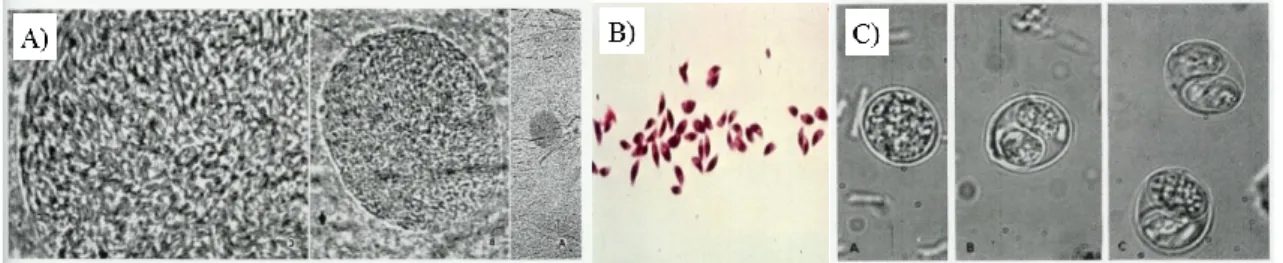
Figure 1.1 Graphic representation of a tachyzoite. 6 Figure 1.2 A) Bradyzoites inside a cyst of a laboratory mouse brain (100 x, 40 x e 10 x objective), B) Free stained tachyzoites on laboratory mouse ascites, C) Sporolated and
unsporulated Oocysts. 6
Figure 1.3 Life cycle of Toxoplasma gondii. 8
Figure 1.4 Mechanism of cell invasion. 10
Figure 1.5 Genetic linkage maps for the 14 chromosomes of Toxoplasma gondii. 12 Figure 1.6 Clinical signs. A) Cerebral calcifications, B) Retina lesions due to ocular
toxoplasmosis C) Newborn with hydrocephalus. 19
Figure 2.1 (A and B) (A) shows the evolution of Toxoplasma gondii seroprevalence in Portugal over the past three decades; (B) shows the sample sizes for the three National Serological Surveys, the precise estimated seroprevalences and respective 95% confidence interval (CI). 34 Figure 2.2 Comparison of Toxoplasma gondii seroprevalence trends, according to the data from
three National Serological Surveys, by age groups. 36
Figure 3.1 Annual distribution of acquired and congenital Toxoplasma gondii infection, 2009-2018. Number of cases by year of diagnosis. 49
Figure 3.2 Annual distribution of Toxoplasma gondii infection acquired by gender, 2009-2018.
Number of cases by year of diagnosis. 49
Figure 4.1 Workflow of RH strain inoculations. 61
Figure 4.2 Experiment A: Continuous RH strain mice propagation. 65 Figure 4.3 Phenotypic evaluation on the course of experiment A. 65 Figure 4.4 Phenotypic evaluation on the course of experiment B. 66 Figure 4.5 Experiment B: Continuous RH strain cell line propagation. 67 Figure 4.6 Experiment C: intercalate passages of the RH strain in the cell line and in mice. 68 Figure 4.7 Comparison of tachyzoites yield between propagation experiments. 68
Table 1.1 Geographical distribution of Toxoplasma gondii genotypes and potencial
relationships with human disease. 15
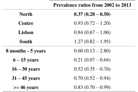
Table 2.1 Trends of Toxoplasma gondii seroprevalence in Portugal by region calculated with
an interval confidence of 95 %. 34
Table 2.2 Multivariate analysis adjusted for gender, age group and region. 35 Table 2.3 Toxoplasma gondii seroprevalence by gender and year of analysis calculated with an
interval confidence of 95 %. 36
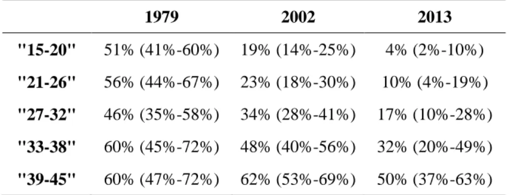
Table 2.4 Toxoplasma gondii seroprevalence in childbearing women by age group calculated
with an interval confidence of 95 %. 37
Table 3.1 Annual distribution of acquired and congenital Toxoplasma gondii infection by age group, 2009-2018. Number of cases by year of diagnosis. 50 Table 3.2 Virulence and genotyping analysis of Toxoplasma gondii strains and laboratorial and
clinical features of the biological samples/cases. 51
Table 4.1 Loci, amplicon length and primers used for PCR. 64 Supplementary Table S 4.1
BioProject PRJEB34235 - European Nucleotide Archive
Throuout this PhD dissertation acronyms are extended upon first usage.
AIDS Acquired Immunodeficiency Syndrome AF Amniotic fluid
AH Aqueous humor
ATCC American Type Culture Collection BAL bronchoalveolar lavage fluid
BB Baby blood
BLAST Basic Local Alignment Search Tool
Bp Base pair CB Cerebral biopsy CI Confidence intervals CSF Cerebrospinal fluid CG Cytosine-guanine cM Centimorgan
DALY’s Disability-adjusted life- years DAT Direct Agglutination test
DMEM Dulbecco's Modified Eagle Medium
DNA Desoxyribonucleic acid DGS Direção Geral de Saúde
ELFA Enzyme-Linked Fluorescent Assay ELISA Enzyme Linked Immunosorbent assay Gra Dense granule protein
HCL Hydrochloric acid
HIV Human Immunodeficiency Virus HFF Human Foreskin Fibroblast
IB Imunoblot
ISAGA Immunoglobulin Immunosorbent Agglutination Assay
Mb Mega base pair
MLST Multilocus Sequence Typing Ms Microsatellites
NB Newborn
NBB Newborn blood
PBS Phosphate Buffered Saline PCR Polymerase Chain Reaction p.i. Post-inoculation
PL Placenta
QALYs Quality-adjusted life-years
RAPD Random amplified polymorphic DNA RFLP Restriction Fragment Length Polymorphisms
Rop Roptry
Sag Surface antigen
SNP Single Nucleotide Polymorphism
TgMA Myosine A
TUB2 Beta Tubulina UCB Umbilical cord blood T. gondii Toxoplasma gondii WHO World Health Organization WGS Whole Genome Sequence
VH Vitreous humor
Laboratory of Parasitic and Fungal Infections, Department of Infectious Diseases, National Institute of Health Doctor Ricardo Jorge, Lisbon, Portugal.
The main body of this Ph.D. dissertation is based on three manuscripts (listed below) that are presented as individual chapters (II to IV). Two of them have already been published (the remaining one is submitted for publication at the time this thesis was completed) in peer reviewed international journal, being presented in this thesis essentially as a reproduction of the content that was published. These three manuscripts reflect the objectives delineated for this Ph.D. thesis and consequently the experimental studies performed throughout the author Ph.D. work, also the order of presentation of these chapters reflet the chronological order of the publication of the respective manuscripts. Besides, these manuscript-based chapters, each one including extensive and specific introduction, material and methods, results and discussion sections. The present doctoral dissertation still encompasses a general introduction (chapter I) and a conclusive overview of the main findings and conclusions, with regards to future perspectives and lines of work (chapter V). In brief, each chapter includes the following contents:
Chapter I
This chapter consists of a general introduction that intents to provide the reader an global overview of the major Toxoplasma gondii features such as, historical background, taxonomy, biology, life cycle, mechanisms of cell invasion, genome and genetic variation; and also the pathophysiology, transmission, clinical features, diagnosis, treatment and epidemiology of
Toxoplasmosis, the infectious disease caused by Toxoplasma gondii. The state of the art that express the reasons why the author selected the objectives of this Ph.D. project and the main objectives of this doctoral dissertation are listed at the end of this chapter.
Chapter II
Toxoplasma gondii seroprevalence in the Portuguese population: comparison of three cross-sectional studies spanning three decades. This chapter corresponds to a manuscript with the following reference: BMJ Open. 2016; 6(10): e011648. “Toxoplasma gondii seroprevalence in the Portuguese population: comparison of three cross-sectional studies spanning three decades.” Maria João Gargaté,Idalina Ferreira,Anabela Vilares, Susana Martins, Carlos Cardoso, Susana Silva, Baltazar Nunes, João Paulo Gomes. Published online 2016 Oct 5. doi: 10.1136/bmjopen-2016-011648.
reference, so some sections constitute a faithful reproduction of what was published: Parasitol Res (2017) 116:979–985. “Molecular and virulence characterization of Toxoplasma gondii strains isolated from humans in Portugal.” Anabela Vilares, Maria João Gargaté, Idalina Ferreira, Susana Martins, João Paulo Gomes. doi: 10.1007/s00436-017-5374-5.
Chapter IV
Contains the following study that was recently submitted for publication in a peer reviewed international journal (Parasitology Research): “Parallel propagation of Toxoplasma gondii in vivo, in vitro and alternate models: towards less dependence on the mice model”. Maria João Gargaté, Anabela Vilares, Idalina Ferreira, Tania Reis, Susana Martins, Vitor Borges, João Paulo Gomes. 2019.
Chapter V
This chapter provides a global overview of the subjects addressed throughout the chapters, where the main findings and conclusions achieved in this Ph.D. thesis are highlighting. Of note, in order to avoid redundancies, this section solely summarizes and discusses the major findings of this work because a detailed discussion of specific results was already provided in each chapter. Future perspectives and follow-up of these investigations are also presented. Considering the different layouts required by the different journals where the manuscripts were published, including tables, figures and references, all chapters were formatted in a unique style, with all references being cited by the name of the first author according alphabetical order and year of the publication and listed in a single section denominated "References". The supplemental material is presented at the final of this Ph.D. thesis, enumerated accordingly with the chapter they concern to in a section referred as "Supplementary material".
Chapter I
General Introduction
1. General Introduction
1.1. Toxoplasma gondii, the parasite
1.1.1. Historical background and taxonomy
The Toxoplasma gondii (T. gondii) is an apicomplexan protozoan parasite and one of the most successful parasites worldwide due to its ability to infect all warm blooded animals including humans. One third of the world’s human population is assumed to be infected with T. gondii (Louis M. Weissa; Weiss and Dubey, 2009; Innes, 2010). This ubiquitous obligate intracellular organism was first discovered in 1908 by Charles Nicolle and Louis Manceaux at the Pasteur Institute in Tunis, who found the parasite in the liver and spleen of a North African rodent, named gundi (Ctenodactylus gundi) and initially presumed it was a species of Leishmania. In the same year, Alfonso Splendore, a Brazilian scientist discovered the parasite in a rabbit (Oryctolagus cuniculus) and again mistakenly identified it as Leishmania (Ferguson D, 2009). However in 1909 following experimental infection and microscopic analysis, the parasite was renamed to Toxoplasma gondii as described by Nicolle and Manceaux (Nicolle and Manceaux, 1909; Ferguson D, 2009) due to the bow shaped morphology of the extracellular stage of the parasite - tachyzoite; Toxo is derived from Greek for bow, plasma meaning life and gondii because its original host was C. gundi. In 1914 Castellani was probably the first to describe a T. gondii–like parasite in smears of the blood and spleen from a 14-year-old boy from Ceylon who died from a disease characterized by severe anemia, fever and spleenomegaly. (Louis M. Weissa; Cheng et al., 2015).
In 1923, Janků observed parasitic cysts in the retina of an eleven month old child who was suffering from hydrocephalus (Janků, 1928). In the same year the first identified case of congenital toxoplasmosis was reported by Wolf and Cowen from a 3 day old child who had developed seizures (Wolf et al., 1939). The baby only survived for one month and following post mortem cerebral calcification, retinochoroiditis, and hydrocephalus were observed. In this same year Sabin isolated T. gondii from two children from Cincinnati, named R.H. (initials of the patients name), aged 6 years old and the other named W.B.D. aged 8 years, with encephalitis (Sabin, 1941). This strain, designated “RH” became the laboratory prototypical Type I strain and since 1938 it has been passed in mice in many laboratories worldwide. In 1941, Pinkerton and Henderson reported atypical pneumonia on two adults who died and in whom they demonstrated T. gondii as the etiological agent. These were the first reports of acute toxoplasmosis in adults without neurological signs. In the 1950’s T. gondii parasites were discovered in enucleated eyes (Wilder, 1952), and this type of ocular toxoplasmosis was presumed to be a consequence of congenital transmission of the parasite. However, more recent studies have also described a
greater number of cases than expected of ocular toxoplasmosis due to postnatal acquired infection (Montoya and Remington, 1996; Burnett et al., 1998; Gilbert et al., 2008).
The development of a serological test, the dye test, in 1948 by Albert Sabin and Harry Feldman was a major advance in the study of toxoplasmosis (Sabin and Feldman, 1948). The ability to identify T. gondii infections based on this serological method allowed epidemiological studies on the incidence of infection, demonstrating the widespread world-wide prevalence of this infection in humans. It also allowed the identification of clinical signs compatible with the diagnosis of congenital toxoplasmosis (Sabin and Feldman, 1948; Feldman and Miller, 1956). As such, Sabin determined that the simultaneous occurrence of clinical signs of hydrocephalus or microcephalus, intracerebral calcification and chorioretinitis, could be used to identify cases of congenital toxoplasmosis, what is nowadays known as the classical triad of symptoms of congenital toxoplasmosis (Sabin, 1941, 1942).
In 1965 Desmonts and colleagues confirmed the transmission by the carnivorous route, which had been previously presupposed by Weinman and Chandler (Weinman and Chandler, 1954) and by Jacobs et al. (1960). In 1972, Wallace and his colleagues began epidemiological surveys in regions where habitants ate raw or undercooked meat and observed a high frequency of infection in humans, demonstrating the transmission route through carnivorism. Anteriorly in 1969, Kean et al. described the first outbreak of toxoplasmosis in Cornell University medical students after eating insufficiently cooked hamburgers. However, in 1959 Rawal in a study in vegetarians observed a frequency equivalent to that found in carnivores, and the diffusion of the parasitizes in this group was not clarified. In 1970 the cycle of this parasite is definitively clarified, with the discovery by Dubey et al. of the sexual development of T. gondii in the intestine of cats (Frenkel et al., 1970), thus felids are still presumed to be the only definitive host. An important step in the history of T. gondii occurred in the 1980’s when AIDS patients were found to develop clinical symptoms of the parasite (Luft and Remington, 1992). T. gondii was identified as a major opportunistic infection for these immunocompromised patients, where either newly acquired infection or recrudescence of latent infection would frequently cause encephalitis (Luft and Remington, 1992).
The most recent developments for T. gondii are its possible effect on behavior changes in both animals (Berdoy et al., 2000; Ingram et al., 2013), and humans. Although the link to T. gondii infection and behavioral problems in humans is not completely clear, several reports have associated infection to schizophrenia (Torrey et al., 2012), increased risk taking and road traffic accidents (Flegr et al., 2009) and an increased risk of suicide (Lester, 2012). Also, the emergence of genetically different strains of the parasite have been linked to several fatal cases of acquired
infection in immuno-competent individuals (Carme et al., 2002, 2009; Ajzenberg et al., 2016), further highlighting the potential public health risk of the parasite.
According to the current taxonomic classification, T. gondii belongs to the Kingdom Protista, sub - Kingdom Protozoa (Goldfuss, 1918), Phylum Apicomplexa (Levine, 1970), Class Sporozoasida (Leukart, 1879), Subclass Coccidiasina (Leukart, 1879), Order Eimeriorina (Leger, 1911), Family Toxoplasmatidae (Biocca, 1956) (Hill et al., 2005; Dubey, 2010) and genus Toxoplasma (Nicolle and Manceaux, 1909). There is only one species, T. gondii; one of the most successful parasitic organisms.
1.1.2. Biology
There are three infective stages of T. gondii: a rapidly dividing invasive tachyzoite (Figure 1.1), a slowly dividing bradyzoite in tissue cysts, and an environmental stage, the sporozoite, protected inside an oocyst (Figure 1.2). Tachyzoites are crescent-shaped cells, approximately 5 m long and 2 m wide, with a pointed apical end and a round posterior end. They are limited by a complex membrane, named the pellicle, closely connected with a cytoskeleton involved in the structural integrity and motility of the cell. They possess a nucleus, a mitochondria, a Golgi complex, ribosomes, an endoplasmic reticulum, and a multiple -membrane-bound plastid-like organelle called the apicoplast (Roos et al., 1999). As other members of the phylum Apicomplexa, they concentrate in their apical area a specialized cytoskeletal structure (the conoid, implicated in cell invasion) and numerous secretory organelles (rhoptries [ROPs], dense granules, and micronemes). Tachyzoites are the dissemination form and they are able to invade cells of all vertebrate, where they multiply in a parasitophorous vacuole. Bradyzoites result from the conversion of tachyzoites into a slow-dividing stage and form tissue cysts. These cysts are spheroid in brain cells or elongated in muscular cells, vary in size and can contain only two bradyzoites or thousands with a latent metabolism. Cysts remain intracellular throughout their lifetime and the death of the host cell may produce the disruption of the cyst wall and the consequent liberation of bradyzoites. The resistance of bradyzoites to the acid pepsin (1- to 2-h survival into pepsin-HCl) allows their transmission through ingestion. Oocysts are ovoid structures with two sporocysts (after sporulation) and with an exceptionally resistant double wall that enables the parasite to survive for long periods in adverse conditions. (Robert-Gangneux and Dardé, 2012).
Figure 1.1 Graphic representation of a tachyzoite.
Image source: VectorStock.com/11563382.
Figure 1.2 A) Bradyzoites inside a cyst of a laboratory mouse brain (100x, 40x e 10x objective), B) Free
stained tachyzoites on laboratory mouse ascites , C) Sporolated and unsporulated Oocysts. Image source: National Reference Laboratory of Parasitic and Fungal Infections.
1.1.3. Life cycle
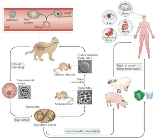
The life cycle of the parasite was fully understood with the discovery of T. gondii oocysts in cat faeces (Hutchison et al., 1969, 1971) it evidenced the central role of the cat as the only definitive host harboring the sexual developmental stages within the small intestine and spreading millions of oocysts through feces to the environment. The life cycle (Figure 1.3) consists of asexual reproduction in the intermediate hosts (including humans) and sexual reproduction in the intestinal mucosa of the definitive host. A unique feature that characterizes T. gondii life cycle is that it can be transmitted not only between intermediate and definitive hosts (sexual cycle) but also between intermediate hosts via carnivorism (asexual cycle) or even between definitive hosts. Oocysts take 3-7 days to sporulate in the environment and become infective by meiosis process leading to the formation of a sporulated oocyst with two sporocysts, each containing four haploid sporozoites (Holliman et al., 2003). Intermediate hosts in nature (including birds and rodents) become infected after ingesting soil, water or plant material contaminated with these oocysts. After oocyst ingestion, sporozoites are released and penetrate the intestinal epithelium. Then, they transform into tachyzoites and become surrounded by a parasitophorous vacuole that protects
them from host defense mechanisms. The tachyzoite multiplies asexually by endodygeny and they spread first to mesenteric lymph nodes and then to the several organs by invasion of the bloodstream (Hill et al., 2005; Jones and Dubey, 2010). After several multiplication cycles, tachyzoites give rise to bradyzoites, and tissue cysts arise as early as 7 to 10 days post infection and may remain throughout lifetime in the hosts, predominantly in the brain or muscles (Jones and Dubey, 2010; Dubey, 2010; Robert-Gangneux and Dardé, 2012). After the ingestion by a cat of cysts present in tissues of an intermediate host, gastric enzymes destroy the cyst wall. Bradyzoites settle within enterocytes, where they undergo a number of asexual multiplicatio ns, with the development of merozoites within schizonts. This process is followed by the formation of male and female gametes (gametogony) (Ferguson, 2002). After fertilization, oocysts formed within enterocytes are released by the disruption of the cell and are excreted as unsporulated forms in cat feces. On the other hand, after the ingestion of the tissue cysts by an intermediate host through raw or undercooked meat, cysts are ruptured as they pass through the digestive tract, causing the release of bradyzoites. The bradyzoites will infect the intestinal epithelium of the new host and differentiate back into the rapidly dividing tachyzoite stage for dissemination throughout the body. In addition, if the acute phase occurs during pregnancy, the parasite can cross the placenta and infect the fetus (congenital transmission) (Robert-Gangneux and Dardé, 2012).
Figure 1.3 Life cycle of Toxoplasma gondii. Cats are the definitive host where sexual replication takes
place. Following replication within enterocytes of the gut (merogony), male and female gametes are formed within the host cell. Fusion of gametes leads to the formation of diploid oocysts that are shed in cat faeces and undergo meiosis in the environment to yield eight haploid progeny. Oocysts contaminate food and water, providing a route of infection for intermediate hosts. In the intermediate host (birds and rodents) asexual replication occurs. Acute infection is characterized by fast replicating tachyzoites that disseminate throughout the body. Differentiation to slow-growing bradyzoites within tissue cysts leads to long-term chronic infection. Ingestion of tissue cysts via omnivorous or carnivorous feeding can lead to transmission to other intermediate hosts or to cats, which re-initiates the sexual phase of the life cycle. Many animals serve as intermediate hosts, including farm animals. Humans become infected by eating undercooked meat containing tissue cysts or by the ingestion of oocysts in contaminated water (de Moura et al., 2006; Jones and Dubey, 2012). T. gondii can infect the brain and other organs as well as the foetus following a congenital infection.
Image adapted from Hunter and Sibley. Nature Reviews Microbiology 2012 Nov; 10:766-778 (Hunter and Sibley, 2012).
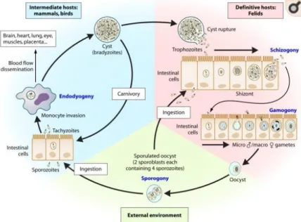
1.1.4. Mechanism of cell Invasion
T. gondii is a successful parasite because it can spread across many biomes and species, and has developed specialized processes to invade and replicate efficiently within cells. It is an obligate intracellular parasite, implying that it cannot survive outside a cell, which provides the parasite a safe, secure home full of nutrients and a refuge from the immune system of the host (Tosh et al., 2016). Invasion is an active process based on parasite motility and the sequential secretion of proteins from specialized secretory organelles, the micronemes, the rhoptries, and the
dense granules. The micronemes and rhoptries are localized at the apical end of the parasite, where they function to form the anchor and tight junction to host cell (Huynh et al., 2006; Dlugonska, 2008). Secreted dense granule synthesizes proteins to facilitate the remodeling of host processes to help replication of the parasite (Bougdour et al., 2014). A fourth organelle, the apicoplast that contains essential metabolic pathways is also envolved in the cell invasion process (Arisue and Hashimoto, 2015). T. gondii invasion starts with parasite attachment to the host cell plasma membrane. First, the parasite contacts its apical end to the plasmatic membrane of a cell and then secretes proteins that will promote parasite adhesion. It requires the calcium-dependent secretion of adhesins from micronemes, such as the microneme protein MIC2, which recognize host cell receptors and promote parasite reorientation and attachment. Cell invasion relies on a complex interaction between the host cell surface and the parasite, a process called gliding motility, a complex motor system promoted by actin-myosin interactions and dynamic rearrangements of the parasite cytoskeleton (Carruthers and Boothroyd, 2007).
Entry is a rapid process (15 to 30 s), apical membrane antigen (AMA1) secreted from micronemes and the secretion of rhoptry (ROP) neck proteins (RONs) inserted into the host cell membrane (Dubremetz, 2007) plays part in the anchoring process and creation of a tight junction between the parasite and the host cell plasma membranes (Blader et al., 2015), called the moving junction. The moving junction complex (AMA1-RON2) allows for host cell membrane invagination and movement of the parasite into the cell (Dobrowolski and Sibley, 1996; Håkansson et al., 2001). When the parasite fully enters the host cell, it is covered by a membrane made of a conglomerate of host lipid bilayer and secreted parasite lipids and proteins, resulting in a specialized vacuole called the parasitophorous vacuole (PV) (Suss-Toby et al., 1996). Host transmembrane proteins and proteins found in lipid rafts are excluded during the formation of the PV (Blader et al., 2015). The PV will be the focal point of interaction with the host cell, through which the parasite will import nutrients and export secreted proteins to create an environment ready for replication (Blader and Koshy, 2014). Soon after invasion, the PV localizes in the perinuclear region and associates with several host organelles, including the endoplasmic reticulum, Golgi complex, and mitochondria (de Melo et al., 1992; Sinai et al., 1997; Walker et al., 2008). The formation of the nascent parasitophorous vacuole membrane (PVM) requires the secretion of proteins from the ROPs. ROP18 one of these proteins is associated with the cytosolic face of the PVM and exerts protein kinase activity, which has a profound effect on parasite growth and virulence (El Hajj et al., 2007), and ROP16 is able to manipulate host gene expression, affecting interleukin secretion (Laliberté and Carruthers, 2008). Within the PV, tachyzoites divide during a 6 to 9 h cycle, by endodyogeny and they exit the cell usually after 64 to 128 parasites have accumulated in the PV (Black and Boothroyd, 2000). The egress of T. gondii tachyzoites from the host cell is performed through the rupture of the PV and the host cell plasma membrane,
releasing free parasites in the medium an active process that is dependent upon a rise in the calcium concentration (Sibley, 2010). This process can be triggered by parasite produced abscisic acid or other vacuole acidification and/or by the NTPases segregation. Egress occurs within minutes and once outside the infected host cell, the parasites use gliding motility to move and invade a new cell (Håkansson et al., 1999; Heintzelman, 2015; Periz et al., 2017) (Figure 1.4).
Figure 1.4: Mechanism of cell invasion.
Image source: Clinical Microbiology Reviews (Robert-Gangneux and Dardé, 2012).
1.1.5. The Genome
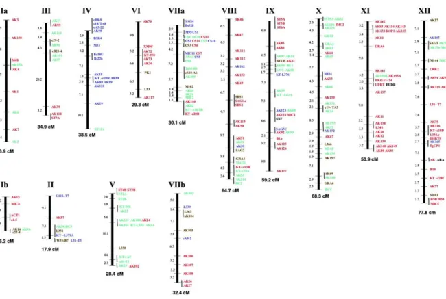
T. gondii has a ~65 Mb genome, comprising of 14 chromosomes which range from approximately 2 Mb – 7.5 Mb as shown in Figure 1.5 (Khan et al., 2005). The genome is closely related to another apicomplexan protozoan parasite, Neospora caninum (N. Caninum), and it is thought that around 28 million years ago the two parasites diverged from a common ancestor, due to the speciation of the definitive hosts (cats - T. gondii, dogs - N. Caninum) (Reid et al., 2012). In comparison to other apiocomplexan parasites, such as Cyptosporidium parvum (C. Parvum) and Theileria parva (T. parva), the genome of T. gondii is significantly larger, it contains more introns, more predicted genes and has a lower gene density (Delbac et al., 2001). One possible reason for the difference in size compared to other apicomplexans can be due to the large number of secondary hosts which this parasite is able to establish within (Roos, 2005). In 2005, a composite genome map was derived from genetic crosses and linkage analysis of the three main archetypal T. gondii lineages (I, II, III) (Khan et al., 2005). The genetic linkage map that was generated identified 250 species specific markers, of which 12 are most commonly used for strain
genotyping by PCR-RFLP. The Figure 1.5 details each of these individual markers and also highlights markers which are strain specific and those which are present on all three archetypal strains. A fourth clonal lineage, designated haplotype 12, is largely confined to North America and more common in wild animals (Khan et al., 2011) was designated. Su and colleagues classified T. gondii strains into 15 different haplotypes, defining six major clades (Su et al., 2012). However, Minot and colleagues disagree with this theory (Minot et al., 2012). The online genome database ToxoDB (http://toxodb.org/toxo/) provides further detailed information about the genome and the functional genomics of T. gondii. It also provides genome sequence information (including the facility to BLAST sequences), gene expression and proteomics data (Gajria et al., 2008), which helps support research on the parasite. The most recent study published in 2016 compared four tissue-cyst forming coccidian parasites and showed that three of these organisms N. caninum , Hammondia hammondi and T. gondii have a similar total genome size of 62–65 Mb while the Sarcocystis neurona genome is larger due to expanded repeats and much larger introns. All four genomes have identical GC compositions and are predicted to encode from 7,000 to more than 8,000 genes located on 14 chromosomes, as with T. gondii. This work reveals that tandem amplification and diversification of secretory pathogenesis determinants is the primary characteristic that distinguishes the closely related genomes of these parasites and also disclosed that the unusual population structure of this parasite is characterized by clade-specific inheritance of large conserved haploblocks that are significantly enriched in tandemly clustered secretory pathogenesis determinants. The shared heritage of these conserved haploblocks, which show a different ancestry than the genome as a whole, may thus influence transmission, host range and pathogenicity (Lorenzi et al., 2016).
Figure 1.5 Genetic linkage maps for the 14 chromosomes of Toxoplasma gondii. Individual markers are
shown to the right of the vertical bar and chromosome numbers are given above each map. Markers that map to the same node are indicated to the right of a solid vertical bar. The corresponding genetic distances between each node are given to the left of each map and the total sizes in cM are shown at the bottom of each chromosome. Polymorphisms that are unique to type I are shown in red, those unique to type II are shown in green, those unique to type III are shown in blue and markers that contain multiple polymorphism are shown in yellow. Maps were constructed using MAPMAKER from the analysis of 71 recombinant progeny using 250 genetic markers. Markers that include data analyzed by Southern blot a re followed by the suffix ‘.c’.
Image source: Nucleic Acids Research, https://doi.org/10.1093/nar/gki604 (Khan et al., 2005).
1.1.6. Genetic variation and disease severity
T. gondii has not only the capacity to propagate asexually but also sexually in its feline definitive host. Therefore, sexual recombination should provide for a high genetic diversity between T. gondii strains worldwide. However, the population structure of this parasite was initially described as being highly clonal and showing a low genetic diversity. Genotyping studies of T. gondii started in the 1990s and were based on a single marker, predominantly Sag2 (Howe et al., 1997; Fuentes et al., 2001; Sabaj et al., 2010) and GRA6 (Fazaeli et al., 2000; Messaritakis et al., 2008), but these methodologies didn’t allow the identification of non-clonal strains. Thus these methodologies were optimized in order to determine more precisely the presence of polymorphisms in the population and with the addition of new PCR-RFLP markers (Su et al., 2006) and by microsatellite analysis (Ajzenberg et al., 2005, 2010), this was achieved. Genetic
studies of isolates from Europe and the United States suggested the presence of a clonal population structure stable in time and space (Darde et al., 1988; Sibley and Boothroyd, 1992; Howe and Sibley, 1995; Ajzenberg et al., 2002a; Khan et al., 2007) where the majority of isolates (> 94 %) are grouped into three main clonal multilocus genotypes I, II and III. This simple clonal structure is accompanied by a low level of genetic divergence among the three lineages (only 1 to 2 % divergence at the DNA sequence level between lineages). However, multilocus and multichromosome genotyping of isolates from other continents revealed a much more complex population structure with a greater genetic diversity, likely reflecting a history of more frequent genetic exchanges and genetic drift (Lehmann et al., 2004; Ajzenberg et al., 2004) (Table 1.1). The majority of isolates from South America, Africa and Asia are not included in the three major lineages (with the exception of type III, which is really cosmopolitan). These deferent genotypes lead to the description of new haplogroups, some of them largely distributed over continents, being considered other successful clonal lineages (Khan et al., 2007). So far, 12 haplogroups (including the 3 initially described lineages, types I, II, and III) have been described (Khan et al., 2007, 2011), based on sequence-based analyses, but these haplogroups are not totally homogenous, and more specific markers revealed subclustering that may be associated with geographical origins and phenotypic characteristics. Based on the classical genotyping, from Northern (Jokelainen et al., 2011) to Southern Europe (De Sousa et al., 2006), the population structure of T. gondii shows a clonal profile, with a predominance of the type II lineage strains. The other two clonal lineages are sporadically found in Europe. While the three clonal lineages predominate in North America and Europe, strains from other regions in the world appear to have genotypes that are more diverse. By analyzing isolates from South America, Asia and Africa by using PCR-RFLP or microsatellite markers, it was revealed that the majority of these isolates have the classical type I, II, and III alleles identical to those in the main three lineages, but some novel alleles were also detected. These ‘new’ genotypes were designated as atypical, exotic, recombinant or non-archetypal genotypes (Grigg et al., 2001a; Ajzenberg et al., 2004). The recombinant genotypes have mixtures of classical alleles, while atypical, unusual, non-archetypical or exotic strains are characterized by the existence of many unique polymorphisms and novel alleles (Grigg et al., 2001a; Su et al., 2003; Ajzenberg et al., 2004). Phylogenetic analysis, based on microsatellites, suggests that these atypical genotypes are phylogenetically disseminated with no clear structure, or association with the main three lineages. Although there is clear divergence among these strains, essentially due to the mixture of alleles, the overall level of sequence polymorphism provided by single nucleotide polymorphisms is modest. (Grigg et al., 2001a; Su et al., 2003; Ajzenberg et al., 2004). The isolation of atypical strains, which do not fit into these three major lineages, is rare in Europe and likely suggests contamination by non-European strains either during residence abroad or after the consumption of imported food
(Ajzenberg et al., 2009). In North America, the population structure appeared similar to that observed in Europe, with a predominance of type II strains (Howe and Sibley, 1995). South America is an area with a high level of diversity for T. gondii and type II seems to be very rare (Dubey et al., 2007a). The high level of genetic diversity observed in this continent is maximal in the wild Amazonian area, with many unique polymorphisms (Ajzenberg et al., 2004). It is believed that, in the Amazonia area, the interpenetration of anthropized and wild rainforest environments leads to hybridization between strains that may represent a potential risk for human health. In Asia, it has been shown that strains have more limited genetic diversity compared to South America (Dubey et al., 2007b). Isolates from Cameroon that were analyzed via microsatellites revealed the existence of fixed combination of type I and III alleles, suggesting a unique clonal African type (Ajzenberg et al., 2004).
Epidemiological studies have shown that type I strains are rare in human and animal infections, however has been linked to reactivation of the parasite in immunocompromised individuals (Khan et al., 2005) and type II strains are considered to be the most common source of human toxoplasmosis (Howe and Sibley, 1995). In North America and Europe, most cases of human toxoplasmosis in AIDS and congenital infections are associated with type II strains (Howe and Sibley, 1995; Howe et al., 1997; Ajzenberg et al., 2002a). However, a study in Spain reported dominance of type II strains in AIDS patients, while type I strains were associated with congenital infections (Fuentes et al., 2001). Another study from the USA revealed the relationship between severe ocular toxoplasmosis in immnunocompetent patients and type I strains and new recombinant genotypes (Grigg et al., 2001a). The severity of T. gondii acute infection is considered to be one of the most significant phenotypes among T. gondii strains. Very little is known concerning circulating strains in Portugal, a small scale study was performed in France with Portuguese sera that reported a majority of type II humans strains (Sousa et al., 2008; Ajzenberg et al., 2009) and other European study that enrolled only two Portuguese isolates (Ajzenberg et al., 2009) revelled that were both type II.
The technological development accompanied with the cost reduction of the Whole Genome Sequence (WGS) methodology will allow in mid-term the sequencing of multiple complete genomes of T. gondii, allowing understanding of the real degree of genetic variability of this protozoan, frequency of recombination and potential genotype - phenotype associations.
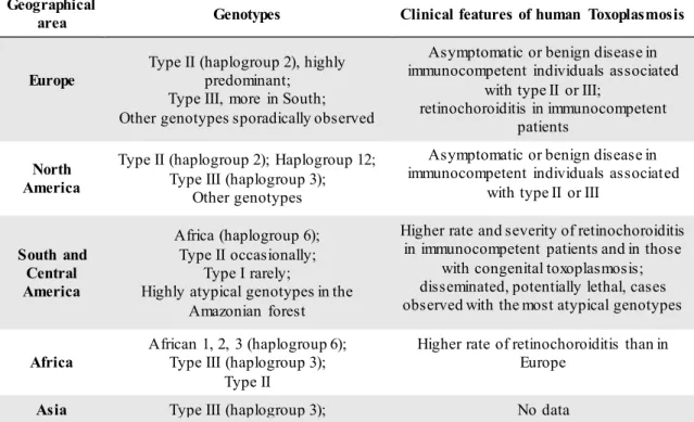
Table 1.1 Geographical distribution of Toxoplasma gondii genotypes and potential relationships with
human disease.
Table Source: Adapted from Clinical Microbiology Reviews (Robert-Gangneux and Dardé, 2012).
Geographical
area Genotypes Clinical features of human Toxoplasmosis
Europe
Type II (haplogroup 2), highly predominant;
Type III, more in South; Other genotypes sporadically observed
Asymptomatic or benign disease in immunocompetent individuals associated
with type II or III;
retinochoroiditis in immunocompetent patients
North America
Type II (haplogroup 2); Haplogroup 12; Type III (haplogroup 3);
Other genotypes
Asymptomatic or benign disease in immunocompetent individuals associated
with type II or III
South and Central America Africa (haplogroup 6); Type II occasionally; Type I rarely;
Highly atypical genotypes in the Amazonian forest
Higher rate and severity of retinochoroiditis in immunocompetent patients and in those
with congenital toxoplasmosis; disseminated, potentially lethal, cases observed with the most atypical genotypes
Africa
African 1, 2, 3 (haplogroup 6); Type III (haplogroup 3);
Type II
Higher rate of retinochoroiditis than in Europe
Asia Type III (haplogroup 3); No data
1.1.7. Assessment of strain virulence
Experimental virulence is usually defined with the mouse model after the intraperitoneal inoculation with a given number of tachyzoites. Type I strains are highly virulent, have a lethal dose (LD100) (the minimal dose which causes 100 % mortality) of a single infectious organism, leading to the death of mice less than 10 days after the inoculation of about 10 tachyzoites. On the other hand, mouse non virulent strains, normally from type II and III, have a lethal dose of > 10 infectious organisms and usually generate chronic infection in the mouse (Sibley et al., 2002). Isolatesfrom other clonal lineages or from atypical strains range from thehighly virulent to the intermediate or non-virulent phenotype, according to differences in the combination of genes that they have inherited. Genotypes with a majority of type I alleles are usually more virulent (Saeij et al., 2005a) The mouse-virulent strains exhibit some characteristics that may explain the rapid dissemination of the parasite and the higher tissue burden observed in mice and other susceptible hosts: improved migration across polarized epithelia or across the extracellular matrix, higher rates of the ex vivo penetration of the lamina propria of mucosa and submucosa (Barragan and Sibley, 2002), and in cell culture, higher growth rates and lower rates of interconversion from tachyzoites to bradyzoites. Experimental crosses between strains with different virulence patterns facilitated the identification of several polymorphic genes coding for
secreted factors of T. gondii associated with differences in the expression of virulence in mice (Taylor et al., 2006; Reese et al., 2011). These key virulence factors are secretory proteins discharged from apical organelles, the rhoptries. The proteins of this rhoptry family (ROP5, ROP16, and ROP18) exert kinase or pseudokinase activity. They are injected directly into the host cell and play a role during the process of parasite invasion or in the induction of interleukin-12 (IL-interleukin-12) secretion by mouse macrophages (Robben et al., 2004).
T. gondii is considered to be a successful organism because it has the ability to cross biological barriers such as blood-brain barrier, placenta, or gut epithelium (Saeij et al., 2005a). However, the expression of virulence in humans is a complex phenomenon due to many other factors that may influence the pathogenicity of a given strain, namely: parasitic factors such as the infectious stage, the inoculums and the genetic background of the strain and immune status of the host. (Robert-Gangneux and Dardé, 2012).
1.2. Toxoplasmosis, the human disease
1.2.1. Pathophysiology
Toxoplasmosis is the infectious disease caused by T. gondii. The pathophysiology of this infection results from the dissemination of tachyzoites throughout the body. Tachyzoites speedily invade monocytes and gain access to the blood flow (after trans-epithelial passage across the intestinal barrier) and from there to all organs (Robert-Gangneux and Dardé, 2012). In acute toxoplasmosis, a host may die due to necrosis (caused by intracellular growth of tachyzoites) of the intestine and mesenteric lymph nodes before severe damage of other organs. Focal areas of necrosis may develop in many organs; the clinical picture is determined by the extent of injury to these organs, especially vital organs such as the eyes, heart, and adrenals (Dubey, 2010). If the host survives, the invasive stages (tachyzoites) convert into a latent form (bradyzoites) within cells and persist as cysts, generally in muscles, retina, and brain, for a lifetime following the onset of an efficient immune response. The immunity is predominantly cell mediated and activated macrophages and T cells play a central role, while interferon gama (y) and other cytokines induce an effective immune response. The specific antibody in the presence of complement eliminates extracellular parasites (Holliman et al., 2003).
1.2.2. Transmission
Human infection is acquired by ingestion of tissue cysts in raw, poorly cooked or cured meat, notably lamb and pork. Consumption of meat from warm blooded animals is considered the major source of infection in Western countries (Cook et al., 2000). The transmission can occur
also by or ingestion of sporulated oocysts derived from contaminating soil, water or inadequately washed vegetables / fruit or directly from cat faeces. Once sporulated, oocysts are resistant to adverse environmental conditions, it can remain viable in a moist environment for more than a year, in very low (icy) or high water temperatures for long periods of time and can resist to chemical and physical treatments currently applied in water treatment plants, including chlorination and ozone treatment (Dumètre et al., 2008). When primary infection is acquired by a pregnant woman, tachyzoites can colonize placental tissues during the dissemination process and access the foetus. The risk of transmission and the severity of disease depend on the gestational stage at which the mother first becomes infected. Congenital infection is the most important part of the disease burden due to T. gondii infection in humans.
Rarely, infection can be acquired via an organ transplant, as cysts can be found potentially in any organ T. gondii infection can be transmitted through a cyst-containing organ from a donor with infection acquired in the past to a nonimmunized recipient (Robert-Gangneux and Dardé, 2012). One the other hand toxoplasmosis associated with bone marrow transplant is a rare event and is usually due to reactivation of the recipients previously quiescent chronic infection. (Holliman et al., 2003). As well as the transmission by blood transfusion because the duration of parasitemia following acute infection is limited. The eye can be reached by T. gondii via the bloodstream in the form of free tachyzoites or as tachyzoites that exist in circulating leukocytes (Roberts and McLeod, 1999). These establish in the retina and form cysts. Pathology occurs due to the release of tachyzoites as a consequence of the rupture of tissue cysts which result in invasion and inflammation of the retina. Another rarely event and not significant from an epidemiological point of view is the transmission by the consumption of unpasteurized goat’s milk (Tenter, 2000).
1.2.3. Clinical Features
1.2.3.1. The immonocompetent patient
Primary infection of T. gondii is asymptomatic and passes unnoticed in the majority of immunocompetent patients. (Holliman et al., 2003) Nevertheless in some cases the symptomatic infection can occur being the most common presentation fever, muscle weakness and painless cervical or occipital lymphadenopathy in the form of lymph node enlargement without tenderness or suppuration for about 4 - 6 weeks or even months in some cases (Ho-Yen, 2009). Infected humans remain infected for their whole lives and cysts persist a life-long threat to the individua l. Parasite reactivation can occur as a result of immunosuppressive factors such as AIDS, or medication for inflammatory disease or transplantations (Porter and Sande, 1992).
1.2.3.2. The immunosuppressed patient
When for any reason an immunosuppression happens, the opportunistic reactivation of a latent T. gondii infection can occur. AIDS patients and transplant recipients constitute examples of this. Toxoplasmosis is the most common cause of focal brain lesions and one of the most frequent opportunistic infections in AIDS patients leading to a fatal outcome and representing a life-threatening disease. Central Nervous System (CNS) lesions are caused by tissue destruction as a result of tachyzoite proliferation yielding immunopathologic effects caused by inflammatory response (Denkers and Gazzinelli, 1998). It has been shown that the existence of encysted bradyzoites in the CNS is associated with high incidence of toxoplasmic encephalitis in these individuals (Denkers and Gazzinelli, 1998). In the beginning of HIV emergence, prior to the development of anti-viral therapy, it was found that toxoplasmic encephalitis developed in about 10-50 % of AIDS patients with chronic T. gondii infection (McCabe and Remington, 1988). In addition, the parasite can reactivate in the eye causing retinochoroiditis, or in the lung causing pneumonitis and acute respiratory failure (Luft and Remington, 1992; Ho-Yen, 2009).
Toxoplasmosis represents a life threading complication to organ graft recipients being the most common transplanted organs, the heart, lung, liver and kidney. The infected recipients develop fever, deterioration of consciousness and signs of respiratory failure usually 3-5 weeks after the surgery.
1.2.3.3. Ocular disease
Ocular toxoplasmosis is one of the most common sequelae of chronic toxoplasmosis and presents in the form of retinochoroiditis with the presence of typical lesions, which are white focal lesions often associated with a vitreous inflammatory reaction. It can occur in congenitally or post-natal acquired infection resulting from acute infection or reactivation of dormant infection. However, ocular toxoplasmosis is more commonly associated with congenital infection (Brézin et al., 1994). Reactivation of infection and retinal disease in individuals with acquired, rather than non-congenital, toxoplasmosis is associated in some cases with reduction of immunity (Nicholson and Wolchok, 1976; Holland et al., 1988). In addition, it has been suggested that ocular toxoplasmosis can occur through transmission from the brain to the eye through the optic nerve (Mets et al., 1996). Furthermore, the development of tissue cysts that contain bradyzoites inside the eye can be involved in the reactivation of toxoplasmosis and is considered to be a significant feature in toxoplasmic retinochoroiditis pathogenesis which causes acute inflammation and can result in retinal scars that might cause blurred vision or blindness (Roberts and McLeod, 1999). Ocular infection can be manifested as pain, tearing, photophobia and finally loss of vision (Montoya and Remington, 1996; Holland, 2003, 2004; Ho-Yen, 2009).
1.2.3.4. Congenital toxoplasmosis
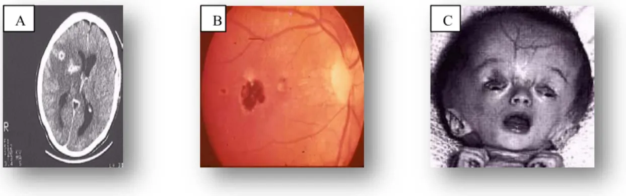
T. gondii infection can be transmitted congenitally if the mother acquires the infection during pregnancy, as parasites cross the placenta and infect the foetus. The risk of infection being passed to the foetus increases as gestation progresses, however, inversely, the severity of disease decreases with an increase in gestation period (Dunn et al., 1999; Cook et al., 2000). Without treatment of the mother during pregnancy, the incidence of acquired foetal infection during the first trimester is 10 % - 15 %, in the second trimester is 30 % and in the third is 60 % (Wong and Remington, 1994). If the mother receives treatment with spiramycin, these incidences decrease to 4.5 %, 17.3 % and 28.9 %, respectively (Wong and Remington, 1994). Congenital infection can occasionally result from reactivation of infection in immunosuppressed women if acquired before pregnancy (Wong and Remington, 1994). The variety of manifestations of congenital infection that occur in the foetus and in infants include spontaneous abortion, still-birth, a live infant with classic signs of congenital toxoplasmosis such as hydrocephalus or microcephalus, cerebral calcifications, mental retardation, seizures and retinochoroiditis (Hill and Dubey, 2002). The majority of cases are asymptomatic at birth, but most will develop neurological or ocular manifestations later in their lives. (Figure 1.6)
Figure 1.6: Clinical signs A) cerebral calcifications , B) Retina lesions due to ocular toxoplasmosis ,C)
Newborn with hydrocephalus .
Image source http://wiki.ggc.edu/wiki/toxoplasma_gondii
1.2.4. The Diagnosis
In most instances the non specific nature of the signs and symptoms of toxoplasmosis do not permit reliable diagnosis based only on the clinical findings. Its detection depends mainly on biological, serological, or histological methods or on the combination of some of these methods (Hill et al., 2005). However due to the diversity of T. gondii infection, investigations must be selected which are appropriated to that patient group. (Holliman et al., 2003)
1.2.4.1. The immonocompetent patient
The diagnosis of T. gondii infection in immunocompetent subjects relies on serology. As the infection is often asymptomatic, serologic diagnosis is usually retrospective and is used to determine the immune status. Available serologic procedures for the detection of T. gondii humoral antibodies include; the Sabin-Feldman dye test (DT), the modified agglutination test (MAT), the indirect hemagglutination test (IHAT) the indirect fluorescent antibody assay (IFA), the direct agglutination test (DAT), the latex agglutination test (LAT), the enzyme-linked immunosorbent assay (ELISA), and the immunosorbent agglutination assay test (IAAT). The IFA, IAAT, and ELISA tests have been modified to detect IgM antibodies (Remington et al., 1995). Immunosorbent agglutination assays (ISAGAs) are also suitable for IgM, IgA, or IgE detection. The methylene blue dye test for the detection of antibodies, introduced in 1948 by Sabin and Feldman, is maintained as a gold standard for serology tests by reference laboratories, but is labor-intensive and requires a continual supply of live organisms.
Immunoglobulin A and IgM are produced during the first week following infection and reach a plateau within one month, these antibodies appear sooner than the IgG antibodies and disappear faster than IgG antibodies after recovery (Remington et al., 1995). Specfic IgE antibodies are also produced early and rapidly disappear. Specific IgM antibodies typically decrease after one to six months and disappear in 25 % of patients within less than seven months but commonly remain detectable for a year or longer with the most sensitive methodologies such as the ISAGA. Concerning that IgG antibodies are synthesized 1 to 3 weeks after the initial rise in IgM levels, IgG synthesis reaches a plateau within 2 or 3 months, then decreases and then persists lifelong as residual titers. Since IgG can persist for the life time, IgM, which typic ally persists for 1 - 6 months, is used as a marker of recent infection, (Wilson and McAuley, 1999).