The MsmX ATPase plays a crucial role in pectin
mobilization by
Bacillus subtilis
Ma´rio J. Ferreira, Aristides L. Mendes¤, Isabel de Sa´-Nogueira*
UCIBIO, REQUIMTE, Departamento de Ciências da Vida, Faculdade de Ciências e Tecnologia, Universidade NOVA de Lisboa, Caparica, Portugal
¤ Current address: Instituto de Tecnologia Quı´mica e Biolo´gica, Anto´nio Xavier, ITQB NOVA, Universidade NOVA de Lisboa, Oeiras, Portugal
Abstract
Carbohydrates from plant cell walls are often found as heteropolysaccharides intertwined with each other. For competitive advantage against other microorganisms, and ability to fully exploit available carbon and energy sources,Bacillus subtilispossesses a high number of proteins dedicated to the uptake of mono- and oligosaccharides. Here, we characterize transporter complexes, belonging to the ATP-binding cassette (ABC) superfamily, involved in the uptake of oligosaccharides commonly found in pectin. The uptake of these carbohy-drates is shown to be MsmX-dependent, assigning a key role in pectin mobilization for MsmX, a multipurpose ATPase serving several distinct ABC-type I sugar importers. Muta-genesis analysis of the transmembrane domains of the AraNPQ MsmX-dependent importer revealed putative residues for MsmX interaction. Interestingly however, although MsmX is shown to be essential for energizing various ABC transporters we found that a secondB.
subtilisATPase, YurJ, is able to complement its function when placed intransat a different locus of the chromosome.
Introduction
The plant cell wall is a highly complex structure, with a variable species- and tissue-dependent composition, and a major reservoir of carbohydrates in the form of cellulose, hemicellulose, and pectin [1,2]. Cellulose is exclusively composed of D-glucose units, linked by β-1,4-glyco-sidic bonds and organized as linear parallel polymers connected via hydrogen bonds [1]. Hemicellulose and pectin are complex mixtures of branched polysaccharides composed of many different sugar monomers such as glucose, galactose, xylose, arabinose, mannose, rham-nose, and galacturonic acid [1,3,4].
Microorganisms have successfully developed concerted mechanisms for the degradation of these complex plant polysaccharides and the subsequent uptake of smaller sugar oligomers and monomers, which can be easily further metabolized. One such concerted mechanism is theBacillus subtilistransport and utilization system for arabinan [5], a hemicellulose polysac-charide commonly found in low-lignin and pectin-rich substrates such as sugar beet pulp and citrus waste [4]. This bacterium in its natural environment–the soil or the gastrointestinal tract a1111111111 a1111111111 a1111111111 a1111111111 a1111111111 OPEN ACCESS
Citation:Ferreira MJ, Mendes AL, de Sa´-Nogueira I (2017) The MsmX ATPase plays a crucial role in pectin mobilization byBacillus subtilis. PLoS ONE 12(12): e0189483.https://doi.org/10.1371/journal. pone.0189483
Editor:Hernaˆni Gero´s, Universidade do Minho, PORTUGAL
Received:May 12, 2017
Accepted:November 27, 2017
Published:December 14, 2017
Copyright:©2017 Ferreira et al. This is an open access article distributed under the terms of the
Creative Commons Attribution License, which permits unrestricted use, distribution, and reproduction in any medium, provided the original author and source are credited.
Data Availability Statement:All relevant data are within the paper and its Supporting Information files.
Funding:This work was supported by Fundac¸ão para a Ciência e a Tecnologia (FCT/MCTEShttp:// www.fct.pt/index.phtml.pt) [PEst-OE/BIA/UI0457/ 2011 to Centro de Recursos Microbiolo´gicos (CREM); UID/Multi/04378/2013 to Unidade de Ciências Biomoleculares Aplicadas (UCIBIO); fellowship SFRH/BD/73039/2010 to M.J.F.].
of animals [6,7]–possesses two endo-α-1,5-arabinanases, AbnA and Abn2, responsible for
breaking down the backbone of the homopolysaccharide arabinan [5,8], releasing arabinose monomers and oligomers. Two intracellularα-L-arabinofuranosidases, AbfA and Abf2 [9], are accountable for further degrading the arabinooligosaccharides, after their uptake through two different types of transport systems. The AraE proton symporter is responsible for the uptake of arabinose and also plays a role in the transport ofα-1,5-arabinobiose [10,11]; the AraNPQ ABC-type system is responsible for the uptake of arabinooligosaccharides up to at least 4 arabinosyl units [11]. This system, along with the ABC-type maltodextrins importer MdxEFG, is energized by the multipurpose ATPase MsmX [11,12]. However, MsmX together with another putative orphan ATPase, YurJ, are predicted to be the nucleotide-binding domain (NBD) that energizes five additional putative ABC-type importers ofB.subtilis[13].
ABC transporters constitute one of the largest and most diverse transporter superfamilies and are found in all three domains of life (Archaea, Bacteria and Eukarya) [14,15]. These transporters use the binding and hydrolysis of ATP to power the directional transport of a wide variety of substrates across membranes, ranging from ions to macromolecules, and all, importers or exporters, share a common structural organization: a modular architecture com-prising two transmembrane domains (TMDs) that form the translocation pore and two NBDs that hydrolyze ATP [14–17]. Although multipurpose ABC ATPases, such as MsmX [11], have been described in other microorganisms, such asStreptomyces lividans[18],Streptococcus pneumoniae[19] andStreptococcus suis[20], the capability to interact with and energize multi-ple transporters is rather uncommon among this type of ATPases. In fact, to date the ability of a single ATPase to energize several ABC-type systems has been exclusively reported in bacte-rial sugar importers.
In previous work, we found that MsmX is crucial for the utilization of arabinan byB.subtilis and the results suggested the existence of other MsmX-dependent ABC importers involved in the uptake plant cell-wall oligosaccharides [11]. Here, we extend our investigation to the role of MsmX in the utilization of distinct plant cell wall polysaccharides byB.subtilisand the bio-logical significance of sharing a single ATPase among multiple ABC transport systems. ABC-type I sugar importers responsible for the uptake of pectin-based polysaccharides were charac-terized and MsmX established as a key player in plant cell wall polysaccharides mobilization. Furthermore, using the AraNPQ importer as model, amino acid residues of the TMDs that are putatively important for the contacts with MsmX were identified by mutagenesis. In addition, we addressed the expression of both ATPases fromB.subtilisMsmX and YurJ. We found that in the tested conditions YurJ is not present in the cell, thus we propose that expression ofyurJ is subjected to post-transcriptional regulation.
Materials and methods
DNA manipulation and sequencing
Cycle Sequencing Kit (Applied Biosystems). The sequencing reaction was purified by gel filtra-tion and resolved in an ABI 3730XL sequencer.
Construction of
B
.
subtilis
strains
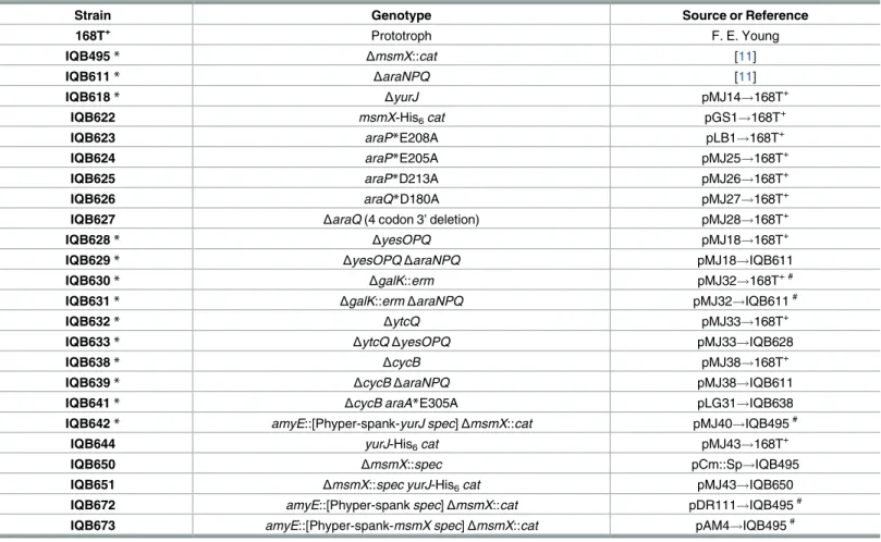
The generation of ayurJclean deletion and in-frame clean deletion ofyesOPQ,ytcQ,cycB, or araQ, in theB.subtilischromosome, were obtained by overlapping PCR followed by allelic replacement using the pMAD vector according to the procedure described by Arnaudet al. [22]. Briefly, plasmids carrying the sequences of the regions immediately upstream and down-stream of the gene(s) targeted for deletion were integrated into the chromosome ofB.subtilis by a single recombination event, promoted by growth at a nonpermissive temperature (42 ˚C) for plasmid replication. A second single recombination event occurs during growth at a per-missive temperature, thus allowing loss of the plasmid and substitution of the targeted allele without introduction of antibiotic resistance determinants. The ectopic regulated expression ofyurJormsmX, was carried out by placing each gene under the control of an IPTG-inducible promoter using the vector pDR111 (S1 Table) and subsequent integration at theamyElocus of theB.subtilischromosome by a double recombination event. The creation of mutations in araA(E305A),araP(E208A, E205A and D213A), andaraQ(D108A), leading to single amino acid substitutions, were obtained by overlapping PCR with mutagenic primers (S2 Table) followed by allelic replacement in theB.subtilischromosome using the pMAD vector as described above. The insertion-deletion mutation ofgalKin the chromosome was performed by insertion of an erythromycin resistance cassette and concomitant partial deletion ofgalKby a double recombination event in theB.subtilischromosome using a derivative of phagemid pBluescript II KS(+). ThemsmX-his6-tagandyurJ-his6-tagalleles were created by a single recombination event, at the respective locus of theB.subtilischromosome, using plasmids har-boring modified versions ofmsmXandyurJthat carry a His6-tag at the 3’ end. TheΔmsmX:: specallele was created using pCm::Sp [23] to switch the chloramphenicol resistance marker of theΔmsmX::catstrain by a spectinomycin resistance marker. Transformations ofB.subtilis strains were performed according to the method described by Anagnostopoulos and Spizizen [24]. AllB.subtilisstrains used in this study are listed inTable 1. A detailed description of plas-mids (S1 Table), primers (S2 Table), and constructions (S1 Appendix), is found in the support-ing information section.
Growth conditions
RNA extraction
B.subtilis168T+was grown in CSK minimal medium supplemented with 0.1% (w/v) of glu-cose, arabinose or arabinotriose, as previously described for growth kinetics parameters and doubling time determinations [11]. When the OD600nmof growing cultures reached 0.7–0.8, cells were collected by centrifuging 1.4 mL of each culture at 6000gand 4 ˚C for 5 minutes. Total RNA extraction was performed using the Absolutely Total RNA Miniprep kit (Agilent Technologies) according to the manufacturer’s instructions and using RNase-free technique. The integrity of RNA was analyzed in a 1% (w/v) agarose gel in TBE 1x. DNA contamination of RNA samples was assessed by PCR using primers ARA422 and ARA423 (S2 Table). Total RNA was quantified using a Nanodrop™1000 Spectrophotometer (Thermo Fisher Scientific) and stored in 10μL aliquots at -80 ˚C.
RT-qPCR experiments and data analyses
The primers used for RT-qPCR experiments–ARA583 and ARA584 (16S gene), ARA638 and ARA639 (yurJ), and ARA640 and ARA641 (msmX)–were designed with the help of the online tool Primer3Plus (http://primer3plus.com/cgi-bin/dev/primer3plus.cgi). Primer efficiency was
Table 1. List ofB.subtilisstrains used or constructed during the course of this work.
Strain Genotype Source or Reference
168T+ Prototroph F. E. Young
IQB495* ΔmsmX::cat [11]
IQB611* ΔaraNPQ [11]
IQB618* ΔyurJ pMJ14!168T+
IQB622 msmX-His6cat pGS1!168T+
IQB623 araP*E208A pLB1!168T+
IQB624 araP*E205A pMJ25!168T+
IQB625 araP*D213A pMJ26!168T+
IQB626 araQ*D180A pMJ27!168T+
IQB627 ΔaraQ(4 codon 3’ deletion) pMJ28!168T+
IQB628* ΔyesOPQ pMJ18!168T+
IQB629* ΔyesOPQΔaraNPQ pMJ18!IQB611
IQB630* ΔgalK::erm pMJ32!168T+ #
IQB631* ΔgalK::ermΔaraNPQ pMJ32!IQB611#
IQB632* ΔytcQ pMJ33!168T+
IQB633* ΔytcQΔyesOPQ pMJ33!IQB628
IQB638* ΔcycB pMJ38!168T+
IQB639* ΔcycBΔaraNPQ pMJ38!IQB611
IQB641* ΔcycB araA*E305A pLG31!IQB638
IQB642* amyE::[Phyper-spank-yurJ spec]ΔmsmX::cat pMJ40!IQB495#
IQB644 yurJ-His6cat pMJ43!168T+
IQB650 ΔmsmX::spec pCm::Sp!IQB495
IQB651 ΔmsmX::spec yurJ-His6cat pMJ43!IQB650
IQB672 amyE::[Phyper-spankspec]ΔmsmX::cat pDR111!IQB495#
IQB673 amyE::[Phyper-spank-msmX spec]ΔmsmX::cat pAM4!IQB495# Arrows indicate transformation and point from donor DNA to the recipient strain.
# Transformation was carried out with linearized plasmid.
*The effect of these mutations in the utilization of several carbon sources is summarized inS3 Table.
assessed using the Rotor-Gene SYBR1
Green PCR Kit (QIAGEN) and RT-qPCR experiments were performed using the SensiFAST™SYBR No-ROX One-Step Kit (Bioline), both in a Rotor-Gene 6000 (Corbett) real-time cycler. RT-qPCR experiments were performed according to the kit manufacturer’s instructions, using 40 ng of total RNA and 0.1μM of each primer, in a volume of 12.5μL. Statistical analyses were performed with GraphPad Prism (version 5.00) usingCtvalues obtained from three independent assays.pvalues were determined using an unpairedttest.
Protein extracts of
B
.
subtilis
and Western Blot analysis
B.subtilisstrains IQB622, IQB644, and IQB651 were grown in CSK minimal medium supple-mented with 0.1% (w/v) of glucose, arabinose or arabinotriose, as previously described for growth kinetics parameters and doubling time determination [11]. Cells of growing cultures were collected as described for RNA extraction (see above). The collected cells were resus-pended in 100μL of Lysis Buffer (500 mM KCl, 20 mM HEPES K+, pH 7.6, 10 mM EDTA, 1 mM DTT, 10% glycerol). Lysozyme (1 mg.mL-1) was added and the mixture was incubated for 10 min at 37 ˚C, followed by three cycles of freezing in liquid nitrogen and thawing for 5 min at 37 ˚C. 10 mM PMSF and 7.5 U of Benzonase1Nuclease (Sigma-Aldrich Co.) were added followed by an incubation for 15 min at 37 ˚C. Total protein content for each extract was determined using Bio-Rad Protein Assay (Bio-Rad Laboratories, Inc.). 20μg of total protein from each extract and 1μg of purified MsmX-His6were loaded in a 12.5% SDS-PAGE and run at constant electrical current (30 mA) for 50 min. The fractionated proteins were then electro-transfered into a nitrocellulose membrane (0.45μm; Bio-Rad), for 90 min at constant voltage (100 V) and 4 ˚C. Protein-blocking to the membrane was then performed using 20 mL of a powdered milk solution in TBS-Tween (5% w/v) for 30 min at room temperature with mild shaking. The membrane was then washed with TBS-Tween for 15 min at room temperature with mild shaking, followed by overnight incubation, at 4 ˚C, with 10 mL of primary antibody solution (mouse monoclonal Anti-6X His-tag1antibody [HIS.H8; Abcam], diluted 1:1000 in PBS-Tween with 4% w/v powdered milk). All subsequent incubation and wash steps were per-formed at room temperature with mild shaking. The membrane was washed twice with 10 mL of TBS-Tween, for 10 min, followed by incubation with 10 mL of secondary antibody solution (HRP-conjugated goat anti-mouse IgG antibody [Jackson ImmunoResearch Europe Ltd.], diluted 1:10000 in TBS-Tween with 4% w/v powdered milk) for 30 min. Finally, the membrane was washed three times with 10 mL of TBS-Tween solution. All subsequent steps were per-formed in a dark room. Blots were developed using 1 mL Western Lightning™ECL Pro kit (PerkinElmer). Amersham Hyperfilm plates (GE Healthcare) were exposed to luminescence for 30 sec inside a closed Hypercassette Autoradiography Cassette (GE Healthcare). The mem-brane was then stripped and reprobed with a second primary antibody solution (mouse mono-clonal Anti-σ70[fromE.coli; a gift from Fujita M.], diluted 1:1000 in PBS-Tween with 4% w/v powdered milk) for loading control. The same secondary antibody was used as described above. Blots were developed using 1 mL Western Lightning™ECL Pro kit (PerkinElmer). Amersham Hyperfilm plates (GE Healthcare) were exposed to luminescence for 3 min inside a closed Hypercassette Autoradiography Cassette (GE Healthcare).
Results
Identification of carbohydrates imported via MsmX-dependent systems
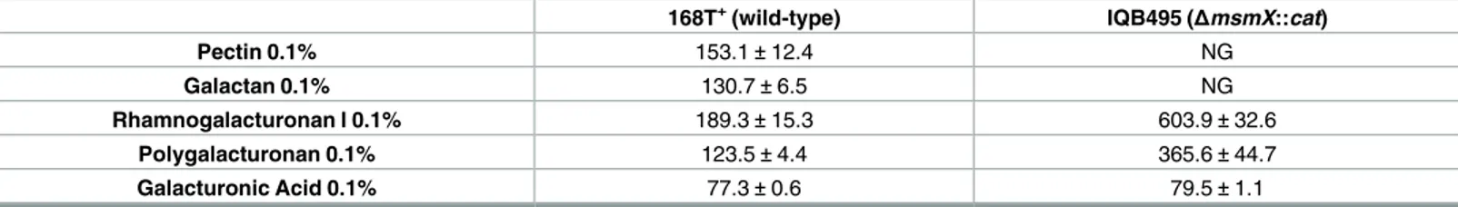
(168T+) and themsmX-null mutant strain (IQB495,S1 Fig) were grown in the presence of pec-tin, galactan, rhamnogalacturonan type I, polygalacturonan and galacturonic acid, and the results are summarized inTable 2.
The ability to utilize pectin was completely abolished in themsmX-null mutant when pared to the wild-type. Because this polysaccharide is highly complex, branched, and com-posed by various sugars, mainly arabinose, galactose, rhamnose and galacturonic acid, this result strongly suggests that MsmX is involved in the uptake of most of these sugars. Growth in the presence of galactan is also abolished when a functional MsmX is missing (Table 2), assigning this ATPase as a critical player in the utilization of galactan. Since galactose is imported by the arabinose proton-symporter AraE [10,27], MsmX is likely to be responsible for energizing an ABC-type importer dedicated to the uptake of galactooligosaccharides result-ing from the extracellular degradation of galactan.
The absence of a functional MsmX also had a significant negative impact on the growth of B.subtilisin minimal medium supplemented with rhamnogalacturonan type I or polygalactur-onan (Table 2). Unlike the complete loss of growth observed when arabinan [11] or galactan were used as sole carbon and energy source, a slower but steady growth rate was detected for themsmX-null strain in the presence of either rhamnogalacturonan type I or polygalacturo-nan. Interestingly, the increase of the doubling-time in the mutant strain (IQB495) when com-pared to the wild-type was very similar for both polysaccharides, about 3-fold (Table 2). Since galacturonic acid is the major component of both polysaccharides, the effect of theΔmsmX:: catmutation in the uptake of this monosaccharide was also assessed and the results show that the utilization of galacturonic acid is MsmX-independent (Table 2). Thus, the negative impact in the growth kinetics parameters observed in the presence of rhamnogalacturonan type I or polygalacturonan is most probably due to the existence of at least one MsmX-dependent ABC-type importer responsible for the uptake of galacturonic acid oligomers and/or rhamnose-galacturonic acid disaccharides but not rhamnose-galacturonic acid.
Functional characterization of ABC-type sugar importers
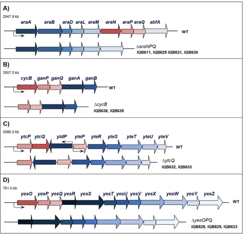
In a previous work by our group [11], it was suggested that AraNPQ was only responsible for the uptake of linearα-1,5-linked arabinooligosaccharides, and nonlinearα-1,2- and α-1,3-linked oligomers were transported by an unidentified MsmX-dependent system, because anaraNPQin-frame deletion (Fig 1A) partially abolished growth on branched arabinan while prevented growth on debranched arabinan. ThecycB-ganPQABoperon (Fig 1B), encodes a putativeβ-galactosidase (GanA), a putative arabinogalactan endo-β-1,4-galactanase (GanB), an ABC-type solute-binding protein (SBP), CycB, two transmembrane domains (TMD), GanP and GanQ, and was proposed to be involved in the utilization of galactan [28,29]. Since the CycB-GanPQ system (formerly YvfKLM) was also suggested to be energized by MsmX [13], and the arabinose proton symporter AraE is able to transport arabinose, xylose and galactose
Table 2. Growth of aB.subtilis msmX-null mutant in the presence of different mono- and polysaccharides.Doubling time (min) of different strains in liquid minimal medium (CSK) using pectin, galactan, rhamnogalacturonan I, polygalacturonan or galacturonic acid as sole carbon and energy source. Results are the averages of three independent assays and their respective standard deviations. NG, no growth.
168T+(wild-type) IQB495 (ΔmsmX::cat)
Pectin 0.1% 153.1±12.4 NG
Galactan 0.1% 130.7±6.5 NG
Rhamnogalacturonan I 0.1% 189.3±15.3 603.9±32.6
Polygalacturonan 0.1% 123.5±4.4 365.6±44.7
Galacturonic Acid 0.1% 77.3±0.6 79.5±1.1
[10,27], we targeted this transporter as the system potentially responsible for the uptake of nonlinear arabinooligosaccharides.
Fig 1. Organization of thearaABDLMNPQ-abfA,cycB-ganPQAB,ytcPQ-ytdP-ytePRSTUVandyesOPQRSTUVWXYZloci of the wild-type (WT) and mutantB.subtilisstrains.The location of the four regions is indicated in kilobase pairs. The genes are represented by arrows pointing in the direction of transcription. The constructs bearing the mutations used in this work are displayed below each region, and the strains harboring each mutation are indicated in front of the construct. The in-frame deletions generated by allelic replacement,ΔaraNPQ,ΔcycB,ΔytcQ, andΔyesOPQ, are depicted below the
araABDLMNPQ-abfA(A),cycB-ganPQAB(B),ytcPQ-ytdP-ytePRSTUV(C), andyesOPQRSTUVWXYZ(D) loci, respectively. All constructions are described in Materials and Methods and supporting information.
An in-frame deletion mutation of thecycBgene was generated by allelic replacement (Fig 1B), and the physiological effect of this mutation in the utilization of arabinan byB.subtilis strains IQB638 (ΔcycB) and IQB639 (ΔcycBΔaraNPQ) was assessed by the determination of growth kinetics parameters (Table 3). Similarly to that observed for theΔaraNPQmutant (188.8±15.3 min; IQB611 [11]), only a marginal increase in the doubling time of theΔcycB mutant was detected (IQB638;Table 3), when compared do the wild-type strain grown on arabinan (149.0±6.8 min; strain 168T+[11]). Nonetheless, the ability of the doubleΔcycB ΔaraNPQmutant (IQB639) to grow on the same substrate was completely abolished (Table 3), suggesting that the putative MsmX-dependent transporter for nonlinear arabinooligosacchar-ides was indeed CycB-GanPQ.
However, we later found out that the branched sugar beet arabinan (Megazyme Interna-tional), contained a level of galactose 5-fold higher than that disclosed (less than 4% (w/w) of galactose). Thus, to investigate if the growth observed in theΔaraNPQmutant was owed to the presence of galactose, or most likely galactooligosaccharides, and not due to nonlinear arabi-nooligosaccharides, an insertion-deletion mutation of the galactose kinase gene (galK) was generated (S1 Fig). In the resulting Gal-strains, IQB630 (ΔgalK::erm) and IQB631 (ΔgalK::erm ΔaraNPQ), an increase in the doubling time was observed for the single mutant and the double mutant is impaired to grow in the presence of branched arabinan as the sole carbon and energy source (Table 3). These results confirmed that the branched arabinan utilized is highly contaminated with galactose/galactooligosaccharides and consequently responsible for main-taining growth ofB.subtiliswhen a functional AraNPQ system is unavailable. Additionally, these results implicate that the CycB-GanPQ system plays a role in the uptake of galactooligo-saccharides, as previously suggested [28,29].
CycB was previously described as a cyclodextrin-binding protein [30]. However, during the course of this work a recent study showed that CycB (renamed GanS) was able to bind sugar oligomers with three and four galactosyl units [31]. Thus, we tested the impact of deleting the cycBgene in the ability to utilize galactan. The obtained results show that theΔcycBmutant (IQB638) displays a 2-fold increase in the doubling time when galactan is the sole carbon and energy source, when compared to the wild-type strain (Table 4). Therefore, CycB is involved in the utilization of galactan byB.subtilis, and is likely to be part of a putative CycB-GanPQ ABC-type importer responsible for the uptake of galactooligosaccharides released by the enzy-matic breakdown of galactan by the extracellular endo-β-1,4-galactanase GanB.
In addition, we also tested a doubleΔcycBΔaraNPQmutant strain (IQB639) in the presence of galactan and its ability to grow was completely lost. To confirm that the observed growth of
Table 3. Uptake of arabinan inB.subtilis.Doubling time (minutes) for different strains in liquid minimal medium (CSK) using glucose or arabinan as sole carbon and energy source. Results are the averages of three independent assays and their respective standard deviations. NG, no growth.
IQB638 (ΔcycB) IQB639 (ΔcycBΔaraNPQ) IQB630 (ΔgalK::erm) IQB631 (ΔaraNPQΔgalK::erm) Glucose 0.1% 52.6±0.1 52.1±1.2 45.8±1.2 53.9±1.7
Arabinan (branched) 0.1% 168.7±39.4 NG 300.4±28.1 NG
https://doi.org/10.1371/journal.pone.0189483.t003
Table 4. Uptake of galactan inB.subtilis.Doubling time (minutes) for different strains in liquid minimal medium (CSK) using glucose or galactan as sole carbon and energy source. Results are the averages of three independent assays and their respective standard deviations. NG, no growth.
168T+(wild-type) IQB638 (ΔcycB) IQB639 (ΔcycBΔaraNPQ) IQB641 (ΔcycB araA*E305A)
Glucose 0.1% 55.5±1.3 52.6±0.1 52.1±1.2 54.2±2.1
Galactan 0.1% 130.7±6.5 258.3±29.6 NG 532.8±2.2
theΔcycBmutant in the presence of galactan was due to the presence of arabinose, we con-structed an Ara-strain by substituting the key catalytic residue E305 of L-arabinose isomerase (AraA) by an alanine. This mutation was previously shown to completely block the isomeriza-tion of L-arabinose to L-ribulose, thus preventing its metabolism [32]. When grown in the presence of galactan, theΔcycB araAE305A strain (IQB641) presented an extremely high
dou-bling time (Table 4), which confirms that the growth observed in theΔcycBmutant strain (IQB638) is most probably due to the presence of arabinose and/or arabinosyl oligomers.
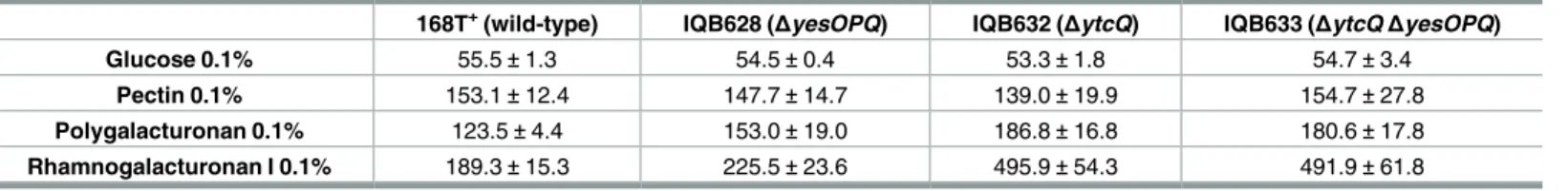
Polygalacturonan and rhamnogalacturonan type I are the two major components of pectin. The former is a linear chain of D-galacturonic acid units, while the latter has a backbone com-posed of alternating L-rhamnose and D-galacturonic acid units highly decorated with mainly arabinans and galactans as side chains [1,33]. Ochiaiet al. [33] previously showed that the genes encoding the putative YesOPQ and YtcQP-YteP ABC-type importers, comprising the predicted solute-binding proteins YesO and YtcQ, and two transmembrane domains each, YesPQ and YtcP-YteP, respectively, were induced in the presence of rhamnogalacturonan type I. Since these transporters were proposed to be energized by MsmX [13], and this ATPase was shown to be important for the utilization of these polysaccharides byB.subtilis(Table 2), we constructed in-frameΔytcQandΔyesOPQmutations (Fig 1C and 1D, respectively) and assessed their effect in the uptake of polygalacturonan and rhamnogalacturonan type I. The results presented inTable 5showed that both mutations have a negative impact in the utiliza-tion of the two polysaccharides, moreover this effect was larger in theΔytcQmutant (IQB632) than in theΔyesOPQstrain (IQB628). These results indicate that YtcQP-YteP is involved in the uptake of polygalacturonan and rhamnogalacturonan, and suggest that YesOPQ may also play a role in the utilization of these polysaccharides. However, the double mutant strain (ΔytcQΔyesOPQ, IQB633) displayed no increase in the doubling time in the presence of either substrate when compared to the singleΔytcQmutant (Table 5). This observation may indicate thatΔyesOPQhas only a marginal effect in the uptake of these substrates, which is likely to be negligible in the doubleΔytcQΔyesOPQmutant since theΔytcQmutation reveals a signifi-cantly higher impact in the utilization of both polysaccharides.
Additionally, for both polysaccharides, the doubling time of the doubleΔytcQΔyesOPQ mutant (Table 5) was lower than that observed for theΔmsmX::catmutant (Table 2). This
Table 5. Uptake of pectin-based polysaccharides inB.subtilis.Doubling time (minutes) for different strains in liquid minimal medium (CSK) using glu-cose, pectin, polygalacturonan or rhamnogalacturonan I as sole carbon and energy source. Results are the averages of three independent assays and their respective standard deviations.
168T+(wild-type) IQB628 (ΔyesOPQ) IQB632 (ΔytcQ) IQB633 (ΔytcQΔyesOPQ) Glucose 0.1% 55.5±1.3 54.5±0.4 53.3±1.8 54.7±3.4
Pectin 0.1% 153.1±12.4 147.7±14.7 139.0±19.9 154.7±27.8
Polygalacturonan 0.1% 123.5±4.4 153.0±19.0 186.8±16.8 180.6±17.8
Rhamnogalacturonan I 0.1% 189.3±15.3 225.5±23.6 495.9±54.3 491.9±61.8
https://doi.org/10.1371/journal.pone.0189483.t005
Table 6. Effect ofyurJdeletion and complementation analysis ofmsmXdeletion inB.subtilis. Doubling time (min) of different strains in liquid minimal medium (CSK) using glucose or arabinotriose as sole carbon and energy source. Results are the averages of three independent assays and their respective standard deviations.#IPTG was added to a final concentration of 1 mM. NG, no growth.
IQB618 (ΔyurJ)
IQB672 (ΔmsmX::cat amyE:: [Phyper-spankspec])
IQB673 (ΔmsmX::cat amyE ::[Phyper-spank-msmX spec])
IQB642 (ΔmsmX::cat amyE:: [Phyper-spank-yurJ spec]) Glucose 0.1% 50.4±5.5 59.8±0.7# 59.1±2.0# 52.9±3.2#
Arabinotriose 0.1%
92.8±10.9 NG# 107.6±3.9# 94.6±0.5#
observation was expected for rhamnogalacturonan I, since the uptake of the oligosaccharides released from the breakdown of its arabinan and galactan side chains is MsmX-dependent, but polygalacturonan is a linear homopolysaccharide composed of galacturonic acid units. Never-theless, our results, together with the previous observations by Ochiaiet al. [33], indicate that the multitask ATPase MsmX most probably has a role in energizing the YesOPQ and YtcQ-P-YteP systems (Table 2andTable 5).
Functional analysis of
yurJ
TheB.subtilischromosome encodes two highly similar ATPases, MsmX and YurJ (>55% amino acid identity), belonging to sub-family 5a of ABC-type importers associated to carbohy-drate uptake [13]. MsmX also displays a high amino acid identity (>40%) to the MalK ATPase [34] belonging to theE.colimaltose/maltodextrins ABC-type importer. The complete Mal-EFGK2transporter structure determination revealed the amino acids responsible for the con-tacts between the TMDs and the NBDs [35]. An alignment of the primary sequence of the three proteins, MsmX, YurJ and MalK, showed that most of the MalK amino acids contacting the TMDs are conserved in both MsmX and YurJ (S2 Fig). Furthermore, the similarity between bothB.subtilisATPases suggests the possibility of YurJ being able to associate to MsmX-energized ABC transporters.
We have previously showed that depletion of MsmX in amsmX-null mutant (strain IQB495) impairs the utilization of arabinose oligomers, which is AraNPQ-dependent, thus indirectly demonstrating that YurJ is unable to functionally substitute MsmX in the conditions tested [11]. Nevertheless, we constructed a deletion ofyurJin theB.subtilischromosome (ΔyurJ; strain IQB618,Table 1andS1 Fig) and tested its ability to grow on arabinotriose as the sole carbon and energy source. The results shown inTable 6indicated that this deletion has no negative impact in the ability to grow on arabinotriose when compared to the wild-type strain 168T+(98.2±10.0 min [11]).
To confirm that MsmX is the sole ATPase responsible for energizing the importer AraNPQ, we placed themsmXallele under the control of an IPTG inducible promoter (Phyper-spank) at theamyElocus of the chromosome in aΔmsmXgenetic background, strain IQB673 (Table 1
andS1 Fig). By comparison to the negative control strain, IQB672, the ectopic controlled expression ofmsmXwas able to re-establish the capacity to utilize arabinotriose in amsmX -null genetic background (Table 6). Interestingly, althoughyurJis not able to energize AraNPQ in amsmX-null background (strain IQB495;Table 1), when theyurJallele is placed at the amyElocus of the chromosome (strain IQB642,Table 6andS1 Fig) complementation of the msmXdeletion is attained. These observations indicate that YurJ is in fact capable to function-ally complement MsmX and energize the uptake of arabinooligosaccharides by AraNPQ, moreover suggest that in the conditions tested YurJ is not present in the cell when theyurJ allele is located in its natural chromosomal genetic context.
Table 7. Quantification ofyurJandmsmXmRNA level in theB.subtiliswild-type strain.The results represent the fold-change of the expression in the target conditions (T) versus the control conditions (C). (glu), (ara) and (A3) indicate the presence of 0.1% of glucose, arabinose or arabinotriose, respectively.
Primers ARA638 and ARA639 were used foryurJand primers ARA640 and ARA641 formsmX. Fold-change was normalized using primers ARA583 and ARA584 for the 16S gene. Statistical analyses were performed with GraphPad Prism (version 5.00) usingCtvalues obtained from three independent assays.
pvalues were determined using an unpairedttest (ns, non-significant difference;*,p<0.05;**,p<0.01;***,p<0.001).
yurJ msmX
(glu)T (A3)T (glu)T (A3)T
(ara)C 0.993±0.054ns 1.782±0.323* 0.013±0.001*** 0.899±0.051ns
(glu)C - 1.794±0.311** - 68.120±3.683***
A post-transcriptional regulatory mechanism controls
yurJ
expression
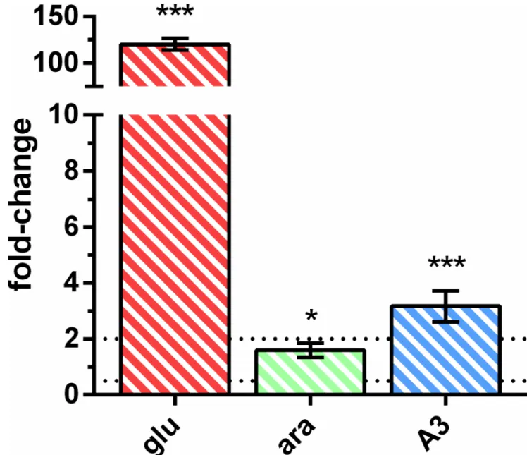
Since YurJ is able to energize the importer AraNPQ in anmsmX-null mutant strain when expressed intransunder the control of an IPTG-inducible promoter, but not from its own locus, we analyzed its expression at the transcriptional level in the wild-type strain. The expres-sion level of bothyurJandmsmXwas determined in the same conditions described above by measuring the mRNA levels of both transcripts using quantitative reverse transcription poly-merase chain reaction (RT-qPCR). The obtained data were used to calculate gene expression fold-change and the results are summarized inTable 7andFig 2.
Fig 2. mRNA level ofyurJversusmsmXin theB.subtiliswild-type strain.The results represent the relative expression ofyurJversus msmX. Upper and lower relative expression threshold of 2.0 and 0.5, respectively, are represented by dotted lines. (glu,red-striped bar), (ara, green-striped bar) and (A3,blue-striped bar) represent the presence of 0.1% of glucose, arabinose or arabinotriose, respectively. Primers ARA638/ARA639 and ARA640/ARA641 were used foryurJandmsmX, respectively. Fold-change was normalized using primers ARA583 and ARA584 for the 16S gene. Statistical analyses were performed with GraphPad Prism (version 5.00) usingCtvalues obtained from three independent assays.pvalues were determined using an unpairedttest (ns, non-significant difference;
*,p<0.05;**,p<0.01;***,p<0.001).
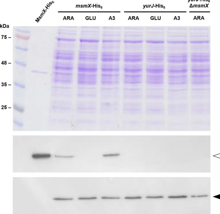
ThemsmXandyurJmRNA level was similar in the presence of both arabinose and arabino-triose, however in the presence of glucosemsmXexpression is repressed about 75-fold while the level ofyurJmRNA was not subjected to glucose repression. These observations correlate with previous results that placemsmX, but notyurJ, in the CcpA regulon [36–38]. Further-more, when directly comparing the transcript levels from both genes it was clear that there was no significant difference in their expression in the presence of arabinose or arabinotriose (Fig 2). In fact, when arabinotriose is used in growth assays a slightly higher expression level (about 3-fold) was detected foryurJwhen compared tomsmX(Fig 2). These results clearly show that in these conditionsyurJis expressed at the transcriptional level in its natural genetic context in theB.subtilischromosome suggesting the existence of a post-transcriptional regulatory mecha-nism responsible for the inability of YurJ to substitute MsmX in amsmX-null mutant. Thus, in order to determine if YurJ is translated, modified versions of both ATPases were constructed by fusing a C-terminal His6-tag tomsmX(IQB622) andyurJ(IQB644) in their own loci, as result of a single recombination event. In addition, a modified version ofyurJ-his6was also generated in amsmX-null background (IQB651). The accumulation of MsmX-His6and YurJ-His6in cells grown in the same conditions used in RT-qPCR experiments was detected by Western blot analysis. Similar amounts of total protein extracts from the three strains, grown in the presence of arabinose, and/or glucose or arabinotriose, were used for the detection of MsmX-His6(42.4 kDa) and YurJ-His6(42.2 kDa), respectively. Additionally, a protein extract of the wild-type strain (168T+) grown in CSK with arabinose was used as negative control and purified recombi-nant MsmX-His6used as positive control for His-tag detection (Fig 3).
The data confirm the presence of MsmX in the cells when grown in minimal medium with either arabinose or arabinotriose, however in identical conditions YurJ was absent (Fig 3). As result of the strong glucose-mediated repression ofmsmX, in accordance to the results obtained by RT-qPCR, no MsmX is detected when glucose is present in the medium (Fig 3). Thus, by combining the RT-qPCR data and Western Blot analysis, we show that although msmXandyurJare being similarly expressed in the presence of arabinotriose, the latter trans-lation product is not detected. Furthermore, in amsmX-null background, or in conditions in which MsmX is not present, YurJ remains undetectable. Together the results indicate that in minimal medium with either glucose, arabinose or arabinotriose as sole carbon and energy source,yurJis transcribed but not translated.
Identification of key residues for the interaction between MsmX and the
TMDs of AraNPQ
features for determining which amino acids should be targeted. Single alanine substitutions of residues E205, E208 and D213 in AraP and D180 in AraQ were generated and their effect in the functionality of AraNPQ was inferred by the impact in arabinotriose uptake (Table 8).
Fig 3. MsmX and YurJ detection by Western Blot.Top panel, SDS-PAGE (12.5%) with a molecular weight marker (NZYColour Protein Marker II) and samples used for Western Blot (20μg of total extract and 1μg of purified MsmX-His6were loaded);Middle panel, Autoradiography plate after
His-tag detection using an anti-His6-specific antibody;Bottom panel, Autoradiography plate afterσAdetection using an anti-σ70-specific antibody.msmX
-His6,yurJ-His6, andyurJ-His6ΔmsmXrepresent strains IQB622, IQB644, and IQB651 respectively. MsmX-His6(42.4 kDa) andσA(42.9 kDa) are
indicated by open or closed arrowheads, respectively. Extracts obtained from cells grown in minimal medium supplemented with arabinose (ARA), glucose (GLU) or arabinotriose (A3) as described in Materials and Methods.
These results showed that the single E205A mutation in AraP had no effect on the growth rate ofB.subtilis, while the E208A substitution resulted in a small negative impact in the uptake of arabinotriose, an increase of about 10% in the doubling time of strain IQB623 (araPE208A;
Table 8) when compared to the wild-type (98.2±10.0 min [11]). This is in accordance with the results published by Mourezet al. [42] for theE.colimaltose transporter, where for each of the TMDs, a single alanine substitution of the residue homologous to E208 from AraP (E401 in MalF and E190 in MalG) had little to no impact in the uptake of maltose. Although E205 and E208 from AraP are still likely to establish contacts with MsmX, they may not be essential for TMD-NBD interaction. On the other hand, the D213A mutation in AraP severely impaired the uptake of arabinotriose as shown by the increase in the doubling time approximately 3.6-fold. Likewise, although not as adversely, the homologous D180A substitution in AraQ had also a negative impact in the growth rate ofB.subtilisin the presence of arabinotriose (Table 8).
In theE.colimaltose transporter the Q loop of MalK establishes additional contacts with the C-terminal end of MalG. This tail, which is not found in MalF, inserts itself along the MalK dimer interface and its four terminal amino acids (-GVKG) interact with residues from the Q loop of both NBDs [35]. Interestingly, the primary sequence of this C-terminal tail from MalG is highly conserved in AraQ, MdxG and GanQ but not in the MsmX-independent OpuAB (Fig 4). Since this feature was only found in systems that are associated with MsmX we deleted the four terminal amino acids of AraQ and assessed the impact of their absence in the uptake of arabinotriose. This deletion resulted in a severe increase of almost 5-fold in the dou-bling time ofB.subtilisstrain IQB627 (ΔaraQ) when compared to that of the wild-type (Table 8). InB.subtilisthis C-terminal tail feature is not found in other sub-families of ABC-type transporters (with the exception of sub-family 5a) and might be a requirement for the interaction between the ATPase MsmX and the TMDs of systems it energizes. However, we cannot exclude the possibility that the small deletion generated in AraQ caused a destabiliza-tion of the protein leading to its degradadestabiliza-tion.
Discussion
ABC transporter genes are the most frequent class of protein coding genes found in the B.subtilisgenome [43]. Three ABC-type I sugar importers have been identified inB.subtilis that are energized by the same ATPase, MsmX: AraNPQ, responsible for the uptake of
Fig 4. The TMDs from the AraNPQ-MsmX system.Membrane-spanning domains in AraP (top) and AraQ (bottom) were predicted using TMpred (http://www.ch.embnet.org/software/TMPRED_form.html). The position of the first and last amino acid of each membrane-spanning domain is indicated. Alignments of the TMDs from theB.subtilisAra, Mdx, Gan, and OpuA and theE.coliMal ABC transporters were obtained using Clustal Omega (http://www.ebi.ac.uk/Tools/msa/clustalo/) and are partially shown. Identical (´*´) and similar (´.´ or ´:´) amino acids are indicated. Gaps in the amino acid sequences inserted for alignment optimization are indicated by a dash (–). The “EAA” sequence motifs in the last cytoplasmic loop are boxed. Identical residues between the four TMDs belonging to sugar transporters are highlighted inred. Similar residues between the four TMDs belonging to sugar transporters are highlighted inpink. Identical or similar residues between the three TMDs belonging to sugar transporters fromB.subtilisare highlighted inorange. Residues selected for mutagenesis in AraP and AraQ are indicated by open arrowheads. MalF and MalG residues known to establish contacts with the MalK dimer are underlined ingreen. Residues selected for deletion in the C-terminal end of AraQ are boxed. Accession numbers: AraP (P94529), AraQ (P94530), MdxF (O06990), MdxG (O06991), GanP (O32261), GanQ (O07011), OpuAB (P46921), MalF (P02916), and MalG (P68183).
https://doi.org/10.1371/journal.pone.0189483.g004
Table 8. Effect of mutations in AraP and AraQ in the uptake ofα-1,5-arabinotriose.Doubling time (min) of different strains in liquid minimal medium (CSK) using glucose or arabinotriose as sole carbon and energy source. Results are the averages of three independent assays and their respective standard deviations.
IQB623 (araP*E208A) IQB624 (araP*E205A) IQB625 (araP*D213A) IQB626 (araQ*D180A) IQB627 (ΔaraQ) Glucose 0.1% 56.5±1.2 53.1±0.1 53.2±1.0 54.9±1.7 56.1±2.3
Arabinotriose 0.1% 114.0±4.8 94.1±5.0 357.0±25.3 149.3±12.8 472.8±22.6
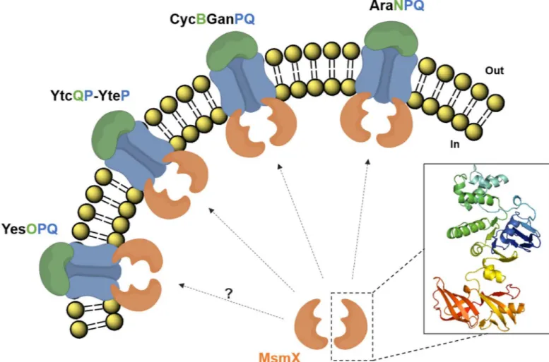
arabinooligosaccharides [11]; MdxEFG, involved in the transport of maltodextrins [12]; and CycB-GanPQ, which is the major transporter of galactooligosaccharides [31]. During the course of this work Watzlawicket al. [31] indirectly demonstrated that MsmX energizes CycB-GanPQ, since its presence was shown to be essential for induction of thecycB-ganPQABoperon in the presence of galactan. The results presented here also suggest that the previously uncharacterized YtcQP-YteP and YesOPQ systems are energized by MsmX, as proposed by the work of Quentin et al. [13]. Furthermore, we provide evidence that these systems play a role in the transport of polygalacturonan (or galacturonic acid oligomers) and rhamnogalacturonan type I (or rham-nose-galacturonic acid disaccharides), as previously hypothesized by Ochiaiet al. [33]. Accord-ing to a microarray analysis on the utilization of carbohydrates by an alkaliphilicBacillussp., genes with high identity toyesOPQandytcQP-ytePwere shown to have increased expression levels in the presence of pectin [44], and further bioinformatics analysis [45], support the role of these transporters in the uptake of polygalacturonan and rhamnogalacturonan. Our work shows that the utilization of arabinan, galactan, rhamnogalacturonan type I and polygalacturo-nan byB.subtilisis completely or partly MsmX-dependent, thus establishing this ATPase as a key player in the uptake of pectin-rich substrates. MsmX is now associated to four different ABC-type I sugar importers involved in the mobilization of pectin (Fig 5).
To apprehend the reason why carbohydrate ABC-type I importers share a common ATPase, in contrast to the majority of ABC-type importers and exporters encoded in theB. subtilischromosome, we may speculate in this particular case it may reflect the fact that in plant biomass sugars are often found as heteropolysaccharides, i.e., sugars like arabinose, galactose or xylose are usually present in the same substrates. Unlike glucose, which is the pre-mier source of energy for most heterotrophic organisms and effectively downregulates the expression of most genes associated with the utilization of other sugars, a clear hierarchy among those is not as well defined. Although a preference of one carbohydrate over another has been reported for bothB.subtilis[37,46] andE.coli[47] that might not be the case for sug-ars transported via the MsmX-dependent ABC importers. Being often simultaneously avail-able, oligomers of arabinose, galactose and galacturonic acid, and rhamnose-galacturonic acid disaccharides are likely to be transported at the same time. Thus, a single ATPase capable of serving the various carbohydrate-specific transport systems, like MsmX, may be a more effec-tive strategy for the cell to respond quickly, adapt and proliferate in a specific ecologic niche.
Additionally, we found that in an engineeredB.subtilisstrain an additional ATPase (YurJ) encoded in its genome is able to functionally replace MsmX when ectopically expressed under the control of an inducible promoter. Moreover, the results presented here show that YurJ is not present in the cell in the conditions tested (Fig 3), despite the presence of similar levels of yurJandmsmXmRNA (Fig 2). Thus, a post-transcriptional regulatory mechanism, is likely to control the synthesis of YurJ in the conditions tested. It has been reported in proteomic studies of theB.subtiliscytosol that YurJ is present in the cell in certain conditions [48]. Furthermore, a condition-dependent transcriptome analysis ofB.subtilis[49] indicated thatyurJis co-tran-scribed together with a putative antisense RNA (S1254) located downstream from the
frlBONMDgenes (formerlyyurPONML) involved in the utilization of Amadori products (fruc-tosamines) [50]. A longer transcription unit includingyurJwas also detected (frlB-frlO-frlN -frlM-frlD-S1254-yurJ), and the putative antisense RNA overlaps and pairs to the convergent regulatory gene of the operon,frlR[49]. All these observations suggest the involvement of the antisense RNA (S1254) in preventingyurJtranslation by an unknown mechanism.
energize its neighboring system. Nevertheless, we may hypothesize that interchangeability between MsmX and YurJ may occur in specific conditions which allow translation of YurJ. In fact, two ABC ATPases ofStreptococcus mutans, MsmK and MalK, are capable of substituting each other and energize both transporters for raffinose/stachyose and for maltodextrins, even though they are encoded by genes located in the vicinity of the remaining components of their partner ABC transporter [51].
In order to understand why some ATPases are able to bind to various ABC-type systems when most are restricted to interactions with a single transporter, several of the residues expected to mediate the TMD-NBD contacts between AraPQ and MsmX were mutated and their effect on sugar uptake assessed. An E208A substitution in AraP displayed a small negative impact in the uptake of arabinotriose, in accordance with previously published results that report a similar substitution in MalF of theE.colimaltose transporter also showing a very lim-ited impact [42]. Likewise, a homologous mutation in MalG had an identical effect, and only when both glutamate residues in MalF and MalG were substituted a significant decrease in the uptake of maltose was observed. Accordingly, aB.subtilisstrain harboring an E208A and a
Fig 5.B.subtilisABC-type sugar importers energized by MsmX and involved in pectin mobilization.AraNPQ, CycB-GanPQ, YtcQP-YteP, and most likely YesOPQ, are energized by MsmX and play a role in the uptake of carbohydrates resulting from the breakdown of pectin. AraNPQ and CycB-GanPQ are respectively responsible for the uptake of arabino- and galactooligosaccharides from the side chains of pectin; YtcQP-YteP and YesOPQ are involved in the uptake of galacturonic acid oligomers and/or rhamnose-galacturonic acid disaccharides. A representation of the predicted structure for an MsmX (Accession number: P94360) monomer, obtained using I-TASSER (http://zhanglab.ccmb.med.umich.edu/I-TASSER/; version 3.0), is shown.
D175A substitution in AraP and AraQ, respectively, is expected to be severely impaired in the transport of arabinooligosaccharides. In contrast to the E208A mutation in AraP, single D213A and D180A substitutions in AraP and AraQ, respectively, displayed a significant negative impact in the uptake of arabinotriose. Based on the crystal structure obtained for the complete maltose transporter ofE.coli[35] and the sequence homology with the AraNPQ system ofB. subtilis, we hypothesize that both D213 in AraP and D180 in AraQ are likely to establish more contacts with MsmX than those expected for E208 of AraP, and consequently more important for maintaining the correct assembly of all components in the AraNPQ-MsmX system. Another putative structural feature in the ABC systems energized by MsmX is a highly-conserved C-ter-minal tail in the second TMD of each system. This feature is also found in MalG of theE.coli maltose system in which the tail is inserted along the ATPase dimer interface, possibly contrib-uting to keep the dimer in a correct position [35]. In fact, upon deletion of this C-terminal tail in AraQ, a severe decrease in the growth rate ofB.subtilisin the presence of arabinotriose was observed (Table 8). This tail is not found in ABC systems belonging to other sub-families, which further emphasizes its putative significance in the specific binding between MsmX and the ABC sugar importers ofB.subtilis. In order to gain insight into AraPQ-MsmX interactions attempts to solve the crystal structure of MsmX are currently in progress.
Supporting information
S1 Table. List of plasmids used or constructed during this work.
(DOCX)
S2 Table. List of all oligonucleotides used during this study.
(DOCX)
S3 Table. Growth of B. subtilis strains in the presence of different carbon sources.The effect of the depicted mutations in the ability to grow in liquid minimal medium (CSK) supple-mented with a single carbon/energy source is represented by ‘++’ (normal growth rate), ‘+’ (slightly to moderately decreased growth rate), and ‘-’ (strongly decreased growth rate or no growth). ‘NA’, no data available.Grown in the presence of 1mM IPTG. Wild-type isB. subti-lis168T+, and all other strains have a 168T+background.
(DOCX)
S1 Fig. Organization of themsmX, amyE, andgalKloci of the wild-type (WT) and mutant B. subtilisstrains.The location of the three regions is indicated in kilobase pairs. The genes are represented by arrows pointing in the direction of transcription. The constructs bearing the mutations used in this work are displayed below each region, and the strains harboring each mutation are indicated in front of the construct. The mutations generated by the ectopic insertion ofmsmXoryurJunder control of an inducible promoter are represented at theamyE locus. The insertion-deletion mutation created by a deletion inmsmXfollowed by the insertion of a chloramphenicol resistance cassette (cat) is shown below themsmXlocus. The insertion-deletion mutation created by a insertion-deletion ingalKfollowed by the insertion of an erythromycin resistance cassette (erm) is shown below thegalKlocus. All constructions are described in Materials and Methods and supporting information.
(TIF)
S2 Fig. MsmX, YurJ and MalK protein sequence alignment.The alignment betweenB. subti-lisMsmX and YurJ, andE.coliMalK was obtained using Clustal Omega (http://www.ebi.ac. uk/Tools/msa/clustalo/). Identical (´´) and similar (´.´ or ´:´) amino acids are indicated. Gaps
Conserved ABC ATPase motifs (Walker A, Q loop, Signature motif, Walker B, D loop, and H loop) are boxed. Identical residues between the three ATPases are highlighted inred. Identical residues between MsmX and YurJ are highlighted inpink. MalK residues involved in interac-tions with the TMDs of theE.colimaltose transporter are underlined ingreen. Accession num-bers: MsmX (P94360), YurJ (O32151), and MalK (P68187).
(TIF)
S3 Fig. Sequence alignment of three ABC-type ATPases.The alignment betweenB.subtilis MsmX and OpuAA, andE.coliMalK was obtained using Clustal Omega (http://www.ebi.ac. uk/Tools/msa/clustalo/). Identical (´´) and similar (´.´ or ´:´) amino acids are indicated. Gaps
in the amino acid sequences inserted for alignment optimization are indicated by a dash (–). MalK residues involved in interactions with the TMDs of theE.colimaltose transporter are highlighted inred(identical in MsmX),pink(similar in MsmX), ororange(not conserved in MsmX). Accession numbers: MsmX (P94360), MalK (P68187), and OpuAA (P46920). (TIF)
S1 Appendix. Construction of plasmids andB. subtilisstrains.
(DOCX)
Acknowledgments
Lia Godinho constructed pLG31, Grac¸a Susete constructed pGS1 and strain IQB622, Liliana Brito constructed pLB1 and strain IQB623, and Joana Lima constructed pJL1. Luı´s Jaime Mota, Adriano O. Henriques, and Mariana G. Pinho provided the antibodies used for immu-noblotting. This work was supported by Fundac¸ão para a Ciência e a Tecnologia (FCT/ MCTES) [PEst-OE/BIA/UI0457/2011 to Centro de Recursos Microbiolo´gicos(CREM); UID/ Multi/04378/2013 to Unidade de Ciências Biomoleculares Aplicadas (UCIBIO); fellowship SFRH/BD/73039/2010 to M.J.F.].
Author Contributions
Conceptualization:Ma´rio J. Ferreira, Isabel de Sa´-Nogueira.
Formal analysis:Ma´rio J. Ferreira.
Funding acquisition:Isabel de Sa´-Nogueira.
Investigation:Ma´rio J. Ferreira, Aristides L. Mendes, Isabel de Sa´-Nogueira.
Methodology:Ma´rio J. Ferreira, Isabel de Sa´-Nogueira.
Project administration:Isabel de Sa´-Nogueira.
Supervision:Isabel de Sa´-Nogueira.
Validation:Ma´rio J. Ferreira, Isabel de Sa´-Nogueira.
Visualization:Ma´rio J. Ferreira, Isabel de Sa´-Nogueira.
Writing – original draft:Ma´rio J. Ferreira, Isabel de Sa´-Nogueira.
Writing – review & editing:Ma´rio J. Ferreira, Aristides L. Mendes, Isabel de Sa´-Nogueira.
References
2. Loque´ D, Scheller HV, Pauly M. Engineering of plant cell walls for enhanced biofuel production. Curr Opin Plant Biol. 2015; 25:151–61. Epub 2015/06/03.https://doi.org/10.1016/j.pbi.2015.05.018PMID: 26051036.
3. Thakur BR, Singh RK, Handa AK. Chemistry and uses of pectin—a review. Crit Rev Food Sci Nutr. 1997; 37(1):47–73.https://doi.org/10.1080/10408399709527767PMID:9067088.
4. Edwards MC, Doran-Peterson J. Pectin-rich biomass as feedstock for fuel ethanol production. Appl Microbiol Biotechnol. 2012; 95(3):565–75. Epub 2012/06/14. https://doi.org/10.1007/s00253-012-4173-2PMID:22695801; PubMed Central PMCID: PMCPMC3396330.
5. Ina´cio JM, de Sa´-Nogueira I. Characterization of abn2 (yxiA), encoding a Bacillus subtilis GH43 arabina-nase, Abn2, and its role in arabino-polysaccharide degradation. J Bacteriol. 2008; 190(12):4272–80. Epub 2008/04/11.https://doi.org/10.1128/JB.00162-08PMID:18408032; PubMed Central PMCID: PMCPMC2446751.
6. Tam NK, Uyen NQ, Hong HA, Duc lH, Hoa TT, Serra CR, et al. The intestinal life cycle of Bacillus subti-lis and close relatives. J Bacteriol. 2006; 188(7):2692–700. https://doi.org/10.1128/JB.188.7.2692-2700.2006PMID:16547057; PubMed Central PMCID: PMCPMC1428398.
7. Serra CR, Earl AM, Barbosa TM, Kolter R, Henriques AO. Sporulation during growth in a gut isolate of Bacillus subtilis. J Bacteriol. 2014; 196(23):4184–96. Epub 2014/09/15.https://doi.org/10.1128/JB. 01993-14PMID:25225273; PubMed Central PMCID: PMCPMC4248874.
8. Leal TF, de Sa´-Nogueira I. Purification, characterization and functional analysis of an endo-arabinanase (AbnA) from Bacillus subtilis. FEMS Microbiol Lett. 2004; 241(1):41–8.https://doi.org/10.1016/j.femsle. 2004.10.003PMID:15556708.
9. Ina´cio JM, Correia IL, de Sa´-Nogueira I. Two distinct arabinofuranosidases contribute to arabino-oligo-saccharide degradation in Bacillus subtilis. Microbiology. 2008;154(Pt 9):2719–29.https://doi.org/10. 1099/mic.0.2008/018978-0PMID:18757805.
10. Sa´-Nogueira I, Ramos SS. Cloning, functional analysis, and transcriptional regulation of the Bacillus subtilis araE gene involved in L-arabinose utilization. J Bacteriol. 1997; 179(24):7705–11. PubMed PMID:9401028; PubMed Central PMCID: PMCPMC179732.
11. Ferreira MJ, de Sa-Nogueira I. A Multitask ATPase Serving Different ABC-Type Sugar Importers in Bacillus subtilis. Journal of Bacteriology. 2010; 192(20):5312–8. PubMed PMID:
WOS:000282281000005.https://doi.org/10.1128/JB.00832-10PMID:20693325
12. Scho¨nert S, Seitz S, Krafft H, Feuerbaum EA, Andernach I, Witz G, et al. Maltose and maltodextrin utili-zation by Bacillus subtilis. J Bacteriol. 2006; 188(11):3911–22.https://doi.org/10.1128/JB.00213-06 PMID:16707683; PubMed Central PMCID: PMCPMC1482931.
13. Quentin Y, Fichant G, Denizot F. Inventory, assembly and analysis of Bacillus subtilis ABC transport systems. J Mol Biol. 1999; 287(3):467–84.https://doi.org/10.1006/jmbi.1999.2624PMID:10092453.
14. Davidson AL, Dassa E, Orelle C, Chen J. Structure, function, and evolution of bacterial ATP-binding cassette systems. Microbiol Mol Biol Rev. 2008; 72(2):317–64, table of contents.https://doi.org/10. 1128/MMBR.00031-07PMID:18535149; PubMed Central PMCID: PMCPMC2415747.
15. Locher KP. Mechanistic diversity in ATP-binding cassette (ABC) transporters. Nat Struct Mol Biol. 2016; 23(6):487–93.https://doi.org/10.1038/nsmb.3216PMID:27273632.
16. ter Beek J, Guskov A, Slotboom DJ. Structural diversity of ABC transporters. J Gen Physiol. 2014; 143 (4):419–35. Epub 2014/03/17.https://doi.org/10.1085/jgp.201411164PMID:24638992; PubMed Cen-tral PMCID: PMCPMC3971661.
17. Lewinson O, Livnat-Levanon N. Mechanism of Action of ABC Importers: Conservation, Divergence, and Physiological Adaptations. J Mol Biol. 2017; 429(5):606–19. Epub 2017/01/16.https://doi.org/10. 1016/j.jmb.2017.01.010PMID:28104364.
18. Hurtubise Y, Shareck F, Kluepfel D, Morosoli R. A cellulase/xylanase-negative mutant of Streptomyces lividans 1326 defective in cellobiose and xylobiose uptake is mutated in a gene encoding a protein homologous to ATP-binding proteins. Mol Microbiol. 1995; 17(2):367–77. PubMed PMID:7494485.
19. Marion C, Aten AE, Woodiga SA, King SJ. Identification of an ATPase, MsmK, which energizes multiple carbohydrate ABC transporters in Streptococcus pneumoniae. Infect Immun. 2011; 79(10):4193–200. Epub 2011/08/08.https://doi.org/10.1128/IAI.05290-11PMID:21825065; PubMed Central PMCID: PMCPMC3187249.
20. Tan MF, Gao T, Liu WQ, Zhang CY, Yang X, Zhu JW, et al. MsmK, an ATPase, Contributes to Utiliza-tion of Multiple Carbohydrates and Host ColonizaUtiliza-tion of Streptococcus suis. PLoS One. 2015; 10(7): e0130792. Epub 2015/07/29.https://doi.org/10.1371/journal.pone.0130792PMID:26222651; PubMed Central PMCID: PMCPMC4519317.
22. Arnaud M, Chastanet A, De´barbouille´ M. New vector for efficient allelic replacement in naturally non-transformable, low-GC-content, gram-positive bacteria. Appl Environ Microbiol. 2004; 70(11):6887–91. https://doi.org/10.1128/AEM.70.11.6887-6891.2004PMID:15528558; PubMed Central PMCID: PMCPMC525206.
23. Steinmetz M, Richter R. Plasmids designed to alter the antibiotic resistance expressed by insertion mutations in Bacillus subtilis, through in vivo recombination. Gene. 1994; 142(1):79–83. PubMed PMID: 8181761.
24. Anagnostopoulos C, Spizizen J. REQUIREMENTS FOR TRANSFORMATION IN BACILLUS SUBTI-LIS. J Bacteriol. 1961; 81(5):741–6. PubMed PMID:16561900; PubMed Central PMCID:
PMCPMC279084.
25. Miller JH. Experiments in Molecular Genetics. Cold Spring Harbor, NY: Cold Spring Harbor Laboratory Press; 1972.
26. Martin I, De´barbouille´ M, Ferrari E, Klier A, Rapoport G. Characterization of the levanase gene of Bacil-lus subtilis which shows homology to yeast invertase. Mol Gen Genet. 1987; 208(1–2):177–84. PubMed PMID:3112519.
27. Krispin O, Allmansberger R. The Bacillus subtilis AraE protein displays a broad substrate specificity for several different sugars. J Bacteriol. 1998; 180(12):3250–2. PubMed PMID:9620981; PubMed Central PMCID: PMCPMC107832.
28. Shipkowski S, Brenchley JE. Bioinformatic, genetic, and biochemical evidence that some glycoside hydrolase family 42 beta-galactosidases are arabinogalactan type I oligomer hydrolases. Appl Environ Microbiol. 2006; 72(12):7730–8. Epub 2006/10/20.https://doi.org/10.1128/AEM.01306-06PMID: 17056685; PubMed Central PMCID: PMCPMC1694227.
29. Chai Y, Beauregard PB, Vlamakis H, Losick R, Kolter R. Galactose metabolism plays a crucial role in biofilm formation by Bacillus subtilis. MBio. 2012; 3(4):e00184–12. Epub 2012/08/14.https://doi.org/10. 1128/mBio.00184-12PMID:22893383; PubMed Central PMCID: PMCPMC3419520.
30. Kamionka A, Dahl MK. Bacillus subtilis contains a cyclodextrin-binding protein which is part of a putative ABC-transporter. FEMS Microbiol Lett. 2001; 204(1):55–60. PubMed PMID:11682178.
31. Watzlawick H, Morabbi Heravi K, Altenbuchner J. Role of the ganSPQAB Operon in Degradation of Galactan by Bacillus subtilis. J Bacteriol. 2016; 198(20):2887–96. Epub 2016/09/22.https://doi.org/10. 1128/JB.00468-16PMID:27501980; PubMed Central PMCID: PMCPMC5038000.
32. Kim JH, Prabhu P, Jeya M, Tiwari MK, Moon HJ, Singh RK, et al. Characterization of an L-arabinose isomerase from Bacillus subtilis. Appl Microbiol Biotechnol. 2010; 85(6):1839–47. Epub 2009/09/02. https://doi.org/10.1007/s00253-009-2210-6PMID:19727704.
33. Ochiai A, Itoh T, Kawamata A, Hashimoto W, Murata K. Plant cell wall degradation by saprophytic Bacil-lus subtilis strains: gene cBacil-lusters responsible for rhamnogalacturonan depolymerization. Appl Environ Microbiol. 2007; 73(12):3803–13. Epub 2007/04/20.https://doi.org/10.1128/AEM.00147-07PMID: 17449691; PubMed Central PMCID: PMCPMC1932723.
34. Gilson E, Nikaido H, Hofnung M. Sequence of the malK gene in E.coli K12. Nucleic Acids Res. 1982; 10 (22):7449–58. PubMed PMID:6296778; PubMed Central PMCID: PMCPMC327017.
35. Oldham ML, Khare D, Quiocho FA, Davidson AL, Chen J. Crystal structure of a catalytic intermediate of the maltose transporter. Nature. 2007; 450(7169):515–21.https://doi.org/10.1038/nature06264PMID: 18033289.
36. Miwa Y, Nakata A, Ogiwara A, Yamamoto M, Fujita Y. Evaluation and characterization of catabolite-responsive elements (cre) of Bacillus subtilis. Nucleic Acids Res. 2000; 28(5):1206–10. PubMed PMID: 10666464; PubMed Central PMCID: PMCPMC102602.
37. Buescher JM, Liebermeister W, Jules M, Uhr M, Muntel J, Botella E, et al. Global network reorganiza-tion during dynamic adaptareorganiza-tions of Bacillus subtilis metabolism. Science. 2012; 335(6072):1099–103. https://doi.org/10.1126/science.1206871PMID:22383848.
38. Marciniak BC, Pabijaniak M, de Jong A, Dűhring R, Seidel G, Hillen W, et al. High- and low-affinity cre boxes for CcpA binding in Bacillus subtilis revealed by genome-wide analysis. BMC Genomics. 2012; 13:401. Epub 2012/08/17.https://doi.org/10.1186/1471-2164-13-401PMID:22900538; PubMed Cen-tral PMCID: PMCPMC3463425.
39. Daus ML, Grote M, Mu¨ller P, Doebber M, Herrmann A, Steinhoff HJ, et al. ATP-driven MalK dimer clo-sure and reopening and conformational changes of the "EAA" motifs are crucial for function of the malt-ose ATP-binding cassette transporter (MalFGK2). J Biol Chem. 2007; 282(31):22387–96. Epub 2007/ 06/01.https://doi.org/10.1074/jbc.M701979200PMID:17545154.
41. Kempf B, Bremer E. OpuA, an osmotically regulated binding protein-dependent transport system for the osmoprotectant glycine betaine in Bacillus subtilis. J Biol Chem. 1995; 270(28):16701–13. PubMed PMID:7622480.
42. Mourez M, Hofnung M, Dassa E. Subunit interactions in ABC transporters: a conserved sequence in hydrophobic membrane proteins of periplasmic permeases defines an important site of interaction with the ATPase subunits. EMBO J. 1997; 16(11):3066–77.https://doi.org/10.1093/emboj/16.11.3066 PMID:9214624; PubMed Central PMCID: PMCPMC1169925.
43. Kunst F, Ogasawara N, Moszer I, Albertini AM, Alloni G, Azevedo V, et al. The complete genome sequence of the gram-positive bacterium Bacillus subtilis. Nature. 1997; 390(6657):249–56.https://doi. org/10.1038/36786PMID:9384377.
44. Song Y, Xue Y, Ma Y. Global microarray analysis of carbohydrate use in alkaliphilic hemicellulolytic bac-terium Bacillus sp. N16-5. PLoS One. 2013; 8(1):e54090. Epub 2013/01/10.https://doi.org/10.1371/ journal.pone.0054090PMID:23326578; PubMed Central PMCID: PMCPMC3542313.
45. Leyn SA, Kazanov MD, Sernova NV, Ermakova EO, Novichkov PS, Rodionov DA. Genomic recon-struction of the transcriptional regulatory network in Bacillus subtilis. J Bacteriol. 2013; 195(11):2463– 73. Epub 2013/03/15.https://doi.org/10.1128/JB.00140-13PMID:23504016; PubMed Central PMCID: PMCPMC3676070.
46. Singh KD, Schmalisch MH, Stu¨lke J, Go¨rke B. Carbon catabolite repression in Bacillus subtilis: quantita-tive analysis of repression exerted by different carbon sources. J Bacteriol. 2008; 190(21):7275–84. Epub 2008/08/29.https://doi.org/10.1128/JB.00848-08PMID:18757537; PubMed Central PMCID: PMCPMC2580719.
47. Desai TA, Rao CV. Regulation of arabinose and xylose metabolism in Escherichia coli. Appl Environ Microbiol. 2010; 76(5):1524–32. Epub 2009/12/18.https://doi.org/10.1128/AEM.01970-09PMID: 20023096; PubMed Central PMCID: PMCPMC2832368.
48. Muntel J, Fromion V, Goelzer A, MaaβS, Ma¨der U, Bu¨ttner K, et al. Comprehensive absolute quantifica-tion of the cytosolic proteome of Bacillus subtilis by data independent, parallel fragmentaquantifica-tion in liquid chromatography/mass spectrometry (LC/MS(E)). Mol Cell Proteomics. 2014; 13(4):1008–19. Epub 2014/01/31.https://doi.org/10.1074/mcp.M113.032631PMID:24696501; PubMed Central PMCID: PMCPMC3977180.
49. Nicolas P, Ma¨der U, Dervyn E, Rochat T, Leduc A, Pigeonneau N, et al. Condition-dependent transcrip-tome reveals high-level regulatory architecture in Bacillus subtilis. Science. 2012; 335(6072):1103–6. https://doi.org/10.1126/science.1206848PMID:22383849.
50. Deppe VM, Klatte S, Bongaerts J, Maurer KH, O’Connell T, Meinhardt F. Genetic control of amadori product degradation in Bacillus subtilis via regulation of frlBONMD expression by FrlR. Appl Environ Microbiol. 2011; 77(9):2839–46. Epub 2011/03/11.https://doi.org/10.1128/AEM.02515-10PMID: 21398478; PubMed Central PMCID: PMCPMC3126415.
51. Webb AJ, Homer KA, Hosie AH. Two closely related ABC transporters in Streptococcus mutans are involved in disaccharide and/or oligosaccharide uptake. J Bacteriol. 2008; 190(1):168–78. Epub 2007/ 10/26.https://doi.org/10.1128/JB.01509-07PMID:17965163; PubMed Central PMCID:


![Table 8) when compared to the wild-type (98.2 ± 10.0 min [11]). This is in accordance with the results published by Mourez et al](https://thumb-eu.123doks.com/thumbv2/123dok_br/16706114.744323/15.918.56.870.998.1058/table-compared-wild-type-accordance-results-published-mourez.webp)