ABSTRACT
Objective: To estimate the prevalence of latent Mycobacterium tuberculosis infection (LTBI) in renal transplant recipients and to assess sociodemographic, behavioral, and clinical associations with positive tuberculin skin test (TST) results. Methods: This was a cross-sectional study of patients aged ≥ 18 years who underwent renal transplantation at the Renal Transplant Center of the Federal University of Minas Gerais Hospital das Clínicas, located in the city of Belo Horizonte, Brazil. We included renal transplant recipients who underwent the TST between January 2011 and July 2013. If the result of the fi rst TST was negative, a second TST was administered. Bivariate and multivariate analyses using logistic regression were used to determine factors associated with positive TST results. Results: The sample included 216 patients. The prevalence of LTBI was 18.5%. In the multivariate analysis, history of contact with a tuberculosis case and preserved graft function (estimated glomerular fi ltration rate ≥ 60 mL/min/1.73 m2) were associated with positive TST results. TST induration increased by 5.8% from the fi rst to the second test, which was considered signifi cant (p = 0.012). Conclusions: The prevalence of LTBI was low in this sample of renal transplant recipients. The TST should be administered if renal graft function is preserved. A second TST should be administered if the fi rst TST is negative.
Keywords: Tuberculosis; Tuberculin test; Immunocompromised host.
Prevalence of latent
Mycobacterium
tuberculosis
infection in renal
transplant recipients
Mônica Maria Moreira Delgado Maciel1,2,a, Maria das Graças Ceccato3,b,
Wânia da Silva Carvalho3,c, Pedro Daibert de Navarro1,d,
Kátia de Paula Farah1,e, Silvana Spindola de Miranda1,f
Correspondence to:
Silvana Miranda. Avenida Alfredo Balena, 190, Santa Efi gênia, CEP 30130-100, Belo Horizonte, MG, Brasil. Tel.: 55 31 9882-17282. E-mail: [email protected]
Financial support: This study received fi nancial support from the Fundação de Amparo à Pesquisa do Estado de Minas Gerais (FAPEMIG, Foundation for the Support of Research in the State of Minas Gerais), the Coordenação de Aperfeiçoamento de Pessoal de Nível Superior (CAPES, Offi ce for the Advancement of Higher Education), and the Brazilian Conselho Nacional de Desenvolvimento Científi co e Tecnológico (CNPq, National Council for Scientifi c and Technological Development).
INTRODUCTION
According to the Brazilian Transplant Registry, the absolute number of renal transplantations from January to December 2016 was 5,492 in Brazil, 563 of which occurred in the state of Minas Gerais. There are 21,264 renal transplant candidates on the waiting list nationwide, 2,297 of whom are from the state of Minas Gerais.(1)
The incidence of tuberculosis in renal transplant recipients compared with that in the general population is approximately 20 to 74 times higher (0.5-15% among kidney recipients)(2) and varies according to the
geographical area (0.5% to 1% in North America).(3)
Current immunosuppressive drugs have more specifi c and potent pharmacological activity to prevent graft rejection, especially in deceased donor recipients at high immunological risk, who require antibody therapy to prevent early humoral rejection.(4) However, these drugs
may cause toxicity effects(5) and predispose patients to
increased risk of infections,(6) such as tuberculosis and
neoplasia.
The most common form of tuberculosis infection after transplantation is reactivation of latent Mycobacterium tuberculosis infection (LTBI). Disease development is
favored by immunosuppression, and most cases of tuberculosis occur in the fi rst year after transplantation.(2,6,7)
In most countries, the tuberculin skin test (TST) is used for diagnosing LTBI, having a sensitivity of approximately 70%, despite various factors that affect its result, such as immunosuppressant pharmacokinetics, induction therapy, previous therapy for cellular or humoral rejection, cytomegalovirus (CMV) infection, time elapsed since transplantation, retransplantation, chronic renal disease (CRD) stage after transplantation, diabetes mellitus (DM), etc.(8)
The TST for detection of LTBI is relevant as a diagnostic assessment test and, consequently, for the prescription of preventive therapy in positive cases, being able to contribute to reducing the rate of tuberculosis in renal transplant recipients.(9,10) However, the TST is not
performed rigorously at transplant centers in Brazil.(11,12)
It is of note that there are few published studies on this topic in the country.
Therefore, the objective of the present study was to estimate the prevalence of LTBI in renal transplant recipients and to assess sociodemographic, behavioral, and clinical associations with positive TST results. 1. Faculdade de Medicina, Universidade
Federal de Minas Gerais, Belo Horizonte (MG) Brasil.
2. Grupo de Transplante Renal, Hospital de Clínicas, Universidade Federal de Minas Gerais, Belo Horizonte (MG) Brasil. 3. Faculdade de Farmácia, Universidade
Federal de Minas Gerais, Faculdade de Farmácia
a. http://orcid.org/0000-0002-3884-9507 b. http://orcid.org/0000-0002-4340-0659 c. http://orcid.org/0000-0002-2575-6352 d. http://orcid.org/0000-0003-3267-4985 e. http://orcid.org/0000-0002-8978-4512 f. http://orcid.org/0000-0001-7245-4472
Submitted: 18 October 2017. Accepted: 10 April 2018.
METHODS
This was a cross-sectional study conducted at the Renal Transplant Center of the Universidade Federal de Minas Gerais (UFMG, Federal University of Minas Gerais) Hospital das Clínicas, located in the city of Belo Horizonte, Brazil. All renal transplant recipients were screened for LTBI between January 2011 and July 2013 by using the TST. The study was approved by the UFMG Research Ethics Committee (Protocol no. 132/10).
Study population
For sample size calculation, we considered as potentially eligible 324 patients at the Renal Transplant Outpatient Clinic of the hospital. Assuming a confi dence interval of 95%, an error of 5%, and an LTBI prevalence of 15% (according to a previous study),(6) we estimated
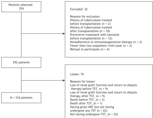
the required sample size to be 160 patients. After adding a refusal rate of 30%, we determined that the minimum sample size was 208 patients. The inclusion criteria were as follows: being ≥ 18 years of age and having undergone transplantation at least three months previously. The exclusion criteria were as follows: 1) history of tuberculosis treated before or after transplantation; 2) preventive treatment with isoniazid before transplantation; 3) renal graft loss and return to dialysis therapy before the fi rst TST (TST1) or second TST (TST2); 4) death; 5) nonadherence to immunosuppressive therapy; 6) having made fewer than two annual visits to the transplant outpatient clinic; or 7) not having given written informed consent (Figure 1).
Screening for LTBI
Participants were screened for LTBI by using the TST with purifi ed protein derivative RT23 (PPD RT23; Statens Serum Institute, Copenhagen, Denmark). The TST was performed by the Mantoux method, which consists of intradermal administration of 0.1 mL (2 tuberculin units) of PPD RT23 on the volar aspect of the forearm. Test results were read within 72-96 h of administration and were recorded in millimeters of induration. TST1 was administered after three months following renal transplantation, and TST2 was administered three weeks later if TST1 was negative, in order to assess reactivation of the immune response. All patients with a TST1 induration ≥ 5 mm were considered to have a positive result; those with a negative result were referred for TST2, which was considered positive if there was a > 10-mm increase in induration compared with the TST1 reading.(13-15) The cumulative frequency
of LTBI was also calculated (N = 216).
Variables and defi nitions
We investigated the following variables: (i) sociodemographic variables (gender, age, individual income, place of residence, and history of contact with tuberculosis); (ii) behavioral variables (smoking, alcoholism, and marital status); (iii) clinical variables (BCG vaccination scar, body mass index [BMI], DM, autoimmune disease, hepatitis B, hepatitis C, and neoplasms); (iv) transplant-related variables (living/deceased donor, double transplantation, retransplantation, immunosuppressive regimen, time
Patients selected:
324 Excluded: 32 Reasons for exclusion: History of tuberculosis treated before transplantation (n = 2) History of tuberculosis treated after transplantation (n = 10) Preventive treatment with isoniazid before transplantation (n = 12)
Nonadherence to immunosuppressive therapy (n = 2) Fewer than two outpatient visits/year (n = 2) Refusal to participate (n = 4)
292 patients
N = 216 patients
Losses: 76 Reasons for losses:
Loss of renal graft function and return to dialysis therapy before TST1 (n = 9)
Loss of renal graft function and return to dialysis therapy after TST1 (n = 9)
Death before TST1 (n = 3)
Death after TST1 (n = 1) Having given WIC but not having undergone any TST (n = 22) Not having undergone TST2 (n = 32)
interval between transplantation and TST, and renal graft function based on the glomerular fi ltration rate).
Patients were classifi ed as “having individual income” (employed, retired, or away from work/medical leave) or as “having no income” (unemployed or never worked). Patients were screened for alcoholism with the Cut down, Annoyed, Guilty, and Eye-opener questionnaire, which was incorporated into the patient interview.(16) Patients were classifi ed as “smokers” or
“nonsmokers” (people who had never smoked or people who had quit smoking one year prior to the study). (17)
BCG vaccination status was determined using the presence or absence of a BCG scar on the right arm. Renal transplant recipient age was categorized on the basis of the median age of the study population. BMI was calculated as recommended by the World Health Organization.(18) Patients were categorized as obese
(BMI > 30 kg/m2) or non-obese (18.5 < BMI ≤ 29.9 kg/m2). A diagnosis of DM was made in accordance with the classifi cation proposed by the American Diabetes Association(19) and the Brazilian Diabetes
Society.(20) Renal graft function was assessed by means
of the estimated glomerular fi ltration rate (eGFR), as calculated by the Modifi cation of Diet in Renal Disease equation.(21) Renal graft function was categorized as
“preserved renal function” (eGFR values ≥ 60 mL/ min/1.73 m2) or as “impaired renal function” (eGFR values < 59 mL/min/1.73 m2).
Statistical analysis
Descriptive statistics (frequency distribution and measures of central tendency and dispersion) were used to analyze the characteristics of the study population. The mean differences for continuous variables were compared by using the Student’s t-test for independent samples, and the proportions of categorical variables were compared by using Pearson’s chi-square test or Fisher’s exact test. For all tests, p values ≤ 0.05 were considered signifi cant. The measure of association in the bivariate analysis was OR and 95% CI.
Explanatory variables with p values ≤ 0.20 in the bivariate analysis were selected for multivariate analysis via a logistic regression model. The level of signifi cance required for inclusion in the fi nal model was 0.05, with adjustment for confounding factors. The goodness of fi t of the fi nal model was assessed by using the Hosmer-Lemeshow test.
The data collected were entered into Microsoft® Excel spreadsheets. All statistical analyses were performed with the IBM SPSS Statistics software package, version 21.0 (IBM Corporation, Armonk, NY, USA), and the R software, version 2.15.1 (The R Foundation for Statistical Computing, Vienna, Austria).
RESULTS
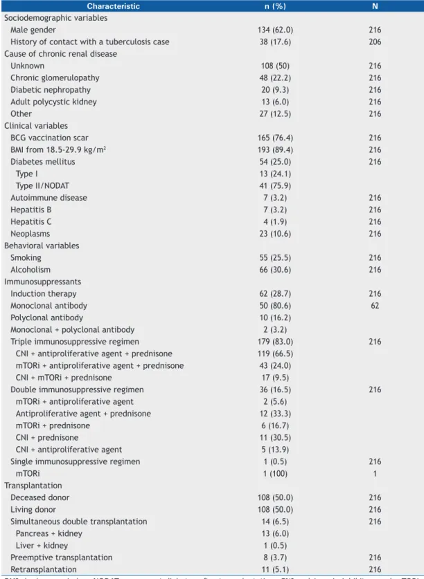
The characteristics of the study population (N = 216) and the causes of CRD are shown in Table 1. Age at the time of the test ranged from 18 to 75 years, with a median of 48 years and a mean of 46.5 ± 12.3 years.
History of contact with tuberculosis was positive in 38 patients (17.6%), negative in 168 (77.8%), and unknown in 10 (4.6%). Obesity was present in 23 patients (10.6%), and 54 patients (25%) had diabetes, of whom 13 were diagnosed with type I DM before transplantation and 41 were diagnosed with type II DM or drug-induced diabetes after transplantation. Seven patients (3.2%) had a previous diagnosis of autoimmune disease. Post-transplant neoplasia, including skin cancer, was present in 23 (10.6%) of the patients.
Of the 216 patients included in the study, 167 (77.3%) reported having income from employment, retirement pension, or medical leave. A total of 152 (70.4%) resided in the greater metropolitan area of Belo Horizonte, close to the transplant center, 63 (29.2%) resided in other areas of the state of Minas Gerais, and 1 (0.4%) resided in the state of Amapá.
The time interval between renal transplantation and TST1 ranged from 3.0 to 360.4 months, with a mean of 86.8 ± 75.6 months and a median of 68.2 months. The time interval between renal transplantation and TST2 ranged from 3.5 to 376.1 months,with a mean of 99.0 ± 78.3 months and a median of 79 months.
The prevalence of LTBI was 18.5%, and 40 individuals had positive TST results. Twenty-nine patients (13.4%) had positive TST1 results, and 11 (5.1%) had positive TST2 results. TST induration increased by 5.8% from the fi rst to the second test, which was signifi cant (p = 0.012).
The cumulative frequency of LTBI in the study population (baseline, TST1, and TST2) was 42.5%, because, of the 216 patients included in the study, 40 had previous positive TST results (18.5%); of the remaining 176 patients, 29 had positive TST1 results (16.5%); therefore, there remained 147 patients to undergo TST2, 11 of whom tested positive (7.5%).
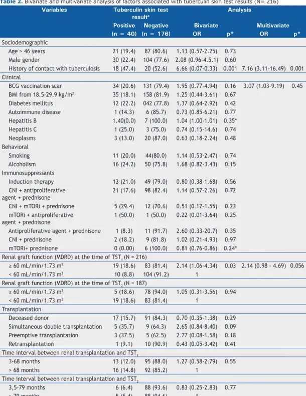
In the bivariate analysis (p ≤ 0.20), the following factors were associated with a diagnosis of LTBI: having a history of contact with a tuberculosis case; alcoholism; presence of a BCG vaccination scar; eGFR ≥ 60 mL/min/1.73 m2; double organ transplantation; and preemptive transplantation (transplantation performed before the initiation of dialysis therapy). In the fi nal logistic regression model, the following variables were statistically signifi cantly associated (p ≤ 0.05) with a diagnosis of LTBI: having a history of contact with a tuberculosis case; presence of a BCG vaccination scar; and eGFR ≥ 60 mL/min/1.73 m2 (Table 2).
DISCUSSION
Various studies have shown a higher prevalence of tuberculosis in patients undergoing renal transplantation in countries with a low, medium, or high prevalence of the disease if these patients are infected with M. tuberculosis.(4,12,22) Therefore, there is a need to diagnose
LTBI, and prescribing preventive therapy is relevant to preventing the development of the disease,(13) although
Although it is recommended that transplant candidates be referred for TST,(13) there have been no studies on
this practice. In the present study, the frequency of LTBI in our population was found to be high (42.5%).
To our knowledge, this is the fi rst time that LTBI and its associations with sociodemographic, behavioral, and clinical characteristics have been assessed in renal transplant recipients at a transplant center in Brazil. Table 1. Sociodemographic, clinical, and behavioral characteristics, immunosuppressive regimen, and transplant-related variables in renal transplant recipients (N = 216).
Characteristic n (%) N
Sociodemographic variables
Male gender 134 (62.0) 216
History of contact with a tuberculosis case 38 (17.6) 206
Cause of chronic renal disease
Unknown 108 (50) 216
Chronic glomerulopathy 48 (22.2) 216
Diabetic nephropathy 20 (9.3) 216
Adult polycystic kidney 13 (6.0) 216
Other 27 (12.5) 216
Clinical variables
BCG vaccination scar 165 (76.4) 216
BMI from 18.5-29.9 kg/m2 193 (89.4) 216
Diabetes mellitus 54 (25.0) 216
Type I 13 (24.1)
Type II/NODAT 41 (75.9)
Autoimmune disease 7 (3.2) 216
Hepatitis B 7 (3.2) 216
Hepatitis C 4 (1.9) 216
Neoplasms 23 (10.6) 216
Behavioral variables
Smoking 55 (25.5) 216
Alcoholism 66 (30.6) 216
Immunosuppressants
Induction therapy 62 (28.7) 216
Monoclonal antibody 50 (80.6) 62
Polyclonal antibody 10 (16.2)
Monoclonal + polyclonal antibody 2 (3.2)
Triple immunosuppressive regimen 179 (83.0) 216
CNI + antiproliferative agent + prednisone 119 (66.5)
mTORi + antiproliferative agent + prednisone 43 (24.0)
CNI + mTORi + prednisone 17 (9.5)
Double immunosuppressive regimen 36 (16.5) 216
mTORi + antiproliferative agent 2 (5.6)
Antiproliferative agent + prednisone 12 (33.3)
mTORi + prednisone 6 (16.7)
CNI + prednisone 11 (30.5)
CNI + antiproliferative agent 5 (13.9)
Single immunosuppressive regimen 1 (0.5) 216
mTORi 1 (100) 1
Transplantation
Deceased donor 108 (50.0) 216
Living donor 108 (50.0) 216
Simultaneous double transplantation 14 (6.5) 216
Pancreas + kidney 13 (6.0)
Liver + kidney 1 (0.5)
Preemptive transplantation 8 (3.7) 216
Retransplantation 11 (5.1) 216
The use of the TST to detect LTBI in pre-renal transplant evaluation is recommended in various countries,(4) including Brazil, where interferon-gamma
release assays (IGRAs) have not been validated for routine use.(13,14) Some factors, such as DM,
immunosuppressant pharmacokinetics, induction
therapy, previous therapy for humoral rejection, CMV infection, etc., may cause false-negative TST results. (10,13) In 2015, the World Health Organization
stated that the IGRAs or the TST can be used to detect LTBI, their use being strongly recommended, but with a low level of evidence.(14)
Table 2. Bivariate and multivariate analysis of factors associated with tuberculin skin test results (N= 216)
Variables Tuberculin skin test resulta
Analysis
Positive Negative Bivariate Multivariate
(n = 40) (n = 176) OR p* OR p*
Sociodemographic
Age > 46 years 21 (19.4) 87 (80.6) 1.13 (0.57-2.25) 0.73
Male gender 30 (22.4) 104 (77.6) 2.08 (0.96-4.5.1) 0.60
History of contact with tuberculosis 18 (47.4) 20 (52.6) 6.66 (0.07-0.33) 0.001 7.16 (3.11-16.49) 0.001 Clinical
BCG vaccination scar 34 (20.6) 131 (79.4) 1.95 (0.77-4.94) 0.16 3.07 (1.03-9.19) 0.45 BMI from 18.5-29.9 kg/m2 35 (18.1) 158 (81.9) 1.25 (0.44-3.61) 0.67
Diabetes mellitus 12 (22.2) 042 (77.8) 1.37 (0.64-2.92) 0.42
Autoimmune disease 1 (14.3) 6 (85.7) 0.73 (0.85-6.21) 0.77
Hepatitis B 1.40(0.0) 7 (100.0) 1.04 (1.00-1.01) 0.35*
Hepatitis C 1 (25.0) 3 (75.0) 0.74 (0.15-14.6) 0.74
Neoplasms 3 (13.0) 20 (87.0) 0.63 (0.18-2.24) 0.48
Behavioral
Smoking 11 (20.0) 44(80.0) 1.14 (0.53-2.47) 0.74
Alcoholism 16 (24.2) 50 (75.8) 1.68 (0.82-3.43) 0.15
Immunosuppressants
Induction therapy 13 (21.0) 49 (79.0) 0.80 (0.38-1.68) 0.56
CNI + antiproliferative agent + prednisone
21 (17.6) 98 (82.4) 1.14 (0.57-2.26) 0.72
CNI + mTORi + prednisone 5 (29.4) 12 (70.6) 0.51 (0.17-1.55) 0.23 mTORi + antiproliferative
agent + prednisone
1 (50.0) 1 (50.0) 0.22 (0.01-3.64) 0.25
Antiproliferative agent + prednisone 1 (8.3) 11 (91.7) 2.60 (0.33-20.7) 0.35
CNI + prednisone 2 (18.2) 9 (81.8) 1.02 (0.21-4.93) 0.97
mTORi+ prednisone 0 (0.00) 6 (100.0) 0.81 (0.76-0.86) 0.24*
Renal graft function (MDRD) at the time of TST1 (N = 216)
≥ 60 mL/min/1.73 m2 19 (18.6) 83 (81.4) 2.14 (1.06-4.34) 0.03 2.14 (0.98 - 4.69) 0.056
< 60 mL/min/1.73 m2 10 (8.8) 104 (91.2) 1
Renal graft function (MDRD) at the time of TST2 (N = 187)
≥ 60 mL/min/1.73 m2 5 (18.6) 78 (94.0) 1.05 (0.31-3.56) 0.94
< 60 mL/min/1.73 m2 19 (18.6) 83 (81.4) 1
Transplantation
Deceased donor 17 (15.7) 91 (84.3) 0.70 (0.35-1.38) 0.29
Simultaneous double transplantation 5 (35.7) 9 (64.3) 2.65 (0.84-8.40) 0.09 Preemptive transplantation 3 (37.5) 5 (62.5) 2.77 (0.08-1.58) 0.18
Retransplantation 1 (9.1) 10 (90.9) 0.43 (0.05-3.42) 0.41
Time interval between renal transplantation and TST1
3-68 months 13 (12.0) 95 (88.0) 1.27 (0.58-2.79) 0.55
> 68 months 16 (14.8) 92 (85.2) 1
Time interval between renal transplantation and TST2
3,5-79 months 6 (6.4) 88 (93.6) 0.83 (0.25-2.83) 0.77
> 79 months 5 (5.4) 88 (94.6) 1
BMI: body mass index; CNI: calcineurin inhibitor; mTORi: mammalian target of rapamycin inhibitor; MDRD:
The predominant etiology of CRD before transplantation was indeterminate, because most patients in the present study did not undergo renal biopsy for histological confi rmation of CRD. It is of note that, in the present study, glomerulopathies were important, as previously mentioned in another study.(7) Alcoholism and smoking
are risk factors for LTBI and for the development of tuberculosis.(23-26) Since, in our study, most patients
did not drink alcohol or smoke, there was no statistical association of alcoholism or smoking (they were not risk factors) with positive TST results.
Patients undergoing organ transplantation are more susceptible to infections because of immunosuppressant use. However, in our study, we found no such association with immunosuppressant use. Therefore, the best strategy is to screen for LTBI before organ transplantation. The World Health Organization recommends that high- and medium-income countries with a low incidence of tuberculosis (< 100 cases per 100,000 population) test for and treat LTBI in patients preparing for organ or hematologic transplantation.(14)
Use of tacrolimus and/or mycophenolate in young recipients, DM,(27) age of recipients,(8) time elapsed since
transplantation,(7,12) hepatitis C,(28) CMV infection, cancer,
and autoimmune diseases(8) have been reported as
factors for reactivation of tuberculosis and development of severe tuberculosis, especially during the fi rst six months after solid organ transplantation.(6) If LTBI is
detected, as occurred in our study, prevention with isoniazid is recommended.(29)
Transplantation of deceased-donor kidneys with increased ischemia times and retransplantation are situations perceived as being of high immunological risk. In these situations, it is recommended that induction therapy consist of higher potency drugs, such as basiliximab, thymoglobulin, or other polyclonal antibodies, in order to prevent acute rejection and reduce the effects of delayed graft function both in the short and long term. This therapy increases the risk of developing tuberculosis after transplantation and may cause negative TST results,(4,6) thereby compromising
the diagnosis of LTBI.(4,7) However, in our study, such
an association with deceased-donor kidneys and retransplantation was not observed. Preemptive renal transplantation and double organ transplantation showed a trend toward higher TST positivity. Nevertheless, it should be taken into consideration that these transplant types represent a small sample, which would lead to an underestimated analysis.
We found that history of contact with a tuberculosis case, presence of a BCG vaccination scar, and preserved renal graft function were associated with positive TST results.
The likelihood of having a positive TST result is 7.16 times higher in patients reporting a history of contact with a tuberculosis case. A history of contact with tuberculosis has long been described as being associated with positive TST results and, therefore, has a direct relationship with a diagnosis of LTBI.(3,9,13)
In the present study, having a history of contact with tuberculosis showed a signifi cant association with positive TST results.
The presence of a BCG vaccination scar increases by 3.07 times the likelihood of a patient having a positive TST result. In contrast, recent BCG vaccination may cause a false-positive TST result.(30) However, studies
have shown that TST results are unaffected if the TST is administered many years after vaccination,(13,31)
given that the response to the TST is almost null and void 8-10 years after vaccination.(15,32) In the present
study, we found a signifi cant relationship between BCG vaccination and TST positivity. All patients in our study who had a BCG vaccination scar had been vaccinated more than 15 years previously (mean age, 46 years). A history of BCG vaccination(13,15,32)
is commonly considered a confounding factor rather than a causal factor.
In our study, a six-month course of isoniazid was used to prevent tuberculosis; some studies recommend that a careful evaluation be made in order to arrive at a decision regarding the use of other drugs to prevent the disease.(2,6,8,33)
In the present study, preserved renal graft function (eGFR ≥ 60 mL/min/1.73 m2) was the only dependent variable that was associated with positive TST results. The immunological effects resulting from uremia, such as changes in phagocytosis, bacterial lability, and lymphocyte transformation, may lead to negative TST results.(2,34) Therefore, in cases of reduced renal
graft function, we observed negative TST results, as reported in another study.(28)
The prevalence of LTBI among the renal transplant recipients in our study (18.5%) was lower than that found by Sester et al.,(35) who obtained positive TST
results in 52.14%, but similar to that reported in the study by Atasever et al. (13.6%).(6) This is probably due
to the fact that the state of Minas Gerais has registered low tuberculosis incidence rates in recent years.(36)
The increase in induration from TST1 to TST2 (signifi cant response) shows that it is advisable to administer a second test if the fi rst one is negative, given that most patients failed to respond to TST1 (81%). Similar results have been reported in other studies in which a TST2 was administered,(5,13) with the
administration of the second test favoring the detection of LTBI in patients receiving immunosuppressants.(8)
The limitation of our study is the use of the TST, which may not refl ect the reality of LTBI because of lymphocyte immunodefi ciency and variation in the prescribed immunosuppressive regimens. Some authors have studied the possibility of new markers for the diagnosis of LTBI and tuberculosis in order to overcome this limitation, but there is still no evidence of the use of new tests in solid organ transplantation.(37,38)
In conclusion, the risk factors observed for positive TST results in screening for LTBI in renal transplant recipients are history of contact with tuberculosis cases
and preserved renal graft function. The prevalence of LTBI was low in renal transplant recipients. A TST2 should be administered to these patients if TST1 is negative. The TST should be administered if renal function is improved.
ACKNOWLEDGMENTS
We would like to thank the Federal University of Minas Gerais School of Medicine and its Mycobacterial Disease Research Group.
REFERENCES
1. Associação Brasileira de Transplante de Órgãos. Dimensionamento dos transplantes no Brasil e em cada estado (2009-2016). Registro Brasileiro de Transplantes [serial on the Internet]. 2016 [cited 2017 Oct 1]. XXII(4):[Adobe Acrobat document, 102p.]. Available from http://www.abto.org.br/abtov03/Upload/file/RBT/2016/RBT2016-leitura.pdf
2. Mu-oz P, Rodríguez C, Bouza E. Mycobacterium tuberculosis infection in recipients of solid organ transplants. Clin Infect Dis. 2005;40(4):581-7. https://doi.org/10.1086/427692
3. Lundin AP, Adler AJ, Berlyne GM, Friedman EA. Tuberculosis in patients undergoing maintenance hemodialysis. Am J Med. 1979;67(4):597-602. https://doi.org/10.1016/0002-9343(79)90240-7
4. Subramanian A, Dorman S; AST Infectious Diseases Community of Practice. Mycobacterium tuberculosis in solid organ transplant recipients. Am J Transplant. 2009; 9 Suppl 4:S57-62. https://doi. org/10.1111/j.1600-6143.2009.02894.x
5. Ergun I, Ekmekci Y, Sengul S, Kutlay S, Dede F, Canbakan B, et al. Mycobacterium tuberculosis infection in renal transplant recipients. Transplant Proc. 2006;38(5):1344-5. https://doi.org/10.1016/j. transproceed.2006.03.029
6. Atasever A, Bacakoglu F, Toz H, Basoglu OK, Duman S, Basak K, et al. Tuberculosis in renal transplant recipients on various immunosuppressive regimens. Nephrol Dial Transplant. 2005;20(4):797-802. https://doi.org/10.1093/ndt/gfh691
7. Singh N, Paterson DL. Mycobacterium tuberculosis infection in solid-organ transplant recipients: Impact and implications for management. Clin Infect Dis. 1998;27(5):1266-77. https://doi.org/10.1086/514993
8. Torre-Cisneros J, Doblas A, Aguado JM, San Juan R, Blanes M, Montejo M, et al. Tuberculosis after solid-organ transplant: incidence, risk factors, and clinical characteristics in the RESITRA (Spanish Network of Infection in Transplantation) cohort. Clin Infect Dis. 2009;48(12):1657-65. https://doi.org/10.1086/599035
9. Subramanian AK, Morris MI; AST Infectious Diseases Community of Practice. Mycobacterium tuberculosis infectious in solid organ transplantation. Am J Transplant. 2013;13 Suppl 4:68-76. https://doi. org/10.1111/ajt.12100
10. Aguado JM, Herrero JA, Gavaldá J, Torre-Cisneros J, Blanes M, Rufí G, et al. Clinical presentation and outcome of tuberculosis in kidney, liver, and heart transplant recipients in Spain. Spanish Transplantation Infection Study Group, GESITRA. Transplantation. 1997;63(9):1278-86. https://doi.org/10.1097/00007890-199705150-00015
11. Fonseca JC, Caiaffa WT, Abreu MN, Farah Kde P, Carvalho Wda S, Spindola de Miranda S. Prevalence of latent tuberculosis infection and risk of infection in patients with chronic kidney disease undergoing hemodialysis in a referral center in Brazil. J Bras Pneumol. 2013;39(2): 214-20. https://doi.org/10.1590/S1806-37132013000200013
12. Guida JP, Bignotto Rosane D, Urbini-Santos C, Alves-Filho G, Ribeiro Resende M, Mazzali M. Tuberculosis in renal transplant recipients: a Brazilian center registry. Transplant Proc. 2009;41(3):883-4. https:// doi.org/10.1016/j.transproceed.2009.01.075
13. Conde MB, Melo FA, Marques AM, Cardoso NC, Pinheiro VG, Dalcin Pde T, et al. III Brazilian Thoracic Association Guidelines on tuberculosis. J Bras Pneumol. 2009;35(10):1018-48. https://doi. org/10.1590/S1806-37132009001000011
14. World Health Organization [homepage on the Internet]. Geneva: WHO; c2017 [updated 2015; cited 2017 Oct 1] Guidelines on the management of latent tuberculosis infection; [about 2 screens]. http://www.who.int/tb/publications/ltbi_document_page/en/
15. Ruffi no-Netto A. Interpretation of the tuberculin test [Article in Portuguese]. Rev Saude Publica. 2006;40(3):546-7. https://doi. org/10.1590/S0034-89102006000300026
16. Mayfi eld D, McLeod G, Hall P. The CAGE questionnaire: Validation of a new alcoholism instrument. Am J Psychiatry. 1974;131(10):1121-3.
17. Reichert J, Araújo AJ, Gonçalves CM, Godoy I, Chatkin JM, Sales MP, et al. Smoking cessation guidelines--2008. J Bras Pneumol. 2008;34(10):845-80. https://doi.org/10.1590/S1806-37132008001000014
18. World Health Organization. Obesity: preventing and managing the global epidemic. Report of a World Health Organization Consultation. Geneva: World Health Organization; 2000.
19. Expert Committee on the Diagnosis and Classifi cation of Diabetes Mellitus. Report of the expert committee on the diagnosis and classifi cation of diabetes mellitus. Diabetes Care. 2003;26 Suppl 1:S5-20. https://doi.org/10.2337/diacare.26.2007.S5
20. Métodos e critérios para o diagnóstico de Diabetes mellitus. In:
Sociedade Brasileira de Diabetes. Tratamento e acompanhamento do Diabetes Mellitus--Diretrizes da Sociedade Brasileira de Diabetes. Rio de Janeiro: a sociedade; 2007. p. 14-5.
21. Buron F, Hadj-Aissa A, Dubourg L, Morelon E, Steghens JP, Ducher M, et al. Estimating glomerular fi ltration rate in kidney transplant recipients: performance over time of four creatinine-based formulas. Transplantation. 2011;92(9):1005-11. https://doi.org/10.1097/ TP.0b013e3182301602
22. Reis-Santos B, Gomes T, Horta BL, Maciel ELN. Prevalência de tuberculose em transplantados renais: revisão sistemática e meta-análise. J Bras Nefrol. 2013;35(3):206-213. https://doi. org/10.5935/0101-2800.20130033
23. Van Zyl Smit RN, Pai M, Yew WW, Leung CC, Zumla A, Bateman ED, et al. Global lung health: the colliding epidemics of tuberculosis, tobacco smoking, HIV and COPD. Eur Respir J. 2010;35(1):27-33. https://doi.org/10.1183/09031936.00072909
24. Bates MN, Khalakdina A, Pai M, Chang L, Lessa F, Smith KR. The risk of tuberculosis from exposure to tobacco smoke: a systematic review and meta-analysis. Arch Intern Med. 2007;167(4):335-42. https://doi.org/10.1001/archinte.167.4.335
25. Menzies D, Pai M, Comstock G. Meta-analysis: new tests for the diagnosis of latent tuberculosis infection: areas of uncertainty and recommendations for research. Ann Intern Med. 2007;146(5):340-54. https://doi.org/10.7326/0003-4819-146-5-200703060-00006
26. Naqvi R, Akhtar S, Noor H, Saeed T, Bhatti S, Sheikh R, et al. Effi cacy of isoniazid prophylaxis in renal allograft recipients. Transplant Proc. 2006;38(7):2057-8. https://doi.org/10.1016/j. transproceed.2006.06.010
27. Dooley KE, Tang T, Golub JE, Dorman SE, Cronin W. Impact of diabetes mellitus on treatment outcomes of patients with active tuberculosis. Am J Trop Med Hyg. 2009;80(4) 634-9. https://doi. org/10.4269/ajtmh.2009.80.634
28. Torres J, Aguado JM, San Juan R, Andrés A, Sierra P, López-Medrano F, et al. Hepatitis C virus, an important risk factor for tuberculosis in immunocompromised: experience with kidney transplantation. Transpl Int. 2008;21(9):873-8. https://doi.org/10.1111/j.1432-2277.2008.00694.x
30. Farhat M, Greenaway C, Pai M, Menzies D. False-positive tuberculin skin tests: what is the absolute effect of BCG and non-tuberculous mycobacteria? Int J Tuberc Lung Dis. 2006;10(11):1192-204.
31. Brasil. Ministério da Saúde. Informe eletrônico da tuberculose. Boletim Eletrônico Epidemiológico. 2009;9(2):1-4.
32. Menzies D, Gardiner G, Farhat M, Greenaway C, Pai M. Thinking in three dimensions: a web-based algorithm to aid the interpretation of tuberculin skin test results. Int J Tuberc Lung Dis. 2008;12(5):498-505.
33. Currie AC, Knight SR, Morris PJ. Tuberculosis in renal transplant recipients: the evidence for prophylaxis. Transplantation. 2010;90(7):695-704. https://doi.org/10.1097/TP.0b013e3181ecea8d
34. Wauters A, Peetermans WE, Van de Brande P, De Moor B, Evenopoel P, et al. The value of tuberculin skin testing in haemodialysis patients. Nephrol Dial Transplant. 2004;19(2):433-8. https://doi.org/10.1093/ ndt/gfg569
35. Sester U, Junker H, Hodapp T, Schütz A, Thiele B, Meyerhans A, et al. Improved effi ciency in detecting cellular immunity towards M. tuberculosis in patients receiving immunosuppressive drug therapy. Nephrol Dial Transplant. 2006;21(11):3258-68. https://doi. org/10.1093/ndt/gfl 416
36. Brasil. Ministério da Saúde. O controle da tuberculose no Brasil: avanços, inovações e desafi os. Boletim Epidemiológico. 2014;45(2):1-13.
37. Kruh-Garcia NA, Schorey JS, Dobos KM. Exosomes: New Tuberculosis Biomarkers – Prospects From the Bench to the Clinic. In: Pere-Joan Cardona, editor. Understanding Tuberculosis: global experiences and innovative approaches to the diagnosis. Rijeka (Croatia): In Tech; 2012. p. 395-410.