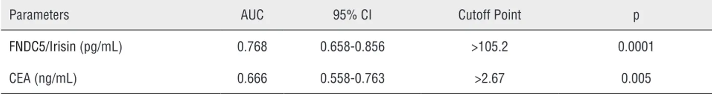
The diagnostic value of fNDC5Irisin in renal Cell Cancer
Texto
Imagem
Documentos relacionados
The prevalence of cholelithiasis was higher in patients with SS homozygous and S β heterozygous thalassemia when compared to patients with sickle cell disease.. In 50% of
Elevated serum levels of S-100beta protein and neuron-specific enolase are associated with brain injury in patients with severe sepsis and septic shock. Crit
Studies in B16-F10 melanoma and MCF-7 breast cancer cell lines indicated that PHOS-S can induce tumor cell apoptosis regardless of blocking of the Bcl-2 protein.. Loss of
A nested case- control study of adjuvant hormonal therapy persistence and compliance, and early breast cancer recurrence in women with stage I-III breast cancer. Pagani O, Gelber
XRCC1 Arg399Gln and RAD51 50UTR G135C polymorphisms and their outcome in tumor aggressiveness and survival of Portuguese breast cancer patients. Breast Cancer
The serum aminotransferase levels were lower in the patients with chronic kidney disease on hemodialysis (with or without viral hepatitis) than in the patients with normal
Entre outros, foram usadas 8 escalas do Children Behavior Questionnaire (CBQ) de Rothbart (Rothbart & Ahadi, 1994) para avaliar características do temperamento
