José Tiago Teixeira de Sousa Moreira
Solanum nigrum L., a fine tool for Phytoremediation of Acetophenone
Master Thesis
Mestrado em Biologia Molecular, Biotecnologia e Bioempreendedorismo em Plantas
Trabalho realizado sob a orientação do Professor Doutor Jorge Teixeira
Acknowledgments
I would like to express my deep gratitude to Professor Doctor Jorge Teixeira, for his patience, support, constant availability and for providing me with useful knowledge, that helped me to develop my scientific knowledge and reasoning.
I also would like to thank Doctor Amalia Calaya, for her help with the quantification of glutathione levels, and to Professor Doctor Manuel Azenha and all his team for their help in the acetophenone quantification. To João Cunha, for his help and contribution to this work.
To my beautiful, loving wife for her patience, help and love, that helped me to surpass every difficulties along the way. Without you, it wouldn´t have been the same. Also to my brother and sister for their fondness throughout this time.
To my mother and father, without whom this work wouldn´t have been possible. Thank you for understanding and supporting my choices.
To Alexandra Sousa and all colleagues in the laboratory for maintaining a good atmosphere in our working place.
Solanum nigrum L., a fine tool for remediation of acetophenone
Abstract
Organic pollutants of several industries constitute an environmental hazard because they may enter food chains, including humans. Phytoremediation is an eco friendly technology for environmental cleanup, and phytodegradtion, a strategy of phytoremediation, consists on the use of plants and associated organisms to render organic pollutants in non-reactive, non-toxic compounds. Acetophenone, a compound used in several industries, is the second most abundant constituent of Ridomil®, which is a systemic fungicide used in horticulture and vineyards and has as its active compound metalaxyl. Moreover, the several industrial uses of acetophenone make it more available and likely to accumulate in the environment. In order to access if acetophenone exerts toxicity towards Solanum nigrum L. and to see if this plant species may be used as a phytoremediation tool for acetophenone-contaminated sites,
S. nigrum plants were exposed for one month to 0.52 and 1.04 ppm of this compound,
which was supplied in the nutrient solution. Acetophenone levels in the nutrient medium were quantified along time, and some oxidative stress markers (H2O2; Chlorophyll a and b; carotenoids; proline and malondialdehyde levels), as well as some of the plant antioxidant system and detoxification mechanisms (catalase (CAT), guaiacol peroxidase (POD), ascorbate peroxidase (APX), Glutathione-S-Transferase (GST), γ-glutamyl-cysteine synthetase (γ-ECS) and glutathione reductase (GR) activities, as well as glutathione- and thiols-related levels) were evaluated at the end of the treatment. Acetophenone exposure impaired plant growth and induced oxidative stress. CAT and APX had enhanced activity in shoots of treated plants, while in roots both POD and APX revealed an increased activity. Increase in non-protein thiol levels revealed the importance of glutathione in acetophenone detoxification, where acetophenone is detoxified partly by conjugation with GSH through GST activity. Changes in GSH/GSSG ratios, alongside with increased γ -ECS activity and absence of statistical differences in GR activity, suggest that S. nigrum used de novo synthesis of GSH in response to acetophenone exposure. The results presented clearly reveal that
S. nigrum is an excellent tool for the rapid remediation of contaminated sites with
acetophenone, as no acetophenone was detected after 7 days of exposure to the plants.
Solanum nigrum L., uma excelente ferramenta de remediação
de Acetofenona
Resumo
Os poluentes orgânicos de várias indústrias constituem um perigo ambiental, podendo ser incorporados em várias cadeias alimentares, incluindo a dos seres humanos. A fitorremediação é uma tecnologia amiga do ambiente de remediação ambiental. A fitodegradação, uma estratégia de fitorremediação, consiste no uso de plantas e organismos associados para tornar poluentes orgânicos em compostos não-reativos e não- tóxicos. Acetofenona, um composto utilizado em diversas indústrias, é o segundo composto mais abundante de Ridomil ®, fungicida sistémico utilizado na horticultura e vinha e tem como composto ativo o metalaxil. Com o objetivos de determinar a toxicidade que acetofenona exerce em Solanum nigrum e para averiguar a utilidade desta espécie como ferramenta fitoremediadora de acetofenona, S. nigrum foi exposta durante um mês a 0,52 e 1,04 ppm deste composto, fornecido a planta na solução nutritiva. Os níveis de acetofenona no meio nutritivo foram quantificados ao longo do tempo, e alguns marcadores de stress oxidativo (H2O2; Clorofila a e b; carotenóides; prolina e níveis de malondialdeído), bem como enzimas do sistema antioxidante e mecanismos de desintoxicação das plantas (catalase (CAT); peroxidase do guaiacol (POD); peroxidase do ascorbato (APX); glutationa-S-transferase (GST); γ-glutamil-cisteína sintetase (γ-ECS); glutationa redutase (GR), bem como os níveis de glutationa e tióis) foram avaliadas no final do tratamento. A exposição à acetofenona prejudicou o crescimento da planta e induziu stress oxidativo. A CAT e a APX revelaram maior atividade na parte aérea das plantas tratadas, enquanto que nas raízes a POD e a APX revelaram um aumento da atividade. O aumento dos níveis de tióis não-proteicos realçou a importância da GSH na desintoxicação da acetofenona, onde esta última é, em parte, neutralizada por conjugação com a GSH pela GST. Alterações na proporção de GSH/GSSG, bem como o aumento da atividade γ-ECS e a ausência de diferenças estatisticamente significativas na atividade de GR, sugerem que S. nigrum sintetizou de
novo GSH em resposta à exposição. Os resultados apresentados revelam que S. nigrum
é uma excelente ferramenta para a rápida recuperação de sítios contaminados com acetofenona, não tendo sido detetada 7 dias após a exposição das plantas.
Index
Acknowledgments ... ii Abstract ... iii Resumo ...iv Index ... v Abreviations ... vii Introduction ... 11. Phytoremediation of organic pollutants ... 1
1.1. Phytodegradation of organic pollutants ... 2
2. Acetophenone ... 3
3. Glutathione ... 3
4. Thiol ... 7
5. Oxidative stress, lipid peroxidation and antioxidant mechanisms ... 9
5.1 ROS ... 10
5.2 Lipid Peroxidation ... 12
5.3 ROS scavenging enzymes ... 14
5.3.1 Catalase (EC 1.11.1.6) ... 14
5.3.2 Ascorbate Peroxidase (EC 1.11.1.11) ... 15
5.3.3 Guaiacol Peroxidase (EC 1.11.1.7) ... 15
5.3.4 Glutathione Reductase (EC1.8.1.7) ... 16
6. Solanum nigrum L. ... 16
Materials and Methods ... 18
1. Seed germination and growth conditions ... 18
2. Oxidative stress biomarkers ... 18
2.1 Hydrogen peroxide levels ... 18
2.2 Photosynthetic pigments quantification ... 19
2.3 Lipid peroxidation... 19 2.4 Proline ... 19 3. Biochemical determinations ... 20 3.1 Catalase ... 20 3.2 Guaiacol Peroxidase ... 20 3.3 Ascorbate Peroxidase ... 20 3.4 Thiol levels ... 21 3.5 Glutathione ... 21
3.6 Glutathione-S-Transferase ... 22
3.7 γ -Glutamyl-Cysteine Synthetase (γ -ECS) ... 23
3.8 Glutathione Reductase ... 23 4. Statistical analysis... 24 Results ... 25 1. Stress biomarkers ... 25 1.1 Biometry ... 25 1.2 Hydrogen peroxide (H2O2) ... 26 1.3 Photosynthetic pigments ... 26 1.4 Lipid Peroxidation ... 27
1.5 Oxidative Stress Index (OSI) ... 27
1.6 Proline contents ... 28
2. Antioxidant mechanisms ... 28
2.1 Catalase activity... 28
2.2 Guaiacol Peroxidase (POD) ... 29
2.3 Ascorbate Peroxidase (APX) ... 29
2.4 Thiol content ... 30
2.5 Glutathione ... 31
3. Acetophenone removal by plants ... 32
4. Glutathione-S-Transferase (GST) ... 32
5. γ-ECS and GR activities ... 33
Discussion ... 34
Conclusion ... 38
Future directions ... 38
Abbreviations
Abs- Absorbance
APX- Ascorbate Peroxidase AsA- Ascorbate
AsA-GSH- Ascorbate-Glutathione cycle ATP- Adenosine Triphosphate
CAT- Catalase
CDNB- Chloro-dinitro-benzene Cys- Cysteine
DHA- Dihydroascorbate
DHAR- Dihydroascrobate reductase DTNB- 5,5'-dithiobis-2-nitrobenzoic acid DTT- Dithiotreitol
EDTA- Ethylenediaminetetraacetic Acid FW- Fresh Weight
GPX- Glutathione Peroxidase GR- Glutathione Reductase
GR1- Glutathione Reductase gene 1
GRX- Glutaredoxin
GS- Glutathione Synthetase
GSH- Glutathione/Oxidized glutathione
GSH1- γ-Glutamylcysteine Synthetase gene GSH2- Glutathione Synthetase gene
GSHt- Total Glutathione GSSG- Reduced Glutathione GST- Glutathione-S-Transferase
IAA- Indoleacetic acid (auxin) Km- Michaelis Constant L•- Lipid Radical
LH- Lipid
LOO•- Lipid Peroxide radical LOOH- Lipid Hydroperoxide LOXs- Lipoxygenates MDA- Malondialdehyde
NADPH- Nicotinamide Adenine Dinucleotide Phosphate OSI- Oxidative Stress Index
Pi- Inorganic Phosphate
PMSF- Phenylmethylsulfonyl fluoride POD- Guaiacol Peroxidase
ppm- Parts per milion PRX- Peroxiredoxin PSII- Photosystem II
RES- Reactive electrophile Species ROS- Reactive Oxygen Species SOD- Superoxide Dismutase TBA- Thiobarbituric acid TCA- Trichloroacetic acid
Tris-HCl- Tris(hydroxymethyl)aminomethane hydrochloride TRX- Thioredoxin
w/v- Weight/Volume
Introduction
1. Phytoremediation of organic pollutants
Man is the cause for the accumulation of most of the organic pollutants in the environment, which are xenobiotics to several species. Many of these organic pollutants are carcinogenic and/or constitute an environmental hazard to other species, which may enter food chains, including humans’. These organic pollutants may be released into the environment via spills (fuel, solvents), military activities (explosives, chemical weapons), agriculture (pesticides, herbicides, fungicides), industry (chemical, petrochemical, etc.) and others .
In order to control fungi, weeds and insect pests, the use of pesticides has become commonly used in agricultural regions throughout the world. In the United States there is a generalized concern that pesticides have become major contaminants of the surface and ground water supplies. In fact, United States Geological Survey data indicate that over 90% of streams and 50% of wells sampled are contaminated with one or more pesticides [1].
Due to their chemical structure, many of these organic pollutants are recalcitrant to chemical and biologic oxygenation and hydrolysis, and, therefore, the remediation of water and soils contaminated by such pollutants is difficult [2]. Also, in modern cities, waste water treatments from municipal, agricultural and industrial sources is of primary concern. Such waters can pose a threat to human health, if the pollutants, ranging from raw sewage to chemical contaminants, are not treated before reaching natural water courses [2].
Hence, there is a need of a cheaper and eco friendly technology for environmental cleanup, like phytoremediation. Phytoremediation is an alternative to current methods of remediation that relies on the use of plants and their associated microorganisms, present on the rhizosphere, for environmental cleanup, and can be used in different strategies such as: Rhizofiltration; Phytostabilization; Phytoextraction; Phytoming; Phytostimulation; Phytodegradation and Phytovolatilization [3]. When organic pollutants are considered, phytodegradation is viewed as the more adequate strategy.
1.1. Phytodegradation of organic pollutants
Phytodegradtion consists on the use of plants and associated organisms to render xenobiotics in non-reactive, non-toxic compounds.
Contrasting with inorganic pollutants, organic pollutants can be degraded or converted to stable compounds or even be mineralized into inorganic compounds like water, carbon dioxide or chlorine, by phytodegradation [3]. Another difference between these two types of pollutants is their uptake: as plants have no transporters for organic xenobiotics, their uptake is mainly driven into and within the plants through simple diffusion, depending on their chemical properties, such as hydrophobicity and volatility, whereas metal uptake and movement within the plant relies on protein transporters and a panoply of metal chelators .
Once inside plant cells, and depending on their chemical structure, organic compounds can suffer enzymatic modifications, becoming promptly available for degradation/use by several enzymes, or they can also be conjugated with glucose or glutathione (GSH) and sequestrated.
GSH has an important role in conferring tolerance and in sequestration of the pollutant agent. Glutathione-S-Transferase (GST) catalyses the nucleophilic addition of a thiol group of GSH into the organic compound, making it more hydrophilic and ready to be sequestrated into the vacuole [4, 5].
Depending on its chemical composition, and before the conjugation of the organic compound with GSH occurs; a chemical modification of its radical groups may have to be performed. Several enzymes can oxidise or reduce these radical groups, and these reactions comprehend phase I of xenobiotic processing, the activation. Phase II is its conjugation to biomolecules, like GSH, and phase III is the conjugates’ compartmentation, which can then be followed by further enzymatic steps leading to xenobiotic degradation, depending on the conjugate formed.
Image 1- Mechanism of detoxification of heavy metals, organic pollutants and oxidative stress in plant cells by glutathione. Cys, cysteine; γ-Glu-Cys, γ-L-glutamyl-L-cysteine; γ-ECS, γ -glutamylcysteine synthetase; GSH, glutathione; GSSG, oxidized glutathione; PC, phytochelatins; HMI, heavy metal ion; HMI-PC, heavy metal–phytochelatin complex; Toxin, xenobiotics; Toxin–SG, toxin–GSH conjugate. (1) γ-Glutamylcysteine synthetase; (2) glutathione synthetase; (3) phytochelatin synthase; (4) glutathione S-transferase
(GST). Adapted from [5].
2. Acetophenone
Acetophenone is the second most abundant constituent in Ridomil® (Syngenta Agro S.A., Portugal), which is a systemic fungicide used in horticulture and vineyards and has as its active compound metalaxyl [6]. Besides, this compound, classified as class D by the United States Environmental Protection Agency (US EPA), is used in a wide range of industries, as a fragrance in perfumes, soaps, detergents, creams, lotions; as a flavouring agent in food, non-alcoholic drinks and tobacco; as a solvent for plastics and resins; as a catalyst for olefin polymerization and in organic synthesis as a photosensitizer1.
The several uses of this compound make it more available and likely to accumulate in the environment, due to these anthropogenic activities, where consequently it can hazard ecosystems.
3. Glutathione
Glutathione is an essential metabolite with multiple functions in plants. The earliest and fundamental roles recognized are the thiol-dissulphide interactions, in which reduced glutathione (GSH) is continuously oxidized to a disulphide form (GSSG)
that is recycled to GSH by NADPH-dependent glutathione reductase (GR), encoded by the gene GR1 [7]. Its role in defence metabolism was firstly established in animals; in plants, the reductive H2O2 metabolism had been link to ascorbate since 1970s, being the role of glutathione as an antioxidant less apparent. However, glutathione depletion in Arabidopsis knockouts lacking the fist enzyme of the committed pathway of GSH synthesis causes embryo lethality, revealing that both plant and mammalian cells rely on at least some of the multifunctional properties of glutathione for survival [7, 8].
Glutathione, like other thiols, can undergo numerous redox reactions, and its oxidized form (GSSG) includes the formation of mixed disulphides with proteins and other thiol molecules through their cysteine residues. This oxidized form can form conjugates with a wide array of thiyl radicals, both endogenous or xenobiotic, electrophilic species, and its interaction with the nitric oxide system makes it a reservoir of signalling potential as well as an antioxidant barrier, scavenging reactive oxygen species (ROS) through the GSH-ascorbate cycle, and as an electron donor to glutathione peroxidase (GPX). GSH is also the storage form and the long-distance transport form of reduced sulphur and it is involved in the pathways for the detoxification of heavy metal and xenobiotic as well [7, 8].
Glutathione´s high concentration (it can accumulate to milimolar concentrations), when compared to other thiols, and rapid recycling of its oxidized form by GR, maintaining its high reduction state (in normal conditions GSH:GSSG ratios are at least 20:1) makes it a buffer that maintains the intracellular environment reduced [7, 8], key characteristics that makes it an important antioxidant molecule. Also a marked characteristic of stress-tolerant genotypes is high ratio of GSH:GSSG, when compared to stress-sensitive genotypes. As a result of ROS scavenging, the high GSH:GSSG ratio shifts towards GSSG when plants are facing stress conditions that compromise the reduction of GSSG to GSH. The ratio of GSH:GSSG may also differ along the developmental stage, as for example, cell proliferation stage requires a higher ratio than somatic embryogenesis [8].
Recently, when considered the redox environmental state, low-molecular weight thiols have also been taken into account, to form a thiol-dissulphide redox environment. Overall, during biotic and abiotic stress the thiol-dissulphide redox state
is a marker of stress and cell viability with an important impact upon signalling mechanisms [8].
The amino acids glutamic acid, cysteine and glycine compose glutathione, and these amino acids are put together to form GSH through two ATP-dependent, enzyme-catalysed steps [7, 9]. The gene GSH1 encodes the enzyme γ -glutamylcysteine synthetase (γ -ECS/GSH1), which catalyses the formation of a covalent bond between y-glutamate and α-cysteine; afterwards, glycine is added to the dipeptide y-glutamyl-α-cysteine with the participation of glutathione synthetase (GS), encoded by the gene
GSH2 [9].
Glutathione synthesis is affected by many factors like glycine and ATP, but the most important factors are considered to be γ -ECS activity and cysteine availability, as demonstrated by the overexpression of the first enzyme of glutathione synthesis or of enzymes involved in cysteine synthesis [7]. Despite the well-described increases in glutathione in situations where hydrogen peroxide (H2O2) is also increased, neither intracellular generated, nor externally applied H2O2 cause marked induction of GSH1 or
GSH2 in Arabidopsis [10, 11]. γ -ECS can be regulated at translation or
post-translationally by redox control. One important factor in the upregulation of glutathione synthesis in response to oxidative stress is the involvement of one of the two disulphide bonds of γ -ECS homodimer in redox regulation [12]. Glutathione homeostasis is also regulated by feedback inhibition of γ -ECS by GSH, which can be alleviated by conditions in which glutathione is being consumed, like in the synthesis of phytochelatins. Nevertheless, both feedback inhibition and thiol/disulphide redox regulation of γ -ECS remains to be elucidated [7].
Glutathione is implied in ROS and ascorbate metabolisms, by reacting chemically with ROS and dehydroascorbate (DHA). Several authors [10, 11, 13-15]; conducted studies in which H2O2 metabolizing enzymes were genetically or pharmacologically inhibited, revealing a close relationship between augmented H2O2 and glutathione status. Queval et al 2009 [11] in their time-coursed analyses with conditional catalase mutants, showed that a several-fold induction of the total glutathione pool is preceded by an initial conversion of GSH to GSSG within the first hours of exposure to H2O2. Mhamdi et al 2010 [16], also showed that at moderate rates of endogenous H2O2 production, the GSH:GSSG ratio decreased from above 20 to
close to 1, this property making the glutathione status a useful marker for oxidative stress triggered by increased H2O2 production [7, 17].
H2O2 metabolism may involve several GSH-dependent and independent
pathways. There are three peroxidases, besides spontaneous GSH reaction with H2O2, that may link peroxide reduction to GSH oxidation: ascorbate peroxidase (APX), certain types of peroxiredoxin (PRX) and Glutathione-S-Transferase (GST) [7]. Haem-based peroxidases found in plants, class III of non-animal peroxidases super-family known as “guaiacol-type” peroxidase (POD), are known to control some biosynthetic processes and/or ROS production [18]. APX reduces H2O2, which will donate its electron to NAD(P)H through the ascorbate-glutathione pathway, linking APX activity to the GSH pathway; on the other hand, APX could also be associated with glutathione independent oxidation of NAD(P)H, via monodehydroascorbate reductase (MDHAR) [7].
Unlike guaiacol-type enzymes, thiol peroxidases such as PRXs are less H2O2 -specific, and can reduce other organic peroxides through thioredoxins (TRX) or similar components. GSH can be used as a reductant power by PRXII via glutaredoxins (GRX) [7]. Another PRX enzyme is GPX, which is implicated in plant stress responses and signalling and, unlike the name indicates, do not peroxidise glutathione, but are thioredoxins (TRX) dependent. This enzyme reduces peroxides involving a formation of a disulphide bridge from the initial sulphenic acid, and the intermediate disulphide is reduced back to the dithiol form by TRX, being therefore not likely to be involved in glutathione mediated peroxide oxidation [7].
On the other hand, GST have GSH peroxidation activity, being, amongst these enzymes, the only ones to appear to act as direct GPX, as all other enzymes require at least one additional protein to link peroxide reduction to GSH oxidation [7].
So, GSH could be linked to H2O2 and/or peroxide reduction by, at least, two ascorbate independent routes, as well as the ascorbate-glutathione pathway [7]. Data from gene expression studies evidenced that certain APX, GPX and GST encoding genes are induced in response to oxidative stress [10]. Additionally, transcriptomics and enzyme and metabolite assays of plants deficient in catalase and/or GR suggest that the ascorbate-glutathione cycle, and enzymes such as GST, may act in concert to remove H2O2 and/or other peroxides [16].
GST´s (EC 2.5.1.18) are a diverse family of proteins composed by two 25-KDa subunits, with a well defined Glutathione-binding domain at their active site [19]. This family of proteins is divided into four families, Phi, Tau, Zeta and Theta, being Phi and Tau plant specific GST, with major roles in herbicide detoxification. The classical reaction catalysed by GST´s comprehends an addition or substitution, with a covalent bond, between the sulphur atom of GSH and an electrophilic centre of a certain molecule [7, 20]. Besides its major role in detoxifying herbicides, this plant specific GST also have less well characterized functions like acting as GSH peroxidases, counteracting oxidative damage; acting as Flavonoid binding proteins and acting as stress signalling proteins [19, 20].
4. Thiol
Recently, there have been emerging evidences that a network of sulphur-containing molecules and related compounds, together with a number of non-protein (low-molecular weight thiols) and protein thiols (high molecular weight thiols) can contribute to plants stress tolerance [8]. Despite of recent progresses, the knowledge of interactions between thiols and other molecules is yet incomplete. Nevertheless, recent evidences indicate that thiols play a central role in conferring stress tolerance in plants [8].
Throughout taxa, from bacteria to higher plants and animals, protein thiols, like thioredoxins (TRX), glutaredoxins (GRX) and glutathionylation of protein sulphydryl groups, are considered most potent protein-based protective and regulatory mechanisms [8]. TRX and GRX are thiol oxireductases that provide reducing power to a variety of stress-related enzymes or exert direct thiol-dissulphide oxireductase activity to various protein targets. Reduction of TRX and GRX depend on different mechanisms: while GRX are directly reduced by 2 GSH molecules to produce GSSG, TRX reduction is dependent on the cellular compartment, requiring different reductases. Cytosolic and mitochondrial TRX require compartment-specific NADPH-dependent TRX reductases and plastid TRX are reduced by ferredoxin/TRX reductase [7, 8].
GRX are also known as thiol transferases and its traditional reaction, as said before, encompasses the reduction of a protein disulphide bond to two thiols oxidizing 2 GSH to GSSG in the process. GRXs comprise a more efficient way in the reduction of
glutathionylated proteins, in contrast to TRXs, that are more involved in the reduction of intra- or inter-molecular disulphide bonds [21]. Some enzymes of this family can also catalyse protein S-glutathionylation or degluthionylation, and are divided into three families that differ on the basis of amino acid motifs in the active site [7].
In proteins, cysteine (Cys) residues are tremendously susceptible to oxidative alteration, such as those associated with ROS production in adverse environmental conditions; these oxidative alterations have also been reported in methionine residues [8]. The three-step oxidation of Cys residues in macromolecules can reach a point of irreversibility, leading to the loss of their native conformation and activity, being further processed to be degraded [8]. The first product of Cys residue oxidation (cysteine sulphenic acid – Cys-SOH) is the only reversible product of the three-step oxidation. Its glutathionylation, a formation of a stable mixed disulphide bond between glutathione and a protein cysteine residue [7, 21], could be produced through a reaction with GSSG or be enzymatically catalysed, leading to a reversible temporary protection of Cys residues [8]. This reaction could modify the conformation, stability or activity of the target protein [7].
The rehabilitation of Cys can be made through reaction of these residues with GRX, which depends on GSH concentration and GR activity [8]. Nonetheless, spontaneous glutathionylation by GSSG is far less likely to occur in vivo. However widespread, glutathionylation and the participation of GRX are likely to enhance stress tolerance, but neither the exact mechanism nor the physiological relevance of protein glutathionylation in plants was established to date [8].
Despite major advances in this field, such as the identification of reversible S-glutathionylation of glutathione-dependent enzymes such as GR, dehydroascorbate reductases (DHAR), and some peroxiredoxins [19, 21, 22], there are still some blanks regarding quantification, significance and specificity: whether the modified Cys residue is glutathionylated in vivo, rather than nitrosylated or a target of TRX, or some combination of these modifications [7].
S-glutathionylation can be mediated through GRX-catalysed thiol-dissulphide
exchange between protein thiol groups and GSSG, conversion of thiol groups to thiyl radicals or sulphenic acids, or be mediated through formation of S-nitrosoglutathione [7].
Glutathionylation of proteins can be seen as a post-translational regulatory mechanism with an important impact on the activity of various antioxidant enzymes and thiol-peroxidases, regulation of transcription TGA factors (basic/leucine zipper-type) and probably deglutathionylation activity [8, 21].
5. Oxidative stress, lipid peroxidation and antioxidant mechanisms
Production of reactive oxygen species (ROS), like superoxide anion (O2•−), hydroxyl radical (•OH), as well as non-radical molecules like hydrogen peroxide (H2O2), singlet oxygen (1O2), and so forth, is an inevitable outcome of a aerobic metabolism [23]. In plants, the electron transport activities from chloroplasts, mitochondria andplasma membrane form ROS by leakage of electrons onto O2; ROS can also be formed
as a by-products of multiple metabolic pathways localized in different cellular compartments [23].
Several biotic and abiotic stresses are able to induce enhanced ROS production, which are known to be extremely harmful to organisms at high concentrations due to the disruption of cellular homeostasis. Hence, the term “oxidative stress” is used to describe when levels of ROS production exceed ROS scavenging mechanisms [23]. However this term still lacks a precise definition, but key marks are: augmented oxidative load (increased ROS production); the possible unrestrained oxidation caused by lower metabolism rates compared to production rates; oxidative damage to cellular components leading to accumulation of damaged cellular components that by some means lead to loss of function and eventual cell death. Enhanced ROS production can pose a hazard to cells by inducing peroxidation of lipids, oxidation of proteins, damage to nucleic acids, enzyme inhibition, activation of programmed cell death (PCD) and eventually lead to death of the cells [23].
On the other hand, ROS can also act as second messengers in a variety of cellular processes, like tolerance towards environmental stresses. It is the fine equilibrium between ROS production and scavenging that defines its damaging or signalling activity [23]. This implicates that ROS levels are tightly controlled by cells, in order to avoid any oxidative injury, rather than complete elimination [23].
Thus, cells have a well-organized antioxidant system, formed of non-enzymatic as well as enzymatic antioxidants, capable of scavenging or detoxifying excess ROS.
Superoxide dismutase (SOD), catalase (CAT), guaiacol peroxidase (POD), as well as
ascorbate peroxidase (APX), monodehydroascorbate reductase (MDHAR),
dehydroascorbate reductase (DHAR) and glutathione reductase, enzymes of the antioxidant ascorbate-glutathione cycle (AsA-GSH), are part of the enzymatic antioxidant system. Molecules such as ascorbate (AsA), glutathione (GSH), carotenoids, tocopherols and phenolics are part of non-enzymatic antioxidants of the cell [23].
Several researchers have stated that increased activity of several enzymes of the antioxidant defence system, as well as maintenance of a high capacity to scavenge ROS in plants, are linked to increased tolerance of plants to several environmental stresses [23]. In scope of this work, hydrogen peroxide (H2O2) and lipid peroxidation; the antioxidant enzymes APX, CAT, POD, GR, as well as the roles of GSH and carotenoids in the antioxidant defence, will be further discussed.
5.1 ROS
ROS are a group of free radicals, reactive molecules and ions that are derived from O2 (molecular oxygen). ROS can act as both deleterious and beneficial, depending on their concentration, as discussed before.
Oxygen is a completely non-hazardous molecule, and, due to its two unpaired electrons with parallel spins, it is unlikely to take part in reactions with organic molecules, unless it is activated. O2 can be activated through absorption of enough energy to reverse the spin on one of the unpaired electrons or by stepwise monovalent reduction. Oxygen singlet (1O2) is formed through energy absorption, while stepwise monovalent reduction can reduce O2 into superoxide anion (O2•−), H2O2 and hydroxyl radical (•OH) [23, 24].
The oxygen singlet has the ability to cause divalent reduction. In other words, it is able to transfer two electrons, due to the absorbance of energy that reverses the spin of one of its electron. It can perform direct oxidation of proteins, unsaturated fatty acids and DNA [23, 24].
Electron spin restriction prevents O2 from receiving four electrons at a time to produce water; it accepts one electron at a time, enabling the formation of stable intermediates during its stepwise reduction. In this stepwise reduction, O2•− is the primary ROS formed in the cell, which begins multiple reactions to generate other ROS. O2•− is a nucleophilic reactant with oxidizing and reducing properties [23]. O2•− has an
anionic charge that enables the transfer of its electrophilic activity onto electron-rich molecules [23]. O2•− oxidizes enzymes containing the [4Fe-4S] cluster and can reduce cytochrome C.
H2O2 is formed by the acceptance of one electron and two protons by O2•−, which is easily dismutated either non-enzymatically or by SOD-catalysed reactions: 2O2•− +2H+−→ H2O2 +O2
2O2•− +2H+ SOD−−→ H2O2 +O2
A wide range of stressful conditions can induce H2O2 generation. Due to not having unpaired electrons, it can cross the membrane, thus causing oxidative damage far from the site of its formation. Being the only ROS that can pass through aquaporins in the membrane and due to its relatively stability compared to other ROS, it has been a target of attention as a signal molecule involved in the regulation of specific biological processes, and to prompt tolerance to environmental stresses at low concentrations. On the other hand, when present in high concentrations, it can oxidize sulfur-containing amino acid residues (cysteine –SH and methionine –SCH3), and inactivate enzymes and transcription factors by oxidizing their thiol groups [23, 24]. Also it orchestrates programmed cell death.
However, both O2•− and H2O2 are only moderately reactive, and the cellular damage induced by ROS appears to be as a result of their conversion into more reactive species [23]. Thus, the hydroxyl radical (•OH) is reliant on O2•− and H2O2, and its formation is dependent on inhibition of SOD and CAT [23].
The Haber-Weiss reaction generates (•OH) from H2O2 and O2•−, and consists of the following reactions:
Fe3+ + O2•−−→ Fe2+ + O2,
In this reaction O2•− reduces Fe(III), and is followed by dihydrogen peroxide (Fenton reaction):
And:
O2•− +H2O2−→ •OH+OH− +O2. •
OH is the most reactive ROS, its single unpaired electron makes it available for interaction with all biological molecules and induces cellular damage, like protein damage, lipid peroxidation and membrane destruction [23].
Oxidation of organic substrates can happen through two reactions, either due to abstraction of hydrogen atoms or through addition of hydroxyl radicals to the substrate [23].
5.2 Lipid Peroxidation
Both enzymatic and non-enzymatic processes can oxidize lipids. A small number of lipidic regulators, such as jasmonates, can be generated by the enzymatic process and are seen as beneficial for survival for their role in defence and reproduction [25]. These enzymatic processes, like enzymatic oxylipin production, lipases and lipoxygenase (LOX) will not be discussed in this work. Instead, non-enzymatic reactions that generate lipid peroxides and their products are going to be subject of description, despite the difficulty to separate both processes as both processes numerous times occur simultaneously and may influence each other.
Innumerous oxygenated lipids are generated through lipid peroxidation, these products of lipid peroxidation, when not dealt with correctly, may interfere with protein function through uncontrolled hydrophobic interactions; also, many of the resulting lipids are reactive electrophile species (RES) and can covalently modify macromolecules [23, 25]. Oxidized lipids are being continuously produced even under optimal conditions, and the machinery that has evolved towards dealing with several lipids produced by non-enzymatic reactions could also be used by plants to handle xenobiotic compounds, such as herbicides and safeners [25].
Lipid peroxidation is widely used as a marker of ROS-mediated injury to cell membranes under stressful conditions, as augmented peroxidation has been reported in plants growing under several environmental stresses. Increase in lipid peroxidation parallels with increased production of ROS [23]. Lipid peroxidation follows three steps: initiation, progression and termination.
The non-enzymatic processes of lipid peroxidation are ROS-related, in which the extent of peroxidation reactions differ quantitatively between shoots and roots, due to the nature of ROS produced in these organs [25]. The first step of lipid peroxidation includes the activation of oxygen, and is rate limiting [23].
In photosynthetic tissues, lipid peroxidation reaction occurring in healthy tissues are initialize by singlet oxygen (1O2), a by-product of light capture at photosystem II (PSII), formed by the transfer of energy from excited chlorophyll to O2 [25]. 1O2 is mainly quenched by reaction center carotenoids to unreactive O2; nevertheless, a part of 1O2 is incorporated into plastid membranes, which occurs alongside O2 insertion catalyzed by LOXs. Oxygenation by 1O2 is non-catalytic, as each
1O
2 only produces one lipid hydroperoxide (LOOH), unlike the LOX mediated
oxygenation [25]. High levels of LOOH present in membranes makes them more vulnerable to fragmentation and this process generates new radicals. Contrasting to
1O
2-related lipid peroxidation, free radical peroxidation of lipids is a catalytic process that may quickly extend through membranes. When the radicals scavengers are over challenged, this process may harshly damage membranes [25].
On the other hand, in underground tissues, there is little 1O2 generation and basal non-enzymatic peroxidation, dominated by radical reactions [25]. Another potential radical source is hydrogen peroxide (H2O2), which is prone to iron-catalysed degradation to produce hydroxyl radicals (•OH) and superoxide anion radicals (O2•−) [25]. Hydroxyl radical, unlike superoxide anion radicals, can remove hydrogen from fatty acids, turning lipids (LH) to lipid radicals (L•), that once are generated are the basis of a diffusion-limited catalytic cycle that consumes O2 and other lipids to produce yet more lipid peroxides [25].
The second step of lipid peroxidation (progression) consists of lipid fragmentation, which is closely linked to radical amplification and supplementary enhancement of the peroxide pool [25]. In this step, a wide array of chemical structures is formed. Single or serial insertion of O2 may take place, before hydrogen abstraction, and thus secondary oxidation events form a large variety of alcohols, acids and reactive epoxides, aldehydes (malonyldialdehyde, acrolein and crotonaldehyde), ketones and cyclopentenones in membrane lipids [23, 25].
The LOOHs formed are more likely to radical-catalysed oxidation than non-oxidized lipids (LH) [25]. This phase of membrane peroxidation is known by the reaction of LOOH with lipid peroxide radicals (LOO•) and O2, after a hydrogen abstraction by LH in membranes occurred. LOOH pool decreases, as membranes become more peroxidised, in favour of L(OOH)2 and higher oxidation products. Lipid peroxide expansion involves the formation of non-radical glycerolipids dimers, which can spontaneously decompose into four fragments, among them there are glycerolipids with an oxidized acyl chain and a hydroxyl radical, which in turn can abstract hydrogens from LH, producing new L• radicals [25].
5.3 ROS scavenging enzymes
The antioxidant system not only allows plants to face stress conditions, it maintains an appropriate balance between ROS production and scavenging under normal conditions, as well. Specific ROS producing and scavenging mechanisms are present in different organelles of plant cells, like chloroplasts, mitochondria and peroxisomes, and these different ROS scavenging pathways are coordinated [23]. ROS production exceeding ROS quenching activity, in stressful conditions, leads to injury in different macromolecules in the cell. To avoid such harmful events, plants raise the levels of the endogenous antioxidant defences. In the scope of this work, only catalase (CAT), ascorbate peroxidase (APX), guaiacol peroxidase (POD) and glutathione reductase (GR) will be referred.
5.3.1 Catalase (EC 1.11.1.6)
This enzyme was the first to be discovered and characterized among the antioxidant enzymes present in plants. This ubiquitous tetrameric heme-containing enzyme, catalyses the dismutation of H2O2, for which has high affinity, into water and oxygen, requiring no cellular reducing equivalents. Despite its affinity for hydrogen peroxide it has poor activity against organic peroxides [23, 24, 26, 27].
When compared to APX, CAT have a much lower affinity to H2O2, but a very fast turnover rate. Based on Willekens et al. 1997 proposed classification, there are three CAT genes. Class I genes are expressed in photosynthetic tissues and are regulated by light, class II have high levels of expression in vascular tissues and class III are abundant in seeds and young seedlings [10].
As it has been discussed, H2O2 is implicated in many stress conditions, and when cells are stressed for energy rapidly generate H2O2 through catabolic processes, which in turn is degraded in an energy efficient manner by CAT [23]. These stresses cause either enhancement or depletion of CAT activity, depending on intensity, duration and type of stress; overall, stresses that reduce protein turnover also reduce CAT activity. Enhanced CAT activity showed accumulation of GSSG and decrease in AsA, demonstrating that CAT is decisive for the redox balance during oxidative stress [10].
5.3.2 Ascorbate Peroxidase (EC 1.11.1.11)
APX participates in the ascorbate-glutathione cycle, and plays a central role in the control of intracellular ROS levels. It uses two molecules of AsA reducing H2O2 to water, generating as well two molecules of monodehydroascorbate [23]. APX is regulated by H2O2 and by redox signals. APX forms are found in cytosol, plastidial stroma and thylakoids, mitochondria and peroxisomes. Whereas the APX found in the organelles scavenges H2O2 found in those organelles, cytosolic APX eliminates H2O2 produced in the cytosol, apoplast or that diffused from organelles [23, 24, 26, 27]. Chloroplastidial and cytosolic APX forms are specific for AsA as electron donor, but the Cytosolic form is less sensitive to AsA depletion than the chloroplastidial isoenzyme [23].
APX correspond to one of the most broadly distributed antioxidant enzymes, and APX forms have much higher affinity for H2O2 than CAT, being thus more efficient in scavenging this ROS under stressful conditions [23].
5.3.3 Guaiacol Peroxidase (EC 1.11.1.7)
POD is implied in many important biosynthetic processes, such as IAA degradation, cell wall lignification, ethylene biosynthesis, wound healing and defence against biotic and abiotic stress [23]. It is widely accepted as a stress enzyme, and can function as quencher of reactive intermediary forms of O2 and peroxy radicals [23]. In addition to scavenge H2O2, POD also serves to detoxify products of lipid peroxidation. Is the major form of protection against low levels of oxidative stress, and, while decomposing peroxides to water or alcohol it simultaneously oxidizes GSH [27].
5.3.4 Glutathione Reductase (EC1.8.1.7)
GSH is oxidized to GSSG through enzymatic and non-enzymatic oxidation-reduction cycles, when acting as an antioxidant. In the AsA-GSH cycle, GSH is oxidized by DHAR, and reduced by GR, a NAD(P)H-dependent enzyme, maintaining high GSH/GSSG ratios [23]. GR belongs to a group of flavoenzymes and contains an important disulphide group [28]. The reduction of GSSG into GSH involves two steps: First, the flavin moiety is reduced by NAD(P)H, the flavin is oxidized and a redox active disulphide bridge is reduced in order to produce a thiolated anion and a cysteine[28]. Afterwards, GSSG is reduced by a thioldisulphide interchange reaction [28]. A reversible inactivation may happen if the enzyme is not deoxidized by GSSG [23].
Most of GR activity in photosynthetic tissues is achieved by chloroplastidial forms, where GSH and GR are involved in detoxification of H2O2 generated by Mehler reaction, but some are located in the cytosol, mitochondria and peroxisomes [23]. Augmented GR activity results in increased GSH levels and ultimately leads to plant tolerance [27].
Nevertheless, because of the complexity of ROS detoxification system, there is no certainty whether or not overexpressing one component of such system will change the ability of the pathway as a whole [23].
6. Solanum nigrum L.
Solanum nigrum L. has good key features to be used as a phytoremediation
tool, such as: wide range global distribution (cosmopolitan), good adaptation to several climacteric conditions, fast growth and great shoot biomass, annual or biannual, and has been demonstrated as being a hyperaccumulator of both organic and non organic pollutants [6, 29-34]. Black nightshade, as it is commonly known, belongs to the family Solanaceae, such as other economically important species: potato (Solanum tuberosum L.); tomato (Solanum lycopersicum L.); eggplant (Solanum
This work follows a previous report by Teixeira et al 2011 [6], in which it was demonstrated that Solanum nigrum L. plants were a good tool for phytoremediation of metalaxyl-contaminated sites. In that work, two concentrations of Ridomil (a fungicide with metalaxyl as its active compound) were used.
Because acetophenone is the second most abundant component in Ridomil, and an organic compound with an extended use in several industries, it was of interest to see how Solanum nigrum L. plants would cope with such compound to further discriminate if the observed toxicity to Ridomil [6] was due solely to metalaxyl, or if it was the result of the combination of metalaxyl plus acetophenone.
So, in order to access if acetophenone exerts toxicity towards Solanum nigrum L. and to see if this plant species may be used as a phytoremediation tool to remediate and detoxify acetophenone, acetophenone levels in the nutrient medium were quantified along time, and some oxidative stress markers as well as some of the plant antioxidant system and detoxification mechanisms were evaluated after one month of exposure. For comparison purposes, the concentrations of acetophenone used were equal to those present when Ridomil was tested in a previous work [6].
Materials and Methods
1. Seed germination and growth conditions
S. nigrum L. seeds were harvested from black nightshade plants in Francos,
Porto, Portugal ( 1º9’55 79’’N 8º38’19 8’’O). They were sterilized in 70% ethanol for three minutes and dried in a laminar flux chamber. Afterwards, they were uniformly laid in Petri dishes with paper embedded in sterile Hoagland solution. No acetophenone was present during seed germination. Two days after incubation at 4 °C in the dark, they were placed in a growth chamber at 23 °C with a 16 h light/8 h dark photoperiod.
Seedlings were transferred to pots, with 2:1 vermiculite:perlite as substrate, 4 weeks after incubation. Plants were fed with a Hoagland solution, to which was added 0.0 (control), 0.52 and 1.04 ppm of acetophenone. Each pot had 2 plants; 6 pots were used for each experimental condition.
Samples of the nutrient solution were taken on a weekly basis, in order to evaluate its acetophenone content. One month after exposure plants were removed from the pots and biomass and length of each organ (roots and shoots) was evaluated. Vegetal material (n≥3 plant organs) was ground to dust with liquid nitrogen, and aliquots of each growth situation were made and stored at -80 °C until needed.
2. Oxidative stress biomarkers
2.1 Hydrogen peroxide levelsShoot aliquots were homogenized with 0.1% (w/v) TCA on ice, and the homogenate was centrifuged at 15 000 x g for 15 minutes. To 0.5 ml of each supernatant, 0.5 ml of 10 mM phosphate buffer (pH 7.0) and 1.0 ml of 1 M KI were added and mixed gently. The blank consisted of 0.5 ml of 0.1% (w/v) TCA instead of extract. Absorbances of these mixtures were read at 390 nm and H2O2 was quantified by using its extinction coefficient (ε390nm=0.28 µM-1cm-1). Results were expressed in nmol H2O2 g-1 fresh weight [36].
2.2 Photosynthetic pigments quantification
Chlorophyll a, b and carotenoids content was measured according to Lichtenthaler 1987 [37], and contents were calculated according to the following formulas:
Chlorophyll a= 12.25×Abs663-2.79×Abs647 ; Chlorophyll b =21.50 x Abs647 – 5.10 x Abs663 ;
Carotenoids =(1000 x Abs470 – 1.82 x Clor a – 85.02 x Clor b)/198 Results were expressed in mg g-1 fresh weight [37].
2.3 Lipid peroxidation
For each 0.2 g of vegetal material 1 ml of TCA 1% (w/v) was added, and the homogenates were agitated with a vortex for 90 seconds. Afterwards, the homogenates were centrifuged for 5 minutes at 10 000 x g, and the supernatant was recovered. To 250 µl of supernatant, 1 ml of TBA 0.5% (w/v) in TCA 20% (w/v) was added, in triplicates. Blanks were made in double with 1 ml of TBA 0.5% (w/v) in TCA 20% (w/v) and 250 µl of TCA 0.1% (w/v), and were given the same treatment as samples. Microtubes were pierced and incubated at 95 °C for 30 min, being cooled down for 10 min in ice before centrifugation at 10 000 x g for 15 minutes. Supernatants were recovered and their absorbance at 532 nm and 600 nm measured. Malondialdehyde (MDA) levels were calculated by subtracting absorbance at 600 nm to that at 532 nm, considering ε to be 155 mM-1.cm-1, results were expressed in nmol MDA.g-1 FW [38].
2.4 Proline
Aliquots of 0.5 g were homogenised in 10 ml of 3% (w/v) sulfosalisylic acid and centrifuged at 500 x g for 10 minutes. The supernatant was recovered and 200 µl of it were mixed with 200 µl of glacial acetic acid and 200 µl of acid nyhydrin. This mixture was incubated at 96 °C, cooled down in ice and 1 ml of toluene was added. Resulting tubes were vigorously agitated and kept at room temperature in order to separate the solution into two phases, the upper phase with reddish colour (organic phase) was used for recording its absorbance at 520 nm, using toluene as a blank. The levels of
proline were determined using a linear pattern with known proline concentrations, and expressed in µg g-1 fresh weight [39] .
3. Biochemical determinations
3.1 CatalaseTissue samples were homogenised with an extraction buffer containing 100 mM phosphate buffer (pH 7.3), 8% glycerol, 1 mM PMSF and 1 mM EDTA. After homogenization, the extract was centrifuged at 26 000 g, at 4 °C for 18 minutes. The resulting supernatant was recovered and kept on ice, for protein quantification by the Bradford method [40].
The activity of catalase was determined by reacting 8 to 12 µg of protein with 4 µl of 30% H2O2 and 100 mM phosphate buffer (pH 7.0) until the final volume was 1 ml. The reaction was followed in a spectrophotometer at 240 nm, for 60 seconds. The activity was calculated by using the coefficient of molar extinction of H2O2, 39.4 mM -1
cm-1 and was expressed as nkat mg-1 of protein. 3.2 Guaiacol Peroxidase
Plant tissue aliquots were homogenised with extraction buffer (100 mM
phosphate buffer (pH=7.3); 1 mM EDTA; 1 mM PMSF; 8% glycerol and 5 mM L-ascorbic acid), on a proportion of 1 g: 2.5 ml and centrifuged at 10 000 x g for 10 minutes at 4 °C. Protein content in the supernatant was quantified by the Bradford method [40]. 250 µl of 200 mM phosphate buffer (pH=7.3), 333 µl of 60 mM guaiacol, 197 µl of water 20 µl of extract and 200 µl of 1 mM H2O2 were pipetted into a cuvette and the increase in absorbance at 470 nm was monitored for 1 minute. The activity of POD was determined using ε=26.6 mM-1cm-1, and results were expressed as nkat mg-1 of protein [41].
3.3 Ascorbate Peroxidase
Tissue samples were homogenised and centrifuged in similar conditions to catalase and protein content was determined by the Bradford method [40]. The oxidation of ascorbate by APX was determine by spectrophotometry, at 300 nm for 30 seconds, by pipetting into a cuvette 100 mM phosphate buffer (pH 7.3) 0.5 mM ascorbate, 0.1 mM H2O2 and 12 µg of protein extract, with a final volume of 1 ml. The
reaction begun after the addition of H2O2, and the activity of the enzyme was determined using ε=0.49 mM-1cm-1 and expressed as nkat mg-1 of protein [41].
3.4 Thiol levels
Tissue aliquots were homogenized in 3 ml (shoots) or 2.5 ml (roots) of extraction buffer (20 mM EDTA and 20 mM of ascorbate), in ice and were centrifuged at 12 000 x g for 20 minutes at 4 °C.
The supernatant was recovered and to 200 µl of it, 960 µl of 200 mM Tris-HCl (pH=8.2) and 40 µl of 10 mM 5,5´-dithiobis(2-nitrobenzoic acid) were added, in triplicates. After 15 minutes at room temperature colour developed and absorbance at 412 nm was measured. Using ε412=13600 M-1cm-1 the total thiols groups were calculated and expressed in nmol g-1 fresh weight.
To determine the non-protein thiol groups, 500 µl of supernatant was mixed with 500 µl of 10% (w/v) sulfosalicylic acid, in duplicates. The solution was incubated at room temperature for 15 minutes and centrifuged for 15 minutes at 3 000 x g. The supernatant of both tubes were recovered and mixed together. To 500 µl of this supernatant 475 µl of 400 mM Tris-HCl (pH=8.9) and 25 µl of 5,5´-dithiobis(2-nitrobenzoic acid) were added, in triplicates. Colour development was allowed for 5
minutes at room temperature and absorbance was read at 412 nm. Using ε412=13600
M-1cm-1 the total thiols groups were calculated and expressed in nmol-1g fresh weight. Proteins thiol levels were calculated by subtracting the non-protein thiol content to the total thiol levels [42].
3.5 Glutathione
The tissue samples were homogenised in a proportion of 1:9 with a homogenization buffer consisting of 10 mM of HCl and 1.3% (w/v) 5-sulfosalicylic acid, and centrifuged at 13 000 x g for 10 minutes at 4 °C.
After centrifugation the supernatant were recovered, and total glutathione (GSHt) levels were determined. For that, 20 µl of supernatant, 20 µl of 63.5 mM phosphate buffer (pH=7.4) and 200 µl of reaction buffer (containing 5 ml of 10 mM 5,5'-dithiobis-2-nitrobenzoic acid (DTNB); 8.5 ml 2 mM NADPH; and 36.5 ml of phosphate buffer pH=7.4, on a final volume of 50 ml) and 40 µl of glutathione reductase (GR) enzyme were pipetted. In this reaction, the GSH is oxidized by the
presence of DTNB, and the all GSSG is reduced to GSH, by GR action. This reaction was measured at 415 nm in a spectrophotometer, and levels of glutathione were calculated with the help of a linear pattern of known GSH concentrations (Attachment 13). Levels were expressed in nmol g-1 fresh weight.
As for oxidized glutathione (GSSG) levels, 100 µl of the supernatant were pipetted together with 6 µl of trietanolamine (TEA) and 2 µl of 2-vinilpiridine and were incubated with agitation at 25 °C for 1 hour. In this reaction, 2-vinilpiridine will form a conjugate with GSH, allowing the measurement of the remaining GSSG. The GSSG is measured by reacting 40 µl of the former solution with 200 µl of reaction buffer and 40 µl of GR. The reaction of GR is measured in a spectrophotometer for 3 minutes at 415 nm. Levels of GSSG were calculated with the help of a linear pattern of known GSSG concentrations (Attachment 13), and were expressed in nmol g-1 fresh weight.
In order to determine the levels of reduced glutathione (GSH), the levels of GSSG were subtracted to those of GSHt [43].
The Oxidative Stress Index was calculated according to the formula: OSI=(2×GSSG /GSHt)×100
3.6 Glutathione-S-Transferase
Aliquots were homogenised with an extraction buffer containing 100 mM phosphate buffer (pH=7.3); 1 mM EDTA; 1 mM PMSF; 8% glycerol and 5 mM L-ascorbic acid, on a proportion of 1 g: 2.5 ml. The homogenised samples were centrifuge at 10 000 x g for 10 minutes at 4 °C and the protein content was quantified using the Bradford method [40]. Seven hundred µl of 71.43 mM of phosphate buffer (pH=7.5); 100 µl of 1.25 mM chlorodinitrobenzene – CDNB; 100 µl of extract and 100 µl of 10 mM GSH (initiates the reaction) were pipetted into a cuvette and the variation in the absorbance (ΔAbs) was read at 340 nm for 2 minutes. In order to determine the non-enzymatic conjugation of CDNB to GSH, 100 µl of extract were substituted by 100 µl of extraction buffer, and this ΔAbs was subtracted to the ΔAbs read with the extract.
The determination of GST activity was made according to the coefficient of extinction of CDNB, 9.6 mM-1cm-1, and results were expressed in nkat mg-1 protein [44].
3.7 γ -Glutamyl-Cysteine Synthetase (γ -ECS)
Aliquots were homogenized with extraction buffer (1 g roots – 2 ml extraction buffer; 1 g shoots – 2.5 ml extraction buffer) containing 100 mM Tris-HCl (pH=8.1); 10 mM MgCl2 and 1 mM EDTA, in ice, and centrifuged for 15 minutes at 10 000 x g (6 °C).
After centrifugation, the supernatant was recovered. Protein content was determined according to the Bradford assay [40]. To a 1.5 ml tube were added 350 µl of reaction mixture, containing: 143 mM Hepes pH=8.7; 1.43 mM MgCl2; 28.57 mM glutamate; 1.43 mM cysteine; 7.14 mM ATP, 7.14 mM phosphoenolpyruvate and 7.14 mM DTT, 10 µl of pyruvate kinase and 140 µl of protein extract (initiates the reaction). For the blank extraction buffer substituted the protein extract. This solution was incubated at 37 °C for 45 minutes, allowing the reaction, which was stopped by the addition of 100 µl of 50% (w/v) TCA. This mixture was centrifuge for 15 minutes at 10 000 x g at 6 °C, and the supernatant was recovered. To 50 µl of this new supernatant 150 µl of colour solution (equal volumes of 12% (w/v) ascorbic acid in 1 M HCl and 2% (w/v) ammonium molybdate tetrahydrate ((NH4)6Mo7O24.4H2O) were mixed) were added in triplicates. The triplicates were incubated for 20 minutes at room temperature, in order to develop a bluish colour (if phosphate is present). At the end of the 20 minutes 0.5 ml of the stop solution, composed by 2% acetic acid and 2% (w/v) sodium citrate tribasic dehydrate, were added. The absorbance at 240 nm was read, and the free phosphate content resulting of the enzyme activity was calculated according to a linear pattern using known phosphate concentrations (Attachment 16).
In order to determine the γ -ECS activity the amount of inorganic phosphate (Pi) determined in the blank was subtracted to that quantified with the samples. Such difference corresponds to the amount of ATP used by γ-ECS during the 45 minutes for 50 µL of reaction. Results were expressed in nkat mg-1 protein [45].
3.8 Glutathione Reductase
0.5 g of plant tissue were homogenized using 1.5 ml of extraction buffer, composed of 100 mM potassium phosphate buffer (pH 7.5); 1 mM EDTA; PMSF 1 mM and 10 mM dithiothreitol (DTT) and centrifuged at 20 000 x g for 30 minutes at 4 °C. The supernatant was recovered, and GR activity was determined by pipetting 500 µl of 200 mM potassium phosphate buffer (pH=7.8); 100 µl of 20 mM EDTA; 100 µl of 2 mM NADPH; 100 µl of extract and 100 µl ofwaterinto a cuvette and reading its absorbance
at 340 nm. Blanks were determined by substituting extract by extraction buffer. The activity of the enzyme was determined using the NADPH extinction coefficient ε= 6.22 mM-1cm-1. Results were expressed as nkat mg-1 protein [46].
4. Statistical analysis
All assays and measurements were performed at least in triplicate (n≥3). Variance analysis was performed by Fisher test and the means were statistically analysed using a two-sided t-test. Statistical significance is assumed at P < 0.05.
Results
1. Stress biomarkers
1.1 BiometryThe biomass and length of shoots and roots of plants exposed to acetophenone treatments were analysed in order to determine if plant growth was affected by the used treatments.
As it can be seen in Graphic 1, there was a 1.63 fold reduced shoot biomass of plants exposed to 1.04 ppm of acetophenone, and these were the only organs affected.
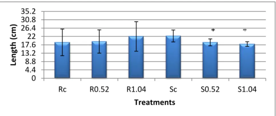
Considering the length of roots and shoots (Graphic 2), the exposure to acetophenone lead to a significant 1.18 and 1.24 fold reduction in shoots, in the 0.52 ppm and 1.04 ppm treatments, respectively, with no changes in roots length being recorded. 0 2.4 4.8 7.2 9.6 12 14.4 16.8 19.2 21.6 24 Rc R0.52 R1.04 Sc S0.52 S1.04 B io m ass (g) Treatments 0 4.4 8.8 13.2 17.6 22 26.4 30.8 35.2 Rc R0.52 R1.04 Sc S0.52 S1.04 Len gth (c m ) Treatments
Graphic 1- Biomass of roots and aerial organs, expressed in grams. Data presented are mean ± SE
(n≥3). Rc- roots of control plants; R0.52- Roots of plants exposed to 0.52 ppm of acetophenone; R1.04- Roots of plants exposed to 1.04 ppm of acetophenone; Sc- Shoots of control plants; S0.52- Shoots of plants exposed to 0.52 ppm of acetophenone; S1.04- Shoots of plants exposed to 1.04 ppm of acetophenone. The asterisk stands for significant statistical difference from control at P = 0.05. Attachment 1
Graphic 2- Length of roots and aerial organs, expressed in centimetres. Data presented are mean ± SE (n≥3). Rc- roots of control plants; R0.52- Roots of plants exposed to 0.52 ppm of acetophenone; R1.04- Roots of plants exposed to 1.04 ppm of acetophenone; Sc- Shoots of control plants; S0.52- Shoots of plants exposed to 0.52 ppm of acetophenone; S1.04- Shoots of plants exposed to 1.04 ppm of acetophenone. The asterisk stands for significant statistical difference from control at P =
1.2 Hydrogen peroxide (H2O2)
There was a significant increase of this ROS in shoots from both treatments, which followed the increase in acetophenone concentration, being 1.7 fold higher in plants exposed to 0.52 ppm and 2.2 times in plants exposed to 1.04 ppm of acetophenone (Graphic 3).
.
1.3 Photosynthetic pigments
The contents of chlorophyll A and B of plants exposed to the different acetophenone concentrations used significantly decreased by 0.9 fold only with the 0.52 ppm treatment (Graphic 4).
There were no differences for carotenoids contents among the different acetophenone concentrations used (Graphic 5).
0 0.5 1 1.5 2 Sc S0.52 S1.04 Ch lo ro p h yl l A +B (m g/ g fw) Treatments 0 1.3 2.6 3.9 5.2 6.5 7.8 Control 0.52 ppm 1.04 ppm H 2O2 ( n g/ g fw) Treatments
Graphic 3- Hydrogen peroxide (H2O2) contents in shoots, expressed as ng/g of fresh weight. Data
presented are mean ± SE (n≥3). 0,52- Plants exposed to 0.52 ppm of acetophenone; 1.04- Plants exposed to 1.04 ppm of acetophenone. The asterisk stands for significant statistical difference from control at P = 0.05. Attachment 3
Graphic 4 - Chlorophyll A and B content in shoots, expressed in mg/g of fresh weight. Data presented are mean ± SE (n≥3). Sc- Shoots of control plants; S0.52- Shoots of plants exposed to 0.52 ppm of acetophenone; S1.04- Shoots of plants exposed to 1.04 ppm of acetophenone. The asterisk stands for significant statistical difference from control at P = 0.05. Attachment 4
1.4 Lipid Peroxidation
The content of malondialdehyde (MDA) was used as an indicator to access membrane damage by lipid peroxidation (Graphic 6). MDA levels in roots clearly increased alongside the increasing acetophenone concentrations used. Thus, in roots of the 0.52 ppm situation it significantly increased 1.4 fold and in the 1.04 ppm situation this increase was 1.5 fold.
In shoots, MDA levels appear to increment, but only in with of 1.04 ppm this increase was statistical significant (1.25 fold).
1.5 Oxidative Stress Index (OSI)
The Oxidative stress index revealed no statistical difference when compared to control situation, nevertheless, it is clear that Roots showed higher stress than Shoots (Graphic 7). 0 0.05 0.1 0.15 0.2 0.25 Sc S0.52 S1.04 Car o te n o id s (m g/ g fw) Treatments 0 5.3 10.6 15.9 21.2 26.5 31.8 Rc R0.52 R1.04 Sc S0.52 S1.04 M D A ( n m o l/ gfw) Treatments
Graphic 5 - Carotenoids content in shoots, expressed in mg/g of fresh weight. Data presented are mean ± SE (n≥3). Sc- Shoots of control plants; S0.52- Shoots of plants exposed to 0.52 ppm of acetophenone; S1.04- Shoots of plants exposed to 1.04 ppm of acetophenone. The asterisk stands for significant statistical difference from control at P = 0.05. Attachment 5
Graphic 6 - Malondialdehyde content in roots and aerial organs, expressed in nmol/g of fresh weight. Data presented are mean ± SE (n≥3). Rc- roots of control plants; R0,52- Roots of plants exposed to 0.52 ppm of acetophenone; R1.04- Roots of plants exposed to 1.04 ppm of acetophenone; Sc- Shoots of control plants; S0.52- Shoots of plants exposed to 0.52 ppm of acetophenone; S1.04- Shoots of plants exposed to 1.04 ppm of acetophenone. The asterisk stands for significant statistical difference from control at P = 0.05. Attachment 6
1.6 Proline contents
Plants exposed to acetophenone suffered significant changes in their proline content, as shown in Graphic 8. In roots, proline levels decreased 1.3 fold when plants were exposed to 0.52 ppm of acetophenone, but increased 2.7 fold with the 1.04 treatment. Considering the shoots, proline levels revealed a dose-dependent increase with the acetophenone concentrations used. Thus, in shoots of plants exposed to 0.52 ppm there was an increase of 1.4 fold and of 1.6 fold with the 1.04 ppm treatment.
2. Antioxidant mechanisms
2.1 Catalase activityAs Graphic 9 depicts, there was a significant decrease of 1.84 fold in CAT activity in roots exposed to 1.04 ppm, while there was no statistical difference in activity in roots exposed to 0.52 ppm. The catalase activity significantly increased in shoots, by 2 fold in plants exposed to 0.52 ppm and a 1.5 fold in plants exposed to 1.04
0 20 40 60 80 100 120 Rc R0.52 R1.04 Sc S0.52 S1.04 OS I (% ) Treatments 0 10.3 20.6 30.9 41.2 51.5 Rc R0.52 R1.04 Sc S0.52 S1.04 Pr o lin e (µ g/ g fw) Treatments
Graphic 7 – Oxidative stress index in roots and aerial organs, expressed in percentage. Data presented are mean ± SE (n≥3). Rc- roots of control plants; R0,52- Roots of plants exposed to 0.52 ppm of acetophenone; R1.04- Roots of plants exposed to 1.04 ppm of acetophenone; Sc- Shoots of control plants; S0.52- Shoots of plants exposed to 0.52 ppm of acetophenone; S1.04- Shoots of plants exposed to 1.04 ppm of acetophenone. Attchment 7
Graphic 8 - Proline content in roots and aerial organs, expressed in µg/g of fresh weight. Data presented are mean ± SE (n≥3). Rc- roots of control plants; R0.52- Roots of plants exposed to 0.52 ppm of acetophenone; R1.04- Roots of plants exposed to 1.04 ppm of acetophenone; Sc- Shoots of control plants; S0.52- Shoots of plants exposed to 0.52 ppm of acetophenone; S1.04- Shoots of plants exposed to 1.04 ppm of acetophenone. The asterisk stands for significant statistical difference from control at P = 0.05. Attachment 8