Janeiro 2015
Universidade do Minho
Escola de CiênciasAndreia Pinto da Costa dos Santos Silva
Identification of new yeast protein
phosphatases involved in the regulation of
Bax
Tese de Mestrado em Bioquímica Aplicada
Trabalho efetuado sob a orientação da
Professora Doutora Manuela Côrte-Real
E coorientação da
DECLARAÇÃO
Nome: Andreia Pinto da Costa dos Santos Silva Endereço eletrónico: [email protected] Telefone: +351 917 753 214
Nº do Bilhete de Identidade: 13611366
Título da Tese de Mestrado:
Identification of new yeast protein phosphatases involved in the regulation of Bax
Orientadores:
Professora Doutora Manuela Côrte-Real Professor Doutora Maria João Sousa
Instituições de Acolhimento:
Centro de Biologia Molecular Ambiental (CBMA)
Ano de Conclusão: 2015
Designação do Mestrado:
Mestrado em Bioquímica Aplicada
1. É AUTORIZADA A REPRODUÇÃO INTEGRAL DESTA TESE, APENAS PARA EFEITOS DE INVESTIGAÇÃO, MEDIANTE DECLARAÇÃO ESCRITA DO INTERESSADO, QUE A TAL SE COMPROMETE.
Universidade do Minho, 19 de Janeiro de 2015 _____________________________________________ Andreia Pinto da Costa dos Santos Silva
Page iii
Agradecimentos
Usufruo desta oportunidade para agradecer a todos os que contribuíram, ajudaram e possibilitaram a realização desta tese de Mestrado, embora seja um trabalho de caracter individual, a mesma não seria possível sem o apoio de algumas pessoas.
Quero agradecer de forma especial às minhas orientadoras de tese, a Professora Doutora Manuela Côrte-Real e a Professora Doutora Maria João Sousa, pois sem a sua disponibilidade para me receber no seu grupo de investigação, este trabalho não seria possível, não teria estado em contacto com diversas técnicas que resultaram no culminar de uma aprendizagem vasta ao longo destes meses. O meu obrigado pela simpatia, disponibilidade, sabedoria, carinho, partilha de conhecimentos e rigor científico.
Segue o meu obrigado a todos os meus companheiros(as) de laboratório de Microbiologia I do CBMA, pela integração e pela disponibilidade no ensinamento das técnicas manipuladas, em especial à Lisandra Castro por toda a ajuda prestada no desenvolvimento deste trabalho.
Ao Departamento de Biologia, ao CBMA e a todos os seus funcionários pela amabilidade com que fui recebida e pelo auxílio nos momentos de aflição.
Agradeço aos meus amigos que me acompanharam não apenas nesta etapa, mas também ao longo deste últimos anos, obrigada pelo carinho e motivação quando mais necessitei.
Ao meu namorado, Diogo Nogueira, pela paciência genuína nos momentos menos bons, pelo companheirismo, pela amizade e por nunca desistires de mim.
Por último, mas os mais importantes, um sentido obrigado aos meus pais, pelo amor, apoio, alegria, confiança e dedicação. Obrigado por me terem proporcionado mais esta oportunidade de crescimento e aprendizagem. Sem vocês nada disto seria possível, ficarei eternamente grata.
O meu muito OBRIGADA a todos!
Este trabalho foi financiado pelo FEDER através do POFC – COMPETE e pela Fundação para a Ciência e Tecnologia através de projetos PEst-OE/BIA/UI4050/2014 e FCT-ANR/BEX-BCM/0175/2012.
Page iv
Identification of new yeast protein phosphatases involved in the
regulation of Bax
Abstract
Apoptosis is a genetically controlled cell suicidal program that is highly orchestrated and which contributes to the elimination of unnecessary or damaged cells in multicellular organisms. In this way the organism is capable to maintain the tissue homeostasis. Key regulators of apoptosis include the Bcl-2 family members, which control the permeabilization of mitochondria and the ensuing release of pro-apoptotic factors. Bax is the major pro-apoptotic member of this family. It is mainly cytosolic and remains inactive in proliferating cells, but is translocated to mitochondria and activated in cells undergoing apoptosis, which ultimately leads to permeabilization of the mitochondrial outer membrane. Bax can be regulated through phosphorylation/dephosphorylation by protein kinases and protein phosphatases that integrate different signaling pathways. Currently, the structure of inactive Bax has been determined, but the structures of its active form are still unsolved.
The yeast Saccharomyces cerevisiae, has a high degree of conservation of many cellular processes that share fundamental aspects with mammalian. For this reason it has been widely used for the study of the function and regulation of different Bcl-2 family members.
In order to identify novel protein phosphatases involved in the dephosphorylation of Bax, we heterologously expressed human Bax in yeast cells lacking non-essential protein phosphatases and determined whether there were differences in the Bax phosphorylation profile. We found several putative protein phosphatase candidates, such as three yeast protein phosphatases (Pph21p/22p and Pct4p), which have as human orthologs PP2A and WIP1 protein phosphatases, respectively. These two human protein phosphatases have been previously described as involved in the dephosphorylation of Bax, which validates the approach developed to identify new candidate protein phosphatases of Bax. The results will be discussed in terms of the consequences of the regulation of Bax by the protein phosphatases identified.
Page v
Identificação de novas fosfatases de proteína em levedura
envolvidas na regulação de Bax
Resumo
A apoptose é um programa de suicídio celular altamente orquestrado e sob controlo genético que contribui para a eliminação das células danificadas ou desnecessárias em organismos multicelulares. Deste modo, o organismo é capaz de manter a homeostasia dos tecidos. Existem alguns reguladores-chave da apoptose, como os membros da família Bcl-2, os quais controlam a permeabilização da membrana mitocondrial externa e a consequente libertação de fatores pró-apoptóticos. A proteína Bax é o principal membro pró-apoptótico da família Bcl-2. Esta proteína é principalmente citosólica e permanece inativa nas células em proliferação, sendo translocada para a mitocôndria e ativada em células apoptóticas, levando à permeabilização da membrana mitocondrial. A proteína Bax pode ser regulada através de processos de fosforilação/desfosforilação por cinases e fosfatases de proteína que integram diferentes vias de sinalização. Atualmente, já se conhecem as estruturas da forma inativa de Bax, mas as estruturas da sua forma ativa ainda não foram determinadas.
A levedura Saccharomyces cerevisiae, tem um elevado grau de conservação de muitos processos celulares partilhando aspetos fundamentais com os mamíferos. Por este motivo, tem sido amplamente utilizada para o estudo da função e regulação de diferentes membros da família Bcl-2.
A fim de identificar novas fosfatases de proteína envolvidas na desfosforilação de Bax, procedemos à expressão heteróloga de Bax humana em células de levedura deficientes em fosfatases não essenciais a fim de determinar diferenças no perfil de fosforilação de Bax. Encontramos várias fosfatases candidatas, entre elas, três fosfatases de levedura (Pph21p / 22p e Pct4p), que têm como ortólogos humanos, as fosfatases PP2A e WIP1, respetivamente. Estas duas fosfatases de proteína humanas foram previamente descritas como estando envolvidas na desfosforilação de Bax, validando assim a abordagem desenvolvida para identificar novas fosfatases candidatas. Os resultados serão discutidos em termos das consequências da regulação de Bax pelas fosfatases identificadas.
Page vi
Index
Agradecimentos ... iii
Abstract ...iv
Resumo ... v
Index of illustrations ... viii
Index of tables ... xi
Abbreviations ... xii
1. INTRODUCTION ... 1
1.1 Historical perspective ... 2
1.2 Apoptosis in the context of regulated cell death ... 3
1.2.1 Main components/regulators of the apoptotic machinery ... 4
1.2.1.1 Caspases ... 4
1.2.1.2 The Bcl-2 family members: structure and function ... 6
1.2.2 Apoptotic pathways ... 9
1.2.2.1 Extrinsic pathway ... 9
1.2.2.2 Intrinsic pathway ... 9
1.3 The Bax Protein ... 11
1.3.1 Conformational studies: The C and N-terminal role ... 11
1.3.2 Regulatory mechanisms by protein interactions and post-translational modifications ... 14
1.4 Contributions of the yeast model to study the function of proteins of the Bcl-2 family 16 1.4.1 Advantages of the yeast model system ... 19
2. OBJECTIVES ... 21
3. MATERIALS AND METHODS ... 23
3.1 Yeast strains and plasmids ... 24
3.2 Transformation of yeast mutants, with the plasmids expressing the forms of Bax and growth conditions ... 26
3.3 Induction conditions for the different Bax forms ... 27
3.4 Protein sample preparation ... 27
3.5 SDS gel electrophoresis/Western blot ... 28
3.6 Viability assays in strains displaying changes in Bax phosphorylation ... 28
Page vii
3.7.1 Mitochondrial membrane potential ... 29
3.7.2 ROS ... 29
3.8 Assessment of Bax content of isolated mitochondria and post mitochondrial fractions by Western blotting. ... 30
3.8.1 Preparation of spheroplasts ... 30
3.8.2 Preparation of mitochondrial and cytosolic fractions ... 30
3.8.3 SDS gel electrophoresis/Western blotting ... 31
3.9 Reproducibility and statistical analysis ... 31
4. RESULTS AND DISCUSSION ... 32
4.1 Screen of yeast protein phosphatase mutants involved in Bax phosphorylation profile…. ... 33
4.2 Effect of the protein phosphatases Ptc4p, Pph21p and Pph22p on Bax-dependent cell death…. ... 38
4.3 Effect of protein phosphatases on Bax-induced mitochondrial dysfunctions (m) ... 46
5. CONCLUSIONS AND FUTURE PERSPECTIVES ... 57
6. REFERENCES ... 64
7. ANNEX ... 74
A.1 - Western Blotting ... 75
A.2 - Western Blotting ... 76
A.3 - Western Blotting ... 76
Page viii
Index of illustrations
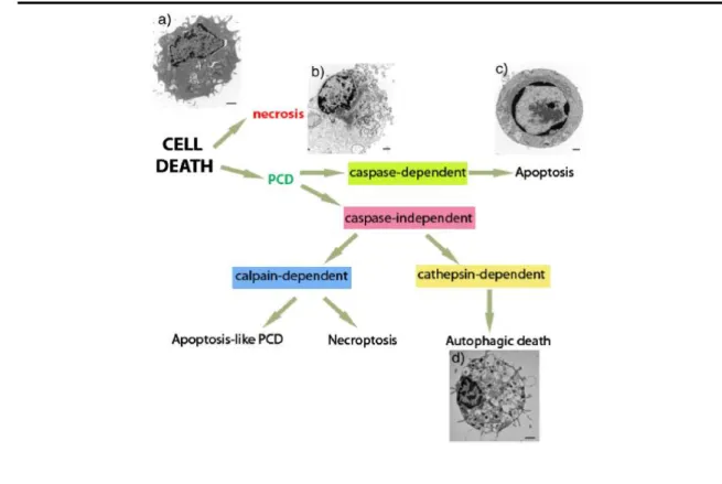
Figure 1.1 - Types of cell death and their morphological hallmarks. Classification diagram of the
different types of cell death. PCD: programmed cell death. Morphological features of a) a healthy cell, b) a necrotic cell, c) an apoptotic cell and d) an autophagic cell. [Taken from (Nikoletopoulou et al, 2013)]. 5
Figure 1.2 - Representation of all the known mammalian Bcl-2 family members. Bcl-2 homology regions
1-4 (BH1-4) are indicated. [Taken from (Er et al, 2006)]. ... 7
Figure 1.3- Diagram showing the intrinsic (left) and extrinsic (right) apoptotic pathways (Gómez-Sintes
et al, 2011). ... 11
Figure 1.4 – A structure of Bax monomer. All the α-helices were indicated ... 14 Figure 1.5- Mitochondrial proteins involved in the regulation of yeast apoptotic cell death. (Taken from
(Pereira et al, 2008). ... 18
Figure 4.1 – S. cerevisiae mutant strains from the Euroscarf collection lacking non-essential protein
phosphatases that have mammalian orthologs, and which were used to express the three forms of Bax (Bax alpha, Bax P168A and Bax c-myc). ... 33
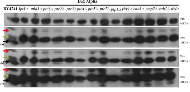
Figure 4.2 - Immunodetection of Bax in S. cerevisiae BY4741 and in the different mutant cells lacking
non-essential protein phosphatases expressing Bax alpha. Pgk1p was used as a loading control. Panels correspond to (a) 5 sec of exposure, (b) 15 sec of exposure and (c) 30 sec of exposure. Three distinct band were identified by three arrows and the meaning of which is explained below. ... 34
Figure 4.3 - Immunodetection of Bax in S. cerevisiae BY4741 and in the different mutant cells lacking
non-essential protein phosphatases expressing Bax alpha. Pgk1p was used as a loading control. Panels correspond to (a) 5 sec of exposure, (b) 15 sec of exposure, (c) 30 sec of exposure and (d) 60 sec of exposure. Three distinct band were identified by three arrows and the meaning of which is explained below. ... 34
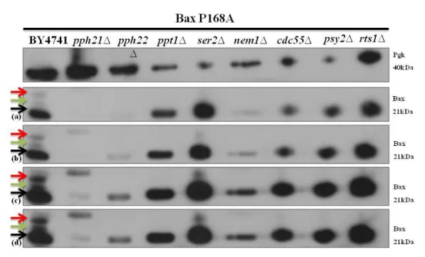
Figure 4.4 - Immunodetection of Bax in S. cerevisiae BY4741 and in the different mutant cells lacking
non-essential protein phosphatases expressing Bax P168A. Pgk1p was used as a loading control. Panels correspond to (a) 5 sec of exposure, (b) 15 sec of exposure, (c) 30 sec of exposure and (d) 60 sec of exposure. Three distinct band were identified by three arrows and the meaning of which is explained below. ... 35
Figure 4.5- Immunodetection of Bax in S. cerevisiae BY4741 and in the different mutant cells lacking
non-essential protein phosphatases expressing Bax P168A. Pgk1p was used as a loading control. Panels correspond to (a) 5 sec of exposure, (b) 15 sec of exposure, (c) 30 sec of exposure and (d) 60 sec of exposure. Three distinct band were identified by three arrows and the meaning of which is explained below. ... 35
Figure 4.6 - Putative protein phosphatases identified in the screening of the different mutant cells lacking
non-essential protein phosphatases expressing Bax alpha or Bax P168A. ... 37
Figure 4.7 - Survival of S. cerevisiae BY4741 and mutant cells lacking Ptc4p protein phosphatase after
(a) 14h of induction of Bax alpha expression in 2% galactose medium in cells pre-grown in glucose and (b) 4h of induction of Bax alpha expression in 0.5% of galactose in cells pre-grown in lactate+ethanol. Cell viability was determined by CFU counts. The percentual values of cell survival were calculated considering 100% at t0. For each bar, mean ± S.E.M. of at least three experiments is represented p <0.05; ** p ≤ 0.01; *** p ≤ 0.001, compared to the empty vector. One-way ANOVA was used to compare cell survival of BY74741 and protein phosphatase mutant cells expressing Bax alpha. The ratio between the percentages of survival of the BY4741 and mutant strain expressing Bax alpha and the respective strains only transformed with the empty vector was also calculated. ... 39
Figure 4.8 - Survival of S. cerevisiae BY4741 and mutant cells lacking Ptc4p protein phosphatase after
(a) 14h of induction of Bax P168A expression in 2% galactose medium in cells pre-grown in glucose and (b) 4h of induction of Bax P168A expression in 0.5% of galactose in cells pre-grown in lactate+ethanol. Cell viability was determined by CFU counts. The percentual values of cell survival were calculated considering 100% at t0. For each bar, mean ± S.E.M. of at least three experiments is represented p <0.05; ** p ≤ 0.01; *** p ≤ 0.001, compared to the empty vector. One-way ANOVA was used to compare cell
Page ix
survival of BY74741 and protein phosphatase mutant cells expressing Bax P168A. The ratio between the percentages of survival of the BY4741 and mutant strain expressing Bax P168A and the strains only transformed with the empty vector was also calculated. ... 40
Figure 4.9 - Survival of S. cerevisiae BY4741 and mutant cells lacking Pph21p protein phosphatase after
(a) 14h of induction of Bax alpha expression in 2% galactose medium in cells pre-grown in glucose and (b) 4h of induction of Bax alpha expression in 0.5% of galactose in cells pre-grown in lactate+ethanol. Cell viability was determined by CFU counts. The percentual values of cell survival were calculated considering 100% at t0. For each bar, mean ± S.E.M. of at least three experiments is represented p <0.05; ** p ≤ 0.01; *** p ≤ 0.001, compared to the empty vector. One-way ANOVA was used to compare cell survival of BY74741 and protein phosphatase mutant cells expressing Bax alpha. The ratio between the percentages of survival of the BY4741 and mutant strain expressing Bax alpha and the strains only transformed with the empty vector was also calculated. ... 41
Figure 4.10 - Survival of S. cerevisiae BY4741 and mutant cells lacking Pph21p protein phosphatase
after (a) 14h of induction of Bax P168A expression in 2% galactose medium in cells pre-grown in glucose and (b) 4h of induction of Bax P168A expression in 0.5% of galactose in cells pre-grown in lactate+ethanol. Cell viability was determined by CFU counts. The percentual values of cell survival were calculated considering 100% at t0. For each bar, mean ± S.E.M. of at least three experiments is represented p <0.05; ** p ≤ 0.01; *** p ≤ 0.001, compared to the empty vector. One-way ANOVA was used to compare cell survival of BY74741 and protein phosphatase mutant cells expressing Bax P168A. The ratio between the percentages of survival of the BY4741 and mutant strain expressing Bax P168A and the strains only transformed with the empty vector was also. ... 42
Figure 4.11 - Survival of S. cerevisiae BY4741 and mutant cells lacking Pph22p protein phosphatase
after (a) 14h of induction of Bax alpha expression in 2% galactose medium in cells pre-grown in glucose and (b) 4h of induction of Bax alpha expression in 0.5% of galactose in cells pre-grown in lactate+ethanol. Cell viability was determined by CFU counts. The percentual values of cell survival were calculated considering 100% at t0. For each bar, mean ± S.E.M. of at least three experiments is represented p <0.05; ** p ≤ 0.01; *** p ≤ 0.001, compared to the empty vector. One-way ANOVA was used to compare cell survival of BY74741 and protein phosphatase mutant cells expressing Bax alpha. The ratio between the percentages of survival of the BY4741 and mutant strain expressing Bax alpha and the strains only transformed with the empty vector was also calculated ... 43
Figure 4.12- Survival of S. cerevisiae BY4741 and mutant cells lacking Pph22p protein phosphatase after
(a) 14h of induction of Bax P168A expression in 2% galactose medium in cells pre-grown in glucose and (b) 4h of induction of Bax P168A expression in 0.5% of galactose in cells pre-grown in lactate+ethanol. Cell viability was determined by CFU counts. The percentual values of cell survival were calculated considering 100% at t0. For each bar, mean ± S.E.M. of at least three experiments is represented p <0.05; ** p ≤ 0.01; *** p ≤ 0.001, compared to the empty vector. One-way ANOVA was used to compare cell survival of BY74741 and protein phosphatase mutant cells expressing Bax P168A. The ratio between the percentages of survival of the BY4741 and mutant strain expressing Bax P168A and the strains only transformed with the empty vector was also. ... 44
Figure 4.13 –Levels of superoxide anion accumulation (a) and changes in mitochondrial membrane
potential (b) in S. cerevisiae BY4741 and mutant cells lacking Ptc4p protein phosphatase expressing Bax alpha after 4h of Bax induction in 0.5% of galactose in cells pre-grown in lactate+ethanol. Values are mean ± S.E.M of at least three independent experiments (b). Values significantly different from BY4741: * P<0.05, ** P<0.01 and *** P<0.001, One-way ANOVA was used. ... 47
Figure 4.14 – Levels of superoxide anion accumulation (a) and changes in mitochondrial membrane
potential (b) in S. cerevisiae BY4741 and mutant cells lacking ptc4∆ protein phosphatase expressing Bax P168Aafter 4h of Bax induction in 0.5% of galactose in cells pre-grown in lactate+ethanol. Values are mean ± S.E.M of at least three independent experiments (b). Values significantly different from BY4741: * P<0.05, ** P<0.01 and *** P<0.001, One- way ANOVA was used. ... 48
Figure 4.15 – Levels of superoxide anion accumulation (a) and changes in mitochondrial membrane
potential (b) in S. cerevisiae BY4741 and mutant cells lacking Pph21p protein phosphatase expressing Bax alpha after 4h of Bax induction in 0.5% of galactose and 0.5% lactate in cells pre-grown in lactate+ethanol. Values are mean ± S.E.M of at least three independent experiments (b). Values
Page x
significantly different from BY4741: * P<0.05, ** P<0.01 and *** P<0.001, One- way ANOVA was used. ... 49
Figure 4.16 – Levels of superoxide anion accumulation (a) and changes in mitochondrial membrane
potential (b) in S. cerevisiae BY4741 and mutant cells lacking Pph21p protein phosphatase expressing Bax P168Aafter 4h of Bax induction in 0.5% of galactose in cells pre-grown in lactate+ethanol. Values are mean ± S.E.M of at least three independent experiments (b). Values significantly different from BY4741: * P<0.05, ** P<0.01 and *** P<0.001, One- way ANOVA was used. ... 50
Figure 4.17 – Levels of superoxide anion accumulation (a) and changes in mitochondrial membrane
potential (b) in S. cerevisiae BY4741 and mutant cells lacking Pph22p protein phosphatase expressing Bax alpha after 4h of Bax induction in 0.5% of galactose in cells pre-grown in lactate+ethanol. Values are mean ± S.E.M of at least three independent experiments (b). Values significantly different from BY4741: * P<0.05, ** P<0.01 and *** P<0.001, One- way ANOVA was used. ... 51
Figure 4.18 - Levels of superoxide anion accumulation (a) and changes in mitochondrial membrane
potencial (b) in S.cerevisiae BY4741 and mutant cells lacking Pph22p∆ protein phosphatase expressing Bax P168Aafter 4h of Bax induction in 0.5% of galactose in cells pre-grown in lactate+ethanol. Values are mean ± S.E.M of at least three independent experiments (b).Values significantly different from BY4741: * P<0.05, ** P<0.01 and *** P<0.001, One- way ANOVA was used. ... 52
Figure 4.19 – Western blot analysis of cytochrome c in S. cerevisiae BY4741 and protein phosphatase
mutant cells expressing Bax alpha before (LAC) and after (GAL) induction of Bax expression after 4h in 0.5% of galactose, in cells pre-grown in lactate+ethanol, in both mitochondrial and cytosolic fractions. Pgk1p and mitochondrial porin (Por1p) levels were used as loading control of cytosolic and mitochondrial fractions, respectively. A representative experiment is shown of at least two independent experiments with similar results. ... 53
Figure 4.20 - Western blot analysis of cytochrome c in S. cerevisiae strains BY4741 and protein
phosphatase mutant cells expressing Bax P168A before (LAC) and after (GAL) induction of Bax expression after 4h in 0.5% of galactose in cells pre-grown in lactate+ethanol, in both mitochondrial and cytosolic fractions (regarding the content of Bax, we analysed a duplicate of the same extracts). Pgk1p and mitochondrial porin (Por1p) levels were used as loading control of cytosolic and mitochondrial fractions, respectively. A representative experiment is shown of at least two independent experiments with similar results. ... 55
Figure 5.1 – Protein Ser/Thr protein phosphatases implicated in both intrinsic and extrinsic apoptotic
pathways. The protein phosphatases are listed in dark blue boxes. Blue arrows indicate direct targets of protein phosphatases, and red arrows indicate indirect targets via MAP kinases and AKT and p53 pathways. Red boxes mark the protein phosphatases studied (Adapted from Sun and Wang, 2012). ... 60
Figure 5.2 - List of protein Ser/Thr protein phosphatases (STPs) involved in cell death regulation and
Page xi
Index of tables
Table 3.1- Yeast protein phosphatase, encoded by non-essential genes and respective human orthologues.
... 24
Table 3.2- List of plasmids used in this study. ... 26 Table 4.1 - Yeast putative protein phosphatases involved in phosphoregulation of Bax alpha and Bax
P168A under different conditions of Bax induction. (n.s.) means that the effect of Bax expression on cell viability is not statistically different from that on the wild type strain and (**) means p <0.05; ** p ≤ 0.01and (***) means p ≤ 0.01; *** p ≤ 0.001. ... 45
Table 4.2 - Comparative table of the phenotypes of the protein phosphatase mutants studied in what
regards Bax-alpha- and Bax-P168A-induced cell death and mitochondrial dysfunctions. Non statistically different (n.s.). Comparatively with the control strain. (-) no release and (+) release of cytochrome c. .... 56
Page xii
Abbreviations
3D – Three Dimensional ACD – Accidental
AIF – Apoptosis Inducing Factor AKT/PKB - protein kinase B AMID – Mitochondria-associated
Inducer of Death
ANT – Adenosine Nucleotide
Translocator
APAF – Apoptosis Protease Activating
Factor
Arg – Arginine
ART – Apoptosis Regulator of
Targetting
ATP – Adenosine Triphosphate BI-1 – Bax-inhibitor 1
BOP – BH3-only Protein BSA - Bovine Serum Albumin Ca2+ - Calcium Ion
CARD – Caspase Recruitment Domain CFU – Colony-Forming Units
Cit. c – Cytochrome c
CK – Creatine Kinase CsA – Cyclosporine A CypD – Cyclophilin D
DED – Death Effector Domain DHE – Dihydroethidium
DISC – Death Inducing Signaling
Complex
DNA – Deoxyribonucleic acid DR – Death Receptors
ER - Endoplasmic Reticulum
FADP – Fas Associated Death Domain
Protein
GFP – Green Flourescent Protein Gly – Glycine
GSK3β – Glycogen Synthase Kinase H2O2 – Hydrogen Peroxide
Hα – α Helix
IMM – Inner Mitochondrial Membrane IMS – Intermembrane space
JNK – Jun N-Terminal Kinase
LB – Luria-Betani Broth
MAC – Mitochondrial
Apoptosis-Induced Channel
MOMP – Mitochondrial Outer
Membrane Permeabilization
mPT – Mitochondrial Permeability
Transition
mPTP - Mitochondrial Permeability
Transition Pore
NADH – Nicotinamide Adenine
Dinucleotide
NCCD - The Nomenclature Committee
on Cell Death
NMR - Nuclear Magnetic Resonance O2- – Superoxide Anion
OD - Optical Density
OMM – Outer Mitochondrial
Membrane
PBR – Peripheral Benzodiazepine
Receptor
PCD – Programmed Cell Death PGK1 – Phosphoglycerate Kinase PGK1 – Phosphoglycerate Kinase 1 Pi – Phosphate PKB – Protein Kinase B PKC – Protein Kinase C PPM – Protein Phosphatase Mg2+ PPP – Phosphoprotein Phosphate Pro – Proline
PTP- Protein Tyr Posphatase PVDF – Polyvinylidene Diflouride RCD – Regulated Cell Death ROS – Reactive Oxygen Species SC – Synthetic Complete
SD – Standard Deviation Ser – Serine
Thr – Threonine
TNF – Tumor Necrosis Factor VDAC – Voltage-Dependent Anion
Channel
YMUC – Yeast Mitochondrial
Unselective Channel
1. INTRODUCTION
Page 2
1.1 Historical perspective
Although the study of cell death started to be reconsidered and addressed more intensively in the eighties, the interest in it was modest and only a decade later it began to grow exponentially, totaling nowadays more than 80.000 publications (Wyllie, 2010). During the early twentieth century cell death was a subject of interest mainly because of its crucial role in tissue homeostasis or in the development of multicellular organisms or even in some isolated studies that recognized its involvement during metamorphosis and embryogenesis. It was assumed that during the developmental, at least, two forms of regulated cell death were present, one that was activated via the mitochondria and a second that was independent of this organelle. The studies pursued and in 1963/64, it was Lockshin and William that introduced the concept of Programmed Cell Death (PCD) (Lockshin and Zakeri, 2001). In 1973 Schweichel and Merker defined three types of cell death, heterophagy or apoptosis, autophagy and non-lysosomal death, which was not associated with any type of digestion. This distinction was mostly based on the location and on the role of lysosomes (Lockshin and Zakeri, 2001; Baehrecke, 2002). Over the years several attempts have been made to classify cell death subroutines based on cell morphological characteristics.
More recently the Nomenclature Committee on Cell Death (NCCD), was created to unify criteria for the definition of cell death and of different cell death morphologies, while formulating several caveats against the misuse of words and concepts that slow down progress in the area of cell death research (Kroemer et al, 2005). The NCCD has formulated several subsequent rounds of recommendations, in Cell Death and Differentiation journal (Kroemer et al, 2005, Galluzzi et al, 2012 and Galluzzi et al, 2014, aiming to define a new systematic classification of cell death based on measurable biochemical features. It was propose that the functional classification of regulated cell death modes should include three major pathways: apoptotic, necrotic and autophagic (Galluzzi et al, 2012). More recently, it was considered that cell death could be generally classified as regulated (RCD) or accidental (ACD) in the sense that can be initiated by a genetically encoded machinery, or not. The term PCD should be used to designate cell death scenarios that take place as part of a developmental program or in the context of physiologic adult tissue homeostasis (Galluzzi et al, 2014).
Page 3
1.2 Apoptosis in the context of regulated cell death
The past decade has witnesses a steady accumulation of findings that suggests apoptosis, necrosis and autophagy are often regulated by similar pathways, involving common organelle and sub-cellular sites or sharing effectors and initiator molecules (Nikoletopoulou et al, 2013). Basically, necrotic death is typically followed by inflammatory reactions and mechanistically is not associated with activation of caspases (Los et al, 2002; Kerr et al, 1972). Morphologically necrotic cells are characterized by the swelling of organelles, such as mitochondria and endoplasmic reticulum (ER), the rupture of the plasma membrane and the lysis of the cell, resulting in damage to neighboring cells (Nikoletopoulou et al., 2013). The process of autophagy or autophagocytosis (from the Greek for ‘self-eating’), first called macroautophagy, is characterized by sequestration of cytoplasmic material like the internal lysis of dysfunctional or unnecessary proteins, organelles, or other sub-cellular components. (Reynolds, 2014), in autophagosomes that are subsequently degraded by lysosomes. The fusion among lysosomes and autophagosomes generates autolysosomes. In turn the acidic lysosomal acid hydrolases will degrade both the autophagosome inner membrane and its luminal content, and this catabolic process marks the completion of the autophagic pathway. But it is not always so linear, and more functional tests are required to investigate the process of autophagy, once an increase in the number of autophagosomes does not necessarily mean that the autophagic route is being induced (Kroemer et al, 2009). “Autophagic cell death" is morphologically defined as a type of cellular death that occurs in the absence of chromatin condensation, but accompanied by massive autophagic vacuolization of cytoplasm (Kroemer et al, 2009). The NCCD has expressed concern that “the term ‘autophagic cell death’ is a linguistic invitation to believe that cell death is occurring through autophagy” rather than occurring with autophagy (Reynolds, 2014). Developmental autophagic cell death of Drosophila salivary glands following growth arrest and autophagy induction is one example of autophagic cell death with a physiological role in the clearance of unnecessary cells during development (Berry and Baehrecke, 2007).
Apoptosis is a type of regulated cell death, which means that cells follow a sequence of controlled steps towards their own destruction, with morphological characteristics distinct from those found in necrosis and autophagy. The term “apoptosis” was introduced by John Kerr and his coworkers Alastair Currie and Andrew
Page 4
Wyllie in 1972 to define a new type of death (Kerr et al, 1972). Morphologically, apoptosis is characterized by marginal condensation of nuclear chromatin, cytoplasmic retraction, modifications of the cytoskeleton and cytoplasmic membranes, nuclear fragmentation, and blebbing of the plasma membrane. Subsequently, the cell breaks up into membrane-enclosed fragments, termed apoptotic bodies, which are rapidly recognized and engulfed by phagocytosis by neighbouring cells or macrophages, allowing the elimination of damaged or infected cells (Orrenius et al, 2011).
In the development context, apoptosis is important, for example in the formation of structure such as fingers and toes of the hand and foot in early life, being responsible for the death of cells from the interdigital membranes. Because of its crucial role in survival of multicellular organism when inappropriately controlled it causes several pathologies leading to a variety of disorders such as degenerative diseases like Alzheimer's disease and Huntington's disease, ischaemic damage, autoimmune disorders and several forms of cancer (Green and Evan, 2002). The execution stage of apoptosis involves the proper function of several enzyme systems activated through elaborate signaling pathways (Vaculova and Zhivotovsky, 2008). Two classical apoptotic pathways called extrinsic and intrinsic have been recognized (Wyllie, 2010).
1.2.1 Main components/regulators of the apoptotic machinery
Pioneering genetic and biochemical studies in mammals and invertebrate models have led to the identification of many apoptotic players from diverse organisms and also have shown that the cell death program has been conserved throughout evolution (Aouacheria et al, 2005). The core of apoptotic program consist of three major components: the Bcl-2 family proteins, the Apoptosis Protease Activating Factor-1 (Apaf-1)/CED-4 protein that relays the signal integrated by Bcl-2 family proteins to caspases and the caspases (Adams and Cory, 1998).
1.2.1.1 Caspases
Caspases belong to a family of cysteine proteases that cleave after aspartic acid residues. Caspases are synthesized as inactive proenzymes, called procaspases, which are activated following cleavage at specific aspartate cleavage sites. In the active site, caspases contain a cysteine residue crucial for their proteolytic activity (Cohen, 1997). Caspase activation plays a central role in the execution of apoptosis. Caspases turn off cell-protective mechanisms and activate pathways that lead to cell destruction (Fischer
Page 5
et al, 2003). On the one hand, it is apparent that cells can survive limited caspase
activation and conversely, that inhibiting caspases will often block the morphological manifestations of apoptosis, but cell death proceeds nevertheless (Alam et al, 1999). Caspases can be divided into executioners (caspases 3, 6, and 7) and initiators (caspases 2, 8, 9 and 10). Executioner caspases have a small pro-domain, whereas initiator caspases have a long pro-domain. In the case of caspases 8 and 10 they have a Death Effector Domain (DED), and in case of caspase 2 and 9 they have a Caspase Recruitment Domain (CARD) (Budihardjo et al, 1999). The two well-studied pathways of caspase activation are the cell surface death receptor pathway and the mitochondria-initiated pathway also known as the extrinsic and intrinsic apoptotic pathways, respectively, as will be discussed below. The following figure presents a diagram classification of the different types of cell death (Figure 1.1).
Figure 1.1 - Types of cell death and their morphological hallmarks. Classification diagram of the
different types of cell death. PCD: programmed cell death. Morphological features of a) a healthy cell, b) a necrotic cell, c) an apoptotic cell and d) an autophagic cell. [Taken from (Nikoletopoulou et al, 2013)].
Page 6
1.2.1.2 The Bcl-2 family members: structure and function
As referred above, the effector processes responsible for apoptosis are now mostly well known, involving the proteolytic activation of caspases, that provides a biochemical basis for the apoptotic phenotypes, and Bcl-2 family members in response to a wide variety of physiological and injury-induced signals (Vaculova and Zhivotovsky, 2008). The Bcl-2 protein which gave the name to the Bcl-2 family was the first member of this family identified on the basis of its involvement in B-cell malignancies (Zavala et al, 1985).Bcl-2 extends cell survival against apoptotic signals induced by a variety of treatments including: growth factor deprivation; ultraviolet and
y-radiation; heat shock; some cytotoxic lymphokines; calcium ionophores; viral
infection and free radicals-promoting agents (Reed, 1994). It was later shown that is oncogenic characteristic stems from its ability to prevent apoptosis rather than promoting proliferation (Tsujimoto, 1998).
It is remarkable that proteins from the same family, constitute an expanding and heterogeneous family divided by their opposite functions into either pro- or anti-apoptotic members, such as anti-anti-apoptotic proteins Bcl-2 or Bcl-xL which tend to prevent the release of apoptogenic molecules from the mitochondria and subsequent caspase activation, while pro-apoptotic proteins, such as Bax and Bak, promote these deleterious events (Aouacheria et al, 2005). Other significant fact about this family is the structural homology of the proteins, because they share a common tertiary structure, even though their primary structures are somewhat distant (Petros et al, 2001). Since the identification of Bcl-2, other members of the family have been identified on the basis of the primary structure homologies in the so-called “Bcl-2 Homology” domains (BH1 to BH4) that are conserved through the whole animal Kingdom (Lucken-Ardjomande and Martinou, 2005; Aouacheria et al, 2005). These proteins can form homo-dimmers and/or hetero-dimmers essentially through the interaction of their BH3 domain (Huang
et al, 1998 and Sattler et al, 1997). The interaction between the BH3-domain of a protein and the BH1/BH2 domain of its partner, is an asymmetric interaction and is one of the bases of the regulation of the apoptotic network (Renault and Manon, 2011). The BH4 domain that corresponds to the first α helix (Hα1) of anti-apoptotic proteins is implicated in the control of their anti-death functions (Huang et al, 1998 and Sattler et
al, 1997). A variety of Bcl-2 family members have been identified in mammalian cells
Page 7
structure and function (Er et al, 2006). Although the understanding of activity and function for several Bcl-2 family proteins (e.g., Bcl-2, Bcl-XL, Bid and Bad), has grown rapidly most members of this family have received only an initial characterization (Aouacheria et al, 2005). Currently, with new results obtained for a sub-group of this family, the BH-3 only proteins (BOP), Bcl-2 family members are divided into four categories: the anti-apoptotic 2 proteins (A1, 2, w, xL and Mcl-1), Bcl-2 family effector proteins, (Bak and Bax), direct activators BOP (Bid, Bim and Puma) and sensitizers/de-repressors BOP (Bad, Bik, Bmf, Hrk and Noxa) (Chipuk et al, 2010). Figure 1.2 shows the homology shared by the Bcl-2 family members. The anti-apoptotic proteins like Bcl-2, Bcl-xL and Bcl-W are characterized by the presence of four Bcl-2 homology domains (BH: BH1, BH2, BH3 and BH4) [Figure 1.2(a)]. The pro-apoptotic proteins, such as Bax, Bak, contain three homology domains BH1, BH2 and BH3 [Figure 1.2(b)] and the BOP only shares one domain [Figure 1.2 (c)].
______________________________________________________________________
______________________________________________________________________
_____________________________________________________________________
______________________________________________________________________
Figure 1.2 - Representation of all the known mammalian Bcl-2 family members. Bcl-2 homology regions
Page 8
Some members of this family are constitutively localized on the outer mitochondrial membrane while other family members can be found in various other sub-cellular locations including the ER, the cytosol and bound to microtubules (Sharpe
et al, 2004). The control over the subcellular localizations of different members of the
Bcl-2 family occurs through heterodimerization, phosphorylation, proteolysis, or interaction with FKBP38 (FK506-binding protein 8 is a protein that in humans is encoded by the FKBP8 gene) (Sharpe et al, 2004). Most of these proteins contain a C-terminal hydrophobic Hα, which is a potential transmembrane domain involved in their localization to the membranes of organelles such as the mitochondria, the ER and the nucleus, as will be explained later (Er et al, 2006). The BOP family members are thought to act as the trigger to initiate apoptosis, and therefore much effort has been devoted to understanding how the cell controls the activity of these lethal proteins (Guscetti et al, 2005). BOP members share only one sequence homology domain with the Bcl-2 family (BH3 domain), an amphipathic helix required to interact with other Bcl-2 family members (Huang et al, 2000). BOP members induce apoptosis by activating pro-apoptotic proteins like Bax or by inhibiting anti-apoptotic proteins like Bcl-2, so they are crucial intermediates that link specific apoptotic stimuli to the permeabilization of mitochondria (Antonsson et al, 2000). Once they reach the mitochondria, they need to cooperate with other mitochondrial proteins to induce the release of apoptogenic proteins (Wang, 2001). Several systems have been described that can keep the protein inactive until the initiation of the apoptotic signal. Examples exist that utilize regulated tethering of BOP to cytoplasm anchors, activation of precursor proteins, or stress-dependent transcriptional induction (Guscetti et al, 2005). Conformational changes that lead to the exposure of key domains within the molecule are required for Bax activation and insertion into the outer mitochondrial membrane and then to kill the cell (Gautier et al, 2011). Nowadays, with the recent completion of the whole-genome sequencing efforts and progress in bioinformatics a great effort has been made towards the identification of all the members of the family.
Page 9
1.2.2 Apoptotic pathways
As referred before, two main apoptotic pathways have been identified. The first, known as the extrinsic pathway, is activated when ligands bind to death receptors on the external surface of the plasma membrane. The second, the intrinsic pathway, is activated by various stress signals, such as growth factor withdrawal, DNA damage, or anoikis (cell detachment), and involves the permeabilization of mitochondria, which then release apoptogenic factors that are normally sequestered in the mitochondrial intermembrane space (Lucken-Ardjomande and Martinou, 2005).
1.2.2.1 Extrinsic pathway
The extrinsic apoptosis pathway is involved in activation of death domain-containing receptors, commonly called death receptors (DR), belonging to the tumor necrosis factor (TNF) receptor family located at the plasma membrane, such as TNF-R1 (also called p55 or CD120a), Fas (also called CD95), DR4 and DR5. These receptors share a common activation mechanism, although each of these receptors is activated by its own ligand. When the ligand binds to the receptor its trimerization is induced and this promotes the connection between the cytoplasmic proteins Fas-associated death domain protein (FADD) and procaspase-8 (or procaspase-10) to form a complex known as the death-inducing signaling complex (DISC) which leads to self-activation of caspase 8, activation of effector caspases 3, 6 and 7 and completion of apoptosis (Gómez-Sintes et al, 2011; Jope, 2006; Peter and Krammer, 2003) (Figure 1.3).
In certain cell types, the direct activation of downstream caspases by the DISC appears to be sufficient for the execution of Fas-mediated apoptosis because Bcl-2 does not protect against Fas killing in these cell types. However, in other cell systems, Bcl-2 or Bcl-xL were reported to protect against Fas mediated apoptosis (Gewies et al, 2000).
1.2.2.2 Intrinsic pathway
Mitochondria have a central role in the induction of apoptosis in the intrinsic pathway which is mediated by mitochondrial outer membrane permeabilization (MOMP). The discovery that mitochondria were able to release apoptogenic factors during apoptosis was a major breakthrough in the understanding of the regulation of this cell death process (Renault and Manon, 2011).
Page 10
MOMP is mediated by Bcl-2 family members, Bax and Bak, through their interaction and formation of pores in the outer membrane of mitochondria (OMM), such as, the mitochondrial apoptosis-induced channel (MAC), a pore that allows the release of pro-apoptotic proteins (Garrido et al, 2006). MOMP is a decisive event in apoptosis not only due to the release of lethal factors, but also because it may lead to cell death by accumulation of lethal Reactive Oxygen Species (ROS), causing the loss of cell homeostasis. Accumulation of ROS along with calcium ion (Ca2+) overload induces mitochondrial permeability transition (mPT) that is associated with opening of the non-selective pathological mitochondrial permeability transition pore (mPTP) at the contact sites between the inner membrane of mitochondria (IMM) and the OMM. Opening of the mPTP is accompanied by loss of the mitochondrial membrane potential and proton gradient across IMM because it allows the passage of solutes and water into the mitochondrial matrix, causing despolarization and osmotic swelling and resulting in the rupture of the OMM and subsequently in the release of proapoptotic proteins that reside in the intermembrane space (Javadov and Kuznetsov, 2013). Though the exact molecular composition of the PTP is not defined it appears to be formed/regulated by the adenine nucleotide translocator (ANT), the voltage-dependent anion channel (VDAC), the peripheral benzodiazepine receptor (PBR), the hexokinase, creatine kinase (CK), and the mitochondrial matrix cyclophilin D (Cyp D) (Garrido et al, 2006). Nowadays recent studies suggest that reconstituted dimmers of the FOF1 ATP synthase
form a channel with properties identical to those of the mitochondrial mega channel (MMC), the electrophysiological equivalent of the PTP (Bernardi, 2013).
VDAC forms a large voltage-gated pore when incorporated into planar lipid bilayers (Shimizu et al, 2000). Mitochondrial PT can occur at low and high conductance leading to reversible or irreversible events (Javadov and Kuznetsov, 2013).
In mammalian cells the MOMP occurs in response to various apoptotic stimuli that can cause the release of cytochrome c (cyt c), a component of the mitochondrial electron transport chain, and other pro-apoptotic proteins like Apoptosis Inducing Factor (AIF) and EndoG, from the intermembrane space of mitochondria into the cytoplasm (Abdelwahid et al, 2012). In the cytoplasm, released cyt c binds to the APAF, ATP and procaspase 9 to form the apoptosome and leading to activation of caspases which finally execute cell death (Linseman et al, 2004; Gómez-Sintes et al, 2011) (Figure 1.3). Only caspase-9 binds to the apoptosome in an energy dependent manner and is able to efficiently cleave and activate downstream executioner caspases,
Page 11
such as caspase 3 and caspase 7 (Rodriguez and Lazebnik, 1999). The Bcl-2 family members have a critical role in determining whether or not the multimeric scaffold/procaspase complex, often termed “apoptosome”, can be assembled (Hengartner, 2000).
_____________________________________________________________________
Figure 1.3- Diagram showing the intrinsic (left) and extrinsic (right) apoptotic pathways (Gómez-Sintes
et al, 2011).
_____________________________________________________________________________
1.3 The Bax Protein
1.3.1 Conformational studies: The C and N-terminal role
In mammalian systems Bax alone or together with other pro-apoptotic members, has proven to be sufficient to induce mitochondrial membrane permeabilization (Shimizu et al, 2000). The first evidence that Bcl-2 family members, namely Bax, displayed this ability was obtained with purified proteins reconstituted in liposomes or in planar lipid bilayers. Currently Bax is the best-studied member of this family of proteins. In healthy cells Bax can adopt at least two stable conformational states: i) inactive Bax with cytosolic localization (Annis et al, 2005 and Pierre-François Cartron
Page 12
morphogenesis and hardly present in the other membrane fractions such as the ER, remains under a closed conformation, making it poorly able to interact with other partners (Amsel et al, 2008; Priault et al, 2003; Arokium, et al, 2004 and Karbowski et
al, 2006) ; ii) fully activated Bax with mitochondrial localization (Annis et al, 2005 and
Pierre-François Cartron et al, 2008). The pro-apoptotic function of Bax depends on its ability to translocate, oligomerize and insert into the mitochondrial membrane following stress (Silva et al, 2011b). Recent studies propose that Bax is converted into an active conformation capable of being inserted into mitochondria by interaction with BH3 proteins, such as Bim (Walensky and Gavathiotis, 2011). Other authors (Lovell et al, 2008) suggests that Bax can only interact with BH3 proteins if both are connected to membranes which implies that mitochondrial targeting of Bax is a prerequisite for its subsequent activation by BH3 binding domain. So Bax targeting to mitochondria turns out to be more complicated than it seems. Furthermore, mitochondrial Bax may be removed back to the cytosol through its interaction with antiapoptotic proteins such as Bcl-xL(Edlich et al, 2011).
Even more recent studies show that Bax is in a dynamic equilibrium between the cytosol and mitochondria. In healthy cells, Bax is not exclusively cytosolic, rather, it is distributed between the cytosol and the mitochondria suggesting that Bax exists in equilibrium between these two subcellular compartments, the proportion of each fraction depending on the level of stress and survival signaling (Schellenberg et al., 2013).
It is thought that oligomerization of Bax and/or Bak facilitates the release of proteins from the Mitochondrial Intermembrane Space (IMS) (Sheridan et al, 2008) and it is known that Bax and Bak promote mitochondrial fission, while dominant-negative interfering mutant forms of proteins, such as Drp1, which antagonize fission of mitochondria, have also been reported to antagonize both cytochrome c release and apoptosis (Sheridan et al, 2008). The dependence of Bax-induced cell death on mitochondrial lipid oxidation reinforces the importance of mitochondria physiology in Bax killing effect (Priault et al, 2002).
Some proteins of the Bcl-2 family have a hydrophobic C-terminal tail that allows their association with membranes (Schinzel et al, 2004). When compared with the full length Bcl-2 and Bcl-xL proteins the C-terminal truncated versions lose their ability to insert into membranes and prevent apoptosis in mammalian cells (Janiak et al, 1994). Movemente of N-terminal domain of Bax, leads to Bax translocation to the
Page 13
mitochondria and insertion into the outer membrane as an alkali-resistant form (Makin
et al, 2001). This N-terminal domain, designated as Apoptotic Regulation of Targetting
(ART), has the ability to lock the proteins under a soluble inactive conformation and its movement is important for mitochondrial translocation of Bax (Goping et al, 1998). The ART of human Bax contains 2 Proline(Pro) residues flanking an Arginine (Arg)-Glycine (Gly)-Gly-Gly sequence, in position 8 and 13, and the function of ART has been further investigated by introducing point mutations. For example the replacement of Pro8 and Pro13 by Gly favored the mitochondrial translocation of Bax both in human and in yeast cells, and apoptosis in human cells (Cartron et al, 2002). Interactions between ART and residues localized in other domains of Bax are central regulators of ART movements and, subsequently, of Bax mitochondrial translocation (Arokium et al, 2004).
The homologous helices in the anti-apoptotic Bcl-2 family members and Bcl-xL are transmembrane anchors and so their complete suppression, prevents the membrane-insertion of these proteins, and only residual loosely-bound Bcl-2 remains attached to mitochondrial and ER membranes (Janiak et al, 1994).
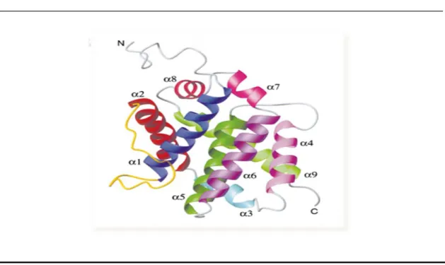
Like other proteins of the Bcl-2 family Bax is formed by alpha helices connected by loops. In aqueous solution Bax is composed by 9 α-helices of which the 2 central helices (Hα5 and Hα6) are mostly hydrophobic (Lalier et al, 2007). The 19 first residues of Bax, that precede the Hα1, are very mobile, as suggested by Nuclear Magnetic Resonance (NMR) data (Suzuki et al, 2000). The Hα1 by itself has been shown to be involved in the translocation of Bax to the OMM (Cartron et al, 2003). The BH-3 domain of Bax roughly corresponds to the Hα2 of the protein (Zha et al, 1996). Movements between Hα1 and Hα2 are involved in Bax activation, and once ART has been moved Bax can be activated (Renault and Manon, 2011). The helices Hα5 and Hα6 may directly participate to the permeabilization processes induced by Bax. Indeed it was shown that the capacity of the protein to promote membrane permeabilization and to trigger apoptosis is altered in mutant proteins lacking these helices (Valero et al, 2011). So, in summary, Hα5, Hα6 and Hα9 helices are likely involved in the interaction of Bax with OMM (Schendel et al, 1998). In the three-dimensional (3D) structure of Bax it is clear that, in the native conformation, Hα9 is tightly sequestered in the hydrophobic pocket and cannot insert into membranes in this conformation (Silva et al, 2011b). Many cell biology studies made with a fusion Green Fluorescent Protein (GFP)-Bax showed that the absence of Hα9 prevent the ability of the fusion protein to be
Page 14
translocated to the OMM following an apoptotic signal (Nechushtan et al, 1999) (Figure 1.4). Therefore the movement of Hα9 seems to be a crucial event in Bax translocation, not because it might be a membrane anchor, but because this movement unmasks the BH domain and the hydrophobic hairpin Hα5/Hα6, that are crucial for the subsequent events of dimerization and oligomerization (Lalier et al, 2007).
______________________________________________________________________
Figure 1.4 – A structure of Bax monomer. All the α-helices were indicated
_____________________________________________________________________________________
1.3.2 Regulatory mechanisms by protein interactions and post-translational modifications
A huge importance has been given to the regulation of Bcl-2 family proteins by post-translational modifications, in the very last years. Proteins of the Bcl-2 family can be regulated by interaction with other members of the Bcl-2 protein family or other proteins, or even by post-translational modifications. A key event in regulation of the Bcl-2 family members is the regulated protein-protein interactions (Shimizu et al, 2001). Members of this family can interact and form heterodimers blocking the activity of each other (Hanada et al, 1995). Inhibiting protein interactions might be a method for pharmacological intervention. A peptide and non-peptide mimetics of the BH3 domain are able to interfere with Bax/Bcl-2 interaction. These BH3 mimetics are small molecule antagonists of the anti-apoptotic Bcl-2 members. They act as a competitive inhibitors of the pro-apoptotic proteins though binding to the hydrophobic cleft of the anti-apoptotic proteins (for a review see Chonghaile and Letai, 2008).
Page 15
It has already been reported that phosphorylation of different Bax residues modulates its activity (Gardai et al, 2004). Phosphorylation is the most common regulation mechanism by post-translational modifications of the members of Bcl-2 protein family and in most cases leads to the loss of the biological function of these proteins (Basu et al, 2006). Bax has been identified as a substrate of different kinases that regulate its activity. As mentioned above, Bax is activated when Hα9 is forced to move away from BH3 domain. So, it would be expected that the phophorylation of Ser184 introduced a size constraint that would also help the movement of Hα9. However Bax phosporylation on Ser184 prevents its translocation to the mitochondria, whereas non-phophorylated Bax was mitochondrial (Suzuki et al, 2000). Consistently, phosphorylation of Ser184 by protein kinase B (AKT/PKB) promotes cell survival (Gardai et al, 2004). The protein kinase Cζ (PKCζ) (Yamaguchi and Wang, 2001 and Xin et al, 2007) promotes cell survival that is prevented by dephosphorylation by the protein phosphatase 2A (Xin and Deng, 2006). Apoptosis modulation-associated proteins may also be regulated through phosphorylation by different proteins kinases, such as protein kinase c (PKC) (Saraiva et al, 2006). Generally, classical and atypical PKCs appear to be associated with cell survival, whereas novel PKCs are associated with apoptosis stimulation. It was shown that different PKC isoforms modulate the Bcl-xL anti-apoptotic activity differently through interference with the phosphorylated forms of Bcl-xL (Saraiva et al, 2006). Akt/PKB also phosphorylates Bad increasing cell survival (Datta et al, 1997), Bcl-2 phosphorylation is required for its anti-apoptotic function (Ito et al, 1997) and Bcl-xL is phosphorylated and inactivated by the Jun N-terminal kinase (JNK) (Fan et al., 2000).
Others residues can also be involved in regulation of Bax by phosphorylation; two examples are the phosphorylation on Ser163 that can be another possible target by glycogen synthase kinase-3β (GSK-3β), and the phosphorylation Thr167 by JNK and p38 kinase, leading to Bax activation and cell death. GSK-3β is a crucial activator of cell death in numerous models of neuronal apoptosis (Linseman et al, 2004; Silva et al, 2011b). Arokium et al, 2007 identified a new putative phosphorylation site at Ser60, which is located in a consensus target sequence for PKA. These phosphorylations results allowed the study of the complex phoshorylation/dephosphorylation events that regulates Bcl-2 protein family activity by heterologous expression of different kinases and protein phosphatases.
Page 16
As it is well known, proteins phosphatases are enzymes whose function is to remove the phosphate group from a substrate, though a dephosphorylation mechanism while kinases have an opposite function, and phosphorylate the substrates through an ATP consuming reaction. Protein phosphatases can be classified according to their specificity regarding the aminoacid residue they dephosphorylate (table below).
Class Example Substrate Reference
Histidine protein
phosphatase PHP Phospho-Histidine (Moorhead, 2007)
Dual specificity protein
phosphatases VHR
Phosphotyrosine/-serine/-threonine (Camps et al, 2000) Serine-/threonine-pecific protein phosphatases PP2C Phosphoserine/-threonine (Moorhead, 2007) Lipid protein phosphatase PTEN
Phosphatidyl-Inositol-3,4,5-Triphosphate (Maehama et al, 2004) Tyrosine-specific protein
phosphatases PTP1B Phosphotyrosine (Zhang, 2002)
Although protein phosphatases can be divided based on their substrate specificity they can also be divided into four main groups based on their catalytic function, structure and sequence. So according to this classification the major classes of protein phosphatases are the protein phosphatase (PPP) family, corresponding to PP1, PP2A, PP2B, PP4, PP5, PP6 and PP7, the protein phosphatase Mg2+ (PPM) family, corresponding to PP2C, the PTP, and the aspartate-based protein phosphatase (Barford, 1996).
1.4 Contributions of the yeast model to study the function of
proteins of the Bcl-2 family
Several studies have examined the response of yeast to the heterologous expression of proteins of the Bcl-2 family, in order to address basic questions on their mechanisms of action. The idea of using yeast as an alternative system for the elucidation of molecular aspects of the function of these apoptotic regulators arose accidentally (Pereira et al, 2008), even before yeast was a well established unicellular eukaryotic model to study cell death. Indeed for many years, apoptosis was assumed to be restricted to multicellular organisms. However, it is now established that several stimuli can induce in unicellular organisms some of typical apoptotic markers that are
Page 17
observed in multicellular organisms. Through this concept initially generated a big controversy due to the difficulty to find an advantage of a cell suicide in an unicellular organism additional evidence showed that apoptotic-like cell death can greatly benefit a population of unicellular organisms, such as yeast, for instance by eliminating virus-infected and damaged cells or by releasing nutrient sources for the fittest individuals (Büttner et al, 2006 and Rego et al, 2012). So far several yeast orthologs of crucial apoptotic regulators have been discovered (for a revision see Carmona-Gutierrez et al, 2010). For instance, a caspase-like protein termed Yca1p has been identified, which revealed to be involved in programmed cell death induced by acetic acid, hydrogen peroxide (H2O2) and ageing. Absence of Yca1p reduces cell death whereas its
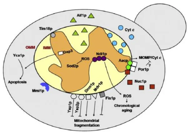
overexpression enhances apoptotic-like death of the cells (Frank Madeo et al, 2002 and Fahrenkrog et al, 2004). It was also recognized in yeast a mitochondria-mediated apoptosis pathway in response to a variety of stimuli, similar to the mammalian intrinsic apoptotic pathway, involving cytochrome c release from mitochondria to the cytosol (Ludovico et al, 2002; Pereira et al, 2008; Giannattasio et al, 2008). The use of yeast mutants defective in genes encoding mitochondrial proteins enabled to identify several other mitochondrial regulators/mediators of apoptosis in this organism (Figure 1.5). The proteins identified are components of the electron transport chain, of the IMM or of OMM. While most of the yeast mitochondrial proteins recognized as implicated in apoptosis have their mammalian counterparts also involved in apoptosis, a few were only identified in yeast (e.g. Ysp1p and Ysp2p).
An ortologue of Apoptosis Inducing Factor (AIF), YNR074C was identified in yeast that displays sequence similarity to AIF and AIF-homologous mitochondrion-associated inducer of death (AMID) and regulates apoptosis in a similar way to that AIF in mammalian cells. However, the inner membrane mitochondrial Nicotinamide Adenine Dinucleotide (NADH) dehydrogenase (Ndi1p), which is the first component of the electron transport chain in yeast, was found to be the closest yeast orthologue to mammalian AMID proteins (Li et al, 2006). Nuc1p, an ortolog of another classical apoptotic regulator, EndoG has also been classified and characterized (Büttner et al, 2007).
Page 18
Figure 1.5- Mitochondrial proteins involved in the regulation of yeast apoptotic cell death. (Taken from
(Pereira et al, 2008).
Some of those yeast mitochondrial proteins, like their mammalian akin, are translocated from the mitochondria to the cytosol or to the nucleus (Aif1p and Nuc1p), others are proposed to be involved in mitochondrial permeabilization and release of the former proteins (Aac1p, Aac2p and Aac3p, and Por1p) or in the fission/fusion of mitochondria (Dnm1p, Mdv1/Net2, Fis1p, Ysp1p and Ysp2p) (Pereira et al, 2008). All these findings firmly established that yeast and metazoan apoptosis have in essence the same cellular program (Li et al, 2006).
Until recently it was thought that yeast lacks obvious homologues of the Bcl-2 protein family members, and it has therefore been used as an “in vivo” system to study several of these apoptotic regulators without the interference of other family members. But in the last decade Büttner et al, 2011 report that the yeast genome encodes a BH3 domain-containing protein (Ybh3p) which interacts with Bcl-xL and shares functional characteristics with the pro-apoptotic members of the mammalian Bcl-2 family. Ybh3p is capable of translocating to mitochondria and regulate the mitochondrial apoptotic pathway in a phylogenetically conserved manner. This study has also proved that the overexpression of Ybh3p enhances stimulus-induced apoptosis, while its knockout
Page 19
reduces cell death. Death mediated by Ybh3p was accompanied by a burst of ROS. The deletion of YBH3 protects against cell death induced by H2O2, acetic acid, mammalian
Bax expression and ageing. Ybh3p was first described as a member of the family of Bax-inhibitor 1 (BI-1) proteins, which operate as antiapoptotic proteins in the endoplasmic reticulum of all phyla, including animals, plants and yeast, and contributes to the development of cancer. BI-1 inhibits Bax induced apoptosis and appears to regulate the concentration of Ca2+ in the ER and the cytosol, in mammalian cells. Plant BI-1 form is able to suppress mammalian Bax induced cell death in yeast (Cebulski et
al, 2011).
1.4.1 Advantages of the yeast model system
Because of the conservation of many cellular processes in yeast and of its simple manipulation and genetic tractability, this organism, among the different cell models exploited to comprehend several mammalian cellular pathways and processes, emerged as a powerful tool and a model of choice to answer important biological questions. Specifically, the easy manipulation of its mitochondria led to an increased interest in using this cell model to unveil unknown features of the mammalian intrinsic apoptotic pathway and its regulation by members of the Bcl-2 family (Silva et al, 2011a).
It was demonstrated that Bcl-2 family members when expressed in yeast are likely to act upon highly conserved mitochondrial components that correspond directly to their apoptotic substrates in mammalian cells, generating similar, if not identical, biochemical and physiological responses (Guscetti et al, 2005). It was described that expression of Bax in S. cerevisiae causes growth arrest and induces cell death with similar characteristics to those observed in apoptotic metazoan cells (Ligr et al, 1998). However, another work showed the occurrence of autophagic features in yeast cells expressing Bax, including increased accumulation of Atg8p and activation of the targeting-deficient mutant of the vacuolar alkaline protein phosphatase. Inactivation of autophagy slightly accelerated Bax-induced cell death showing a protective role for this process (Kissová et al, 2006).
Bax was also shown to induce cell death in other yeast species namely in
Schizosaccharomyces pombe, Pichia pastoris and Candida albicans (Ink et al, 1997 and
Greenhalf et al, 1996). The budding yeast S. cerevisiae has proved particularly useful for studying Bax-induced mitochondrial changes, because Bax is able to induce the release of cyt c from mitochondria (Manon et al, 1997) and induces apoptotic
![Figure 1.2 shows the homology shared by the Bcl-2 family members. The anti-apoptotic proteins like Bcl-2, Bcl-xL and Bcl-W are characterized by the presence of four Bcl-2 homology domains (BH: BH1, BH2, BH3 and BH4) [Figure 1.2(a)]](https://thumb-eu.123doks.com/thumbv2/123dok_br/17686317.827021/19.892.124.760.514.1087/figure-homology-members-apoptotic-proteins-characterized-presence-homology.webp)