Allelopathic activity of the picocyanobacterium Synechococcus sp. on
1coexisting microalgae
2 3 Sylwia Śliwińska-WilczewskaA, Aldo Barreiro FelpetoB,D, Jakub MaculewiczA, Amanda
4
SobczykA, Vitor VasconcelosB,C and Adam LatałaA
5 6
A
University of Gdansk, Institute of Oceanography, Av. Pilsudskiego 46, 81-378 Gdynia, 7
Poland 8
B
Interdisciplinary Center of Marine and Environmental Research–CIMAR/CIIMAR, 9
University of Porto, Av. General Norton de Matos s/n, 4450-208 Matosinhos, Portugal 10
C
Department of Biology, Faculty of Sciences, Porto University. Rua do Campo Alegre, 4069-11
007 Porto, Portugal 12
D
Corresponding author. Email: [email protected] 13
14
Abstract. The production and release of allelopathic compounds is an important adaptation
15
by which some species of cyanobacteria can achieve a competitive advantage over other 16
primary producers. In this study, we tested the allelopathic activity of the picocyanobacterium 17
Synechococcus sp. against the following coexisting microalgae: Porphyridium purpureum,
18
Stichococcus bacillaris, Prymnesium parvum and Nitzschia dissipata. We demonstrated that
19
both addition of Synechococcus sp. cell-free filtrate and co-culture inhibited the growth of P. 20
purpureum and S. bacillaris. Conversely, the growth of P. parvum was positively affected. By
21
contrast, N. dissipata was unaffected by either the picocyanobacterial filtrate or co-culture. 22
These results showed that Synechococcus sp. allelopathy should be considered when 23
estimating the potential interactions between picocyanobacteria and coexisting microalgae in 24
aquatic ecosystems. 25
26
Additional keywords: allelopathy, cyanobacterial bloom, growth, microalgae,
27 picocyanobacteria, Synechococcus sp. 28 29 Introduction 30
Allelopathy can be found in aquatic environments and primary producers can release active 31
compounds which can affect the functioning of the marine, brackish and freshwater 32
ecosystems (e.g., Fistarol et al. 2004; Ji et al. 2011; Poulson-Ellestad et al. 2014; Brutemark 33
et al. 2015). Allelopathy is also considered among the potential drivers of massive blooms
34
throughout many aquatic environments. The number of reports on the allelopathic effects of 35
cyanobacteria and microalgae has been steadily increasing (e.g., Barreiro and Hairston 2013; 36
Żak and Kosakowska 2015; Dias et al. 2017; Wang et al. 2017). 37
Picocyanobacteria play an important role in aquatic ecosystems (Beardall 2008; Callieri 38
2010; Costa et al. 2015; Worden and Wilken 2016) but little is known about their allelopathic 39
activity again coexisting microalgae. In previous works, we demonstrated the allelopathic 40
effect of Synechococcus sp. on diatom Navicula perminuta (Śliwińska-Wilczewska et al. 41
2016) and we also showed a significant inhibitory effect of cell-free filtrate obtained from 42
Synechococcus sp. against the filamentous cyanobacteria Nostoc sp. and Phormidium sp.
43
(Śliwińska-Wilczewska et al. 2017a) and against different groups in a natural plankton 44
community (Śliwińska-Wilczewska et al. 2017b). There are also some reports of allelopathic 45
effects such as growth inhibition or stimulation caused by other species of cyanobacteria (e.g., 46
Suikkanen et al. 2004, 2005; Antunes et al. 2012; Barreiro and Vasconcelos 2014; Rzymski et 47
al. 2014) but there is no information about allelopathic potential of Synechococcus sp. on
48
coexisting microalgae like Porphyridium purpureum, Stichococcus bacillaris, Prymnesium 49
parvum and Nitzschia dissipata. The issue of picocyanobacterial allelopathy needs more
50
researcher’s attention, since this group is the major contributor to marine primary production. 51
The effect of allelochemicals depends on the nature of the interaction between donor 52
and target organisms (direct cell to cell contact, no contact) and activity of the chemical 53
compounds responsible of this interaction. In aquatic ecosystems, donor organisms may affect 54
the target species in different ways, and, in most cases, allelopathic compounds can reduce the 55
growth rate and eventually cause the death of targeted organisms (Rzymski et al. 2014). 56
However, it is difficult to find direct evidence of allelopathic interactions between 57
cyanobacteria and microalgae in natural communities (see Keating 1977, 1978). Before this 58
can be achieved, it is very important to characterize allelopathic interactions under controlled 59
laboratory conditions, in order to investigate in detail the nature of released substances and 60
their impact on target organisms. 61
The main aim of this study was to determine the influence of allelopathic compounds 62
produced by picocyanobacterium Synechococcus sp. on growth of Porphyridium purpureum, 63
Stichococcus bacillaris, Prymnesium parvum and Nitzschia dissipata in both co-cultures and
64
bioassays employing cell-free filtrates. These species are widely geographically distributed 65
and relevant in terms of abundance in phytoplankton communities, so allelopathic interactions 66
between them could play a significant role in aquatic ecosystems. Based on this, further 67
research could be done to identify the chemical structures of the allelopathic compounds 68
involved in the interaction, as well as their mechanisms and modes of action on target 69
organisms. 70
71
Materials and methods
72
Material and culture conditions
The experiments were conducted with the picocyanobacterium Synechococcus sp. (BA-124) 74
and the following microalgae: the red algae Porphyridium purpureum (MA-03), the green 75
algae Stichococcus bacillaris (BA-09), the golden algae Prymnesium parvum (AA-69) and the 76
diatom Nitzschia dissipata (BA-40). These strains were isolated from the coastal zone of the 77
Norwegian Sea and the Baltic Sea and were maintained as unispecies cultures in the Culture 78
Collection of Baltic Algae (CCBA) at the Institute of Oceanography, University of Gdańsk, 79
Poland. 80
Cultures were grown in f/2 medium (Guillard 1975) in 25-mL glass Erlenmeyer flasks 81
that were swirled daily during the experiment. Culture media was prepared with Baltic Sea 82
water filtered through glass microfiber filters (Whatman GF/C) and autoclaved. The salinity 83
was 8 PSU, as measured with a salinometer (inoLab Cond Level 1, Weilheim in Oberbayern, 84
Germany). The picocyanobacteria and microalgae were incubated under a 16:8 h light:dark 85
cycle at Photosynthetically Active Radiation (PAR) irradiance 10 μmol photons m–2s–1 and 86
temperature 20°C. The intensity of PAR was measured using a quantum-meter (LI-COR, 87
Nebraska, USA) with a cosine collector. 88
Prior to the experiments, the concentrations of macronutrients (i.e., nitrate, N-NO3 and 89
P-PO4 orthophosphates) in the picocyanobacterial cultures were measured to set them to the 90
standard levels of f/2 medium. Nutrients were determined using spectrophotometric methods 91
as described by Grasshoff (1976). Then, the concentrations of N-NO3 and P-PO4 in the 92
controls and all treatments were adjusted to the same level as in the f/2 growth medium. The 93
microalgae in the controls showed active growth during the course of the experiment. 94
Therefore, the effects of major nutrients, microelements and vitamin limitations in the control 95
and allelochemical treatments can be excluded. On the first and the last days of the 96
experiment, the pH of all experimental flasks was measured using a pH-meter (Elmetron CP-97
401, Zabrze, Poland). pH values were similar across treatments and durations and ranged 98
from 8.2 to 8.6. 99
100
Test of the allelopathic effect of cell-free filtrates
101
Allelopathic effects were tested following a modified version of the method proposed by 102
Suikkanen et al. (2004). Cultures of the donor picocyanobacterium Synechococcus sp. and the 103
target microalgae were maintained in active growth during 7 days in standard conditions (see 104
above) and then employed in the experiments. The picocyanobacterium culture was gently 105
filtered through a 0.45-µm filter (Macherey-Nagel MN GF-5) using a vacuum pump (400 106
mbar). The filtrate obtained was analyzed under an epifluorescence microscope (Nikon 107
Eclipse 80i, Nikon, Tokyo, Japan) in order to confirm the absence of picocyanobacteria cells. 108
Experimental treatments were prepared by adding 4 mL of this cell-free filtrate to 25-mL 109
Erlenmeyer flasks containing 20 mL of cell suspensions of the target microalgae. These cell 110
suspensions were obtained from culture aliquots. Controls were prepared by adding 4 mL of 111
filtered f/2 medium to 25-mL Erlenmeyer flasks containing 20 mL of cell suspensions of the 112
same microalgae species. These tests were conducted in triplicate. In all experiments, the ratio 113
of donor picocyanobacteria to target species was adjusted to 1:1 based on the chlorophyll a 114
(Chl a) content (the initial Chl a concentration in the experimental cultures was 0.8 µg Chl a 115
mL-1). The experiments lasted 7 days. 116
117
Test of the allelopathic effect in co-cultures
118
Allelopathic interactions in co-cultures were determined following a modified version of the 119
method proposed by Ji et al. (2011). Employed cultures maintained in the conditions 120
described above, 4 mL aliquots from the donor picocyanobacteria were added to 25-mL 121
Erlenmeyer flasks containing 20 mL of cell suspensions of the target microalgae. Controls 122
were prepared by adding 4 mL of filtered f/2 medium to 25-mL Erlenmeyer flasks containing 123
20 mL of cell suspensions of the target microalgae. These tests were conducted in triplicate. 124
In all these experiments, the initial ratio of donor picocyanobacteria to target species in 125
Erlenmeyer flasks was adjusted to 1:1 based on the Chl a content (the initial chlorophyll a 126
concentration in the experimental cultures was 0.8 µg Chl a mL–1 for donor and target 127
species). The experiments lasted 7 days. 128
129
Determination of cell abundances
130
The number of cells (N) in cultures was counted with flow cytometer BD Accuri™ C6 Plus 131
(BD Biosciences, San Jose, CA, USA). Events were recorded in list mode. Samples were run 132
at a flow rate of approximately 14 µl min–1. Flow was daily calibrated with Spherotech 6- and 133
8- Peak Validation Beads (BD, San Jose, USA). This ensures that the cytometer is working 134
properly before running experimental samples. FITC, PE, and PE-Cy5 detectors were daily 135
calibrated with SPHERO™ Rainbow Calibration Particles (BD, San Jose, USA), and the APC 136
channel was calibrated with SPHERO 6-peaks Allophycocyanin Calibration Particles (APC). 137
Detectors FL1, FL2, and FL3 read fluorescence emissions excited by the blue laser (480 nm), 138
while detector FL4 reads emissions excited by the red laser (640 nm). In the co-cultures, the 139
populations of Synechococcus sp. and the corresponding target microalgae were completely 140
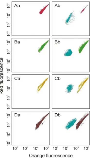
discriminated using a combination of red and orange fluoresecences (Fig. 1). N was 141
determined in all the experiments at times 0 (1h) and 1st, 3rd and 7th day of the experiments. 142 143 Fig. 1 144 145 Statistical analyses 146
Different ANOVAs were performed in order to test the effect of picocyanobacterial filtrates 147
or co-culture on the growth of the targeted microalgae. A post-hoc Dunnett’s test was used to 148
determine significant differences between the control and the other treatment levels. These 149
statistical analyses were performed with Statistica® 13.1 software. 150
151
Results
152
Allelopathic effect of cell-free filtrates
153
The addition of cell-free filtrate obtained from Synechococcus sp. significantly affected the 154
number of cells of Porphyridium purpureum, Stichococcus bacillaris and Prymnesium 155
parvum (ANOVA, F7,16 = 280.3, P < 0.001, ANOVA, F7,16 = 459.5, P < 0.001 and ANOVA, 156
F7,16 = 279.5, P < 0.001, respectively, Fig. 2A, B, C). Considering individual days, the 157
allelopathic effect of the picocyanobacterium significantly reduced the number of cells of P. 158
purpureum at day 7 (85% relative to the control; Dunnett, P < 0.01), and of S. bacillaris at
159
day 3 (87% relative to the control; Dunnett, P < 0.05) (Fig. 2A, B). In contrast, the addition of 160
cell-free filtrate obtained from Synechococcus sp. had a significantly positive effect on the 161
growth of P. parvum, which, at day 7 was higher than the control by 19% (Fig. 2C; Dunnett, 162
P < 0.001). The addition of the cell-free filtrate obtained from Synechococcus sp. had no
163
effect on the target diatom Nitzschia dissipata (ANOVA, F7,16 = 0.5, P = 0.7, Fig 2D). 164
165
Fig. 2 166
167
Allelopathic effects in co-cultures
168
After the addition of Synechococcus sp. cells, it was observed not only a reduction, but also a 169
decline with time in the number of cells of Porphyridium purpureum (ANOVA, F7,16 = 170
1526.6, P < 0.001, Fig. 3A). Significant differences were found on the first, third and seventh 171
day of the experiment, when cell numbers of P. purpureum constituted 38% (Dunnett, P < 172
0.001), 17% (Dunnett, P < 0.001) and 1% (Dunnett, P < 0.001), of control. For Stichococcus 173
bacillaris, it was observed a reduction in the number of cells (ANOVA, F7,16 = 599.8, P < 174
0.001, Fig. 3B). By the first, third and seventh day of the experiment the growths of S. 175
bacillaris were reduced by 80% (Dunnett, P < 0.001), 86% (Dunnett, P < 0.01) and 94%
176
(Dunnett, P < 0.05), respectively, relative to the control treatment. On the other hand, 177
Synechococcus sp. cells had significantly positive effect on the growth of Prymensium
178
parvum (ANOVA, F7,16 = 491.1, P < 0.001, Fig. 3C). The number of cells of P. parvum 179
significantly increased after third (Dunnett, P < 0.01) and seventh (Dunnett, P < 0.001) day of 180
the experiment and constituted 114% and 131%, respectively, of control (Fig. 3C). No effects 181
were detected in Nitzschia dissipata cultures (ANOVA, F7,16 = 1.2, P = 0.3, Fig. 3D). 182 183 Fig. 3 184 185 Discussion 186
There are very few reports of allelopathic compounds from phytoplankton that affect red 187
algae. Just very recently, García-Espín et al. (2017) showed that both cyanobacteria extracts 188
obtained from Rivularia haematites and Rivularia biasolettiana as well as pure microcystin 189
affected the photosynthetic activity of Chroothece richteriana. In this sense, our report of the 190
strong negative effect of Synechococcus sp. on P. purpureum, particularly evident in the co-191
culture, is also a novel finding. P. purpureum was more strongly inhibited in co-culture 192
compared to cell-free filtrate addition. This could be explained simply by the renewal of 193
allelochemical compounds in the presence of cells. 194
Some studies have shown that green algae are a phytoplankton group that are 195
particularly sensitivity to allelopathic compounds from cyanobacteria (Schlegel et al. 1999; 196
Schagerl et al. 2002; Żak et al. 2012). The data presented here constitutes, to our knowledge, 197
the first report of an allelopathic effect of a picocyanobactera against a specific species of 198
chlorophyte. These inhibitory effects on S. bacillaris in monoculture and co-culture were 199
stronger during the first days of exposition both with cell-free filtrates and co-culture. This 200
suggests that this species may become resistant to these compounds through adaptation. 201
Previous studies did not find allelopathic effects from different cyanobacteria 202
(Nodularia spumigena, Aphanizomenon flos-aquae and Anabaena lemmermannii) against 203
Prymnesium parvum (Suikkanen et al. 2004). Our study shows that Synechococcus sp. affects
204
P. parvum positively both through cell-free filtrates and co-culture. Suikkanen et al. (2005)
205
also noted that cyanobacterial filtrates could promote the growth of some microalgae due to 206
stimulatory allelochemicals. This could explain our results in the cell-free filtrate treatment. 207
Also, osmotrophic feeding by P. parvum on exudates from Synechococcus sp. could be an 208
explanation. In the co-culture treatment, the stronger positive effect could be explained by the 209
higher concentration of exudates in the presence of Synechococcus sp. cells, but, it is also 210
possible the occurrence of phagotrophic feeding by P. parvum, (Tillmann 1998). 211
Many authors showed different sensitivity of diatoms for the allelopathic compounds 212
produced by cyanobacteria. In this work, the diatom Nitzschia dissipata was found to be 213
tolerant to Synechococcus sp. cell-free filtrate and co-culture. Suikkanen et al. (2004) showed 214
that Thalassiosira weissflogii was inhibited by cell-free filtrates of three cyanobacteria: N. 215
spumigena, A. flos-aquae and A. lemmermannii. The tolerance of our species could be due to
216
its relatively large cell size, as suggested by Lyczkowski and Karp-Boss (2014). 217
Our results suggest that allelopathic effects of Synechococcus sp. may have a 218
differential effect in phytoplankton communities, due to the contrasting responses found 219
among target species. The effects of picocyanobacteria on coexisting phytoplankton species is 220
very important, since they account for the majority of primary producer biomass in the 221
oceans, and their relative abundance is even expected to increase in scenarios predicted by 222
global change (Dutkiewicz et al. 2015). 223
224
Acknowledgments
225
This study was supported by BMN grants, Poland, No. 538-G245-B568-17 and FCT Project
226 UID/Multi/04423/2013. 227 228 References 229
Antunes, J. T., Leão, P. N., and Vasconcelos, V. M. (2012). Influence of Biotic and Abiotic 230
Factors on the Allelopathic Activity of the Cyanobacterium Cylindrospermopsis 231
raciborskii Strain LEGE 99043. Microbial Ecology 64, 584–592. doi:
10.1007/s00248-232
012-0061-7 233
Barreiro, A., and Vasconcelos, V. M. (2014). Interactions between allelopathic properties and 234
growth kynetics in four freshwater phytoplankton species studied by model simulations. 235
Aquatic Ecology 48, 191–205. doi: 10.1007/s10452-014-9475-2
236
Barreiro, A., and Hairston Jr, N. G. (2013). The influence of resource limitation on the 237
allelopathic effect of Chlamydomonas reinhardtii on other unicellular freshwater 238
planktonic organisms. Journal of Plankton Research 35, 1339–1344. doi: 239
10.1093/plankt/fbt080 240
Beardall, J. (2008). Blooms of Synechococcus: An analysis of the problem worldwide and 241
possible causative factors in relation to nuisance blooms in the Gippsland Lakes. 242
Monash university, 1–8. 243
Brutemark, A., Vandelannoote, A., Engström-Öst, J., and Suikkanen, S. (2015). A less saline 244
Baltic Sea promotes cyanobacterial growth, hampers intracellular microcystin 245
production, and leads to strain-specific differences in allelopathy. PloS one 10, 246
e0128904. doi: 10.1371/journal.pone.0128904 247
Callieri, C. (2010). Single cells and microcolonies of freshwater picocyanobacteria: A 248
common ecology. Journal of Limnology 69, 257–277. doi: 10.3274/JL10-69-2-08 249
Costa, M. S., Costa, M., Ramos, V., Leão, P. N., Barreiro, A., Vasconcelos, V., and Martins, 250
R. (2015). Picocyanobacteria from a clade of marine cyanobium revealed bioactive 251
potential against microalgae, bacteria, and marine invertebrates. Journal of Toxicology 252
and Environmental Health, Part A 78, 432–442. doi: 10.1080/15287394.2014.991466
253
Dias, F., Antunes, J. T., Ribeiro, T., Azevedo, J., Vasconcelos, V., and Leão, P. N. (2017). 254
Cyanobacterial Allelochemicals But Not Cyanobacterial Cells Markedly Reduce 255
Microbial Community Diversity. Frontiers in Microbiology 8, 1495. doi: 256
10.3389/fmicb.2017.01495 257
Dutkiewicz, S., Morris, J. J., Follows, M. J., Scott, J., Levitan, O., Dyhrman, S. T., and 258
Berman-Frank, I. (2015). Impact of ocean acidification on the structure of future 259
phytoplankton communities. Nature Climate Change 5, 1002–1006. doi: 260
10.1038/NCLIMATE2722 261
Fistarol, G. O., Legrand, C., Selander, K., Hummer, C., Stolte, W., and Granéli, E. (2004). 262
Allelopathy in Alexandrium spp.: effect on a natural plankton community and on algal 263
monocultures. Aquatic Microbial Ecology 35, 45–56. 264
García-Espín, L., Cantoral, E. A., Asencio, A. D., and Aboal, M. (2017). Microcystins and 265
cyanophyte extracts inhibit or promote the photosynthesis of fluvial algae. Ecological 266
and management implications. Ecotoxicology 26, 658–666. doi: 10.1007/s10646-017-267
1798-z 268
Grasshoff, K. (1976). Methods of seawater analysis, Verlag Chemie Weinheim, New York. 269
Guillard, R. R. L. (1975). Culture of phytoplankton for feeding marine invertebrates. In: W. 270
L., Smith, M. H., Chanley, (eds), Culture of Marine Invertebrate Animals, pp. 26–60. 271
Plenum Press, New York, USA. 272
Ji, X. Q., Han, X. T., Zheng, L., Yu, Z. M., Yang, B. J., and Zou, J. Z. (2011). Allelopathic 273
interactions between Prorocentrum micans and Skeletonema costatum or Karenia 274
mikimotoi in laboratory cultures. Chinese Journal of Oceanology and Limnology 29,
275
840–848. doi: 10.1007/s00343-011-0512-x 276
Keating, K. I. (1977). Allelopathic Influence on Blue-Green Bloom Sequence in a Eutrophic 277
Lake. Science 196, 885–887. doi: 10.1126/science.196.4292.885 278
Keating, K. I. (1978). Blue-green algal inhibition of diatom growth: transition from 279
mesotrophic to eutrophic community structure. Science 199, 971–973. doi: 280
10.1126/science.199.4332.971 281
Lyczkowski, E. R., and Karp-Boss, L. (2014). Allelopathic effects of Alexandrium fundyense 282
(Dinophyceae) on Thalassiosira cf. gravida (Bacillariophyceae): a matter of size. 283
Journal of Phycology 50, 376–387. doi: 10.1111/jpy.12172
284
Poulson-Ellestad, K., Mcmillan, E., Montoya, J. P., and Kubanek, J. (2014). Are offshore 285
phytoplankton susceptible to Karenia brevis allelopathy? Journal of Plankton Research 286
36, 1344–1356. doi: 10.1093/plankt/fbu064
287
Rzymski, P., Poniedziałek, B., Kokociński, M., Jurczak, T., Lipski, D., and Wiktorowicz, K. 288
(2014). Interspecific allelopathy in cyanobacteria: Cylindrospermopsin and 289
Cylindrospermopsis raciborskii effect on the growth and metabolism of Microcystis
290
aeruginosa. Harmful Algae 35, 1–8. doi: 10.1016/j.hal.2014.03.002
291
Schagerl, M., Unterrieder, I., and Angeler, D.G. (2002). Allelopathy among cyanoprokaryota 292
and other algae originating from Lake Neusiedlersee (Austria). International Review of 293
Hydrobiology 87, 365–374. doi: 10.1002/1522-2632(200207)87:4<365::AID-294
IROH365>3.0.CO;2-B 295
Schlegel, I., Doan, N. T., de Chazal, N., and Smith, G. D. (1999). Antibiotic activity of new 296
cyanobacterial isolates from Australia and Asia against green algae and cyanobacteria. 297
Journal of Applied Phycology 10, 471–479.
298
Suikkanen, S., Fistarol, G. O., and Granéli, E. (2004). Allelopathic effects of the Baltic 299
cyanobacteria Nodularia spumigena, Aphanizomenon flos-aquae and Anabaena 300
lemmermannii on algal monocultures. Journal of Experimental Marine Biology and
301
Ecology 308, 85–101. doi: 10.1016/j.jembe.2004.02.012
302
Suikkanen, S., Fistarol, G. O., and Granéli, E. (2005). Effects of cyanobacterial 303
allelochemicals on a natural plankton community. Marine Ecology Progress Series 287, 304
1–9. 305
Śliwińska-Wilczewska, S., Pniewski, F., and Latała, A. (2016). Allelopathic activity of the 306
picocyanobacterium Synechococcus sp. under varied light, temperature and salinity 307
conditions. International Review of Hydrobiology 101, 69–77. doi: 308
10.1002/iroh.201501819 309
Śliwińska-Wilczewska, S., Maculewicz, J., Barreiro Felpeto, A., Vasconcelos, V., and Latała, 310
A. (2017a). Allelopathic activity of the picocyanobacterium Synechococcus sp. on 311
filamentous cyanobacteria. Journal of Experimental Marine Biology and Ecology 496, 312
16–21. doi: 10.1016/j.jembe.2017.07.008 313
Śliwińska-Wilczewska, S., Maculewicz, J., Tuszer, J., Dobosz, K., Kalusa, D., and Latała, A. 314
(2017b). First record of allelopathic activity of the picocyanobacterium Synechococcus 315
sp. on a natural plankton community. Ecohydrology & Hydrobiology 17, 227–234. doi: 316
10.1016/j.ecohyd.2017.05.001 317
Tillmann, U. 1998. Phagotrophy by a plastidic haptophyte, Prymnesium patelliferum. Aquatic 318
Microbial Ecology 14, 155–160.
Wang, L., Zi, J., Xu, R., Hilt, S., Hou, X., and Chang, X. (2017). Allelopathic effects of 320
Microcystis aeruginosa on green algae and a diatom: Evidence from exudates addition
321
and co-culturing. Harmful Algae 61, 56–62. doi: 10.1016/j.hal.2016.11.010 322
Worden, A. Z., and Wilken, S. (2016). A plankton bloom shifts as the ocean warms. Science 323
354, 287–288. doi: 10.1126/science.aaj1751
324
Żak, A., and Kosakowska, A. (2015). The influence of extracellular compounds produced by 325
selected Baltic cyanobacteria, diatoms and dinoflagellates on growth of green algae 326
Chlorella vulgaris. Estuarine, Coastal and Shelf Science 167, 113–118. doi:
327
10.1016/j.ecss.2015.07.038 328
Żak, A., Musiewicz, K., and Kosakowska, A., 2012. Allelopathic activity of the Baltic 329
cyanobacteria against microalgae. Estuarine, Coastal and Shelf Science 112, 4–10. doi: 330
10.1016/j.ecss.2011.10.007 331
332 333
334
Fig. 1. Cytograms obtained with a monocultures (a) and co-cultures with Synechococcus sp.
335
(b) of four microalgae: P. purpureum (A), S. bacillaris (B), P. parvum (C) and N. dissipata 336
(D) analyzed using a BD Accuri™ C6 Plus flow cytometer. 337
338 339
340
Fig. 2. The number of cells (N) for P. purpureum (A), S. bacillaris (B), P. parvum (C) and N.
341
dissipata (D) for controls and experiments with additions of cell-free filtrate obtained from
342
Synechococcus sp. cultures after 0 (1 h), 1, 3 and 7 days of exposure. The values refer to
343
means (n = 3, mean ± SD). Asterisk indicates significant difference compared with control. 344
Levels of significance were: * P < 0.05; ** P < 0.01; *** P < 0.001. 345
346 347
348
Fig. 3. The number of cells (N) for P. purpureum (A), S. bacillaris (B), P. parvum (C) and N.
349
dissipata (D) for controls and experiments with additions of Synechococcus sp. cultures after
350
0 (1 h), 1, 3 and 7 days of exposure. The values refer to means (n = 3, mean ± SD). Asterisk 351
indicates significant difference compared with control. Levels of significance were: * P < 352
0.05; ** P < 0.01; *** P < 0.001. 353