UNIVERSIDADE DA BEIRA INTERIOR
Ciências da Saúde
Preclinical performance of vaginal semisolid
products: technological and safety evaluations
assuming physiologic parameters
Rita Solange Monteiro Machado
Thesis for Doctoral Degree in
Pharmaceutical Sciences
(3
rdcycle of studies)
Supervisor: Professor José António Martinez Souto de Oliveira, PhD
Co-supervisor: Professor Rita Manuela Palmeira de Oliveira, PhD
The experimental work presented in this thesis was carried out at the Health Sciences Research Center laboratories, University of Beira Interior (CICS-UBI) and, at Labfit – Health Products Research and Development, Lda facilities, under the supervision of Professor José António Martinez Souto de Oliveira and Professor Rita Manuela Palmeira de Oliveira.
The work herein presented was supported by FCT, the Portuguese Foundation for Science and Technology, and Labfit – Health Products Research and Development, Lda. in a collaborative academia-company project (SFRH/BDE/111544/2015). Additionally, CICS-UBI was supported by FEDER funds through the POCI - COMPETE 2020 - Operational Programme Competitiveness and Internationalization in Axis I - Strengthening research, technological development and innovation (Project POCI-01-0145-FEDER-007491), National Funds by FCT - Foundation for Science and Technology (Project UID/Multi /00709/2013).
“Tenho em mim todos os sonhos do mundo.”
Depois de percorrido este caminho, que com certeza não faria sozinha, resta-me agradecer a todos aqueles que me apoiaram e inspiraram a lutar pelos meus objetivos.
Aos meus orientadores,
agradeço a oportunidade que me deram de integrar este projeto. Posso agora dizer que a caminhada não foi sempre fácil, mas foi certamente um desafio gratificante. Ao Prof. Doutor José Martinez de Oliveira agradeço a simpatia, o entusiasmo e o seu notável contributo e orientação científica. À Prof. Doutora Rita Palmeira de Oliveira, pela sua constante boa disposição e otimismo, pela disponibilidade e ensinamentos transmitidos. Acima de tudo, agradeço a ambos, as ferramentas do pensar e comunicar ciência que me foram incutidas ao longo destes anos.
À Labfit-HPRD, Lda,
e a todos os seus colaboradores, pelo apoio técnico e científico, e pelo simpático acolhimento e amizade durante as fases em que o trabalho laboratorial foi lá desenvolvido. Em especial agradeço à Prof. Doutora Ana Palmeira de Oliveira que acompanhou de perto este trabalho, pela sua disponibilidade e contributo científico.
Ao CICS-UBI,
pelo acolhimento laboratorial e por ser a minha casa desde que comecei na investigação científica. Agradeço aos professores, investigadores, colegas e funcionários. Todos deram um pequeno contributo no desenvolvimento deste trabalho.
À Prof. Doutora Adriana Santos,
pela oportunidade que tive de colaborar nas suas aulas práticas. Agradeço os seus contributos científicos, e esclarecimentos no decorrer destes anos.
À Prof. Doutora Luiza Breitenfeld,
pela disponibilidade e pelo contributo científico que prestou na fase final deste trabalho. À Dr. Catarina Ferreira,
pela simpatia, disponibilidade e profissionalismo e sobretudo pela ajuda na histologia e microscopia.
À Oviger, SA,
pelo exemplo que são para mim, pelo amor e carinho, pelo apoio incondicional e força para seguir em frente, sem vocês nada teria conseguido.
À minha irmã,
e colega de casa durante estes últimos anos, pela amizade, companheirismo, pelo o que caminhámos juntas até agora e todo o caminho que temos pela frente, para toda a vida. À minha avó,
por todo o amor, pelos conselhos e recomendações, pelo carinho e boa disposição. À restante minha família,
madrinha, padrinho, tias e tios, primas e primos, pelo vosso carinho e apoio incondicional. Ao Artur,
por teres entrado na minha vida nesta fase e mesmo assim teres ficado. Pelos nossos momentos, pelos mimos, pelo conforto, pelo apoio, pelo companheirismo e acima de tudo, pelo amor. Aos meus Amigos,
que o destino colocou no meu caminho e nunca mais tirou. Sou grata pela vossa amizade e por tudo o que já vivemos e viveremos juntos. Esta caminhada, apesar das dificuldades, tornou-se mais fácil e gratificante com a amizade que nos uniu.
A avaliação pré-clínica de produtos semi-sólidos de administração vaginal quando é realizada tendo em consideração as particularidades intrínsecas desta via de administração, pode constituir-se como um ponto-chave na previsão do desempenho in vivo destes produtos. De facto, os medicamentos semi-sólidos vaginais são largamente prescritos e utilizados pelas mulheres, sendo também a classe de formas farmacêuticas de administração vaginal preferida por estas. Antes de entrarem nas fases clínicas do desenvolvimento, os semi-sólidos vaginais devem ter previamente demonstrado um perfil tecnológico adequado e devem ter dado provas de serem seguros. Deste modo, será possível obter maiores taxas de sucesso nas fases seguintes: pré-clínicas em animais (in vivo) e clínicas em humanos. Os métodos compendiais tradicionais de caracterização galénica e tecnológica não consideram as especificidades da via de administração vaginal, uma vez que não têm em conta, por exemplo, o pH vaginal, a composição e volume do fluido vaginal e a temperatura corporal. Além disso, mesmo na literatura científica é escassa a informação sistematizada de quais os parâmetros significativos a avaliar neste tipo de formulações. Avaliações precoces dos perfis de segurança e eficácia de medicamentos, são amplamente aconselhados, e, embora poucas, algumas estratégias e métodos encontram-se já descritas e validadas por agências reguladoras, contudo, concentram-se principalmente em modelos celulares. Adicionalmente, concentram-se por um lado a concentram-segurança pode concentram-ser abordada em testes de toxicidade, por outro, pode ainda ser mais amplamente caracterizada através da conjugação com resultados provenientes de estudos de libertação e permeação de fármacos. É um facto que a via de administração vaginal permite não só a administração de fármacos com vista a efeitos localizados, mas também efeitos sistémicos. Sendo que, uma ou outra pode ser desejada e a forma farmacêutica deve permitir que o fármaco seja confinado ao local de administração pretendido, para que se obtenha a máxima eficácia, evitando os efeitos secundários. O objetivo deste trabalho consistiu na realização de uma avaliação pré-clínica completa em formulações já comercializadas, a fim de estabelecer novas abordagens metodológicas que possam ser facilmente aplicadas no desenvolvimento e caracterização de novos produtos.
Neste contexto, medicamentos semi-sólidos vaginais antimicrobianos, tais como, o Gino-canesten®, Sertopic®, Dermofix®, Gyno-pevaryl®, Lomexin®, Gino Travogen®, Dalacin V®; estrogénios, como o Ovestin®, Blissel® e Colpotrophine®; e ainda duas formulações de referência, o Placebo Universal e o Replens® foram amplamente avaliadas. Em termos tecnológicos foram testados o pH, a capacidade tampão, a osmolalidade, alguns parâmetros de textura e viscosidade, seguindo um ponto de vista fisiológico, em que se consideraram a temperatura corporal e a diluição num simulante de fluido vaginal (SFV). Foi ainda otimizado, um modelo vaginal suíno ex vivo para estudar estas formulações no que respeita à bioadesão e à reologia num ambiente pós-administração. Quanto ao estudo da segurança, a toxicidade
(uterinas), e para tal, foram realizados os ensaios de viabilidade que utilizaram o MTT (brometo de 3-(4,5-dimetiltiazol-2-il)-2,5-difenil tetrazólio) e o NRU (captação de vermelho neutro). Além disso, tecidos epiteliais ex vivo, foram excisados a partir da vagina suína, e nestes, também foi aplicado o ensaio do MTT e, ainda uma análise histológica. Para incluir uma avaliação ainda mais avançada da toxicidade in vitro, os semi-sólidos vaginais foram testados num modelo organotípico: HET-CAM (ensaio que é realizado na membrana corio-alantóide de embriões de galinhas Henn) e que se encontra em validação para substituir o teste in vivo em animais para testes de irritação ocular. Deste modo, foi transposto este ensaio para a avaliação da irritação vaginal. Um método de quantificação de HPLC-DAD (Cromatografia Líquida de Alta Pressão acoplada a um detetor de díodos) para as moléculas presentes nos semi-sólidos vaginais incluídos neste trabalho, foi desenvolvido e validado de acordo com os requisitos das normas da FDA (Food and Drug Administration - EUA), da EMA (Agência Europeia de Medicamentos) e da ICH (Conferência Internacional para a Harmonização). Depois disso, este método foi aplicado na quantificação de fármacos nos estudos de libertação in vitro e nas experiências de permeação de fármacos ex vivo. Estas duas técnicas foram realizadas utilizando células de difusão dinâmicas verticais de Franz, tendo todas as configurações experimentais sido especificamente otimizadas para as moléculas em estudo (estriol, clotrimazol, econazole, isoconazol, sertaconazol e fenticonazol).
Quanto às características tecnológicas, as formulações antimicrobianas apresentaram menor pH do que os estrogénios tópicos. A capacidade tampão no SFV conduziu a melhores previsões do que acontece in vivo. Após diluição em volumes fisiológicos de SFV, a osmolalidade da maioria dos produtos esteve conforme o recomendado pela Organização Mundial da Saúde (OMS), claramente dependente da composição. Os antimicrobianos apresentaram comportamentos de textura semelhantes entre eles, enquanto os estrogénios tópicos variaram entre eles, no que respeita aos parâmetros de textura. Com efeito, neste grupo encontrava-se uma formulação de base polimérica em gel (Blissel®), sendo que os restantes, eram cremes (Ovestin® e Colpotrophine). Regra geral, para todos os produtos observou-se uma ligeira diminuição da viscosidade após aplicação de fatores fisiológicos, como a diluição em SFV e a temperatura (37ºC), mas os seus comportamentos pseudoplásticos mantiveram-se. No entanto, cada formulação demonstrou ter o seu próprio perfil reológico, possivelmente conduzido pela sua composição, tanto qualitativa como quantitativa. Contudo, a viscosidade das formulações foi mais elevada quando testada num modelo de administração ex vivo, comparando com a viscosidade obtida nos ensaios em SFV a 37ºC. Este dado releva, que podem existir outros fatores determinantes na viscosidade adquirida após a administração de formulações semi-sólidas. Quanto aos modelos in vitro, as células VK2 E6/E7 apresentaram viabilidades relativamente superiores às células HeLa e HEC-1A nas concentrações de produto testadas. Os resultados de viabilidade no tecido foram muito superiores aos obtidos para nos modelos celulares. Isto revela que este modelo poderá ser mais robusto e mais próximo do que acontece in vivo. Em todos os modelos, as formulações antimicrobianas mostraram viabilidades
apresentaram perfis relacionados com os efeitos celulares estrogénicos reconhecidos (ou seja, foram dependentes da dosagem da formulação e da concentração testada). Os produtos de referência apresentaram os perfis de viabilidade mais estáveis e mais elevados em todas as concentrações. Nos estudos de permeação ex vivo, foi questionada a existência de diferenças na utilização de secções de tecido vaginal caudal ou cranial. Não obstante, em termos de permeação de fármacos, não foram encontradas extensas diferenças significativas entre estes dois tipos de epitélio, mas a vagina caudal parece ser mais adequada para experiências de permeação vaginal, uma vez que conduz a resultados mais reprodutíveis e consistentes. De facto, a região mais cranial da vagina suína apresenta, histologicamente, um epitélio mais fino, e anatomicamente, uma superfície que contém mais rugas. O que conduz a desvios-padrão entre experiências mais elevados. Além disso, observou-se que a permeação de fármacos não é apenas dependente da libertação do fármaco a partir formulação. A sua afinidade pela formulação e/ou pelo tecido, é determinante para a sua permeação até à câmara recetora.
Concluindo, a avaliação pré-clínica integrada para semi-sólidos vaginais, proposta nesta tese, representa uma abordagem importante a ter em consideração, no desenvolvimento de novos produtos, uma vez que pode reduzir custos com o desenvolvimento de novas formulações, prevendo precocemente a sua eficácia in vivo e os seus perfis de segurança. Estas metodologias têm, assim, um grande potencial não só para serem aplicadas na indústria cosmética, farmacêutica e de dispositivos médicos, mas também pode ser aplicada em investigação mais fundamental ao nível da academia.
Palavras-chave
Administração vaginal de fármacos, semi-sólidos vaginais, avaliação pré-clínica, caracterização tecnológica, in vitro, ex vivo, segurança, permeação de fármacos, libertação de fármacos
Vaginal semisolid products preclinical evaluations, when performed considering the particularities of the target organ, may represent key tools to predict in vivo performance. Before heading to clinical phases, vaginal semisolids must demonstrate to have an adequate technological and safety profile, in order to achieve higher success rates in the human testing stage. Traditional characterization methods currently used for vaginal semisolids do not undertake an integrative approach, since they do not address, for example, vaginal pH, fluid and temperature. Moreover, early safety assessment methods are largely described and validated not only on scientific literature, but also by regulatory agencies, although they are still mainly focused on cellular-based models. This safety profile of products can be further improved by combining toxicity testing, with drug release and permeation studies. Indeed, the vaginal administration route allows for local and systemic delivery of drugs, depending on the therapeutic purpose. Consequently, the drug should be confined to the chosen location of administration, to obtain maximum efficacy while avoiding side effects. The aim of this work was to develop a full set of assessment methods for characterization of vaginal semisolid products. Commercialized formulations were used to establish new methodological approaches that could be applied in new products development and characterization.
Therefore, antimicrobials, Gino-canesten®, Sertopic®, Dermofix®, Gyno-pevaryl®, Lomexin®, Gino Travogen®, Dalacin V®; oestrogens, Ovestin®, Blissel® and Colpotrophine®; and, two reference formulations, Universal Placebo and Replens® were extensively evaluated. Technologically, they were tested in terms of pH, pH-buffering capacity, osmolality, textural parameters and viscosity, using a physiologic standpoint that considered the body temperature and dilution in a physiologic volume of vaginal fluid simulant (VFS); and even an ex vivo porcine model to infer bioadhesion and rheology on an after-administration environment. In terms of safety investigation, cellular toxicity was disclosed on VK2 E6/E7, HeLa and HEC-1A cell-lines, using MTT (3-(4,5-dimethylthiazol-2-yl)-2,5-diphenyltetrazolium bromide) and NRU (Neutral Red Uptake) assays. Tissue explants, collected from the ex vivo porcine vaginal model, were also tested concerning toxicity, through MTT and histological analysis. Moreover, to include an advanced in vitro toxicity evaluation, the HET-CAM (Hen's Egg Test – Chorioallantoic Membrane), already in validation for eye irritation testing, was applied to vaginal irritation. A HPLC-DAD (High Performance Liquid Chromatography with Diode Array Detector) quantification method for the molecules present in semisolids included in this work, was developed and validated according to FDA (Food and Drug Administration - USA), EMA (European Medicines Agency) and ICH (International Conference for Harmonization) requirements. Further, this method was applied in drug quantification on in vitro drug release and ex vivo drug permeation experiments. These two techniques were performed using dynamic vertical Franz diffusion cells, having all experimental setting being specifically designed and optimized concerning the
fenticonazole).
Concerning technological characteristics, antimicrobial formulations exhibited lower pH than topical oestrogens. Buffering capacity in a vaginal fluid simulant conducted to better predictions of what happens in vivo. Characterization was performed also for those less acidic products to assess their ability to gain physiologic pH after mixing with simulated vaginal fluids. Products osmolality after dilution in VFS were below the upper limit advised by the World Health Organization (WHO). The antimicrobials had similar textural behaviours, while topical oestrogens varied in textural parameters. A slight decrease in viscosity was observed after application of dilution and temperature factors, showing the influence of the surrogate vaginal environment, while maintaining their pseudoplastic behaviour. However, each formulation had its own profile, possibly driven by their composition. Formulations’ viscosity was higher when tested using the ex vivo administration model than when only diluted in VFS at 37ºC. Concerning the in vitro models, VK2 E6/E7, presented relatively higher viabilities than HeLa and HEC-1A cells over the tested product concentrations. Tissue viability results were much higher than those obtained for the in vitro cellular models, revealing that this model could be more robust and closer to the in vivo situation. Across models, antimicrobials showed concentration-dependent viabilities. While oestrogens presented odd profiles, depending on the formulation and concentration tested. Reference products led to the most stable and higher viability profiles across concentrations. On ex vivo permeation studies we have investigated if there were differences in performing ex vivo permeation studies using the porcine vaginal model, when collecting a proximal or a distal tissue within the vaginal tube. No extensive significant differences between these tissues were found, but the caudal vagina could be more suitable for vaginal permeation experiments since it conducted to more reproducible and consistent results. Furthermore, it was observed that drug permeation is not directly dependent on drug release from the formulation.
To sum up, the conduction of this integrative preclinical assessment for vaginal semisolids can be a valuable approach in new products development or characterisation, since it could optimize cost-efficiency of new formulations development by predicting in vivo efficacy and safety profiles. In addition, these methodologies have great potential not only to be applied in cosmetics, medical devices and medicines industry, but also in academical research.
Keywords
Vagina drug delivery, vaginal semisolids, preclinical evaluation, technological characterization, in vitro, ex vivo, safety, drug permeation, drug release
This doctoral thesis is structured in eight chapters and appendices, herein summarized:
CHAPTER I consists on a brief introduction over the vaginal environment, the vaginal drug
delivery systems focusing on semisolids and the characterization methods applied for these fomulations. At the end of this chapter the main objectives of this thesis are presented.
CHAPTER II provides a methodologic background for vaginal semisolid formulations, and,
consequently, proposes adaptations to traditional characterization methods, based on results obtained experimentally.
CHAPTER III comprises the development of in vitro and ex vivo models. A preclinical
safety characterization was accomplished through cell-based and organ-based testing.
CHAPTER IV embraces the topic of drug release and permeation. Primarily, a
chromatographic quantification method for all drugs included in this study was validated.
CHAPTER Vdiscusses and integrates all the results obtained presented in the previous
chapters.
CHAPTER VI describes the contributions that the results of this thesis had for enhancing
the competitiveness of the hosting company, Labfit-HPRD, Lda.
CHAPTER VIIpresents the conclusions of this thesis and future remarks.
CHAPTER VIIIdetails the bibliographic references used to construct this thesis.
APPENDICES contain information that, despite being important for the development of
this work, were not essential in the main manuscript. They are composed of: A) a published review article comprising methodologies for vaginal drug permeation; B and C) two manuscripts in the format that was submitted concerning in vitro and ex vivo toxicity evaluations; D) supplementary material that was gathered to support the experiementsal setup of Chapter IV.
Publications in Peer-Reviewed International Scientific Journals
included in this thesis
Machado RM, Agonia AS, Borges L, de-Oliveira A, Martinez-de-Oliveira J,
Palmeira-de-Oliveira R, “Vaginal drug delivery: in vitro release and ex vivo permeation of six active pharmaceutical ingredients from commercial semisolids, quantified by a sole HPLC-DAD validated method”
(submitted manuscript)
Palmeira-de-Oliveira R, Machado RM, Palmeira-de-Oliveira A, Martinez-de-Oliveira J, “Testing vaginal irritation with the HET-CAM assay: an in vitro alternative to the in vivo method” (accepted manuscript)
Machado RM, Oliveira A, Breitenfeld L, Martinez-de-Oliveira J,
Palmeira-de-Oliveira R, “Optimization and application of in vitro and ex vivo models for vaginal semisolids safety evaluation”
(submitted manuscript)
Machado RM, Palmeira-de-Oliveira A, Martinez-de-Oliveira J, Palmeira-de-Oliveira R, “Vaginal
semisolid products: Technological performance considering physiologic parameters”. European Journal of Pharmaceutical Sciences. 2017; doi: 10.1016/j.ejps.2017.09.009
Machado RM, Oliveira A, Silva Gaspar C, Martinez-de-Oliveira J,
Palmeira-de-Oliveira R, “Studies and methodologies on vaginal drug permeation”, Advanced Drug Delivery Reviews. Special Issue: Vaginal Drug Delivery. 2015; 92:14-26; doi: 10.1016/j.addr.2015.02.003
Machado RM, Palmeira-de-Oliveira R, Agonia AS, Martinez-de-Oliveira J, Palmeira-de-Oliveira
A, “Development and characterization of a new Vaginal Estriol Cream (VEC) for vaginal atrophy – a biocompatible and bioadhesive approach”
(submitted manuscript)
Palmeira-de-Oliveira R, Palmeira-de-Oliveira A, Machado RM, Elvas AR, Amorim I, Gartner F, Martinez-de-Oliveira J, Amaral MH, Breitenfeld L, “Porcine vaginal model for drug permeation: new insights”
(submitted manuscript)
Machado RM, Palmeira-de-Oliveira A, Martinez-de-Oliveira J, Palmeira-de-Oliveira R,
“Development and preliminary characterization of a new vaginal dosage form: a vaginal sheet” (submitted manuscript)
Palmeira-de-Oliveira R, Macedo M, Machado RM, Pacheco AF, Palmeira-de-Oliveira A, Martinez-de-Oliveira J, Duarte P, “Pharmaceutical Compounding in Portuguese Community Pharmacies: Characterization and Future Perspectives”, International Journal of Pharmaceutical Compounding, 2016; 20(2):114-122
Cunha AR, Machado RM, de-Oliveira A, Martinez-de-Oliveira J, das Neves J, Palmeira-de-Oliveira R, “Characterization of Commercially Available Vaginal Lubricants: A Safety Perspective”. Pharmaceutics. 2014; 6(3):530-542; doi: 10.3390/pharmaceutics6030530
Machado RM, Palmeira-de-Oliveira A, Martinez-de-Oliveira J, Palmeira-de-Oliveira R. “Vaginal
Films for Drug Delivery”, Journal of Pharmaceutical Sciences. 2013; 102(7):2069-2081; doi: 10.1002/jps.23577
Palmeira-de-Oliveira A, Silva BM, Machado RM, Palmeira-de-Oliveira R, Martinez-de-Oliveira J, Salgueiro L, “Potencial terapêutico de extractos de plantas em infecções genitais”. Revista de Fitoterapia. 2012; 12 (2): 135-144
Machado RM*, Palmeira-de-Oliveira A, Martinez-de-Oliveira J, Palmeira-de-Oliveira R, “The
porcine ex vivo model for vaginal drug permeation: being more selective”, XII Annual CICS Symposium, Covilhã, Portugal (2017)
Machado RM*, Palmeira-de-Oliveira A, Martinez-de-Oliveira J, Palmeira-de-Oliveira R,
“Pre-validation of an ex vivo model for vaginal formulations safety evaluation”, II International Congress in Health Sciences Research towards innovation and entrepreneurship: Trends in Biotechnology for Biomedical Applications, Covilhã, Portugal (2017)
Machado RM*, Palmeira-de-Oliveira A, Martinez-de-Oliveira J, Palmeira-de-Oliveira R, “Design
of innovative vaginal semisolid formulations: physiologic characterization methods as a starting point”, EUPAT 8 - EUFEPS, University College Cork, Ireland (2016)
Machado RM*, Palmeira-de-Oliveira A, Martinez-de-Oliveira J, Palmeira-de-Oliveira R,
“Physiologic characterization as a starting point towards the design of innovative vaginal products”, XI Annual CICS Symposium, Covilhã, Portugal (2016)
Machado RM*, “1 slide, 3 minutes – Your PhD thesis”, University of Beira Interior, Covilhã,
Portugal (2015)
Machado RM*, “Professional Pathways on Pharmaceutical Sciences – Research”, UBIPharma
Career Opportunities Day, Covilhã, Portugal (2015)
Palmeira-de-Oliveira R*, Palmeira-de-Oliveira A, Machado RM, et al., “Development and Characterization of Vaginal Probiotic Films”, Pharmabiotics Conference, Paris, France (2014)
Machado RM*, Palmeira-de-Oliveira R, Gaspar C, et al., “Development and characterization of
a new Estriol Cream for vaginal atrophy”, IX Annual CICS Symposium, Covilhã, Portugal (2014) Cunha AR*, Palmeira-de-Oliveira A, Machado RM, et al. “Technological characterization of commercially available vaginal products: a safety perspective”, IX Annual CICS Symposium, Covilhã, Portugal (2014)
Machado RM, Palmeira-de-Oliveira A*, Martinez-de-Oliveira J, Palmeira-de-Oliveira R,
“Formulation, technological characterization and in vitro toxicity of a new vaginal estriol cream (VEC), 2nd ISIDOG CONGRESS, Vienna, Austria (2017)
Machado RM, Palmeira-de-Oliveira A*, Martinez-de-Oliveira J, Palmeira-de-Oliveira R, “Vaginal
semisolid products: technological performance and in vitro evaluation under a safety perspective”, 2nd ISIDOG CONGRESS, Vienna, Austria (2017)
Machado RM*, Palmeira-de-Oliveira A, Martinez-de-Oliveira J, Palmeira-de-Oliveira R,
“Predicting vaginal drug delivery: an ex vivo model for rheological behaviour evaluation after administration”, XII Annual CICS Symposium, Covilhã, Portugal (2017)
Machado RM*, Palmeira-de-Oliveira A, Martinez-de-Oliveira J, Palmeira-de-Oliveira R,
“Preclinical evaluation of vaginal semisolid products: development, optimization and analysis of methods considering physiologic parameters”, Encontro Ciência 2017, Lisbon, Portugal (2017)
Machado RM*, Palmeira-de-Oliveira A, Martinez-de-Oliveira J, Palmeira-de-Oliveira R,
“Optimization and application of in vitro and ex vivo models for vaginal semisolids safety evaluation”, II International Congress in Health Sciences Research towards innovation and entrepreneurship: Trends in Biotechnology for Biomedical Applications, Covilhã, Portugal (2017)
Machado RM*, Palmeira-de-Oliveira A, Martinez-de-Oliveira J, Palmeira-de-Oliveira R, “Design
of innovative vaginal semisolid formulations: physiologic characterization methods as a starting point”, EUPAT 8 - EUFEPS, University College Cork, Ireland (2016)
Batista J, Machado RM*, Palmeira-de-Oliveira R, et al. “The anti-Candida biofilm effect of Clotrimazole and Trypsin combination tested upon an ex vivo vaginal model”, XI Annual CICS Symposium, Covilhã, Portugal (2016)
Machado RM*, Palmeira-de-Oliveira A, Costa A, et al., “Physiologic approaches in
characterization methods for vaginal products towards drug delivery optimization, safety and women´s compliance”, 11th BioBarriers, Saarland University, Germany (2016)
Machado RM*, Palmeira-de-Oliveira R, Gaspar C, et al., “Cytotoxicity assessment using a
commercial in vitro reconstructed human vaginal epithelial model versus an experimental ex vivo vaginal tissue porcine model”, II Iberian Toxicology Journeys, Covilhã, Portugal (2014)
C
HAPTERI
–
I
NTRODUCTORYO
VERVIEW ... 1 I.1. Background ... 3 I.1.1. Historical perspective ... 3 I.1.2. Vaginal administration route ... 4 I.2. Vagina: anatomy, histology and physiology ... 5 I.3. Vaginal dosage forms ... 7 I.3.1. Traditionally used in therapeutics ... 7 I.3.2. Main evolutions on vaginal therapeutical strategies ... 7 I.3.3. Less used/advanced dosage forms ... 9 I.3.4. “Mind the gap!” Marketed products vs R&D products ... 11 I.4. Women’s acceptability for vaginal products ... 13 I.5. Early safety assessment ... 15 I.5.1. Technological characterization ... 15 I.5.1.1. pH and acid-buffering capacity ... 16 I.5.1.2. Osmolality ... 16 I.5.1.3. Texture ... 17 I.5.1.4. Bioadhesion... 17 I.5.1.5. Viscosity... 19 I.5.2. Alternative non-animal methods for safety characterization ... 19 I.5.2.1. In vitro cell-based assays ... 20 I.5.2.2. Ex vivo tissue-based assays ... 21 I.5.2.3. Ex vivo organ-based assays ... 21 I.5.3. Drug performance: from in vitro release to ex vivo permeation ... 21 I.5.3.1. Drug absorption from the vagina ... 22 I.5.3.2. Vaginal drug permeation methodologies ... 23 I.5.3.2.1. In vitro models ... 24 I.5.3.2.2. Ex vivo models ... 25 I.5.3.2.3. In vivo models ... 27 I.5.3.2.4. Release and permeation systems ... 28 I.5.3.2.4.1. Franz cells ... 29 I.5.3.3. In vitro drug release ... 30 I.5.3.4. Quantification methods development and validation ... 31 I.6. Aims of this thesis ... 33C
HAPTERII
–
T
ECHNOLOGICALC
HARACTERIZATION:
C
HEMICALP
HYSICAL ANDP
HYSIOLOGICALM
ETHODOLOGICALI
NSIGHTS ... 35 II.1.GENERAL CONSIDERATIONS ... 37II.2.EXPERIMENTAL ... 41
II.2.1. Materials and Methods ... 43 II.2.1.1. Tested products ... 43 II.2.1.2. Materials ... 43 II.2.1.3. Organoleptic characteristics ... 43 II.2.1.4. pH and buffering capacity ... 45 II.2.1.5. Osmolality ... 45 II.2.1.6. Texture: Firmness and Adhesiveness ... 45 II.2.1.7. Bioadhesion ... 46 II.2.1.8. Viscosity ... 46 II.2.1.9. Data processing and statistical analysis ... 47 II.2.2. Results and Discussion ... 48 II.2.2.1. Organoleptic characteristics ... 48 II.2.2.2. pH and buffering capacity ... 48 II.2.2.3. Osmolality ... 52 II.2.2.4. Firmness and Adhesiveness ... 53 II.2.2.5. Bioadhesion ... 55 II.2.2.6. Viscosity ... 57 II.2.3. Conclusions ... 63
C
HAPTERIII
–
P
RECLINICALS
AFETYC
HARACTERIZATION:
C
ELL-
BASED ANDO
RGAN-
BASED(
IN VITRO AND EX VIVO)
TESTING ... 65 III.1.GENERAL CONSIDERATIONS ... 67III1.1. Objectives ... 71 III.2.EXPERIMENTAL ... 73
III.2.1. Materials and Methods ... 75 III.2.1.1. Tested Formulations ... 75 III.2.1.2. Materials ... 75 III.2.1.3. Epithelial cells ... 75 III.2.1.4. Vaginal tissues explants ... 76 III.2.1.5. Testing products preparation ... 76 III.2.1.6. Cellular toxicity ... 77 III.2.1.6.1. MTT assay ... 77 III.2.1.6.2. NRU assay ... 77 III.2.1.7. Tissue toxicity ... 78 III.2.1.7.1. Method optimization ... 78 III.2.1.7.2. MTT assay ... 78 III.2.1.7.3. Histological analysis ... 78 III.2.1.8. Data processing and statistical analysis ... 79
III.2.2.1. Cellular toxicity ... 80 III.2.2.2. Tissue toxicity ... 82 III.2.2.2.1 Method optimization ... 82 III.2.2.2.2. MTT assay ... 83 III.2.2.2.3. Histological analysis ... 83 III.2.2.3. Models comparison ... 85 III.2.3. Discussion ... 88 III.2.4. Conclusions ... 93 III.3.EXPERIMENTAL ... 95
III.3.1. Materials and Methods ... 97 III.3.1.1. Chemicals and testing products ... 97 III.3.1.1.1 Eggs and incubation conditions ... 97 III.3.1.1.2. HET-CAM assay ... 97 III.3.2. Results ... 99 III.3.3. Discussion ... 103 III.3.4. Conclusions ... 106
C
HAPTERIV
–
P
RODUCTP
ERFORMANCE ANDD
RUGD
ELIVERY:
IN VITRO RELEASE AND EX VIVOPERMEATION ... 107 IV.1.GENERAL CONSIDERATIONS ... 109 IV.1.1. Objectives... 111 IV.2.EXPERIMENTAL ... 113
IV.2.1. Materials and Methods ... 115 IV.2.1.1. Chemicals ... 115 IV.2.1.2. Testing formulations ... 115 IV.2.1.3. Quantification method ... 115 IV.2.1.3.1. Instrument and chromatographic conditions ... 115 IV.2.1.3.2. Preparation of stock solutions, calibration standards and quality control samples
115
IV.2.1.3.3. Method validation ... 116 IV.2.1.4. Solubility testing ... 117 IV.2.1.5. In vitro drug release ... 118 IV.2.1.6. Ex vivo drug permeation ... 118 IV.2.1.7. Data processing and statistical analysis ... 119 IV.2.2. Results and Discussion ... 120 IV.2.2.1. Quantification method validation ... 120 IV.2.2.2. Drug solubility ... 124 IV.2.2.3. In vitro drug release ... 125 IV.2.2.4. Ex vivo drug permeation ... 126
C
HAPTERV
–
G
ENERALD
ISCUSSION ... 133C
HAPTER VI – CONTRIBUTIONS FOR ENHANCING THE COMPETITIVENESS OF THE HOSTING COMPANY(L
ABFIT-HPRD,L
DA)
... 141C
HAPTERVII – C
ONCLUSIONS ANDF
UTURER
EMARKS ... 145C
HAPTERVIII
-
R
EFERENCES ... 149C
HAPTERIX
-
A
PPENDICES ... 179APPENDIX A ... 181
APPENDIX B ... 197
APPENDIX C ... 233
C
HAPTERI
Figure I.1: Schematic representation of nonkeratinized stratified squamous vaginal tissue, also
representing local and systemic drug delivery39. Adapted from: Schnell, Citologia y
Microbiologia de la vagina40. ... 5
Figure I. 2: Comparison between H&E-stained vaginal epithelium of: human (a), rabbit (b),
rhesus monkey (c), pig (d), mouse (e), EpiVaginal from MatTek Corporation (Ashland, MA, USA) (f) and HVE — Human vaginal Epithelium from SkinEthic (Nice, France) (g). Reproduced from42
with kind permission from FRAME. ... 27
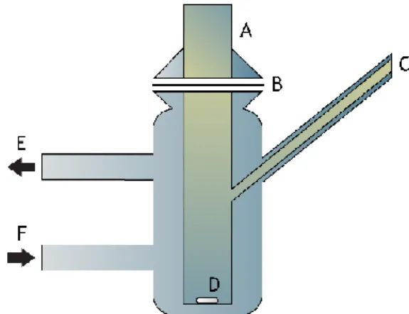
Figure I. 3: Schematic representation of a Franz cell. (A) donor compartment; (B) membrane;
(C) sampling port; (D) stirbar; (E) water outlet; (F) water inlet. ... 29
C
HAPTERII
Figure II. 1: (A) Relevant and (B) Absolute pH-buffering capacity expressed as NaOH meq for
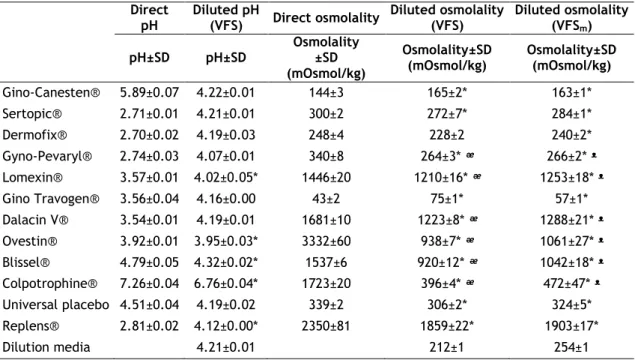
the vaginal products included in this study. For Gino-Canesten®, Colpotrophine® and Control in NS, the addition was made with HCl, since their pH were higher than 5. Results are means and bars represent standard deviations (n=3). NS=Normal saline; VFS=Vaginal Fluid Simulant; NS (HCl)=Normal saline tritrated with HCl. * represents statistically different from the NS control and ** represents statistically different from the VFS control (one way-ANOVA, p < 0.05). ... 51
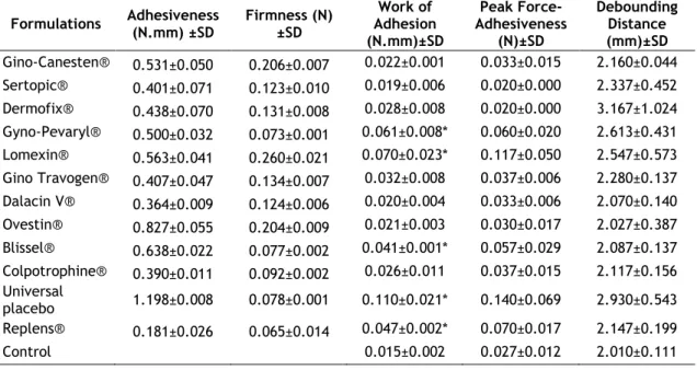
Figure II. 2: Viscosity represented as Shear Stress (Pa) vs Shear Rate (Pa) demonstrating
thixotropic behaviour for (A) antimicrobials and Replens® gel; (B) topical oestrogens; and, (C) Low viscosity formulations. Results represent the mean of 3 independent determinations. .. 58
Figure II. 3: Viscosity (as Shear stress (Pa)) comparisons for direct measurements and diluted
measurements at temperatures of 25ºC and 37ºC. Within formulations the same Shear rate (1/s) was considered. Results correspond to the mean and bars to the standard deviation of 3 determinations. Viscosity comparisons were assessed only within formulation. * represents statistically different from direct viscosity; γ represents statistically different from the dilution
at 25ºC and σ represents statistically different from the dilution at 37ºC (two way-ANOVA, p < 0.05, Tukey’s multiple comparisons test). ... 59
C
HAPTERIII
Figure III. 1: Cellular viability profiles for all the testing formulations at dilutions from 0.1 to
20% (w/v). Results for the NRU and the MTT assay. Viabilities are represented as percentage of the control treated only with culture media. Results are means and bars represent standard deviations from 2 independent experiments in which each condition was tested in triplicate
multiple comparisons test, p < 0.05). ... 81
Figure III. 2: Tissue viability profiles for all the testing formulations at dilutions from 0.1 to
20% (w/v). Viabilities as percentage of the control tissue treated only with culture media. Results are means and bars represent standard deviations from 2 experiments in which each condition was tested in triplicate (total n=6). * represents NO statistical difference from the control (two-way ANOVA, Dunnett’s multiple comparisons test, p < 0.05). ... 84
Figure III. 3: Impact of the tested formulations on the porcine ex vivo vaginal epithelium after
24 h of exposure. Histological images are representative of the higher concentration tested (20%), i.e. the worst-case scenario for the dilutions tested for tissue toxicity. H&E staining. Magnification 100x... 85
Figure III. 4: Irritation Scores (IS) for N-9 (nonoxynol-9) according to categories A and B. Results
are presented as mean values and bars as standard deviation (n=3). * denotes statistical difference between the two scales IS (A) and IS (B) (two way-ANOVA, p < 0.05, Sidak’s multiple comparisons test). ... 99
Figure III. 5: Irritation Scores (IS) for therapeutic vaginal products (a) and vaginal lubricants
(b) according to the categories A and B. Results are presented as mean values and bars as standard deviation (n=3). * denotes statistical difference between the two scales IS (A) and IS (B) (two way-ANOVA, p < 0.05, Sidak’s multiple comparisons test). ... 100
C
HAPTERIV
Figure IV. 1: Representative chromatogram of the quantification method in the full validation
process, HPLC-DAD, measured at 210 nm (standard sample from the calibration curve at 50 µg/ml). The six molecules were identified and quantified as separated peaks in a single run. ... 122
Figure IV. 2: in vitro drug release (µg/cm2) profiles of Ovestin® (EST), Blissel® (EST),
Gino-Canesten® (CLT), Lomexin® (FEN) and Gino Travogen® (ISO) as function of square root of time (h). The release rate was inferred by the slope obtained for each profile. Results are means and bars represent standard deviations (n=6). ... 126
Figure IV. 3: Histological images of the porcine ex vivo model used in the permeation
experiments. Tissues collected at the end of the experiments (t48h) where no formulation was
applied are shown of the middle and right column. The left column shows specimens right-after tissue preparation (t0h). ... 127
Figure IV. 4: ex vivo permeation (µg/cm2) profiles of Ovestin® (EST), Gino-Canesten® (CLT),
Gino Travogen® (ISO), Gyno-Pevaryl® (ECO), Sertopic® (SER) and Dermofix® (SER) as function of time (h). Results are means and bars represent standard deviations (n=3 for each tissue type). * represents statistically different values between caudal and cranial vagina (two-way ANOVA, p < 0.05, Tukey’s multiple comparisons test). ... 128
C
HAPTERI
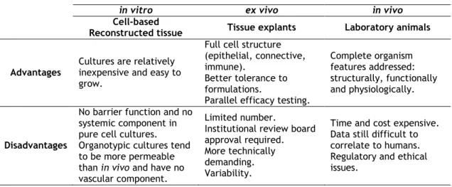
Table I. 1: Advantages and disadvantages of models applied for predicting vaginal drug
permeation. Adapted from Costin et al. (2011)42. ... 23
C
HAPTERII
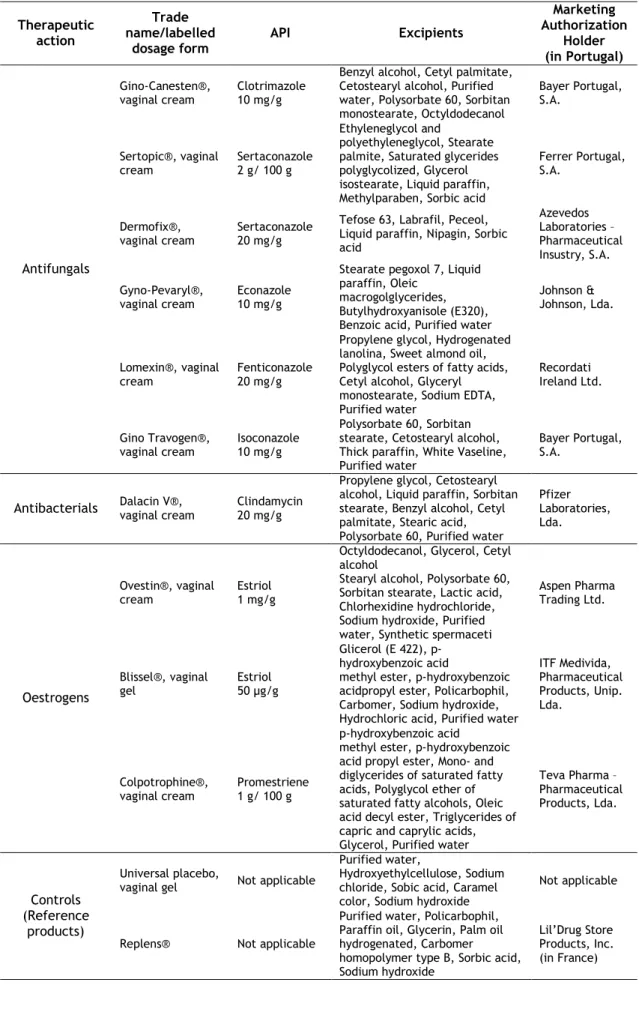
Table II. 1: Studied formulations general characteristics (information provided by the
manufacturer). ... 44
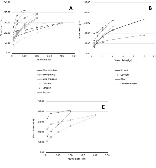
Table II. 2: pH and osmolality studies of vaginal products included in this study. Results are
means ± SD (standard deviation) (n=3). * represents statistically different from the respective dilution media; ᵆ represents statistically different between dilutions with VFS and the undiluted formulation; ᵜ represents statistically different between dilutions with VFSm and the undiluted
formulation (two-way ANOVA, p < 0.05). ... 50
Table II. 3: Mechanical (adhesiveness (N.mm) and firmness(N)) and bioadhesive parameters
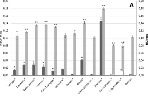
(work of adhesion (N.mm), peak force-adhesiveness (N) and debounding distance (mm)) determined for the products in study. Results are means ± SD (standard deviation) (n=3). * represents statistically different from the control (one-way ANOVA, p < 0.05). ... 55
C
HAPTERIII
Table III. 1: Optimization and pre-validation studies on tissues viability and toxicity. The
viability study corresponds to a negative control (tissues plus culture media) and the toxicity study was performed using SDS 5%, recognized to have a toxic effect on epithelial vaginal tissue. *Denotes significance on One-way ANOVA Dunnett’s multiple comparisons test (p < 0.05). ... 83
Table III. 2: Half-maximal Toxic Concentrations (% - TC50) and confidence intervals (95%)
calculated for cellular and tissue models using three different cell lines (HeLa, HEC-1A and VK2 E6/E7) and porcine vaginal explants. Within the cellular models, MTT and NRU assay were issued, as for tissues only MTT assay was performed. ... 87
Table III. 3: Irritancy classification. Classification on the in vitro HET-CAM assay concerning
Irritation Score (IS) analysis methods A and B. ... 98
Table III. 4: Irritation potential. Irritation scores, IS (A) and (B) determined for vaginal semisolid
C
HAPTERIV
Table IV. 1: Chemical characteristics of the drugs included in this study. Adpated from
PubChem365, Drug Bank366, and ChEMBL from the European Bioinformatics Institute367. ... 112
Table IV. 2: Calibration parameters for CLT, ECO, EST, FEN, ISO and SER after the full validation
and partial validations for the receptor media used on in vitro drug release and ex vivo permeation studies. ... 121
Table IV. 3: Stability of CLT, ECO, EST, FEN, ISO and SER in a methanolic solution considered
in the full validation process. Results are presented as percentual variation from t0 samples.
... 121
Table IV. 4: Within and Between Run precision (% CV) and accuracy (% bias) obtained for CLT,
ECO, EST, FEN, ISO and SER at concentrations of the Lower Limit of Quantification (QC1) and at the low (QC2), middle (QC3) and high (QC4) concentrations representative of the calibration range. ... 123
Table IV. 5: Solubility (µg/mL) for CLT, ECO, EST, FEN, ISO and SER in solutions tested as
possible receptor media for in vitro drug release and ex vivo permeation studies. The receptor media selected to be used in further experiments were marked with *. ... 124
Table IV. 6: Drug recovery from the formulation (%), ex vivo permeation and in vitro release
parameters. Papp (apparent permeability coefficient); Jss (steady-state flux); NC (<LLOQ) (Not
calculable because concentrations were below the lower limit of quantification); * represents statistically different from the histological tissue in comparison (One-way ANOVA, Tukey's multiple comparisons test, p < 0.05). n=3 tissues in ex vivo permeation experiments and n=6 in in vitro drug release studies. When drug quantification was <LLOQ, a 0 µg/mL concentration was asssumed. ... 131
A
ABC Absolute pH-Buffering Capacity
ATCC American Type Culture Collection
B BV Bacterial Vaginosis C CI Confidence Interval CLT Clotrimazole CV Coefficient of Variation D
DAD Diode Array Detector
DMEM Dulbecco’s Modified Eagle’s Medium
DMSO Dimethyl Sulfoxide
E
ECO Econazole
EDTA Ethylenediaminetetraacetic Acid
e.g. exempli gratia meaning “for example”
EMA European Medicines Agency
EU European Union
ECVAM European Centre for the Validation of Alternative Methods
F
FBS Foetal Bovine Serum
FDA Food and Drug Administration
FEN Fenticonazole
Fmax Maximum Force
H
HBSS Hanks’ Balanced Saline Solution
HET-CAM Hen's Egg Test-Chorioallantoic Membrane
HPLC High Pressure Liquid Chromatography
I
ICH International Conference on Harmonisation
ICCVAM Interagency Coordinating Committee on the Validation of Alternative Methods
ISO Isoconazole; International Organization for Standardization
L
LLOQ Lower Limit of Quantification
LOD Limit of Detection
M
MTT 3-[4,5-Dimethylthiazol-2-yl]-2,5-diphbenyltetrazolium bromide
N
NS Normal Saline (NaCl 0.9%)
O
OECD Organisation for Economic Co-operation and Development
OTC Over-The-Counter
P
PBS Phosphate Puffer Solution
Q
QC Quality Control
R
RPMI Roswell Park Memorial Institute
RBC Relevant pH-Buffering Capacity
S
SD Standard-Deviation
SER Sertaconazole
spp. Species
U
USA United States of America
UV Ultraviolet
V
VFS Vaginal Fluid Simulant
VFSm Modified Vaginal Fluid Simulant
VVC Vulvovaginal Candidosis
W
Wad Work of Adhesion
C
HAPTER
I
The content of this chapter is partially published in:
Machado RM, Oliveira A, Silva Gaspar C, Martinez-de-Oliveira J,
Palmeira-de-Oliveira R, “Studies and methodologies on vaginal drug permeation”, Advanced Drug Delivery Reviews. Special Issue: Vaginal Drug Delivery. 2015; 92:14-26.
I.1. Background
Researchers are now devoted to find new forms or to re-discover safer and more effective alternative routes for the administration of drugs that are poorly absorbed orally or suffer precociously metabolization1–3. The vaginal route has been considered of great interest for drug
delivery, since it enables both local and systemic drug delivery4,5, allowing for the absorption
of peptide and other macromolecules, and even micro and nanoparticles6–8.
The vaginal route provides different advantages over the oral one but it is not deprived of inconvenients9,10. Its large surface area, rich blood supply, ability to bypass hepatic
first-passage, avoidance of gastrointestinal side effects, and relatively high permeability to drugs with a wide range of molecular weights drugs are some of its physiological characteristics that contribute to its pharmacokinetic advantages11–14. However, drug absorption through the vagina
may be affected by variations of epithelial thickness and by changings in the vaginal milieu composition, that occur as a consequence of age dependent physiological conditions or sexual intercourse; leakage and the self-cleaning action of the vaginal tract may result in low drug bioavailability5,15. Furthermore, general disadvantages of vaginal drug delivery include its
obvious gender specificity, cultural background limitations and personal hygienic care interference12,14.
Vaginal drug delivery systems include solutions, semisolids (creams, ointments and gels) and solid formulations (tampons, capsules, pessaries, suppositories, films, sponges, powders and special controlled release devices like the intravaginal ring) as well as other types of formulations such as aerosols and particulate systems integrated in adequate drug delivery systems16. Efficacy of drug delivery systems will rely on their ability to promote adequate drug
concentrations at the targeted site of action, being it local or distant. When a systemic effect is the objective through this route, drugs must be transported across the epithelium to gain access to dermal vessels and the systemic circulation17. On the other hand, when a local effect
is the goal, as is the case for some antimicrobials, microbicides and contraceptive effects, retention of the drug at the surface of the vagina is desirable18–21.
Concerning all these particularities added to women’s preference and acceptability patterns for vaginal semisolid formulations22,23, a safety- and suitability-driven study can
conduct to a better characterization of the already marketed products in the perspective of developing new products.
I.1.1. Historical perspective
The first records of vaginal administration of preparations date back to Ancient Egypt. In the Kahun Papyrus, the oldest of medical papyri (ca. 1850 B.C.), refers to vaginal “preparations”, which contained natural substances such as mud, frankincense, oil, malachite, ass urine, myrrh, crocodile dung, honey, and sour milk, intended to be used in female genitalia for specific vaginal conditions and contraception24. Some decades after, the Ramesseum
1300 B.C.) also described drug formulations to be administered in the vagina. The administration of preparations through the vaginal route was historically succeeded by other civilizations such as Greece, Rome, the Arabic and Oriental cultures, Renaissance and throughout Modern Era until nowadays25,26.
It is believed that primarily the vaginal route of administration was consistently been used for local applications. However, it was only in 20th century that its recruitment for systemic
delivery was scientifically hypothesized and studied. Firstly, Macht reported the vaginal absorption of morphine, atrophine and potassium iodide27. In the early twenties, an obstetric
surgeon from Westminster and West London Hospitals, conducted an experimental study to determine the vagina’s aptitude to absorb some substances. In this study, Robison Drummond concluded that potassium iodide and sodium salicylate solutions were rapidly absorbed and found in urine an hour after being placed in the vagina. Also quinine, cane sugar and phenol red were clinically found to be absorbed and excreted by the kidneys28.
Since then, several drugs have been approved for vaginal administration by medicines’ authorities all over the world, the majority being proposed for the treatment of local conditions. In fact, the vaginal is presently considered a well-established alternative administration route to formulations intended both for local or systemic delivery4.
I.1.2. Vaginal administration route
Vaginal drug delivery represents an important approach for the treatment of both local and systemic diseases1,4,5. As already stated, the vaginal route has several advantages due to
its large surface area, rich blood supply, avoidance of the hepatic first-pass effect, relatively high permeability to many drugs, and allowance of self-insertion12. These particular
characteristics of the vagina provide significant potential for the delivery of a wide range of bioactive compounds, including peptides and proteins, and offer an alternative to the parenteral route of administration1. In the case of local disease or disorder, using a vaginal
product frequently avoids drug absorption in systemically relevant amounts and thus prevents side effects. The vaginal route may be of particular importance in the case of drugs undergoing extensive hepatic metabolism since it avoids the hepatic first-pass effect and allows reducing the amount of administered drugs (e.g. estrogens).
On the other hand, several drawbacks, including cultural background, personal hygiene, gender specificity, local irritation, and influence on sexual intercourse, need to be addressed during the design of a vaginal formulation5,29. Inconsistent drug absorption behavior may also
be a concern due to the physiological variability observed during different stages of women development and hormonal status (e.g. childhood, pre- or post-menopausal, pregnancy).
Ideally, vaginal drug delivery systems should not interfere with vaginal physiology and daily life, while allowing obtaining high drug bioavailability (either local or systemic) with little variability and low incidence of side effects.
I.2. Vagina: anatomy, histology and physiology
The vagina is described as an expandable, longitudinally S-shaped, fibromuscular, collapsed canal showing at transverse cross-section an H configuration, with the anterior and posterior walls contacting each other in current conditions. It extends from the cervix of the uterus to the vestibule2,30,31, presenting approximately 7-10 cm in length, more than 4 cm in
width and 150-200 µm in thickness. The posterior wall is longer than the anterior, a consequence of the asymmetrical position of the cervix at the vaginal vault32–35.
Though it normally does not have secreting glands, the vagina is usually referred as a mucosa. In fact despite not having a secreting role, the vaginal epithelial surface is actually coated by a thin layer of fluid that includes endometrial, cervical and vestibular secretions, tissue transudate, residues of urine and products of cellular autolysis36,37. Additionally, the
composition of the vaginal fluid varies according to age, menstrual cycle phase and health status condition32,38. Generally, it is considered that around 0.50-0.75 g of vaginal fluid are
present in the vaginal cavity of a healthy reproductive aged women, representing a total daily production of 6 g37. For instance, the vaginal pH is acidic (3.5-4.5) in healthy women during the
reproductive age but it fluctuates along the different stages of the menstrual cycle and it is also dependent on coitus frequency, the amount of cervical mucus present in the vagina, the amount of vaginal transudate which also varies along the vagina (being higher close to the cervix and lower at the anterior fornix)2,37. The maintenance of the pH is accomplished by lactic
acid bacteria, mainly Lactobacillus spp., microorganisms that metabolize into lactic acid the mono and di-saccharides that result from the autolytic breakage of vaginal cells glycogen32.
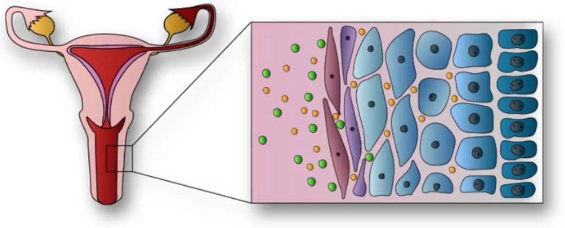
Figure I.1: Schematic representation of nonkeratinized stratified squamous vaginal tissue, also
representing local and systemic drug delivery39. Adapted from: Schnell, Citologia y Microbiologia de la
The vaginal wall consists of various cell layers: nonkeratinized stratified squamous epithelium, lamina propria, muscular layer and tunica adventitia covering only their proximal segments (Figure I.1). The lamina propria is constituted of connective tissue rich in blood and lymphatic vessels draining to the internal iliac vein, explaining why the absorbed products do avoid the hepatic circulation as an initial passage31. The vaginal cell turnover is estimated to
replace 10-15 layers in a week41. The non-keratinized stratified squamous epithelium, settled
on glycogen containing keratinocytes but also integrating other cell types (such as macrophages and Langerhans’ cells), is grounded on the lamina propria, that includes elastic fibers, polymorphonuclear leukocytes and occasional lymph nodules42. The vaginal epithelial cells are
disposed according to different stages of differentiation, identifiable through different keratins expression, such as K10 and K13, being the differential expression arrangement function of the cell location within the epithelium43. Numerous folds and microrridges called “ruggae” are
present in the epithelium, largely increasing the vagina’s surface area and providing distensibility7.
The vaginal innervation depends on two types of sources: a peripheral one providing a highly sensible lower quarter segment and an autonomic fiber network in the upper tract, which is more sensitive to stretch than to touch or pain. This explains why women do not feel discomfort when using continuous intravaginal drug delivery systems7.
Several conditions influence vaginal physiology: hormonal balance, pregnancy, pH, microflora and age, being the last one the best current biomarker for epithelium layer thickness, enzyme concentrations and vaginal fluid production44. Variation in these vaginal
characteristics changes influence drug permeation as it depends mainly on the superficial layer characteristics, such as thickness, cell tightness, and lipids composition and organization in the intercellular space34,45,46.
I.3. Vaginal dosage forms
I.3.1. Traditionally used in therapeutics
According to Portuguese Decree-Law no 176/2006 of 30th August, corresponding to the
Medicines Statute, a pharmaceutical dosage form is defined as "the final state that active substances present after undergoing the necessary pharmaceutical operations in order to facilitate its administration and get the most desired therapeutic effect". Furthermore, the European Pharmacopoeia 8.0 states that vaginal preparations are liquid preparations, semisolid or solid intended to be administered vaginally, usually to a local action. They contain one or more active substances into an appropriate excipient. Containers intended for the packaging of vaginal preparations must meet the specified requirements. Various categories of vaginal preparations are defined: pessaries; vaginal tablets; vaginal capsules; vaginal solutions, emulsions and suspensions; tablets for vaginal solutions and suspensions; semisolid vaginal preparations; vaginal foams and medicated vaginal tampons47.
In terms of production, it is stated that, during development, it must be demonstrated that the nominal contents can be withdrawn from the container of liquid and semisolid vaginal preparations presented in single-dose containers. In the manufacturing, packaging, storage and distribution of vaginal preparations, suitable measures are taken to ensure their microbial quality. Uniformity of dosage unit, uniformity of content, uniformity of mass and dissolution or disintegration profile constitute the specified testing for quality control of these dosage forms47.
Generally, the most commonly administered dosage forms by the vaginal route are ovules, tablets and semisolids (in which category, creams). They are used as astringent drugs, antimicrobials (antibacterials, antiprotozoals, antifungals, antivirals), keratoplastics, as wound healers, spermicides, prostaglandins and steroids48. However, these traditional commercial
preparations have shown to have a short retention time due to the vaginal depuration mechanism, which results in discomfort and runoff feeling; are uncomfortable to apply; often require multiple daily administrations in order to obtain the desired therapeutic concentration and do not provide a uniform distribution of the drug5,49–51. These characteristics generally
entail a decrease in the acceptability of women in relation to these dosage forms, and so, leading to less reliability on the treatment.
I.3.2. Main evolutions on vaginal therapeutical strategies
The vaginal route is now considered an option for several therapeutic strategies. Hormones and antibiotics have been largely included in vaginal dosage forms but recently other therapeutic purposes have also been explored, such as prevention of infection and immunization. Also, the possibility of systemically delivering through the vaginal route, molecules presenting high molecular weight, such as calcitonin and insulin, has been explored52,53.
The vaginal route is being increasingly used for hormone administration as it exhibits the great advantage of preventing gastrointestinal side effects and the hepatic first-pass effect. This is clearly useful for molecules that undergo a high degree of hepatic metabolism such as natural estrogens. While vaginal administration of estriol is indicated for atrophic vaginitis treatment in postmenopausal women, ethinylestradiol and etonogestrel are included in a vaginal ring for combined contraception. Vaginal progesterone is very effective in hormone replacement treatment (HRT) for postmenopausal women, in assisted reproductive technologies protocols and in supporting early and pre-term pregnancy54,55. In pre-term labor
prevention, indomethacin administered intravaginally has also been found useful and even advantageous when compared to its oral administration56. The vaginal route is also considered
the best option when the opposite aim is intended: labor induction. Vaginal misoprostol or dinoprostone are widely used for this purpose57.
Over the last years, hormonal contraception has been achieved mainly by the oral or transdermal routes. However, the introduction of oral hormone pills into the vagina was shown to have relatively good efficacy and acceptability rates, opening ways for vaginal hormonal contraception58,59. Nonetheless, it was the introduction of the vaginal ring which mainly
promoted the vaginal route for hormonal contraception60. Vaginal rings for steroids release
have also been proposed when both local and systemic effects are intended, as is the case for estradiol to treat atrophic conditions including vaginitis61,62. Moreover, distinct applications
may emerge in the future for vaginal rings. For instance, it may contribute for the development of a new delivery approach for spermicides that are classically administered through semisolid dosage forms and sponges, and even microbicides. New spermicides molecules have also been developed and classic dosage forms have been proposed63,64.
Regarding vaginal antimicrobial treatments, therapeutic strategies for the most common infections, namely bacterial vaginosis (BV) and vulvovaginal candidosis (VVC), include drug products for vaginal application. Clindamycin cream and metronidazole gel are two available therapeutic options to treat BV65. However, due to increasing bacterial resistance and
infection-related complications, acid-buffering gels and vitamin C tablets for vaginal application have been proposed, alone or combined with oral therapy66–68. Additionally,
recognized antimicrobial molecules such as fenticonazole, garenoxacin and rifaximin have been re-visited and their possible topical application for BV treatment considered69–71. Additionally,
classical local therapies, namely with gentian violet solution and boric acid vaginal capsules, have been revised72,73.
Vaginal administration of natural products to control and eradicate genital infections is very popular among women and arises as a possible alternative to overcome antibiotic resistance. Distinct plant extracts and essential oils have been proposed as valuable therapeutic alternatives for both BV and VVC, and have been studied in vitro and in animal models74–76.
Immunomodulation through the vaginal route is another important investigation field encouraging scientific work. Intravaginal administration of vaccines was shown to promote local immunoglobulin production, standing-up as a valuable route for sexually transmitted diseases
prevention77,78. Also, a combined an anti-allergic and antifungal therapy based on oral cetirizine
and fluconazole was tested and showed to be helpful in women suffering from recurrent VVC with persistent pruritus79. Vaginal application of these two drugs may arise as a possible
therapy.
Local therapy is also very common in human papillomavirus (HPV) lesions, as systemic therapy is highly ineffective80. Podophyllin and podophyllotoxin, trichloroacetic acid solution
and 5-fluorouracil have been used for vulvar infections. However, they must be very carefully used when applied in the vagina so that applications are restricted to diseased tissue81.
Recently, new possibilities for the management of HPV infection have been proposed such as cidofovir, lopinavir, polyphenon E and an extract from green tea (Camellia sinensis)82–84.
Immune response modifier molecules, such as immunostimulatory oligonucleotides and resiquimod, are also anticipated as a valuable vaginal topical therapeutic strategy to treat HVS genital infections and reduce the frequency of recurrences85,86.
Topical administration of protective microorganisms, especially Lactobacillus spp., has been proposed to restore the vaginal microbiota after an insult and as an alternative or coadjuvant treatment for urogenital infections87,88. Clinical trials have shown vaginal probiotics
formulations to be safe and having high rates of acceptability but data on efficacy of these formulations are still controversial, mostly related to limitations such as small samples studies and lack of product stability89,90.
I.3.3. Less used/advanced dosage forms
The major challenge in vaginal dosage forms design is the ability to fulfill functional criteria such as product dispersion throughout the vagina, prolonged residence time, adequate physicochemical interaction with vaginal content, release profile of active ingredients and effects on targets91.
Bioadhesion has been stated as a target characteristic for the design of new vaginal products since it is defined as the attachment of synthetic and natural macromolecules to a biological tissue and mucoadhesion is considered a particular case of bioadhesion when the biological tissue is covered by mucus92. Mucoadhesive polymers have been explored in the
development of either semisolid and solid vaginal drug delivery systems in order to circumvent some of the outlined limitations of traditional dosage forms while gathering user compliance and improving therapeutic outcomes93. Moreover some polymers have shown microbicidal
activity on their own, as is the case for kappa carrageenan (absorption inhibitor, in vitro), carbomers (interruption of HIV cell binding, in vivo), cellulose acetate phthalate (inactivation of HIV and HSV, in vivo), and polystyrene sulfonate (activity against HIV and HSV, in vitro)1.
Herein this study, and regarding the pharmacopeia background, tablets, pessaries, capsules, gels, creams and solutions are considered as the traditional dosage forms for vaginal administration26. In addition, over the last decades, new vaginal drug delivery systems (such as
rings, films, sponges and diaphragms or cervical caps) have gained popularity among pharmaceutical developers, clinicians and users, and are now being the focus of intense study.