ASSESSMENT OF ANTICANCER PROPERTIES OF ESSENTIAL
OILS FROM AROMATIC PLANTS OF THE SAND DUNES OF
PENICHE (PORTUGAL)
Juliana Poças Almeida
ASSESSMENT OF ANTICANCER PROPERTIES OF ESSENTIAL
OILS FROM AROMATIC PLANTS OF THE SAND DUNES OF
PENICHE (PORTUGAL)
Juliana Poças Almeida
Trabalho de Relatório de Estágio apresentado à Escola Superior de Turismo e Tecnologia do Mar do Instituto Politécnico de Leiria para a obtenção do grau de Mestre em Biotecnologia Aplicada, realizado sob a orientação científica do Doutor Marco Filipe Loureiro Lemos, Professor Adjunto da Escola Superior de Turismo e Tecnologia do Mar do Instituto Politécnico de Leiria, da Doutora Isabel Pires, Professora na School of Biological, Biomedical and Environmental Sciences da Universidade de Hull (UK) e da Doutora Célia Cabral, Investigadora do Centro de Estudos Interdisciplinares do Séc. XX, Instituto de Investigação Interdisciplinar da Universidade de Coimbra.
Title: Assessment of Anticancer Properties of Essential Oils from aromatic plants
of the sand dunes of Peniche (Portugal).
Título: Avaliação das propriedades anticancerígenas de óleos essenciais de
plantas aromáticas das dunas de Peniche (Portugal).
Copyright © Juliana Poças Almeida
A Escola Superior de Turismo e Tecnologia do Mar e o Instituto Politécnico de
Leiria têm o direito, perpétuo e sem limites geográficos, de arquivar e publicar
este trabalho de relatório de estágio através de exemplares impressos
reproduzidos em papel ou de forma digital, ou por qualquer outro meio conhecido
ou que venha a ser inventado, e de a divulgar através de repositórios científicos e
de admitir a sua cópia e distribuição com objetivos educacionais ou de
investigação, não comerciais, desde que seja dado crédito ao autor e editor.
v
“The journey of a thousand miles begins with a single step”
Lao Tzu
vii
Acknowledgements
Em primeiro lugar, quero agradecer à minha mãe, Ana Poças. Sou quem sou graças a ti. Obrigada pelo teu esforço, pelo apoio diário e principalmente pela educação que me proporcionaste. Sem ti este projeto não seria possível. À minha irmã, Mariana Poças, por acreditar sempre em mim, mesmo quando eu não o faço, e pelo apoio incondicional. Ao meu irmão, Frederico, por estar sempre do meu lado e pela força que me dá para seguir em frente. A todos um Muito Obrigada pela compreensão pelos meus momentos de maiores nervos durante esta fase.
Aos meus orientadores Doutor Marco Lemos e Doutora Célia Cabral por todas as apreciações feitas e por ajudarem a este projeto ter tido um rumo, bem como à Doutora Isabel Pires por me ter recebido tão bem no seu laboratório em Hull, pela hospitalidade e disponibilidade. Foi sem dúvida uma experiência muito gratificante. Obrigada aos três por me terem apoiado sempre ao longo desta etapa da minha vida e pela paciência que tiveram durante a escrita desta tese.
Muito obrigada aos amigos Luís Alves, Tânia Serreira e Joana Paiva por estarem sempre presentes de alguma forma. Sem dúvida que o vosso apoio e força que me deram ajudaram nos momentos difíceis e com maior saudade durante o estágio. Ao Ricardo Costa por estar presente e se disponibilizar na recolha das plantas (ainda que tenha sido “voluntário à força”). Obrigada ao Proença, por me “ter dado na cabeça” (diversas vezes), mas também pelos bons momentos que partilhou comigo. Muito Obrigada pelo vosso apoio, sem vocês esta aventura teria sido mais difícil. Á minha housemate, Cecília por termos partilhado momentos em Hull e por toda a ajuda prestada. Obrigada aos restantes portugueses em Hull, pelos momentos de diversão e descontração.
Aos meus avós, Fátima e António, que sempre disseram que eu ia ser “uma grande senhora”. Obrigada por acreditarem sempre em mim.
Aos meus tios, Adérito e Umbelina, que estiveram presentes na minha vida desde a minha infância até aos dias de hoje. Obrigada por todos os momentos que partilham comigo e por todos os conselhos que me dão.
I want thank you to the girls with I worked in Hull, Flore-Anne, Hannah, Becky and Anna for received me so well and for helped me in the lab. Thank you for your time, I really enjoyed it! Thank you to Stephen Maher and Elena Rosca for the discussions on the Lab meetings.
Um agradecimento ao Consórcio Erasmus pela oportunidade de estagiar noutro país. Sem dúvida que foi crucial para eu expandir os meus conhecimentos e conhecer uma nova cultura. Obrigada ao Dr. João Assis pela ajuda e eficácia em todo o processo.
ix
Resumo
O cancro é um problema de saúde crescente no mundo e é a segunda causa de morte depois das doenças cardíacas. De acordo com a Agência Internacional de Investigação em Cancro (IARC) existem atualmente mais de 10 milhões de casos de cancro por ano no mundo.
Os produtos naturais oferecem oportunidades de inovação na descoberta de novos fármacos. Neste sentido, os compostos naturais isolados a partir de plantas medicinais, como potenciais fontes de novas drogas anticancerígenas, têm tido um interesse crescente.
Os Óleos Essenciais (OEs) são sintetizados pelas plantas e têm sido estudados pelas suas inúmeras atividades biológicas, incluindo anticancerígena, anti-inflamatória, antimicrobiana, antiviral, antioxidante e repelente de insetos.
Este estudo tem como objetivos determinar a eficácia de OEs de seis espécies de plantas das dunas de Peniche (Portugal), como potenciais agentes terapêuticos anticancerígenos em linhas celulares de cancro da mama (MCF7) e do colo-rectal (RKO), assim como perceber o mecanismo de ação destes OEs.
Neste estudo, partes aéreas de Artemisia campestris subsp. maritima, Crithmum maritimum, Eryngium maritimum, Juniperus turbinata subsp. turbinata, Otanthus maritimus e Seseli tortuosum foram colhidas na praia da Consolação, em Peniche (Portugal), e os seus OEs isolados através de hidrodestilação. A composição química dos OEs foi investigada por cromatografia gasosa (GC) e por cromatografia gasosa com espetrofotometria de massa (GC-MS) e os compostos maioritários foram descritos para cada óleo.
Para avaliar a atividade anticancerígena nas linhas celulares MCF7 e RKO, o método MTS (3- (4, 5-dimethyl- 2 -thiazolyl) - 2, 5-dyphenyl-2H-tetrazolium bromide) foi usado e a viabilidade celular avaliada, através de diluições sucessivas, a concentrações iniciais de 5 µL/mL e 1 µL/mL, com diluição de 1:2 e 1:10, respetivamente, comparando com o controlo (DMSO). De todos os OEs testados, a atividade anticancerígena foi descrita, em ambas as linhas celulares, como observado pela diminuição da viabilidade/proliferação celular – exceto o OE Eryngium maritimum a uma concentração inicial de 5 µL/mL.
Com o objetivo de avaliar o mecanismo biológico de ação dos OEs, foi realizado um western blot para marcadores relativos ao bloqueio do ciclo celular e apoptose (p53, p21 e caspase 3 clivada), para Seseli tortuosum e Otanthus maritimus. Foi observado um aumento do nível proteína p53 nas células tratadas com estes OEs, sugerindo a indução de stress celular nas células cancerígenas testadas. No entanto, não foi observada caspase 3 clivada, sugerindo que a apoptose não terá sido a causa para a diminuição da viabilidade/proliferação celular observada. Foi ainda observado o aumento da expressão da p21 com os OEs selecionados, sugerindo que o tratamento com OE está associado ao bloqueio do ciclo celular. Para validar estas observações, a análise realizada por FACS, depois do tratamento indica um possível bloqueio do ciclo celular na fase G1.
Concluindo, a concentração inicial de 5 µL/mL revelou ser muito tóxica para as linhas celulares testadas. No entanto, a uma concentração final de 1 µL/mL foi demonstrada uma diminuição da viabilidade/proliferação celular para todos os OEs.
No estudo preliminar do mecanismo de ação dos OEs, foi demonstrado, face à presença da p21, que os óleos de Seseli tortuosum e Otanthus maritimus atuam bloqueando o ciclo celular. Para comprovar estes resultados, o FACS realizado (apenas no OE de Seseli tortuosum) revelou que este bloqueio pode ocorrer, pelo aumento da percentagem de células observadas, na fase G1.
Estes resultados demonstram o interesse destes OEs de Peniche na procura de novos agentes quimo preventivos contra a progressão do cancro da mama e colo-rectal.
xi
Abstract
Cancer is a growing health concern around the world and is the second leading cause of death after heart disease. According to the International Agency for Research on Cancer (IARC), there are now more than 10 million cases of cancer per year worldwide.
Novel natural products offer opportunities for innovation in drug discovery. In this sense, natural compounds isolated from medicinal plants, as potential sources of novel anticancer drugs, have been of increasing interest.
Essential Oils (EOs) are synthesised by plants and have been studied regarding their numerous biological activities, including anticancer, anti-inflammatory, antimicrobial, antiviral, antioxidant and insect repellent.
This study aims to determine the efficacy of the EOs from six selected plant species from the dunes of Peniche (Portugal), as potential anticancer therapeutic agents in breast (MCF7) and colorectal (RKO) cancer cell lines, as also understand the mechanism of action of these EOs.
In this work, aerial parts of Artemisia campestris subsp. maritima, Crithmum maritimum, Eryngium maritimum, Juniperus turbinata subsp. turbinata, Otanthus maritimus and Seseli tortuosum were harvested in Consolação beach, in Peniche (Portugal), and the EOs isolated by hidrodistillation. The chemical composition of EOs was investigated by gas chromatography (GC) and gas chromatography–mass spectrometry (GC–MS) and major compounds were reported for each EO.
To evaluate anticancer activity in MCF7 and RKO cell lines, MTS (3-(4, 5-dimethyl-2-thiazolyl)-2, 5-dyphenyl-2H-tetrazolium bromide) assay was carried out and cell viability was evaluated, through successive dilutions, at starting concentrations 5 µL/mL and 1 µL/mL with a dilution 1:2 and 1:10, respectively, comparing to vehicle (DMSO). For all the EOs tested, anticancer activity was observed in both cell lines, as indicated by a decrease in cell viability/proliferation – except for Eryngium maritimum EO at starting concentration of 5 µL/mL.
With the aim to evaluate the mechanism of biological effect of the EOs, a Western blot was carried out for markers of cell cycle arrest and apoptosis (p53, p21 and cleaved caspase 3), for Seseli tortuosum and Otanthus maritimus. Were observed an increase of p53 protein levels in cells treated with these EOs, suggesting that cellular stress was
induced in the cancer cells tested. However, cleaved caspase 3 was not observed, suggesting that apoptosis was not the cause for the observed decrease in cellular viability/proliferation. It was also observed the increased of p21 expression with the selected EOs, suggesting that EO treatment is associated with cell cycle arrest. To validate these observations, FACS analysis performed, indicating a possible G1 phase cell
cycle arrest after treatment.
In conclusion, the starting concentration 5 µL/mL revealed to be very toxic to the cell lines tested. However, at a starting concentration 1 µL/mL showed a decrease in cellular viability/proliferation for every EOs.
In the preliminary study of the mechanism of action of EOs, was demonstrated, due to presence of p21, Seseli tortuosum and Otanthus maritimus EOs can act by arresting cell cycle. To comprove this results, the FACS performed (just for Seseli tortuosum EO) revealed this arrest may be occur, by the increase of percentage of cells observed, in G1
phase.
Our results show the interest of these EOs from Peniche in search of new chemo preventive agents against breast and colorectal cancer progression.
xiii
Table of contents
Resumo ... ix
Abstract ... xi
List of figures ... xv
List of tables ... xxiii
List of abbreviations ... xxv
1. Introduction ... 27
1.1 Essential Oils ... 29
1.1.1 Plant metabolic pathways... 29
1.1.2 Secretory structures in plants ... 29
1.1.3 Chemical Composition of Essential Oils ... 31
1.1.4 Factors influencing the production and quality of essential oils ... 35
1.1.5 Extraction techniques ... 36
1.1.5.1 Hydrodistillation ... 36
1.2 Biological Properties of Essential Oils ... 37
1.2.1 Antinociceptive Effect ... 38
1.2.2 Insect Repellent Activity ... 39
1.2.3 Antiviral Activity ... 39
1.2.4 Antioxidant Activity ... 40
1.2.5 Anticancer Activity ... 41
1.3 Key concepts in cancer biology ... 43
1.3.1 Cell cycle regulation ... 44
1.3.2 The DNA Damage response ... 46
1.3.3 Study of cell cycle Progression ... 48
1.4 Dune Plant Species selected in this study ... 50
1.4.1 Crithmum maritimum L. ... 50
1.4.2 Seseli tortuosum L. ... 51
1.4.3 Artemisia campestris subsp. maritima (DC.) Arcang. ... 52
1.4.4 Juniperus turbinata Guss. subsp. turbinata ... 53
1.4.5 Otanthus maritimus (L.) Hoffmans. & Link ... 54
1.4.6 Eryngium maritimum L. ... 55
1.5 Aim of this study ... 56
2.1 General materials and reagentes ... 59
2.2 Plant material collection ... 59
2.3 Essential Oils isolation ... 59
2.4 Chemical characterization of essential oils ... 59
2.4.1 Gas chromatography (CG) ... 59
2.4.2 Gas chromatography-mass spectrophotometry (GC-MS) ... 60
2.4.3 Qualitative and quantitative analysis ... 60
2.5 Cell Culture ... 60
2.6 Treatment of cancer cells with EOs ... 61
2.7 MTS (3-(4,5-dimethylthazol-2-yl)-2,5-diphenyl tetrazolium bromide) assay for cellular viability ... 62
2.8 Half maximal inhibitory concentration (IC50) calculations ... 62
2.9 Protein Lysis and Protein Concentration Determination ... 63
2.10 SDS-PAGE and Western Blot ... 63
2.11 Determination of DNA content by Fluorescence-activated cell sorting (FACS) .. 65
3. Results ... 66
3.1 Essential oils composition ... 67
3.2 Antitumor activity ... 67
3.3 Evaluation of mechanism of biological effects of EOs ... 78
4. Discussion ... 86
4.1 Overview of this work ... 87
4.2 Chemical composition of EOs tested ... 88
4.3 Cell Viability by MTS assay ... 92
4.3.1 Crithmum maritimum L. ... 93
4.3.2 Seseli tortuosum L. ... 94
4.3.3 Artemisia campestris subsp. maritima (DC.) Arcang. ... 95
4.3.4 Juniperus turbinata Guss. subsp. turbinata ... 96
4.3.5 Otanthus maritimus (L.) Hoffmans. & Link ... 96
4.3.6 Eryngium maritimum L. ... 97
4.4 Viability determination in experimental cancer biology ... 99
4.5 Study of mode of action of EOs ... 100
4.5.1 Mechanism of action of Seseli tortuosum and Otanthus maritimus EOs 101 4.5.2 Cell cycle analysis of Seseli tortuosum EO ... 105
5. Conclusion and Future Work ... 111
xv
List of Figures
Figure 1.1.2.1 – Schematic diagram of a glandular trichome illustrating the placement of
this epidermal structures and the relationship of the disc of secretory cells to the stalk, basal cells and to the subcuticular storage space (Turner et al. 2014) ... 30
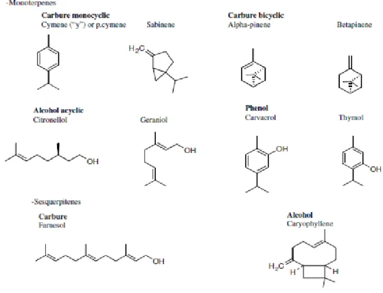
Figure 1.1.3.1 – Chemical structures of terpenoids (selected compounds of essential oils).
Monoterpenes have a variety of structures, for example - Carbures, Alcohols, Aldeydes, Ketone, Esters and Ethers. The structures of the sesquiterpenes - Carbures, Alcohols, Ketones and Epoxide - are similar comparing to the monoterpenes (modified after Bakkali et al. 2008) ... 33
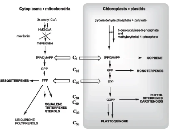
Figure 1.1.3.2 – The two different terpenoid classes in the mitochondria and plastids,
respectively (from Baser and Demirci 2007) ... 34
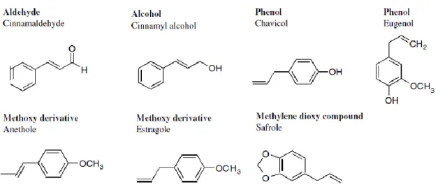
Figure 1.1.3.3 – Chemical structures of nonterpenoids compounds (selected compounds
of essential oils) (modified after (Bakkali et al. 2008) ... 35
Figure 1.1.5.1.1 – Clevenger apparatus. Dimensions are indicated in millimetres
(Farmacopeia Portuguesa, 2005)... 37
Figure 1.3.1 - Benign versus malignant tumours. A benign glandular tumour (pink cells;
an adenoma) remains inside the basal lamina (yellow) that marks the boundary of the normal structure (a duct, in this example). In contrast, a malignant glandular tumour (red cells; an adenocarcinoma) can develop from a benign tumour cell, and it destroys the integrity of the tissue, as shown (from Alberts et al. 2008) ... 44
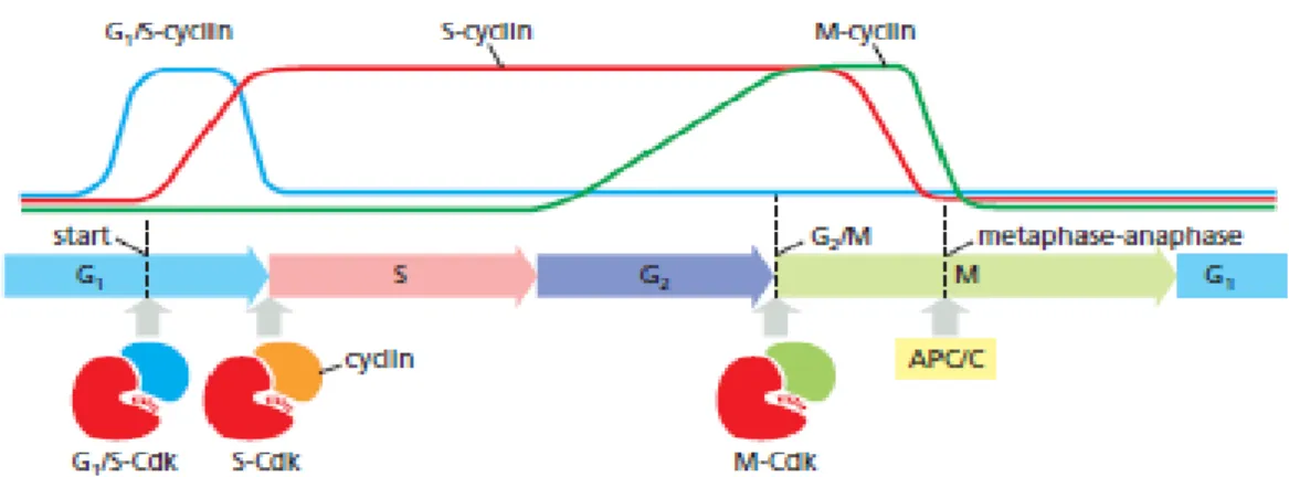
Figure 1.3.1.1 - Cyclin–Cdk complexes of the cell-cycle control system. The
concentrations of the three major cyclin types oscillate during the cell cycle, while the concentrations of Cdks (not shown) do not change and exceed cyclin amounts. In late G1, rising G1/S-cyclin levels lead to the formation of G1/S-Cdk complexes that trigger progression through the Start transition. S-Cdk complexes form at the start of S phase and trigger DNA replication, as well as some early mitotic events. M-Cdk complexes form during G2 but are held in an inactive state; they are activated at the end of G2 and trigger entry into mitosis at the G2/M transition. A separate regulatory protein complex, the APC/C, initiates the metaphase- to- anaphase transition (from Alberts et al. 2008) ... 45
Figure 1.3.2.1 - How DNA damage arrests the cell cycle in G1 phase. When DNA is damaged, various protein kinases are recruited to the site of damage and initiate a
signalling pathway that causes cell-cycle arrest. The first kinase at the damage site is either ATM or ATR, depending on the type of damage. Additional protein kinases, called Chk1 and Chk2, are then recruited and activated, resulting in the phosphorylation of the transcription regulatory protein p53. Mdm2 normally binds to p53 and promotes its ubiquitylation and destruction in proteasomes. Phosphorylation of p53 blocks its binding to Mdm2; as a result, p53 accumulates to high levels and stimulates transcription of numerous genes, including the gene that encodes the CKI protein p21. The p21 binds and inactivates G1/S-Cdk and S-Cdk complexes, arresting the cell in G1. In some cases, DNA damage also induces either the phosphorylation of Mdm2 or a decrease in Mdm2 production, which causes a further increase in p53 (not shown) (from Alberts et al. 2008) ... 47
Figure 1.3.3.1 - Analysis of DNA content with a flow cytometer. This graph shows typical
results obtained for a proliferating cell population when the DNA content of its individual cells is determined in a flow cytometer. A flow cytometer, also called a fluorescence-activated cell sorter, or FACS, can also be used to sort cells according to their fluorescence. The cells analysed here were stained with a dye that becomes fluorescent when it binds to DNA, so that the amount of fluorescence is directly proportional to the amount of DNA in each cell. The cells fall into three categories: those that have an unreplicated complement of DNA and are therefore in G1, those that have a fully replicated complement of DNA (twice the G1 DNA content) and are in G2 or M phase, and those that have an intermediate amount of DNA and are in S phase. The distribution of cells indicates that there are greater numbers of cells in G1 than in G2 + M phase, showing that G1 is longer than G2 + M in this population (from Alberts et al. 2008) ... 49
Figure 1.4.1.1 – Crithmum maritimum. A) Photography of Crithmum maritimum in Peniche
and B) geographic distribution map of this species in Portugal (extracted from Flora-On: Flora de Portugal Interactiva, Sociedade Portuguesa de Botânica, http://www.flora-on.pt/#/1crithmum+maritimum) ... 51
Figure 1.4.2.1 – Seseli tortuosum. A) Photography of Seseli tortuosum in Consolação
beach, Peniche and B) geographic distribution map of this species in Portugal (extracted from Flora-On: Flora de Portugal Interactiva, Sociedade Portuguesa de Botânica, http:// www.flora-on.pt/#wSeseli+tortuosum) ... 52
Figure 1.4.3.1 - Artemisia campestris subsp. maritima. A) Photography of Artemisia
xvii
map of this species in Portugal (extracted from Flora-On: Flora de Portugal Interactiva, Sociedade Portuguesa de Botânica, http:// www.flora-on.pt/#wArtemisia+campestris) ... 53
Figure 1.4.4.1 – Juniperus turbinata subsp. turbinata. A) Photography of Juniperus
turbinata in Consolação beach, Peniche and B) geographic distribution map of this species in Portugal (extracted from Flora-On: Flora de Portugal Interactiva, Sociedade Portuguesa de Botânica, http:// www.flora-on.pt/#wJuniperus+turbinata) ... 54
Figure 1.4.5.1 – Otanthus maritimus. A) Photography of Otanthus maritimus in
Consolação beach, Peniche and B) geographic distribution map of this species in Portugal (extracted from Flora-On: Flora de Portugal Interactiva, Sociedade Portuguesa de Botânica, http:// www.flora-on.pt/#wOtanthus+maritimus) ... 55
Figure 1.4.6.1 – Eryngium maritimum. A) Photography of Eryngium maritimum in
Consolação beach, Peniche and B) geographic distribution map of this species in Portugal (extracted from Flora-On: Flora de Portugal Interactiva, Sociedade Portuguesa de Botânica, http:// www.flora-on.pt/#wEryngium+maritimum) ... 56
Figure 2.6.1– Diagram relative to the preparation of dilutions of EOs. Initially, the working
solution was prepared at a ratio 1:4 (EO per DMSO). Was pipetted 80 µL for 4 mL of me-dium, thus making the final concentration 5 µL/mL. The serial dilutions (until 0, 16 µL/mL) was made in dilution 1:2. The last samples was prepared by 80 µL DMSO added with 4 mL of medium and just medium, respectively. Previously, RKO and MCF7 cell lines were seeded into 96 well microtiter plates, at a density of 1x10⁴ and 5x10³ cells per well,
re-spectively. For each EO and cancer cell line was made two intra replicas ... 61
Figure 2.6.2 – Diagram relative to the preparation of dilutions of EOs. Initially, the working
solution was prepared at a ratio 1:4 (EO per DMSO). Was pipetted 16 µL for 4 mL of medium, thus making the final concentration 1 µL/mL. The serial dilutions (until 10-5
µL/mL) was made in dilution 1:10. The last samples was prepared by 16 µL DMSO added with 4 mL of medium and just medium, respectively. Previously, RKO and MCF7 cell lines were seeded into 96 well microtiter plates, at a density of 1x10⁴ and 5x10³ cells per well,
respectively. For each EO and cancer cell line was made two intra replicas. ... 61
Figure 2.7.1 – Structures of MTS tetrazolium and its formazan product (CellTiter 96®
AQueous One Solution Cell Proliferation Assay). ... 62
Figure 3.2.1 - Effect of EO treatment on RKO cells on cell viability (serial dilutions 1:2
dunar plants A) Crithmum maritimum (n=3), B) Seseli tortuosum (n=3), C) Artemisia campestris (n=2), D) Juniperus turbinata (n=4), E) Otanthus maritimus (n=4) and F) Eryngium maritimum (n=3). The starting concentration of each EO was 5 µL/mL, further serially diluted 1:2 up to 0.16 µL/ml. RKO cells were seeded in a 96 well plate at a density 1x10⁴ cells per well and treated with the diluted oil solutions. After 72 hours of incubation,
an MTS assay was performed for each plate, according to manufacturers instructions. Values represent the % of cellular viability as assessed by the MTS assay ... 69
Figure 3.2.2 - Effect of EO treatment on MCF7 cells on cell viability (serial dilutions 1:2
starting at 5 µL/mL concentration). Oils used in this study were extracted from the Peniche dunar plants A) Crithmum maritimum (n=3), B) Seseli tortuosum (n=3), C) Artemisia cam-pestris (n=2), D) Juniperus turbinata (n=4), E) Otanthus maritimus (n=4) and F) Eryngium maritimum (n=3). The starting concentration of each EO was 5 µL/mL, further serially di-luted 1:2 up to 0.16 µl/ml. RKO cells were seeded in a 96 well plate at a density 5x10³
cells per well and treated with the diluted oil solutions. After 72 hours of incubation, an MTS assay was performed for each plate, according to manufacturers instructions. Values represent the % of cellular viability as assessed by the MTS assay ... 70
Figure 3.2.3 - Effect of EO treatment on RKO cells on cell viability (serial dilutions 1:10
starting at 1 µL/mL concentration). Oils used in this study were extracted from the Peniche dunar plants A) Crithmum maritimum (n=3), B) Seseli tortuosum (n=3), C) Artemisia campestris (n=2), D) Juniperus turbinata (n=4), E) Otanthus maritimus (n=4) and F) Eryngium maritimum (n=2). The starting concentration of each EO was 1 µL/mL, further serially diluted 1:10 up to 10-6 µl/ml. RKO cells were seeded in a 96 well plate at a density
1x10⁴ cells per well and treated with the diluted oil solutions. After 72 hours of incubation,
an MTS assay was performed for each plate, according to manufacturers instructions. Values represent the % of cellular viability as assessed by the MTS assay ... 72
Figure 3.2.4 - Effect of EO treatment on MCF7 cells on cell viability (serial dilutions 1:10
starting at 1 µL/mL concentration). Oils used in this study were extracted from the Peniche dunar plants A) Crithmum maritimum (n=3), B) Seseli tortuosum (n=3), C) Artemisia campestris (n=2), D) Juniperus turbinata (n=4), E) Otanthus maritimus (n=4) and F) Eryngium maritimum (n=2). The starting concentration of each EO was 1 µL/mL, further serially diluted 1:10 up to 10-6 µl/ml. RKO cells were seeded in a 96 well plate at a density
5x10³ cells per well and treated with the diluted oil solutions. After 72 hours of incubation,
an MTS assay was performed for each plate, according to manufacturers instructions. Values represent the % of cellular viability as assessed by the MTS assay ... 73
xix
Figure 3.2.5 – Estimated dose-response parameter of each EO tested against RKO and
MCF7 cancer cell lines. The species used were A) Crithmum maritimum, B) Seseli tortuosum, C) Artemisia campestris, D) Juniperus turbinata, E) Otanthus maritimus and F) Eryngium maritimum Essential Oils, against RKO (●) and MCF7 (■) cell lines viability detected by MTS Assay, at a concentration 5 µL/mL and dilution 1:2. MTS assay was carried out as described before (Figures 3.2.1 and 3.2.2) and Graph pad prism software was used to plot the curve fitting for each oil ... 75
Figure 3.2.6 – Estimated dose-response parameter of each EO tested against RKO and
MCF7 cancer cell lines. The species used were A) Crithmum maritimum, B) Seseli tortuosum, C) Artemisia campestris, D) Juniperus turbinata, E) Otanthus maritimus and F) Eryngium maritimum Essential Oils, against RKO (●) and MCF7 (■) cell lines viability detected by MTS Assay, at a concentration 1 µL/mL and dilution 1:10. MTS assay was carried out as described before (Figures 3.2.3 and 3.2.4) and Graph pad prism software was used to plot the curve fitting for each oil ... 77
Figure 3.3.1 – Western blot analysis for Seseli tortuosum EO treatment in MCF7 (A) and
RKO (B). MCF7 and RKO cells were treated with 8.5 µL/mL and 6 µL/mL (IC50 values
represented in table III), respectively. Cells were incubated for 8, 24, 48 and 72 hours before harvesting. Western blotting was carried out for p53, p21, cleaved Caspase-3 and β-Actin (loading control). Bar charts represent densitometric analysis of the protein bands was performed using ImageJ software (calculated relative to β-Actin band intensity) for MCF7 (C) and RKO (D) cell lines, of the proteins bands in p53 and p21 in 1) DMSO at 8 hours (Control); 2) Seseli tortuosum EO at 8 hours; 3) DMSO at 24 hours; 4) Seseli tortuosum EO at 24 hours; 5) DMSO at 48 hours; 6) Seseli tortuosum at 48 hours; 7) DMSO at 72 hours and 8) Seseli tortuosum at 72 hours treatments ... 79
Figure 3.3.2 – Western blot analysis for Otanthus maritimus EO treatment in MCF7 (A)
and RKO (B). MCF7 and RKO cells were treated with 7.5 µL/mL and 4 µL/mL (IC50 values
represented in table IV), respectively. Cells were incubated for 8, 24, 48 and 72 hours before harvesting. Western blotting was carried out for p53, p21, cleaved Caspase-3 and β-Actin (loading control). Bar charts represent densitometric analysis of the protein bands was performed using ImageJ software (calculated relative to β-Actin band intensity) for MCF7 (C) and RKO (D) cell lines, of the proteins bands in p53 and p21 in 1) DMSO at 8 hours (Control); 2) Otanthus maritimus EO at 8 hours; 3) DMSO at 24 hours; 4) Otanthus maritimus EO at 24 hours; 5) DMSO at 48 hours; 6) Otanthus maritimus at 48 hours; 7) DMSO at 72 hours and 8) Otanthus maritimus at 72 hours treatments ... 80
Figure 3.3.3 – DNA content histograms from RKO cell line after treatment with Seseli
tortuosum. RKO cells was treated with 6 µL/mL, respectively, of Seseli tortuosum EO and DMSO (vehicle). The cells were incubated for 8, 24, 48 and 72 hours and fixed in 70% ethanol. After PI staining, FACS analysis (section 2.11) was done for all samples. Sample key: A) Control; B) DMSO – 8 hours; C) Seseli tortuosum – 8 hours; D) DMSO for 24h; E) Seseli tortuosum – 24 hours; F) DMSO – 48 hours; G) Seseli tortuosum – 48 hours; H) DMSO for 72h; I) Seseli tortuosum – 72 hours. The different phases of the cell cycle were represented by M1: G1 Phase; M2- S Phase and M3 – G2 and M Phase. In the FL2-H (x axis) the intensity of emitted fluorescent from PI (DNA dye) is represented, as a surrogate of DNA content ... 82
Figure 3.3.4 - DNA content histograms from MCF7 cell line after treatment with Seseli
tortuosum. RKO cells was treated with 8.5 µL/mL, respectively, of Seseli tortuosum EO and DMSO (vehicle). The cells were incubated for 8, 24, 48 and 72 hours and fixed in 70% ethanol. After PI staining, FACS analysis (section 2.11) was done for all samples. Sample key: A) Control; B) DMSO – 8 hours; C) Seseli tortuosum – 8 hours; D) DMSO for 24h; E) Seseli tortuosum – 24 hours; F) DMSO – 48 hours; G) Seseli tortuosum – 48 hours; H) DMSO for 72h; I) Seseli tortuosum – 72 hours. The different phases of the cell cycle were represented by M1: G1 Phase; M2- S Phase and M3 – G2 and M Phase. In the FL2-H (x axis) the intensity of emitted fluorescent from PI (DNA dye) is represented, as a surrogate of DNA content ... 84
Figure 4.5.1 – The six hallmarks of cancer proposed by Hanahan and Weinberg
(Hanahan & Weinberg 2011) ... 101
Figure 4.5.1.1 - Downstream targets of the p53 transcription factor mediate its different
biological outcome. The genes in p53 activated network initiate one of three programs that result in cell cycle arrest (G1 or G2 blocks are observed), cellular senescence or apoptosis.
(Harris & Levine, 2005) ... 103
Figure 4.5.2.1 - EOs and their constituents target multiple pathways in cancer cells. EOs
by virtue have cell membrane permeability and act on different cellular targets involved in various pathways. EOs increase intracellular ROS/RNS levels which results in apoptosis in cancer cells. Inhibition of Akt, mTOR, and MAPK pathways at different steps by EOs leads to corresponding up-/down regulation of various key biomolecules (and correspond-ing genes which are not shown in the figure). Alteration in expression of NF-𝜅B by EOs and further binding of NF-𝜅B to DNA result in apoptosis in cancer cells.
Dephosphoryla-xxi
tion of Akt by the action of EOs results in overexpression of p21, which either induces apoptosis by increasing caspases level or results in cell cycle arrest by binding to cyclins. In addition, EOs-induced mitochondrial stress leads to activation of Bcl-2 and membrane depolarisation resulting in enhanced release of cytochrome-C to the cytoplasm which in-duces apoptotic cell death in cancer cells. EOs also modulate DNA repair mechanisms by acting as DNA polymerase inhibitors and led to PARP cleavage which also results in apoptosis in cancer cells (Gautam et al. 2014) ... 110
xxiii
List of Tables
Table I – List of antibodies used for western blot technique ... 64 Table II - Sand dune species with respective yield and main compounds of the essential
oils ... 67
Table III - IC50 mean values (± SD) for Crithmum maritimum, Seseli tortuosum, Artemisia campestris, Juniperus turbinata (leaves), Otanthus maritimus, and Eryngium maritimum EOs from Peniche (Portugal), at a final concentration 5 µL/mL with dilution 1:2. To calculate the IC50 values, MTS assay was carried out and was used the Graph Pad Prism
software ... 74
Table IV - IC50 mean values (± SD) for Crithmum maritimum, Seseli tortuosum, Artemisia campestris, Juniperus turbinata (leaves), Otanthus maritimus and Eryngium maritimum EOs from Peniche (Portugal), at a final concentration 5 µL/mL with dilution 1:2. To calculate the IC50 values, MTS assay was carried out and was used the Graph Pad Prism
software. ... 76
Table V – Flow cytometry analysis of % of cells per stage of the cell cycle in RKO cells
treated with Seseli tortuosum EO. The different phases of the cell cycle were represented by M1: G1 Phase; M2- S Phase and M3 – G2 and M Phase. ... 83
Table VI – Flow cytometry analysis of % of cells, in MCF7 cell line, with Seseli tortuosum
EO treatment. The different phases of the cell cycle were represented by M1: G1 Phase; M2- S Phase and M3 – G2 and M Phase. ... 85
Table VII – Major constituent(s) for each specie studied with respective author and year of
xxv
List of Abbreviations
ADP - Adenosine diphosphate APS – ammonium persulphate
ATM – Ataxia Telangiectasia mutated ATR – ATM and rad3 related
BSA - Bovine Serum Albumin Cdc25 – Cell division cycle 25 Cdk – Cyclin dependent kinase Chk1 – checkpoint kinase 1 Chk2 – checkpoint kinase 2
DMAPP- dimethylallyldiphosphate DMSO - Dimethyl sulfoxide
DNA - Deoxyribonucleic acid DOX - doxorubicin
DPPH - 2,2-diphenyl-1-1-picrylhydrazyl DXP - deoxyxylulose phosphate patways ECL - Enhanced Chemiluminescence EO – Essential Oil (s)
EtOH - Ethanol
FACS – Fluorescence-activated cell sorting FBS – Foetal bovine serum
FDA - Food and Drug Administration FITC - Fluorescein Isothiocyanate G0- Gap phase 0 of cell cycle G1 – Gap phase 1 of cell cycle G2 – Gap phase 2 of cell cycle HSV-1 - Herpes simplex virus type 1 HSV-2 - Herpes simplex virus type2 IC50 – Median inhibitory concentration
IPP - Isopentenyl diphosphate M- Mitosis
M-Cdk – Mitosis/cyclin-dependent kinase complex MCF7 AdrR - MCF7 multi-drug resistant
MCF7 WT – MCF7 Wild Type
Mdm2 - Mouse double minute 2 homolog MDR - multi-drug resistant
MTS - 3-(4,5-dimethylthazol-2-yl)-2,5-diphenyl tetrazolium bromide MTT - 3-[4,5-dimethylthiazol-2-yl]-2,5 diphenyl tetrazolium bromide MVA - mevalonate
PAGE - Polyacrylamide Gel Electrophoresis PARP - Poly(ADP-ribose)-polymerase PI – Propidium Iodide
PBS – phosphate buffered saline S – S phase of cell cycle
SDS – Sodium Dodecyl Sulphate TBS-T - Tris-Buffered Saline Tween TEMED – tetramethylethylenediamine
27
29
1.1 Essential Oils
1.1.1 Plant metabolic pathways
Plants possess metabolic pathways through which sugars, amino acids, fats, nucleotides are synthesised (Evans 2002). These pathways constitute the plants primary metabolism since the compounds produced have an essential role in the plant metabolism and are universally present in all plants — these molecules are designated as primary metabolites (Azcon-Bieto & Talon 1996).
Plants can also have other metabolic pathways that lead to the production of compounds characteristic of a taxonomic group (i.e. family, genus and species). These pathways con-stitute the secondary metabolism and their products are designated as secondary me-tabolites. The biosynthesis of these metabolites is restricted to specific stages of the de-velopment and specialized cells of the plant, and can be induced by the stress caused by nutrient deficiency or by the attack by organisms (Azcon-Bieto & Talon 1996).
Unlike primary metabolites, secondary metabolites do not have a direct role in the cells that produce them. Secondary metabolites are involved in functions such as providing characteristic odors, pungencies and colors; plant-plant relations and plant-animal interac-tion, providing culinary, medicinal or poisonous properties to plants. Although the func-tions of many secondary metabolites are already well known, for the greatest part of these the value to the plant is still unknown (Evans 2002).
Essential oils (EOs) are secondary metabolites and they are produced in a wide range of plant species, called aromatic plants.
1.1.2Secretory structures in plants
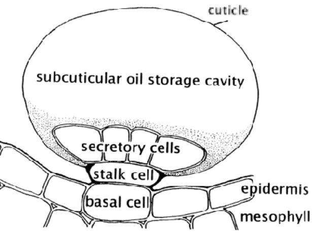
EOs are found in only 10% of the plant kingdom and can be synthetized by all plant organs, including buds, flowers, leaves, stems, twigs, seeds, fruits, roots and wood or bark (Gershenzon 1994; Ahmadi et al. 2002; Bezić et al. 2009; Djilani & Dicko 2010). Different organs can share the same kind of secretory structures or possess different types (Fahn 1979). These structures can be classified into six groups: secretory cells, osmophores, secretory cavities, secretory ducts, glandular trichomes (figure 1.1.2.1) and epidermal cells (Svoboda & Svoboda 2000; Fahn 1979).
Figure 1.1.2.1 – Schematic diagram of a glandular trichome illustrating the placement of this epidermal structures and the relationship of the disc of secretory cells to the stalk, basal cells and to the subcuticular storage space (Turner et al. 2014).
Secretory cells are the simplest secretory structures comprising a single
secretion-containing cell where it is only the actual content that distinguishes it from adjacent non-secretory cells (Svoboda & Svoboda 2000). This cell type is found in many different plant tissues, such as in the rhizome pith and cortex of ginger, and in the perisperm and embryo of nutmeg (Svoboda & Svoboda 2000).
Osmophores are areas of flower tissues with secretory cells differing structurally from
adjacent cells (e.g. isodiametric in orchids) (Fahn 1979).
Secretory cavities are spherical structures that can be formed in two ways. In one
mechanism, the parenchyma cells (thin-walled, relatively undifferentiated cells that may vary in structure and function) can separate one from another, leaving intercellular spaces called lumina or lacuna. The other, an actual cell can disintegrate and leave a cavity within the tissue (Fahn 1979; Svoboda & Svoboda 2000). These spaces are lined with secretory cells or an epithelium that produces the essential oils. In high oil-yielding plants, several layers of these secretory cells are formed. The cavities continually enlarge and some become filled with cells with thin, convoluted walls that also store the oil produced from within their plastids (a class of cytoplasmic organelles) (Fahn 1979; Azcon-Bieto & Talon 1996; Svoboda & Svoboda 2000).
Secretory ducts are elongated cavities. They can often branch to create a network
31
composed of an epithelium that surrounds a central cavity. Several predisposed cells within the parenchyma undergo asynchronous division and in doing so, expand the initial space in the middle, where the cells are adjacent, to form a cavity. Some of the cells forming the wall of the cavity will change into secretory epithelial cells. The oils are biosynthesized within their leucoplasts and move via the endoplasmic reticulum into the cavity. These cavities join up to form ducts (Fahn 1979; Svoboda & Svoboda 2000).
Glandular trichomes are modified epidermal hairs and can be found covering leaves,
stems, and even flower parts (Svoboda & Svoboda 2000). The secretory cells are attached by a single stem or basal cell in the epidermis. The outer surface of the gland is heavily cutinised (Fahn 1979). A cuticle, in which no pores or perforations are present, usually completely covers the trichome (Svoboda & Svoboda 2000). The EOs accumulate in subcuticular spaces and are thought to diffuse outwards through the cuticle (Fahn 1979). The glandular cells differ from normal plant cells in that they have a very large nucleus and dense protoplasm that lacks a large central vacuole (Svoboda and Svoboda 2000). There are numerous plasmodesmata (cytoplasmic threads running through cell walls, connecting cytoplasm of adjacent cells) across the walls of the gland cells, especially between the stalk cell and the collecting cell (Fahn 1979). In the very young gland the intracellular organization is almost identical to that of the adjacent cells, but as the secretory cells develop, complex changes occur (Svoboda & Svoboda 2000). The membrane system progressively degenerates and in the fully developed glands only a thin granular cytoplasm (the living cell parts within the membrane, except the nucleus) remains (Fahn 1979; Svoboda & Svoboda 2000).
Secretory cells also occur in flower petals – for example in jasmine and rose. As in this cases glandular hairs and glandular trichomes are not present, the volatile oil diffuses through the epidermal cells and the cuticle to reach the atmosphere (Svoboda & Svoboda 2000).
1.1.3 Chemical composition of essential oils
Since ancient times, EOs have been recognised for their medicinal value and are considered very interesting and powerful natural plant products (Baris et al. 2006). EOs continue to be of paramount importance until the present day (Margaris et al. 1982; Tisserand 1997). Recording findings in Mesopotamia, China, India, Persia and ancient Egypt show their uses for many treatments in various forms (Guenther 1948; Wei &
Shibamoto 2010). For example, in ancient Egypt, the population extracted oils by infusion for medicinal purposes (Burt 2004; Peeyush et al. 2011).
EOs naturally occur in aromatic plants, which make these species very valuable. Among many other well-known families rich in species bearing EOs are Apiaceae, Asteraceae, Geraniaceae, Lamiaceae, Pinaceae, Solanaceae, Verbenaceae (Figueiredo et al. 1997; Ahmadi et al. 2002; Elias et al. 2003).
According to the International Standard Organization on Essential Oils, ISO 9235 (1997) of the ISO/TC and the Portuguese Standard,NP 90 (1987) of the IPQ-CT 5, the definition of EO is restricted to the products obtained exclusively by distillation of plant material, with or without water steam, or by mechanical processes applied to the pericarp of fruits of the genus Citrus L. EOs are volatile, natural, complex compounds characterised by a strong odour. They are hydrophobic, soluble in alcohol, DMSO and methanol, non-polar or weakly polar solvents, waxes and oils (Gupta et al. 2010). EOs are limpid and rarely coloured, most are liquid and of lower density than water (Ciccarelli et al. 2008; Martín et al. 2010). Due to their molecular structures (presence of olefenic double bonds and functional groups such as hydroxyl, aldehyde, and ester) EOs are readily oxidizable by light, heat and air (Gershenzon 1994; Liolios et al. 2010; Djilani & Dicko 2010).
Essential oils are very complex natural mixtures that can contain about 20-60 components at varied concentrations (Miguel 2010). They are composed by two or three major components at fairly high concentrations (20-70%), with other components present in trace amounts. Generally, these major components determine the biological properties of the EOs (Skaltsa et al. 2003; Sell 2006; Thomar 2011).
These oils may comprise volatile compounds that have carbon, hydrogen, and oxygen as their building blocks. The major volatile constituents are: hydrocarbons, alcohols, acids, aldehydes, cyclic aldehydes, ketones, lactones, phenols, phenolic ethers, oxides and esters (Bakkali et al. 2008). These are subdivided into two categories: of terpenoid
origin, for compounds composed almost exclusively of terpenes (monoterpenes,
sesquiterpenes and diterpenes), and nonterpenoid origin, for compounds containing phenylpropanoid and aliphatic skeletons. Some compounds may also contain nitrogen (antharanilates, pyrazines) or sulphur (thiols, sulphides) (Bakkali et al. 2008; Baser & Demirci 2007; Kalemba & Wajs 2011). All of them are hydrocarbons and their oxygenated derivatives (Proença da Cunha et al. 2003).
33
Terpenoids (figure 1.1.3.1) are the most structurally varied class of plant natural products
(Bakkali et al. 2008). In nature, they play significant roles in plant-environment interactions, plant-plant communication and plant-animal interactions (Pichersky & Gershenzon 2002). They are commercially important due to their wide application in a vast number of industrial products, such as flavouring agents, pharmaceuticals, perfumes, insecticides and anti-microbial agents (Martin et al. 2003).
Figure 1.1.3.1 – Chemical structures of terpenoids (selected compounds of essential oils). Monoterpenes have a variety of structures, for example - Carbures, Alcohols, Aldeydes, Ketones, Esters and Ethers. The structures of the sesquiterpenes - Carbures, Alcohols, Ketones and Epoxides - are similar comparing to the monoterpenes (modified after Bakkali et al. 2008).
Terpenes result from the condensation of the pentacarbonate unit, 2-methilbutadiene or
isoprene (Dubey et al. 2003). For this reason they are also called isoprenoides. These are classified according to the isoprene units (five carbons) in their structure: hemiterpenes C5 (1 isoprene unit), monoterpenes C10 (2 isoprene units), sesquiterpenes C15 (3 isoprene units), diterpenes C20 (4 isoprene units), sesterpenes C25 (5 isoprene units), triterpenes C30 (6 isoprene units), tetraterpenes C40 (8 isoprene units) and polyterpenes (C5) n (Dubey et al. 2003). Dimers and trimers of isoprene, monoterpenes with 10
carbons and sesquiterpenes with 15 carbons, respectively, are predominant in essential oils (Gershenzon & Kreis, 1999; Proença da Cunha et al. 2003).
In higher plants, terpenes are synthesised via two isopentenyl diphosphate (IPP) generating pathways. Isopentenyl diphosphate (IPP) and dimethylallyldiphosphate (DMAPP) serve as universal precursors for the biosynthesis of terpenes. They are biosynthesised through the mevalonate (MVA) and the non-mevalonate or deoxyxylulose phosphate pathways (DXP) (Croteau 2000; Sangwan et al. 2001; Dubey et al. 2003; Baser & Demirci 2007) (Figure 1.1.3.2).
Figure 1.1.3.2 – The two different terpenoid classes in the mitochondria and plastids , respectively (from Baser & Demirci 2007).
Nonterpenoids or aromatic compounds (Figure 1.1.3.3) are derived from phenylpropane
and occur less frequently than the terpenes. The biosynthetic pathways concerning terpenes and phenylpropanic derivatives generally are separated in plants but may coexist in some, with one major pathway taking over. Nonterpenoid compounds comprise: Aldeyde; alcohol, phenols, methoxy derivatives and methylene dioxy compounds (Bick & Lange 2003; Baser & Demirci 2007; Bakkali et al. 2008)
35
Nitrogenous or sulphured components such as glucosinolates or isothiocyanate derivatives (garlic and mustard oils) are also characteristic as secondary metabolites of diverse plants or of torrefied, grilled or roasted products (Bick & Lange 2003; Baser & Demirci 2007).
Figure 1.1.3.3 – Chemical structures of non-terpenoids compounds (selected compounds of essential oils) (modified after Bakkali et al. 2008).
1.1.4 Factors influencing the production and quality of essential oils
The fragrance and chemical composition of essential oils may vary according to the geo-climatic location and growing conditions (soil type, climate, altitude and amount of water available), season (before and after flowering), and time of day of harvesting (Djilani & Dicko 2010). Another important factor is the genetic composition of the plant. All these factors (genetic and epigenetic) influence the biochemical synthesis of EOs in a given plant (Bakkali et al. 2008; Adorjan & Buchbauer 2010). In this way, specimens from same species of plant can produce a similar essential oil but with different chemical composition, resulting in different therapeutic activities (Djilani & Dicko 2010).
1.1.5 Extraction techniques
The two main industrial processes used to obtain EOs are distillation and expression. However, there are more methodologies to obtain other aromatic extracts from aromatic plants, such as extraction by solvents, and adsorption techniques (Lawrence 1995; Başer & Demirci 2007; Figueiredo et al. 2007; Proença da Cunha et al. 2003).
The main method, at a laboratory scale that is used to obtain EOs from plant material is the distillation with water (hydrodistillation), using a Clevenger apparatus (figure 1.5.1.1). However, if not enough plant material is available to obtain the EOs by hydrodistillation, solvent extraction or solid phase micro-extraction can be used as alternative extraction processes to obtain a small amount of the volatile compounds and determine the chemical composition.
1.1.5.1Hydrodistillation
The principle of this process is to boil a suspension of an aromatic plant material in water so that its vapour can be condensed. The oil, which is immiscible with the water, is then separated. In this extraction process, the plant material is always in direct contact with water. This technique has the advantage of only dragging volatile substances, and is simple and cheap (Lawrence 1995).
The apparatus (figure 1.1.5.1.1) described in the Portuguese Pharmacopoeia (2005), for dosage of essential oils in aromatic plants comprises the following parts: (1) a suitable round-bottomed flask with a short, ground-glass neck having an internal diameter of about 29 mm at the wide end, where the plants and the water boil; (2) a condenser assembly that closely fits the flask; (3) a suitable heating device, allowing a fine control; and (4) a vertical support with a horizontal ring covered with insulating material (Farmacopeia Portuguesa 2005).
37
Figure 1.1.5.1.1 – Clevenger apparatus. Dimensions are indicated in millimetres (Farmacopeia Portuguesa 2005).
1.2 Biological Properties of Essential Oils
Approximately 3000 EOs have been described known, 300 of which are commercially important especially for the pharmaceutical, agronomic, food, sanitary, cosmetic and perfume industries (Djilani & Dicko 2010). For example, limonene, geranyl acetate or d-carvone are employed in perfumes, creams, soaps, as flavor additives for food, as fragrances for household cleaning products and as industrial solvents (Hajhashemi et al. 2003; Perry et al. 2003). However, the information available on the estimation of precisely number EOs is scarce (Silva et al. 2003; Djilani & Dicko 2010).
Essential oils are used in massage vegetable oil mixtures or added to baths but most frequently in aromatherapy (Perry et al. 2003). Importantly, EOs are also extremely useful as therapeutic agents for several pathologies. The use of EOs in medical applications has become very popular, both for the complex oil mixes, as well as the individual constituents as single compounds (Hajhashemi et al. 2003; Perry et al. 2003; Djilani & Dicko 2010).
Essential oils have been largely employed for their properties observed in nature, i.e. for their anti-nociceptive effect, insect repellent activity, antiviral activity, as well as other proprieties, such as antioxidant activity and anticancer activity.
1.2.1 Antinociceptive Effect
A nociceptor is a sensory receptor that responds to potentially damaging stimuli by sending nerve signals to the spinal cord and brain (Hucho & Levine 2007; Maham et al. 2013). The antinociceptive effect is a reduction in pain sensitivity made within neurons when endorphin or a similar opium-containing substance combines with a receptor (Maham et al. 2013).
Sousa and collaborators analysed the antinociceptive and anti-inflammatory effects of the EO extracted from Eremanthus erythropappus (DC.) McLeish (Asteraceae) leaves. In in vivo studies using mice, treatment with the Eremanthus EO led to a significant increase of the reaction time (reduction in pain sensitivity) after 30, 60 and 90 minutes of treatment, at doses of 200 mg/kg and 400 mg/kg (Sousa et al. 2008).
In 2008, the antinociceptive activity of the volatile oils of Hyptis pectinataI L. Pois (Lamiae) genotypes were analysed by Arrigoni-Blank and co-workers (Arrigoni-Blank et al. 2008) The use of H. pectinata is very common in Brazilian folk medicine for the treatment of inflammations, bacterial infections and pain. Six genotypes of volatile oil were investigated. The main compounds of all genotypes are sesquiterpenes. In the models used, all genotypes showed antinociceptive effect, for both central and peripheral nervous system (Arrigoni-Blank et al. 2008).
Takaki and colleagues investigated the anti-inflammatory and anti-nociceptive effects of Rosmarinus officinalis L. (Lamiaceae) EO in experiment in animal models (Takaki et al. 2008). In this study, it was shown that Eos extracted from Rosmarinus officinalis, at a dose of 500 mg/kg, led to a significant reduction of the volume of pleural exudate and slightly decreased the number of cells that had migrated in treated animals, when compared with control animals (not treated) (Takaki et al. 2008).
39
1.2.2 Insect Repellent Activity
Currently, the use of synthetic chemicals to control insects and arthropods raises several concerns related to the environment and human health. Therefore, there is a growing demand for alternative repellents or natural products. These products should possess good efficacy and be environmentally friendly (Nerio et al. 2010). Essential oils from several species have been extensively tested to assess their repellent and even insecticidal properties as a valuable natural source (Nerio et al. 2010).
In 2007, Rajkumar and colleagues investigated the repellent effect of selected EO against the malaria fever mosquito Anopheles stephensi in mosquito cages (Rajkumar et al. 2007). The five EOs tested were from Centella asiatica; Ipomoea cairica; Momordica charantia; Psidium guajava and Tridax procumbens. The oils were tested at three concentrations: 2, 4 and 6% (v/v). A dose-dependent effect was noticed and the highest concentration (6%) led to the highest repellent effect (Rajkumar et al. 2007).
In a study looking at the repellent effects of catmint (Nepeta cataria L., Lamiaceae) oil formulations against black flies (Simulium decorum Walker) and mosquitoes (primarily Aedes intrudens Dyar) in Maine and Florida showed that tested all formulations led to protection against mosquitoes for more than 4 hours (Rajkumar et al. 2007).
1.2.3 Antiviral Activity
A virus is a small infectious particle (20-300 nm) that is able to infect cells of another living organism, in which it can replicate itself (Saddi et al. 2007). Viruses cannot reproduce on their own: a virus is composed of genes and a protein coat, and some have an envelope of fat that surrounds them. Viruses can lead to infections, which provoke an immune response that usually eliminates the infecting virus (Saddi et al. 2007; Loizzo et al. 2008). In 2007, Saddi and colleagues investigated the activities of the EOs from Artemisia arborescens L. (Asteraceae) against herpes simplex virus 1 and 2 (HSV-1 and HSV-2), since new prophylactic and therapeutic tools are needed. Using MTT assay, the result of this study showed that the IC50 values were 2.4 and 4.1 µg/mL for HSV-1 and HSV-2,
respectively. Furthermore, the study showed that the antiviral activity of the EOs was principally due to direct virucidal effects, but also led to inhibition of the cell-to-cell virus diffusion of both HSV-1 and HSV-2 (Saddi et al. 2007).
An in vitro evaluation of the biological activity against HSV-1 was carried out with Cedrus libani A. Rich EO and ethanol extracts of cones, by Loizzo and collaborators (Loizzo et al. 2008). In this study, it was shown that ethanol extracts and EO possess anti-viral activity with IC50 values of 0.50 mg/mL and 0.44 mg/mL, respectively.
1.2.4 Antioxidant Activity
Antioxidant compounds in food play an important role as a health-protecting factor (Saeed et al. 2012). Antioxidants reduce the risk for chronic diseases including cancer and heart diseases (Camire et al. 2005; Saeed et al. 2012).
Primary sources of naturally occurring antioxidants are whole grains, fruits and vegetables (Saeed et al. 2012). Plant sources of food antioxidants like vitamin C, vitamin E, carotenes, phenolic acids, phytate and phytoestrogens have been recognized as having the potential to reduce disease risk (Camire et al. 2005; Saeed et al. 2012). Most of the antioxidant compounds in a typical diet are derived from plant sources and belong to various classes of compounds with a wide variety of physical and chemical properties. Some compounds, such as gallates, have strong antioxidant activity, while others, such as the monophenols are weak antioxidants (Camire et al. 2005; Saeed et al. 2012).
The main characteristic of an antioxidant is its ability to trap free radicals. Highly reactive free radicals and oxygen species are present in biological systems from a wide variety of sources. These free radicals may oxidize nucleic acids, proteins, lipids or DNA and can initiate degenerative diseases (Saeed et al. 2012). Antioxidant compounds like phenolic acids, polyphenols and flavonoids scavenge free radicals such as peroxides, hydroperoxides or lipid peroxyl, and thus inhibit the oxidative mechanisms that lead to degenerative diseases (Saeed et al. 2012).
In 2007, Sharififar and colleagues investigated the antioxidant and free radical scavenging activities of the EOs from flowers of Otostegia persica Boiss (Lamiaceae) (Sharififar et al. 2007). This study showed that this EO possesses high antioxidant and radical scavenging activity in both DPPH (2,2-diphenyl-1-1-picrylhydrazyl) free radical scavenging test and ammonium thiocyanate tests. In the DPPH free-radical scavenging protocol, the EO showed antioxidant activity with an IC50 value of 19.8 µg/mL. In the second protocol, the
EO exhibited an inhibition rate of oxidation of linoleic acid of 93.5% (percentage of total inhibition of oxidation) (Sharififar et al. 2007)
41
Chaieb and colleagues investigated the antioxidant properties of the EO in clove (Eugenia caryophyllata (L. Myrtaceae) (Chaieb et al. 2007). The antioxidant activity was evaluated by the DPPH free-radical scavenging test and the IC50 value was 0.2 µg/mL, revealing that
the oil has a very strong radical scavenging activity (Chaieb et al. 2007).
1.2.5 Anticancer Activity
Despite recent advances in treatment modalities, cancer remains a major source of morbidity and mortality throughout the world (Edwards et al. 2005; Hesketh 2013). In the United States, cancer is the leading cause of death for individuals less than 85 years of age (Jemal et al. 2006). Moreover, the incidence of many cancers, including cancers of the skin, prostate, breast, and kidney, continues to increase (Edwards et al. 2005). A recent study, led by Cancer Research UK (CRUK), indicated that one in two individuals born after 1960 will be diagnosed with cancer at some point in their lifetime (Ahmad et al. 2015)
Cancer is, in fact, a general term that refers to over 100 distinct diseases affecting many different tissues and cell types. However, all forms of cancer are characterised by abnormal cell growth resulting from a relatively small number of inherited or environmentally-induced genetic mutations (Hesketh 2013).
In their seminal review, Hanahan and Weinberg have argued that in order for a cell to become cancerous, it must acquire six unique traits as a result of altered cell physiology (Hanahan & Weinberg 2011). These defining traits of cancer cells are: (1) the ability to generate their own growth signals or respond to weak growth signals that are ignored by healthy cells; (2) insensitivity to anti-proliferative signals; (3) resistance to cell death by apoptosis; (4) the capacity for limitless replication; (5) the ability to stimulate new blood vessel development in order to allow for tumour growth; and (6) the capacity to invade tissues, at first locally, and later to spread or metastasize throughout the body (Hanahan & Weinberg 2011).. Although localized cancers can often be successfully treated by surgery and/or radiation therapy, chemotherapy remains the usual treatment of choice for advanced or metastatic disease (Espinosa et al. 2003). However, the use of conventional chemotherapeutic agents that typically target rapidly dividing cancer cells is often associated with deleterious side effects caused by inadvertent drug-induced damage to healthy cells and tissues (Cassidy et al. 2002).
Moreover, cancer cells that are quiescent or slowly proliferating are refractory to the cytotoxic effect of chemotherapeutic drugs that act at the level of DNA synthesis (Naumov et al. 2003). Cancer cells also frequently become resistant to chemotherapy as a consequence of cellular changes that include increased expression of drug-detoxifying enzymes and drug transporters, altered interactions between the drug and its target, altered DNA damage capacity, and defects in the cellular machinery that mediate apoptosis (Gatti & Zunino 2005).
Therefore, the development of a new class of anticancer drugs that lack the toxicity of conventional chemotherapeutic agents and are unaffected by common mechanisms of chemoresistance would be a major advance in cancer treatment (Gatti & Zunino 2005). Some data strongly support the view that EOs have potential therapeutic applications in the prevention of cancer, but EOs remain mainly poorly researched.
Ravizza and colleagues investigated the anticancer properties of linalool, a plant-derivated monoterpene alcohol that is found in the essential oils from many aromatic plants (Ravizza et al. 2008). The cancer modules for this study were two human breast adenocarcinoma cell lines, MCF7 WT (wild type) and multi-drug resistant MCF7 AdrR (selected as being resistant to treatment with the chemotherapy agent Adriamycin, also known as doxorubicin). Linalool was used either as single agent or in combination with doxorubicin. Linalool treatment alone only mildly inhibited cell proliferation. However, at sub-toxic concentrations when in combination with doxorubicin, it led to increased doxorubicin-induced cytotoxicity and pro-apoptotic effects in both cell lines. It was therefore concluded that combination with linalool could lead to an improved therapeutic index of anthracyclines (such as doxorubicin) in the treatment of breast cancer, especially in multi-drug resistant tumours (Ravizza et al. 2008).
Another study analysed the induction of mitochondria-associated (or intrinsic) apoptosis by EO extracted from Tanacetum gracile Hook., (Asteraceae), an alpine aromatic herb that contains about 40 components (Verma et al. 2008). This EO led to an inhibition the proliferation of the HL-60 leukaemia cell line, with an IC50 value of 27 µg/mL, associated
with induction of apoptosis. Furthermore, the study showed that this effect was mediated by the mitochondria, since it led to a decrease of mitochondrial membrane potential, release of cytochrome C, activation of caspases-9 and -3, and an increase poly(ADP-ribose)-polymerase (PARP) cleavage.
43
In another report, Medina-Holguín and colleagues reported on the chemo-typical variation of EOs in medicinal plant Anemopsis californica (Nutt.) Hook., Saururaceae (Medina-Holguín et al. 2009). The authors extracted EOs from roots/rhizomes of the Anemopsis californica (ACEO) plant by steam distillation. It was then analysed for its effect on AN3CA (uterine) and HeLa (cervical) human cancer cell lines. The study showed an antiproliferative activity of the EO against both cell lines in vitro, with IC50 values of
0.056% and 0.052% (v/v) for AN3CA and HeLa cells, respectively. The three main compounds, thymol, piperitone and methyleugenol, were tested independently for growth inhibition against the AN3CA and HeLa cells and also inhibited cell growth. The IC50
values for these three compounds against each cell line was determined and compared with the concentration of these compounds in the root oil of A. californica (Medina-Holguín et al. 2009). The inhibition may be the result of a synergistic relationship between the combined abundant compounds piperitone and methyleugenol, or also with a minor component in the oil. In conclusion, the study showed the specific bioactivity against uterine and cervical cancer cell lines of steam-distilled oil of Anemopsis californica root tissue, thus supporting the traditional and cultural use of ACEO to treat uterine cancer. Finally, another study analysed the antibacterial, antifungal, and anticancer activities of volatile oils and extracts from stems, leaves and flowers of Eucalyptus siderxylon A. Cunn and Eucalyptus torquata grown in Egypt (Ashour 2008). To analyse the anticancer activity, the sulforhodamine B (SRB) assay was used, an evaluation of cell density based on the measurement of cellular protein content (Ashour 2008). The in vitro cytotoxic activities of the essential oils and extracts were evaluated using the hepatocellular carcinoma cell line HEPG2 and the breast adenocarcinoma cell line MCF7. The results indicated that the EOs extracted from E. torquata leaves and stems and of E. sideroxylon leaves exert cytotoxic effect activity against MCF7 cells, but no effect on HEPG2 cells (Ashour 2008).
1.3 Key concepts in cancer biology
An abnormal cell that grows (increases in mass) and proliferates (divides) out of control will give rise to a tumour, or neoplasm (Knowles & Selby 2005). As long as the neoplastic cells have not yet become invasive, however, the tumour is said to be benign. For most types of such neoplasms, removing or destroying the mass locally usually achieves a complete cure. A tumour is considered a true cancer if it is malignant; that is, when its cells have acquired the ability to invade the surrounding tissue, and invasiveness is an