Cloning, Characterization, and Tissue Distribution of
Prolactin Receptor in the Sea Bream (Sparus aurata)
C. R. A. Santos,* P. M. Ingleton,† J. E. B. Cavaco,* P. A. Kelly,‡
M. Edery,‡ and D. M. Power*
*Centre of Marine Sciences (CCMAR), Universidade do Algarve, Campus de Gambelas, 8000-810 Faro, Portugal; †Institute of Endocrinology, Division of Biochemical and Musculo-skeletal Medicine, Medical School,
Sheffield S10 2RX, United Kingdom; and ‡Faculte´ de Medecine Necker Enfants Malades, INSERM Unite´ 344-Endocrinology Moleculaire, 75730 Paris, France
Accepted August 15, 2000
The prolactin receptor (PRLR) was cloned and its tissue distribution characterized in adults of the protandrous hermaphrodite marine teleost, the sea bream (Sparus
au-rata). An homologous cDNA probe for sea bream PRLR
(sbPRLR) was obtained by RT-PCR using gill mRNA. This probe was used to screen intestine and kidney cDNA libraries from which two overlapping clones (1100 and 2425 bp, respectively) were obtained. These clones had 100% sequence identity in the overlapping region (893 bp) and were used to deduce the complete amino acid sequence of sbPRLR. The receptor spans 2640 bp and encodes a protein of 537 amino acids. Features character-istic of PRLR, two pairs of cysteines, WS box, hydropho-bic transmembrane domain, box 1, and box 2, were iden-tified and showed a high degree of sequence identity to PRLRs from other vertebrate species. SbPRLR is 29 and 32% identical to tilapia (Oreochromis niloticus) and gold-fish (Carassius auratus) PRLRs, respectively. In the sea bream two PRLR transcripts of 2.8 and 3.2 kb were de-tected in the intestine, kidney, and gills and a single tran-script of 2.8 kb was detected in skin and pituitary by Northern blot. Spermiating gonads (more than 95% male tissue; gonado–somatic index of 0.6) contained, in addi-tion to the 2.8-kb transcript, three more transcripts of 1.9, 1.3, and 1.1 kb. RT-PCR, which is a far more sensitive method than Northern blot, detected PRLR mRNA in gills, intestine, brain, pituitary, kidney, liver, gonads, spleen, head-kidney, heart, muscle, and bone.
Immuno-histochemistry using specific polyclonal antibodies raised against an oligopeptide from the extracellular do-main of sbPRLR detected PRLR in several epithelial tis-sues of juvenile sea bream, including the anterior gut, renal tubule, choroid membrane of the third ventricle, saccus vasculosus, branchial chloride cells, and branchial cartilage. © 2001 Academic Press
Key Words: prolactin receptor; marine teleost;
osmo-regulation; reproduction.
INTRODUCTION
Prolactin receptors (PRLRs) belong to an extensive family of hormone and cytokine receptors (class 1
cytokine receptor superfamily), which includes
growth hormone (GH) and several interleukin recep-tors (Finidori and Kelly, 1995). These receprecep-tors share several structural features in common: an extracellular domain (ECD) with two pairs of cysteines and a con-served motif (WS box) composed of a concon-served amino acid sequence, Trp-Ser-X-Trp-Ser, where X can be any amino acid, a single-transmembrane domain composed largely of hydrophobic amino acids, and an intracellular domain (ICD). The latter domain contains a proline-rich region (box 1), lying immediately adja-cent to the membrane, which is composed largely of negatively charged residues and is followed by
an-0016-6480/01 $35.00 Copyright © 2001 by Academic Press
other consensus region (box 2), comprising a series of hydrophobic residues followed by negatively charged residues (Bole-Feysot et al., 1998; Kelly et al., 1993, 1991b). These two highly conserved motifs are essen-tial for receptor activity (Goupille et al., 1997).
A PRLR cDNA was first cloned from rat liver (Bou-tin et al., 1988) and shown to encode a protein of 291 amino acid residues; subsequently, a longer form (591 aa) of the receptor was identified in rat liver and ovary. PRLR cDNAs have now been cloned from spe-cies in all vertebrate groups except reptiles (for review see Bole-Feysot et al., 1998). Short and long forms of the PRLR have been identified in rat, mouse, and sheep (Davis and Linzer, 1989; Moore and Oka, 1993; Bignon et al., 1997). These two receptors differ only in the ICD, which is 357 amino acids in the long form and 57 amino acids in the short form which lacks box 2 (Shirota et al., 1990; Bole-Feysot et al., 1998). A third form of PRLR has also been identified in the rat PRL-dependent immune cell line (Nb2); this form contains a partial deletion of the cytoplasmic domain (Ali et al., 1991). Short and long forms of PRLRs originate from alternative splicing of the mRNA transcribed from a single gene (Ormandy et al., 1998) and only long forms are capable of inducing the transcription of milk pro-teins, whereas short forms are capable of inducing cell proliferation (Das and Vonderhaar, 1995). A single form of PRLR has been identified in birds, which most closely resembles the long form found in mammals, with the exception of a tandem repeat within the ECD (Chen and Horseman, 1994; Zhou et al., 1996). In tet-rapods, PRLR has a widespread distribution and has been identified in brain and pituitary, liver, kidney, lungs, skin, gonads and accessory glands, skeletal muscle, and tissues of the immune system (for review Bole-Feysot et al., 1998); it has also recently been de-tected in bone (Clement-Lacroix et al., 1999).
PRLRs have also been cloned in an amphibian (Yamamoto et al., 1998), in a euryhaline teleost, Nile tilapia (Oreochromis niloticus) (tiPRLR) (Sandra et al., 1995), and in a fresh water teleost, the goldfish
(Car-assius auratus) (gf PRLR) (Tse et al., 2000). In tilapia
there is a single transcript of 3.2 kb which shares greatest similarity with the long forms of mammalian PRLRs, although the overall homology is low (37%) (Sandra et al., 1995, 2000). In the goldfish two tran-scripts, of 3.5 and 4.6 kb, were identified. The 4.6-kb transcript encodes a 600-amino acid protein 48.3%
identical to tiPRLR (Tse et al., 2000). The expression of PRLR was analyzed in tilapia by Northern blot and the results obtained demonstrated that the osmoregu-latory organs, gills, kidney, and intestine expressed PRLR at higher levels. The ovary, testis, and head-kidney (which has hematopoietic function in fish) also expressed PRLR but at much lower levels (Sandra et
al., 1995, 2000). A similar distribution of PRLR was
also observed in the goldfish (Tse et al., 2000). These results confirmed the results obtained with homolo-gous ligand binding studies in the gills and kidney of tilapia (Auperin et al., 1994) and identified alternative target tissues for PRL in fish. The high levels of PRLR expression in osmoregulatory tissues of fish corrobo-rate the well-documented action of PRL in the main-tenance of hydromineral balance in euryhaline teleosts (Bern, 1975).
The function of PRL in marine teleosts is unclear, particularly since in euryhaline fish its principal role appears to be in freshwater adaptation and it inhibits seawater adaptation. High levels of expression of PRL have been reported in the sea bream pituitary gland (Santos et al., 1999) but relatively little is known about its physiological role in marine fish. In the present study the cloning of PRLR cDNA from a marine te-leost, the sea bream (Sparus aurata), is described and its expression and tissue localization in adult fish is char-acterized. In addition, part of the sbPRLR gene was cloned and compared with the gene for mouse PRLR.
MATERIALS AND METHODS
Animals and Tissues
Adult sea bream (⬃350 g) maintained in
through-flow seawater tanks at 17⫾ 2° under natural
photo-period for winter in the Algarve, Portugal were killed by stunning and decapitation. Liver, kidney, intestine, brain, pituitary, skin, gills, skeletal muscle, heart, bone, spleen, head-kidney, and mature gonadal tissue (more than 95% male tissue; gonado-somatic index of 0.6) were frozen in liquid nitrogen and stored at⫺70°.
Total RNA and mRNA Purification
Total RNA was extracted from the tissues men-tioned above using “TRI reagent” (Sigma, St. Louis,
MO). The poly(A)⫹RNA fraction was obtained from total RNA by chromatography on columns of oli-go(dT) cellulose (Aviv and Leder, 1972).
Generation of a Homologous sbPRLR cDNA Probe by RT-PCR
PCR primers were designed within the most highly conserved regions of PRLRs identified after multiple sequence alignments of all PRLR sequences available (GenBank). A forward primer based on a highly con-served region of the ECD domain of PRLR
(TFTCWW), 5⬘ ACA TTC ACC TGC TGG TGG3⬘
(Pharmacia Biotech, Uppsala, Sweden) (primer PF1), was synthesized and the reverse primer, in which inosine (I) was introduced to minimize degeneracy at
the 3⬘ end 5⬘ GAT CTT TGG CAC IGG IGG3⬘ (primer
PR1), was located within the proline-rich motif (PPVPGPKI).
cDNA was synthesized from 1 g of gill RNA
poly(A)⫹ in a 30-l reaction containing 50 mM Tris–
HCl, pH 8.3, 75 mM KCl, 3 mM MgCl2, 10 mM
dithio-threitol (DTT), 1 mM dNTP, 15 pmol oligo(dT) primer, RNase inhibitor (3.2 U; Pharmacia Biotech), and M-MLV reverse transcriptase (20 U; Gibco BRL, Barce-lona), for 2 h at 37°. PCR was carried out in a 50-l
reaction containing 5 l of gill cDNA, 10 mM Tris–
HCl, pH 9.0, 50 mM KCl, 0.1% Triton X-100, 3 mM
MgCl2, 1 mM dNTP, 40 pmol of forward primer, 200
pmol of reverse primer, and Taq DNA polymerase (1.25 U; Promega, Madison, WI); sterile water substi-tuted for cDNA in control reactions.
The reaction was initially denatured for 5 min and then submitted to 45 cycles of denaturing (94°, 1 min), annealing (52°, 2 min), and extension (72°, 1 min) in a thermocycler (Robocycler; Stratagene, La Jolla, CA). The amplified products were analyzed on a 1.5%
aga-rose gel containing ethidium bromide (0.5 g/l),
excised from the gel, and purified using a column containing a silica– gel membrane (Quiagen).
The purified DNAs were ligated into pGEM-T vec-tor (Promega) in a 10-l reaction containing purified
DNA (7 l), 1 l of T4 DNA ligase (3 U/ml), 1 l of
pGEM-T (50 ng), and 1 l of 10⫻ buffer (300 mM
Tris–HCl, pH 7.8, 100 mM MgCl2, 100 mM DTT, and
10 mM ATP), incubated overnight at 4°, and used to
transform Escherichia coli (XL1B MRF⬘ strain). DNA
was isolated from clones and analyzed in 1.5% agarose
gels after restriction digest. Sequencing was carried out using a modification of the dideoxy chain termi-nation method (Sanger et al., 1977; T7 sequencing Kit; Pharmacia Biotech).
Construction of Sea Bream Intestine and Kidney cDNA Libraries
cDNA libraries were constructed from 5 g of sea
bream intestine and kidney poly(A)⫹ RNA using the
Lambda ZAP cDNA and UNI-ZAP XR cDNA cloning kits (Stratagene), respectively. The double-stranded cDNA was ligated into the corresponding vectors and packaged into Gigapack Gold III packaging extracts (Stratagene).
Screening of cDNA Libraries
The sea bream intestine library was screened with a
[␣-32P]dCTP-labeled sbPRLR probe (Rediprime,
ran-dom labeling kit; Amersham, Little Chalfont, UK) gen-erated by RT-PCR. Filters were hybridized overnight
at 65° and washed at 65° in 0.1⫻ SSC/0.1% SDS. Two
positive plaques were obtained from 400,000 recombi-nants, isolated, automatically excised into pBluescript (Stratagene), and sequenced using an adaptation of the dideoxy chain termination method (Sanger et al., 1977) with the universal primers within the flanking regions of the polylinker of pBluescript. This clone, named I3a2.2, was then used to screen the kidney cDNA library, and one positive plaque was isolated from 400,000 recombinants. This clone, named
KA13.1, was also sequenced from the 3⬘ and 5⬘ ends
using internal walking primers designed from the newly achieved sequence (Fig. 1).
Northern Blot Analysis
Two micrograms of mRNA from sea bream liver, kidney, intestine, brain, pituitary, skin, gill,
head-kid-ney, spleen, and skeletal muscle and 10g of gonad
mRNA were fractionated on a 5.5% formaldehyde/ 1.5% agarose gel, transferred to a nylon filter (Hy-bond-N; Amersham) with 10⫻ SSC, and cross-linked at 80° for 2 h. Prior to hybridization the filter was washed at 60° for 20 min in 1⫻ SSC, 0.1% SDS and
prehybridized in 50% formamide, 50 mM NaPO4, 5⫻
Denharts, 0.1% SDS, 5⫻ SSC, 50g/ml DNA from calf
thymus for 4 h at 42°. Hybridization was allowed to proceed overnight at 42° in fresh prehybridization solution containing sbPRLR clone I3A2.2 labeled with
[␣-32P]dCTP. Filters were then washed for 30 min at
42° in prehybridization solution and stringency washes carried out at 55° for 15 min in 1⫻ SSC, 0.1% SDS and then at 60° for 30 min. The membrane was exposed to Biomax MS film (Kodak, Rochester, NY)
with intensifying screens at⫺70° for 5 days. To
eval-uate the relative amounts of mRNA for each tissue, the blot was stripped with a boiling solution of 0.1% SDS
and hybridized with a sea bream-actin probe (Santos
et al., 1997) following the same protocol as before but
reducing exposure time to 1 h.
Tissue Distribution by RT-PCR
The tissue expression of sbPRLR was also analyzed with the more sensitive method of RT-PCR. Specific primers were designed using the Primer Premier com-puter program (Version 4.04; Premier Biosoft Interna-tional) based on the sbPRLR sequence. Forward
primer (PF2) (5⬘AGTCCGGCTGGGTCACCATTA)
and reverse primer (PR2)
(5⬘GGTGGCGACCAA-GATCCAAAAC3⬘) were synthesized by
MWG-Bio-tech GmbH (Germany) and were predicted to amplify a fragment of 249 bp.
cDNA was synthesized in a 30-l reaction as
previ-ously described from total RNA (0.5 g) from the
following tissue samples: liver, kidney, intestine, brain, pituitary, skin, gills, heart, skeletal muscle, bone, ovary, and testis. As a negative control, a PCR which contained all the reagents, but in which cDNA was omitted, was run simultaneously. RT-PCR was carried out with this cDNA (5l) in a 50-l reaction as previously described, with the exception that a lower
concentration of MgCl2 (1.5 mM), 50 pmol of each
primer, and Taq DNA polymerase (1.25 U; Promega) were used. The thermocycling protocol (Robocycler; Stratagene) was as follows: initial denaturing step (94°, 2 min) followed by 35 cycles of denaturation (94°, 1 min), annealing (56°, 1 min), and extension (72°, 30 s).
Southern Blot Analysis
PCR products were separated on a TBE/agarose gel (1.5%). The gel was soaked in denaturing solution (0.5
M NaOH, 1.5 M NaCl) for 15 min with gentle agita-tion, washed twice for 2 min in sterile water, and soaked in neutralizing solution (1.5 M NaCl, 0.5 M Tris–HCl, pH 8.0) for 30 min. Transfer to a nitrocellu-lose membrane (Hybond C; Amersham) was carried out using 10⫻ SSC as transfer buffer. The membrane was baked for 2 h at 80° and hybridized using sbPRLR clone I3A2.2 as the probe and the same procedure described for cDNA library screening. The blot was exposed for 5 min and 2 h at room temperature (24°).
Partial Cloning of sbPRLR Gene
Genomic DNA was extracted using TRI reagent (Sigma). The PCR protocol already described above for analyzing sbPRLR expression in adult tissue was applied to approximately 10 ng of sea bream genomic DNA using primers PF2 and PR2. The product ob-tained was cloned into pGEM-T vector using the pro-tocol already described for cloning the sbPRLR probe and its sequence was determined.
Tissue Distribution by Immunohistochemistry (IHC)
The methodology used for antibody production is detailed in Nevalainen et al. (1996). Briefly, an oligo-peptide from the extracellular domain of sbPRLR was synthesized by Sheffield University Krebs Institute Molecular Synthesis Service using Applied Biosys-tems Model 476A. The chosen sequence, 35– 49 (RLYY-ERERLEGVHEC) from the extracellular domain of the sbPRLR, was conjugated to bovine thyroglobulin (Sigma T-1001) using carbodiimide. The oligopeptide was chosen on the basis of amino acid composition and because a similar region of rat prolactin receptor has been found to induce high-quality antisera used for detecting PRLR in many mammalian tissues. The conjugate was dissolved in normal saline, emulsified in Freund’s Complete Adjuvant, and injected intra-muscularly into rabbits. A second injection of antigen, emulsified in Freund’s Incomplete Adjuvant, was given 2 months later, and after an initial test bleed the serum was collected 14 days after the second injection. Immunohistochemistry was carried out using a modification of Sternberger (1974). Briefly, tissues were fixed in sublimated Bouin–Hollande (Kraicer et
al., 1967). Sections were cut at 4m and mounted on
in xylene and rehydrated in graded alcohols, and heavy metal ions were removed by immersion in 1% iodine in 70% ethanol and reduced in 5% sodium thiosulphate. For the IHC procedure, sections were immersed first in 1% hydrogen peroxide in phosphate buffer (100 mM, pH 7.4) containing 20% methanol to destroy endogenous peroxidase; then nonspecific binding sites were blocked using 4% swine serum in 1% bovine serum albumin (BSA) in phosphate buffer. Sections were then covered in the primary rabbit an-tiserum to sea bream PRLRs diluted in phosphate-buffered saline (PBS)/BSA (1/200) and incubated at 4° overnight; control slides received normal rabbit serum instead of specific primary antiserum. After washing, the sections were incubated at room temperature (ca. 20°) in swine anti-rabbit serum diluted 1/50 (Dako, Denmark) and then in peroxidase/anti-peroxidase re-agent (Dako). Color was generated by immersion in diaminobenzidine in Tris–HCl buffer, pH 7.6. Sections were dehydrated, cleared, and mounted in Depex. Cross-reaction to bovine thyroglobulin was checked
by absorption of diluted antiserum with 500g/ml of
bovine thyroglobulin and testing on sections of larvae containing thyroid follicles.
RESULTS
sbPRLR Probe
A cDNA fragment spanning 429 bp was obtained by RT-PCR using primers PF1 and PR1. The se-quence of this fragment was compared to sese-quences available in the EMBL databases (NCBI, BLASTX) and was found to have the highest homology with tiPRLR (43% identity). The sea bream RT-PCR prod-uct was found to align with tiPRLR in the region predicted from primer localization and consisted of
part of the ECD and part of the ICD. The PCR product was of a smaller size than predicted as the forward primer annealed downstream to the region for which it was designed. In addition to the overall sequence similarity to tiPRLR, other structural fea-tures characteristic of PRLR were also present in this fragment, such as the WS box (WSEWT) and a 24-amino acid transmembrane domain with a high con-tent of hydrophobic amino acids.
cDNA Library Screening
The RT-PCR product was used as a probe to screen a sea bream intestine cDNA library containing 450,000 primary recombinants. Two clones were isolated from 400,000 recombinants; the clone inserts were excised and sized by agarose gel electrophoresis, where they were observed to be of a similar size (1100 bp). Se-quencing revealed that one insert was 23 bp longer at the 5⬘ end (clone I3A2.2). Both inserts shared 100% identity in the overlap. The deduced amino acid se-quence showed that they encoded the complete ECD, the transmembrane domain, and part of the cytoplas-mic domain (Fig. 1). The longer clone (I3A2.2) was then used to screen the sea bream kidney cDNA
li-brary (2 ⫻ 106 primary recombinants) from which
another clone, 2425-bp long, was isolated from 400,000 recombinants. This clone was incomplete at the 5⬘ end. Alignment of the sequences of the clones obtained from the intestine and kidney libraries showed that both were 100% identical in the overlap (893 bp) and, in combination, permitted elucidation of the full-length sequence of sbPRLR.
SbPRLR spans 2.6 kb and contains an open reading frame (ORF) encoding a protein of 537 amino acids (Fig. 1). This ORF consists of a signal peptide of 20 amino acids followed by a mature protein of 517 amino acids. The 5⬘ untranslated region (UTR)
com-FIG. 1. Nucleotide sequence and deduced amino acid sequence of the sea bream PRLR. The sequence underlined comprises the overlapping region between clones I3A.2.2. and KA13.1. The cysteine residues are circled; the four potential N-glycosylation sites are enclosed by pentagons. The WS motif is boxed and the transmembrane domain is in italics. Within the intracellular domain, box 1 (next to the transmembrane domain) and box 2 are boxed. Single tyrosine residues are enclosed by a star. In the 3⬘ untranslated region three potential polyadenylation signals are printed in boldface and underlined. Nucleotide sequences printed in boldface correspond to the primers used to analyze tissue distribution by RT-PCR. Walking primers used to elucidate the complete nucleotide sequence of sbPRLR are enclosed by an arrow.
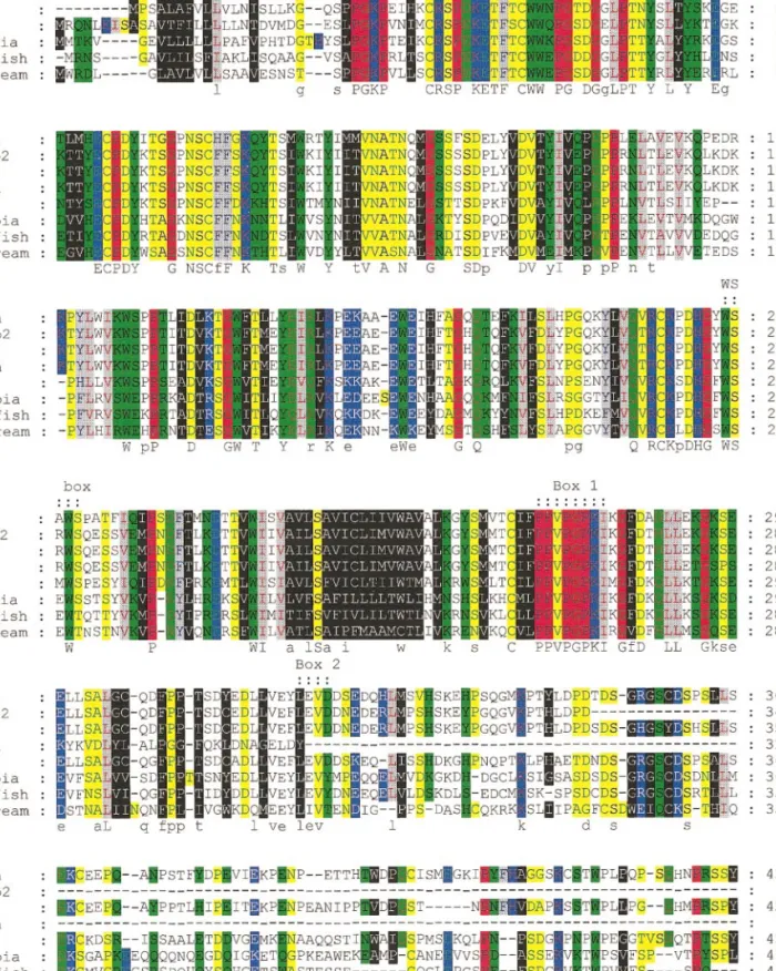
FIG. 2. Multiple sequence alignment of sea bream PRLR (sbPRLR) with PRLR from several other vertebrates. Colored blocks refer to the physicochemical properties of the amino acids according to the key. Short form of rat PRLR, ratsh; intermediate form of rat PRLR, ratnb2; long form of rat PRLR, rat1; newt PRLR, newt; tilapia PRLR, tilapia; goldfish PRLR, goldfish. On the last line of each block, amino acids identical in all species are indicated by capital letters; small letters represent the most common amino acid in that position. WS box, box 1, and box 2 are indicated.
prises 120 nucleotides and the 3⬘UTR comprises 906 nucleotides. Three clear polyadenylation signals are present at positions 1687, 1919, and 2004 and precede the poly(A) tail.
All the characteristic structural features of PRLRs are present (Fig. 1): two pairs of cysteine residues in the ECD at positions 12, 22, 51, and 62, a WS box in which the last serine was conservatively substituted by threonine, the transmembrane domain compris-ing 24 hydrophobic amino acids and box 1, and a proline-rich region (PPVPGPKI) in the transmem-brane proximal to ICD at position 243. Four poten-tial N-linked glycosylation sites were identified in
the ECD. Within the ICD tyrosine residues were found at positions 289, 361, 390, 402, 422, 448, 462, and 507 (Fig. 1).
Multiple sequence alignment of sbPRLR with other previously isolated PRLRs (Fig. 2) shows that the overall identity of sbPRLR with tilapia and gold-fish PRLR is 29 and 32%, respectively, and that with mammalian and amphibian counterparts is 23–27% (Table 1). However, if the physicochemical proper-ties of the amino acids are taken into consideration, receptor sequence conservation is remarkable, not only between fish but also among all vertebrates (Fig. 2).
Partial Genomic Sequence of sbPRLR
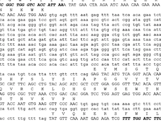
PCR of sea bream genomic DNA using primers PR1 and PR2 yielded a 1000-bp fragment. This fragment contains three exons separated by two introns of 540 and 38 bp (Fig. 3). The intron– exon boundaries follow the gt/ag rule (Mount, 1982) and the organization appears to be conserved compared with mouse PRLR gene (Ormandy et al., 1998).
Expression Analysis of sbPRLR by Northern Blot
Northern blot analysis using 2g of mRNA (Figs.
4A and 4B) demonstrated that PRLR is encoded by two transcripts of 2.8 and 3.2 kb in the intestine, kidney, and gills. In the pituitary and skin a single
transcript of 2.8 kb was observed. When 10 g of
mRNA extracted from spermiating gonads, which contained more than 95% male tissue (Fig. 4C), was
TABLE 1
Amino Acid Identities (%) in PRLRs from Several Species
Human Ratnb2 Rat1 Ratsh Newt Tilapia Goldfish Seabream
Human — 49 66 33 48 35 33 26 Ratnb2 — — 67 68 35 28 28 25 Rat1 — — — 46 46 34 33 26 Ratsh — — — — 25 22 22 23 Newt — — — — — 34 36 27 Tilapia — — — — — — 46 29 Goldfish — — — — — — — 32
Note. Short form of rat PRLR, ratsh; intermediate form of rat PRLR, ratnb2; long form of rat PRLR, rat1; newt PRLR, newt; tilapia PRLR,
tilapia; goldfish PRLR, goldfish.
FIG. 3. Nucleotide sequence and deduced amino acid sequence of the PCR-amplified genomic sequence of sea bream PRLR. The primers used are indicated by boldface/italic. The nucleotides corresponding to coding regions are in capitals and those within the noncoding regions are in lowercase letters.
analyzed, the major transcripts identified were 1.3 and 1.1 kb. The 2.8-kb transcript, which was the principle transcript in larvae, skin, kidney, gill, and pituitary, was present, but at low levels, in gonads and a further transcript of 1.9 kb was observed in gonads. Intestine, gill, kidney, and pituitary had the highest levels of PRLR expression and no signal was detected in the other tissues analyzed using the same amount of mRNA (Fig. 4A).
Tissue Distribution by RT-PCR
RT-PCR with sea bream receptor-specific primers of approximately equivalent quantities of cDNA from kidney, skin, gill, intestine, brain, testis, ovary, liver, skeletal muscle, heart, and bone resulted in detection of transcripts in gill and intestine (Figs. 5A
and 5B). The Southern blot of these reactions also permitted detection of PRLR transcripts in skin, kid-ney, brain, testis, ovary, and liver (Fig. 5C). Al-though no signal was visible in skeletal muscle, heart, and bone after the blot had been exposed for 5 min (Fig. 5C), longer exposure time (2 h) permitted detection of PRLR transcripts in these tissues (Fig. 5D).
Immunohistochemistry of Sea Bream Tissues
Only tissues from juvenile sea bream were exam-ined for PRLR distribution by specific immunohis-tochemistry using antiserum to an oligopeptide of the external domain of the receptor; some of these are illustrated in Fig. 7. The antiserum used for these studies was prepared by conjugation of a spe-cific oligopeptide to bovine thyroglobulin. When tested for the presence of antibodies to thyroglobu-lin, the antiserum produced only a weak reaction with colloid of thyroid follicles in sea bream. This cross-reaction could be eliminated by incubation with bovine thyroglobulin without significant effect on specific reaction with PRLR in other tissues; this effect is illustrated in Figs. 6A– 6D. Figure 6A shows
FIG. 5. (A) Distribution of PRLR in adult tissues by RT-PCR: 1, brain; 2, testis; 3, heart; 4, muscle; 5, bone; 6, kidney; 7, skin; 8, gills; 9, ovary; 10, liver; 11, intestine; 12, PCR negative control. (B) The cDNA used to determine PRLR tissue distribution were also ampli-fied with sea bream-actin primers to evaluate the relative amount of cDNA used in each PCR. (C) Southern blot of the reactions shown in A (5 min exposure). (D) Heart, muscle, and bone samples after 2 h exposure. The sensitivity of RT-PCR coupled with Southern blotting permitted detection of PRLR transcripts in brain, testis, kidney, skin, gills, ovary, liver, and intestine after 5 min exposure and in heart, muscle, and bone after 2 h exposure.
FIG. 4. (A) Northern blot analysis of approximately equivalent quantities of mRNA (2g) from several sea bream tissues. With the exception of sample 2, all are from adult fish: 1, head-kidney; 2, larvae; 3, skin; 4, brain; 5, kidney; 6, muscle; 7, gill; 8, spleen; 9, liver; 10, intestine; 11, testis; 12, ovary; 13, pituitary. Hybridization was carried out with sbPRLR (clone I3A2). (B) Results obtained from the hybridization of the same filter with sea bream-actin. (C) Northern blot analysis of mRNA (10g) extracted from mature (spermiating) male gonads. Hybridization was carried out with sbPRLR (clone I3A2). The arrows on the right side of the filter indicate transcript size.
reaction with unabsorbed antiserum with colloid in thyroid follicles, and in Fig. 6B there is strong reac-tion with the proventricular gland cells on the same section. Figures 6C and 6D show the same areas of tissue as those from the adjacent section but reacted with antiserum absorbed with bovine thyroglobu-lin. The weak reaction in thyroid colloid was ab-sorbed by thyroglobulin but the reaction in the proventricular cells was not reduced.
Figure 7 shows immunoreactions in renal tubules (Fig. 7B) and gut epithelium, with the most intense reaction in the basal glandular region of the proven-triculus (Fig. 7A); epithelia of other regions of the gut contained only low levels of receptor protein. In the gill, the chloride cells of the filament epithelium re-acted strongly (Fig. 7E) but stromal cells of the fibrous cartilage of the gill bars appeared to contain abundant receptor protein (Fig. 7C). The single layer of epithelial cells of the choroid plexus was rich in receptor protein (Fig. 7D) and it is interesting that the epithelial cells of the saccus vasculosus also contained prolactin recep-tors (Fig. 7F).
DISCUSSION
The present study describes the cloning and char-acterization of a PRLR in a marine teleost. Conserva-tion of all the structural and funcConserva-tional features char-acteristic of PRLRs, the physicochemical properties, both in the ECD and in the ICD, and gene organiza-tion, substantiate the identification of this protein as a PRLR and make it improbable that it is a receptor for another member of the growth hormone/PRL gene family.
The tissue distribution of the receptor was deter-mined in adult fish both by Northern blotting, which permitted the identification of size and number of transcripts, and by the more sensitive method of RT-PCR, which allowed detection of PRLR in tissue with low transcript number. Cellular localization of PRLR protein was determined by IHC with a sea bream-specific receptor antiserum. In addition, part of the genomic organization of sbPRLR was elucidated and compared to the PRLR gene of mammals.
PRLR in the sea bream exhibits all the characteristic features of long forms of PRLRs. In common with other species sbPRLR contains a single transmem-brane spanning domain and an ECD and ICD. The ECD comprises 208 amino acids and contains two pairs of cysteines which were identified at positions topologically equivalent to those of the mammalian, amphibian, and other fish PRLR, representing areas of high homology. In mammals these two pairs of cys-teines form ligation pockets specific for each ligand and mutational studies demonstrated that they are important for receptor structure and function (Roza-kis-Adcock and Kelly, 1991, 1992). The similar consti-tution and organization in sbPRLR, as well as in the other fish, suggest a common function. In the WS box of the ECD, there has been a conservative substitution of the last serine by threonine; an identical substitu-tion is present in goldfish (Tse et al., 2000) and in mouse (Davis and Linzer, 1989) serine has been re-placed by glycine. The WS box can be found in all the members of the class 1 superfamily of cytokine tors with the exception of the growth hormone recep-tor (GHR), in which conservative substitutions have occurred (Kelly et al., 1991a). The high degree of con-servation of the WS box suggests that it has an impor-tant role in receptor function, and mutations in this
FIG. 6. Immunohistochemistry of thyroid follicles of sea bream (A and C) and proventricular gland cells (B and D). (A and B) show immunoreaction with unabsorbed antiserum to PRLR peptide anti-body conjugated to bovine thyroglobulin, and (C and D) are adja-cent sections reacted with the same antiserm absorbed with bovine thyroglobulin. Immunoreaction has been abrogated only in thyroid follicle colloid (co) by thyroglobulin absorption (cf. A and C).
FIG. 7. Immunohistochemistry of some tissues from juvenile sea bream using rabbit antiserum to an oligopeptide in the external domain of the prolactin receptor. (A) The proventricular region of the gut showed the strongest reaction of all the gut epithelia; moreover, the basal glandular cells (g) of this region appear to have greater content of receptor protein. There is also immunostaining in the epithelial cells lining the lumen of the gut (lu), but no reaction in the smooth muscle (m). (B) A section through the renal kidney showing immunostaining in epithelial cells of some tubules (t), but no reaction in smaller proximal tubules (nt). The reaction in most epithelial cells is spread throughout the cytoplasm but in others it is concentrated in the basal region of the cell as punctate bodies (p). (C) Fibrous cartilage of the gill bars showing strong reaction in the stromal cells (s). (D) There is a strong prolactin receptor immunoreaction in the single layer of epithelial cells of the choroid (ec) of the midbrain. (E) Section of a gill filament showing prolactin receptors in the chloride cells (c). (F) Epithelial cells of the saccus vasculosus showed two patterns of immunostaining similar to the kidney tubule cells in B. In some cells prolactin receptors appeared to be spread throughout the cytoplasm (vc) but in others there was also a concentration around the nucleus (nc) in the basal portion of the cell adjacent to the capillary (cap).
region are detrimental to receptor affinity (Rozakis-Adcock et al., 1991), suggesting a role in ligand bind-ing. It seems that the WS motif is required for main-tenance of the correct folding of the molecule (Goffin
et al., 1998). In common with mammalian PRLR, the
ECD of sbPRLR contains four potential N-linked gly-cosylation sites, although their relative positions are not conserved. In tiPRLR only two N-linked glycosyl-ation sites were found. The importance of glycosyla-tion sites for adopglycosyla-tion of an active conformaglycosyla-tion of PRLR has been demonstrated in rat (Buteau et al., 1998) but is unknown for other species, including fish. Unlike in avian species, tandem repeats (Chen and Horseman, 1994; Zhou et al., 1996) were not identified within the ECD of sbPRLR. The transmembrane do-main of sbPRLR comprises 24 amino acids and shares the hydrophobic properties described for previously characterized PRLRs. The ICD of sbPRLR comprises 284 amino acids and contains the proline-rich motif (box 1), which shares 100% identity with all PRLRs known so far. This is by far the most highly conserved region within the PRLRs ICD among cytokine recep-tors and it plays an important role in the signal trans-duction pathway (Dinerstein et al., 1995; Pezet et al., 1997). The ICD of different PRLR forms varies in size and several isoforms of PRLR which differ from each other in length and composition have been found in the same species (Boutin et al., 1989, 1988; Shirota et al., 1990). Additional important structural and functional features are also present in sbPRLR ICD and include box 2 and several tyrosine residues. In general sbPRLR resembles the long form of PRLR, although six dele-tions scattered throughout the ICD, which lacks 6 –21 amino acids, make it 62–92 residues shorter than all the other long forms of PRLR so far identified. Most of these deletions are also present in gfPRLR but do not occur in tiPRLR.
Despite the presence of all the characteristic fea-tures of PRLRs, the amino acid sequence of sbPRLR shares a relatively low level of identity with tiPRLR (Sandra et al., 1995) and gfPRLR (Tse et al., 2000). However, sequence identity is higher (46%) in tila-pia and goldfish PRLR. Whether the sequence dif-ferences observed in fish PRLR are important for specific functions of the PRLR and related to differ-ences in the environment that they inhabit remains to be examined; for example, tilapia is euryhaline and goldfish is a freshwater stenohaline species.
Nevertheless, it appears that PRLR are more vari-able (29 – 46% sequence identity) among fishes than among mammals, birds, and amphibia, which share 71– 83% sequence identity. However, since there are many more species of teleost fish than all the rest of the vertebrates, it is too early to make general com-parisons. It is important to note, however, that the motifs known to be essential for the mechanisms of signal transduction are conserved, suggesting that similar mechanisms operate throughout the verte-brates. If the physicochemical properties of the amino acids are taken into consideration, conserva-tion is remarkable, principally in the ECD but also within the ICD. Even though the amino acid com-position of the receptors varies, substitutions, which occurred during evolution, appear to have pre-served the overall physicochemical properties of the receptor. Analysis of a partial sequence of genomic DNA for sbPRLR shows that, within the region analyzed, gene organization also appears to have been conserved and resembles that of the mouse (Ormandy et al., 1998). In particular, the sea bream WS box and transmembrane domain, in common with the situation reported in mouse, are coded by different exons.
Northern blot analysis of several sea bream tis-sues revealed the presence of two differently sized transcripts, which had differential levels of expres-sion in the diverse tissues analyzed. In gills, intes-tine, and kidney, both transcripts could be
ob-served, though the 2.8-kb transcript was
predominant in gills and kidney, whereas the 3.2-kb transcript appears to be more abundant in the intes-tine. In the pituitary and skin, only the smaller transcript was observed. In addition, the gonads contained three further transcripts of 1.9, 1.3, and 1.1 kb, suggesting that more than one form of PRLR is transcribed in the sea bream gonad. It is possible that these three transcripts encode a short form of PRLR. The expression of PRLR in sea bream differs from that reported in tilapia, in which a single tran-script of 3.2 kb was identified, and although more than one transcript of gfPRLR has been identified, they are much larger, 4.6 and 3.5 kb. The existence of alternative forms of PRLR in the sea bream remains to be clarified but expression may be more like that of mouse, in which several transcripts encode the long and short forms of PRLR in a tissue-specific
pattern (Buck et al., 1992) as a consequence of alter-native splicing of the same gene (Ormandy et al., 1998). Moreover, in sea bream, Northern blotting demonstrated that the intestine, gill, kidney, and pituitary are the tissues expressing sbPRLR at the highest levels. PRL has long been recognized as a freshwater-adapting hormone in fish. In fish adapted to hyperosmotic environments, PRL is known to modulate the absorption of NaCl in the gastrointestinal tract (Morley et al., 1981) and in hyposmotic environments, PRL reduces branchial sodium efflux and water permeability of gills, renal tubules, intestine, and urinary bladder; some of
these actions are due to a reduction to Na⫹K⫹
AT-Pase activity (Flick et al., 1994; Shepherd et al., 1997). Adult sea bream spend their life in a marine envi-ronment without a severe hydromineral challenge; nevertheless, the high level of expression of receptor in the osmoregulatory organs suggests that these are important targets for PRL and raises intriguing questions about the function of PRL at these sites in this marine teleost. In tilapia, somewhat paradoxi-cally, there was an increase in PRLR numbers in fish transferred from fresh to brackish water (Auperin et
al., 1995), suggesting that they are prepared for
un-expected changes in external ionic or osmotic con-centrations. The expression of PRLR in skin may suggest a function at the level of hydromineral bal-ance or at the level of fish immunoprotection. Fish skin is a major barrier against external aggressions and pathogens and PRL enhances the production of mucus, which further protects the animal. This mechanism of protection has already been described in other species of fish (Sage, 1970). The presence of PRLR transcripts in the gonads suggests a likely involvement of PRL in the reproductive cycle of this species. Sea bream is a protandrous hermaphrodite (Hape and Zohar, 1988). The mechanisms underly-ing its sex reversal are still poorly understood, al-though it appears that estrogens induce feminiza-tion of the gonads, whereas androgens induce masculinization (Badura and Friedman, 1988; Hape and Zohar, 1988; Condec¸a and Canario, 1999). The involvement of PRL in the production of steroid hormones and the onset of gonadal development has already been reported in cichlid species (Rubin and Specker, 1992; Tan et al., 1988), suggesting that PRL may be involved in the process of sex reversal
through control of steroid production and may also be involved in control of gonad maturation.
The analysis of receptor tissue distribution carried out by RT-PCR supports the results obtained by Northern blotting and IHC, and due to its higher sensitivity, PRLR expression was detected in other tissues, such as the liver, muscle, bone, and brain, suggesting that PRL may also be involved in growth and behavior, other putative functions already de-scribed in cichlid fish (Blum and Fiedler, 1965; Lam and Hoar, 1967; Shepherd et al., 1997).
Immunohistochemistry of PRLR in tissues of sea bream using the antibody to an oligopeptide of the external domain of the receptor showed specific reac-tion in several tissues. Reacreac-tion with thyroglobulin, the carrier protein of the peptide immunogen, was shown to be weak and occurred only with the thyro-globulin-rich colloid within the thyroid follicles.
Distribution of specific PRLR was noted in tissues concerned with control of water and ion balance, in-cluding gills, intestine, and renal tubule epithelium. Although PRL is associated with survival of euryha-line fish in water of low osmotic potential, principally by inhibition of sodium loss, PRL receptors have also been shown to increase in gills of Oreochromis niloticus when transferred from fresh water to brackish water (Auperin et al., 1995). The presence of PRLR in these epithelial cells may suggest roles for PRL other than ion and water regulation since PRL influences the cell cycle, through inhibition of apoptosis and differentia-tion (Clarke and Bern, 1980; Hirano, 1986). The pres-ence of PRLR in the saccus vasculosus and in the choroid plexus may be related to the function of these epithelia in monitoring the composition of cerebro-spinal fluid in relation to plasma composition. PRLR have been described in the choroid plexus epithelia of other vertebrates (Posner et al., 1983; Buntin and Walsh, 1988; Muccioli et al., 1988) and their presence in fish suggests that they are an earlier evolutionary development. PRLR also occur in mesodermally de-rived connective tissues, having been described in mammalian chondrocytes of developing cartilage and mesenchymal precartilage (Freemark et al., 1997), and their recent detection in osteoblasts (Clement-Lacroix
et al., 1999) shows that they do have a role(s) in skeletal
tissues. Only further physiological studies will deter-mine the functions of PRL through receptor binding in these various tissues.
REFERENCES
Ali, S., Pellegrini, I., and Kelly, P. A. (1991). A prolactin-dependent immune cell line (Nb2) expresses a mutant form of prolactin receptor. J. Biol. Chem. 266, 20110 –20117.
Auperin, B., Rentier-Delrue, F., Martial, J. A., and Prunet, P. (1994). Characterization of a single prolactin (PRL) receptor in tilapia (Oreochromis niloticus) which binds both PRLI and PRLII. J. Mol.
Endocrinol. 13, 241–251.
Auperin, B., Rentier-Delrue, F., Martial, J. A., and Prunet, P. (1995). Regulation of gill prolactin receptors in tilapia (Oreochromis
nil-oticus) after a change in salinity or hypophysectomy. J. Endocrinol.
145,213–220.
Aviv, H., and Leder, P. (1972). Purification of biologically active globin messenger RNA by chromatography on oligothymidylic acid– cellulose. Proc. Natl. Acad. Sci. USA 69, 1408 –1412. Badura, L. L., and Friedman, H. (1988). Sex reversal in female Betta
splendens as a function of testosterone manipulation and social
influence. J. Comp. Psychol. 102, 262–268.
Bern, H. A. (1975). Prolactin osmoregulation. Am. Zool. 15, 937–948. Bignon, C., Binart, N., Ormandy, C., Schuler, L. A., Kelly, P. A., and Djiane, J. (1997). Long and short forms of the ovine prolactin receptor: cDNA cloning and genomic analysis reveal that the two forms arise by different alternative splicing mechanisms in rumi-nants and in rodents. J. Mol. Endocrinol. 19, 109 –120.
Blum, V., and Fiedler, K. (1965). Hormonal control of reproductive behavior in some cichlid fish. Gen. Comp. Endocrinol. 5, 186 –196. Bole-Feysot, C., Goffin, V., Edery, M., Binart, N., and Kelly, P. A. (1998). Prolactin (PRL) and its receptor: Actions, signal transduc-tion pathways and phenotypes observed in PRL receptor knock-out mice. Endocr. Rev. 19, 225–268.
Boutin, J. M., Edery, M., Shirota, M., Jolicoeur, C., Lesueur, L., Ali, S., Gould, D., Djiane, J., and Kelly, P. A. (1989). Identification of a cDNA encoding a long form of prolactin receptor in human hepatoma and breast cancer cells. Mol. Endocrinol. 3, 1455–1461. Boutin, J. M., Jolicoeur, C., Okamura, H., Gagnon, J., Edery, M.,
Shirota, M., Banville, D., Dusanter-Fourt, I., Djiane, J., and Kelly, P. A. (1988). Cloning and expression of the rat prolactin receptor, a member of the growth hormone/prolactin receptor gene family.
Cell 53, 69 –77.
Buck, K., Vanek, M., Groner, B., and Ball, R. K. (1992). Multiple forms of prolactin receptor messenger ribonucleic acid are specif-ically expressed and regulated in murine tissues and the mam-mary cell line HC11. Endocrinology 130, 1108 –1114.
Buntin, J. D., and Walsh, R. J. (1988). In vivo autoradiographic analysis of prolactin binding in brain and choroid plexus of the domestic ring dove. Cell Tissue Res. 251, 105–109.
Buteau, H., Pezet, A., Ferrag, F., Perrot-Applanat, M., Kelly, P. A., and Edery, M. (1998). N-glycosylation of the prolactin receptor is not required for activation of gene transcription but is crucial for its cell surface targeting. Mol. Endocrinol. 12, 544 –555.
Clarke, W. C., and Bern, H. A. (1980). Comparative endocrinology of prolactin. In “Hormonal Proteins and Peptides” (C. H. Li, Ed.), Vol. VIII, pp. 105–197. Academic Press, New York.
Chen, X., and Horseman, N. D. (1994). Cloning, expression, and mutational analysis of the pigeon prolactin receptor.
Endocrinol-ogy 135, 269 –276.
Clement-Lacroix, P., Ormandy, C., Lepescheux, L., Ammann, P., Damotte, D., Goffin, V., Bouchard, B., Amling, M., Gaillard-Kelly, M., Binart, N., Baron, R., and Kelly, P. A. (1999). Osteoblasts are a new target for prolactin: Analysis of bone formation in prolactin receptor knockout mice. Endocrinology 140, 96 –105.
Condec¸a, J. B., and Canario, A. V. M. (1999). The effect of estrogen on the gonads and on in vitro conversion of androstenione to testosterone, 11-ketotestosterone, and estradiol-17 in Sparus
au-rata (Teleostei, Sparidae). Gen. Comp. Endocrinol. 116, 59 –72.
Das, R., and Vonderhaar, B. K. (1995). Transduction of prolactin’s (PRL) growth signal through both long and short forms of the PRL receptor. Mol. Endocrinol. 9, 1750 –1759.
Davis, J. A., and Linzer, D. I. (1989). Expression of multiple forms of the prolactin receptor in mouse liver. Mol. Endocrinol. 3, 674 – 680. Di Carlo, R., Muccioli, G., Papotti, M., and Bussolati, G. (1992). Characterization of prolactin receptor in human brain and cho-roid plexus. Brain Res. 570, 341–346.
Dinerstein, H., Lago, F., Goujon, L., Ferrag, F., Esposito, N., Finidori, J., Kelly, P. A., and Postel-Vinay, M. C. (1995). The proline-rich region of the GH receptor is essential for JAK2 phosphorylation, activation of cell proliferation, and gene transcription. Mol.
Endo-crinol. 9, 1701–1707.
Dusanter-Fourt, I., Gaye, P., Belair, L., Petridou, B., Kelly, P. A., and Djiane, J. (1991). Prolactin receptor gene expression in the rabbit: Identification, characterization and tissue distribution of several prolactin receptor messenger RNAs encoding a unique precursor.
Mol. Cell. Endocrinol. 77, 181–192.
Dusanter-Fourt, I., Kelly, P. A., and Djiane, J. (1987). Immunological recognition of the prolactin receptor: Identification of a single binding unit of molecular weight approximately 42,000. Biochimie
69,639 – 646.
Edery, M., Jolicoeur, C., Levi-Meyrueis, C., Dusanter-Fourt, I., Petri-dou, B., Boutin, J. M., Lesueur, L., Kelly, P. A., and Djiane, J. (1989). Identification and sequence analysis of a second form of prolactin receptor by molecular cloning of complementary DNA from rabbit mammary gland. Proc. Natl. Acad. Sci. USA 86, 2112–2116. Finidori, J., and Kelly, P. A. (1995). Cytokine receptor signalling
through two novel families of transducer molecules: Janus ki-nases, and signal transducers and activators of transcription. J.
Endocrinol. 147, 11–23.
Freemark, M., Driscoll, P., Maaskant, R., Petryk, A., and Kelly, P. A. (1997). Ontogenesis of prolactin receptors in the human fetus in early gestation—Implications for tissue differentiation and devel-opment. J. Clin. Invest. 99, 1107–1117.
Flick, G., Rentier-Delrue, F., and Wendelaar Bonga, S. E. (1994). Calcitropic effects of recombinant prolactins in Oreochromis
mossambicus. Am. J. Physiol. 15, R1302–R1308.
Goffin, V., Bouchard, B., Ormandy, C. J., Weimann, E., Ferrag, F., Touraine, P., Bole-Feysot, C., Maaskant, R. A., Clement-Lacroix, P., Edery, M., Binart, N., and Kelly, P. A. (1998). Prolactin: A hormone at the crossroads of neuroimmunoendocrinology. Ann.
N. Y. Acad. Sci. 840, 498 –509.
Goupille, O., Daniel, N., Bignon, C., Jolivet, G., and Djiane, J. (1997). Prolactin signal transduction to milk protein genes: Carboxy-terminal part of the prolactin receptor and its tyrosine phosphor-ylation are not obligatory for JAK2 and STAT5 activation. Mol.
Cell. Endocrinol. 127, 155–169.
Happe, A., and Zohar, Y. (1988). Self-fertilization in the protandrous hermaphrodite Sparus aurata: Development of the technology. In “Reproduction in Fish—Basic and Applied Aspects in Endocri-nology and Genetics” (Y. Zohar and B. Breton, Eds.), pp. 177–180. Les Colloques de l’INRA, No. 44. INRA, Paris.
Hirano, T. (1986). The spectrum of prolactin action in teleosts. In “Comparative Endocrinology: Developments and Directions” (C. L. Ralph, Ed.), pp. 53–74. A. R. Liss, New York.
Kelly, P. A., Ali, S., Rozakis, M., Goujon, L., Nagano, M., Pellegrini, I., Gould, D., Djiane, J., Edery, M., Finidori, J., et al. (1993). The growth hormone/prolactin receptor family. Recent Prog. Horm.
Res. 48, 123–164.
Kelly, P. A., Djiane, J., Banville, D., Ali, S., Edery, M., and Rozakis, M. (1991a). The growth hormone/prolactin receptor gene family.
Oxf. Surv. Eukaryot. Genes 7, 29 –50.
Kelly, P. A., Djiane, J., Postel-Vinay, M. C., and Edery, M. (1991b). The prolactin/growth hormone receptor family. Endocr. Ver. 12, 235–251.
Kraicer, J., Herlant, M., and Duclos, P. (1967). Changes in adenohy-pophyseal cytology and nucleic acid content in the rat 32 days after adrenalectomy and the chronic injection of cortisol. Can.
J. Pharmacol. Physiol. 45, 947–956.
Lam, T., and Hoar, W. (1967). Seasonal effects of prolactin on fresh-water osmoregulation of the marine form (trachurus) of the stickleback Gasterosteus aculeatus. Can. J. Zool. 45, 509 –516. Moore, R. C., and Oka, T. (1993). Cloning and sequencing of the
cDNA encoding the murine mammary gland long-form prolactin receptor. Gene 134, 263–265.
Morley, M., Chadwick, A., and El Tounsy, E. M. (1981). The effect of prolactin on water absorption by the intestine of the trout (Salmo
gairdneri). Gen. Comp. Endocrinol. 44, 64 – 68.
Mount, S. M. (1982). A catalogue of splice junction sequences.
Nu-cleic Acids Res. 10, 459 – 472.
Muccioloi, G., Bellussi, G., Ghe, C., Pagnini, G., and Di Carlo, R. (1988). Regional distribution and species variations of prolactin binding-sites in the brain. Gen. Comp. Endocrinol. 69, 399 – 405. Nevalainen, M. T., Valve, E. M., Ingleton, P. M., and Ha¨rko¨nen, P. L.
(1996). Expression and hormone regulation of prolactin receptors in rat dorsal and lateral prostate in organ culture. Endocrinology
137,3078 –3088.
Ormandy, C. J., Binart, N., Helloco, C., and Kelly, P. A. (1998). Mouse prolactin receptor gene: Genomic organization reveals alternative promoter usage and generation of isoforms via alter-native 3⬘-exon splicing. DNA Cell. Biol. 17, 761–770.
Pezet, A., Buteau, H., Kelly, P. A., and Edery, M. (1997). The last proline of Box 1 is essential for association with JAK2 and func-tional activation of the prolactin receptor. Mol. Cell. Endocrinol.
129,199 –208.
Posner, B. I., van Houten, M., Patel, B., and Walsh, R. J. (1983). Characterisation of lactogen binding sites in choroid plexus. Exp.
Brain Res. 49, 300 –306.
Rozakis-Adcock, M., and Kelly, P. A. (1991). Mutational analysis of the ligand-binding domain of the prolactin receptor. J. Biol. Chem.
266,16472–16477.
Rozakis-Adcock, M., and Kelly, P. A. (1992). Identification of ligand binding determinants of the prolactin receptor. J. Biol. Chem. 267, 7428 –7433.
Rubin, D. A., and Specker, J. L. (1992). In vitro effects of homologous prolactins on testosterone production by testes of tilapia
(Oreo-chromis mossambicus). Gen. Comp. Endocrinol. 87, 189 –196.
Sage, M. (1970). Control of prolactin release and its role in color change in the teleost Gillichthys mirabilis. J. Exp. Zool. 173, 121–127. Sandra, O., Le Rouzic, P., Cauty, C., Edery, M., and Prunet, P. (2000). Expression of the prolactin receptor (tiPRL-R) gene in tilapia
Oreochromis niloticus: Tissue distribution and cellular localization
in osmoregulatory organs. J. Mol. Endocrinol. 24, 215–224. Sandra, O., Sohm, F., de Luze, A., Prunet, P., Edery, M., and Kelly,
P. A. (1995). Expression cloning of a cDNA encoding a fish prolactin receptor. Proc. Natl. Acad. Sci. USA 92, 6037– 6041. Sanger, F., Nicklen, S., and Coulson, A. R. (1977). DNA sequencing
with chain-terminating inhibitors. Proc. Natl. Acad. Sci. USA 74, 5463–5467.
Santos, C. R. A., Power, D. M., Kille, P., Llwellyn, L., Ramsurn, V., Wigham, T., and Sweeney, G. E. (1997). Cloning and sequencing of a full length sea bream (Sparus aurata) -actin cDNA. Comp.
Biochem. Physiol. B 117, 185–189.
Santos, C. R. A., Brinca, L., Ingleton, P. M., and Power, D. M. (1999). Cloning, expression, and tissue localization of prolactin in adult sea bream (Sparus aurata). Gen. Comp. Endocrinol. 114, 57– 66. Shepherd, B. S., Sakamoto, T., Nishioka, R. S., Richman, N. H., 3rd,
Mori, I., Madsen, S. S., Chen, T. T., Hirano, T., Bern, H. A., and Grau, E. G. (1997). Somatotropic actions of the homologous growth hormone and prolactins in the euryhaline teleost, the tilapia, Oreochromis mossambicus. Proc. Natl. Acad. Sci. USA 94, 2068 –2072.
Shirota, M., Banville, D., Ali, S., Jolicoeur, C., Boutin, J. M., Edery, M., Djiane, J., and Kelly, P. A. (1990). Expression of two forms of prolactin receptor in rat ovary and liver. Mol. Endocrinol. 4, 1136 – 1143.
Sternberger, L. A. (1974). “Immunocytochemistry.” Prentice-Hall, Englewood Cliffs, NJ.
Tan, C., Wong, L., Pang, M., and Lam, T. (1988). Tilapia prolactin stimulates estradiol-17b synthesis in vitro in vitellogenic oocytes of the guppy Poecilia reticulata. J. Exp. Zool. 248, 361–364. Tse, D. L. Y., Chow, B. K. C., Chan, C. B., Lee, L. T. O., and Cheng,
C. H. K. (2000). Molecular cloning and expression studies of a prolactin receptor in goldfish (Carassius auratus). Life Sci. 66, 593–605. Yamamoto, T., Nakayama, Y., Matsuda, Y., and Abe´, S-I. (1998). Cloning and expression of a cDNA encoding a prolactin receptor of the japanese red-bellied newt, Cynops pyrrhogaster. Zool. Sci. 15, 741–747.
Zhou, J. F., Zadworny, D., Guemene, D., and Kuhnlein, U. (1996). Molecular cloning, tissue distribution, and expression of the pro-lactin receptor during various reproductive states in Meleagris