UNIVERSIDADE DA BEIRA INTERIOR
Ciências da Saúde
Establishment of a method to evaluate the
plasticity and maturation of the dopaminergic
nerve terminal
Diogo António Bessa Neto
Dissertação para obtenção do Grau de Mestre em
Ciências Biomédicas
(2º ciclo de estudos)
Orientador: Prof.ª Doutora Graça Maria Fernandes Baltazar Coorientador: Prof. Doutor Ramiro Daniel Carvalho de Almeida
Dedicatória
Numa outra situação nunca escreveria esta secção pois a todos aqueles que acreditaram em mim eu dedico esta dissertação que representa um pequena amostra de tudo o que passei neste ano. Contudo, durante este ano houve uma situação particular que me marcou de uma forma especial.
Desde criança que estava habituado à tua presença. Sempre me deste carinho e sempre estiveste pronto a me ajudar. Lembro-me perfeitamente toda a ajuda que me davas na escola primária com os trabalhos escolares, fora os exercícios extras que me davas para que eu fosse sempre melhor e melhor. Ajudaste-me muito nas matemáticas e o gosto que criei por elas foi graças a ti. O tempo passou, envelhecemos e sempre estiveste lá a dar-me apoio até que há cinco anos candidatei-me ao Ensino Superior e acabei por entrar na cidade da neve. Uma cidade nova, um ambiente novo, sem dúvida um mundo completamente novo para mim, e mais uma vez, pude contar com o teu apoio logo desde o primeiro dia.
Nunca escondeste o quanto gostaste de eu ter conseguido entrar na Universidade e sei perfeitamente que uma das coisas que mais querias era conseguires ver-me a terminar os estudos e conseguir um emprego a fazer algo que eu realmente gostasse. Então praticamente três anos se passaram e o tão esperado dia chegou. Era o dia da minha bênção e mais uma vez não te importaste de fazer algo como 300 km para estares presente num dia tão importante para mim enquanto estudante, ainda que na realidade viesses só para me ver a abanar uma pasta cheia de fitas coloridas.
Porém, ainda não foi dessa que me viste a terminar os estudos. Logo a seguir entrei em mestrado e uma vez mais sei que ficaste contente por ter conseguido alcançar algo ainda maior e gratificante. Sei que uma vez mais adorarias ver-me a terminar esta nova etapa, contudo não fui rápido o suficiente e o tempo foi ainda mais ingrato contigo. Ainda há umas semanas quando consegui ter tempo para ir aí a casa, estive com a avó e estávamos a falar desta minha etapa estar a terminar, pelo que ela diz-me que algo que tu querias mesmo ver era eu a terminar esta etapa. Assim, uma vez que não vais conseguir estar pessoalmente, a ti te dedico em especial esta minha etapa.
Desculpa não ter conseguido chegar a tempo, desculpa não terminar a tempo, desculpa por não teres conseguido ver-me a terminar os estudos, desculpa avô… mas isso nunca terias conseguido, eu não nasci para parar de estudar. Esta é a vida que escolhi para mim, ainda que de uma ou outra maneira as coisas mudem, não tenciono parar de estudar.
Agradecimentos
Como não podia deixar de ser, em primeiro lugar agradeço aos meus pais por toda a ajuda e força que sempre me deram e por me terem proporcionado tal experiência, não podendo esquecer nunca o apoio e afeto dos meus irmãos.
Agradeço também a todos aqueles que na minha família estiveram sempre prontos a ajudar-me e que sempre acreditaram em mim.
Um profundo agradecimento à Doutora Graça Baltazar e ao Doutor Ramiro de Almeida por me terem aceite como orientando e me proporcionarem trabalhar na área que realmente me apaixona, as neurociências. Agradeço-vos ainda por me terem proporcionado condições para que nada me faltasse dentro dos possíveis.
Um muito obrigado a vós, Filipa Campos, Julieta Oliveira, Mónica Sardinha e Rita Videira, por todo o apoio e por me terem conseguido aturar durante todo este tempo. Foi sem dúvida, muito agradável ter-vos como colegas e amigos de trabalho.
Rita Videira, um especial obrigado a ti por toda a ajuda dada ao longo deste ano. Tornaste a minha primeira passagem no mundo da investigação muito mais fácil.
Um grande agradecimento ao David Oppolzer por toda a ajuda que me deu na parte da cromatografia.
Agradeço também a todos os restantes colegas de laboratório pela ajuda prestada e terem animado o ambiente de laboratório nos dias menos bons.
Por fim, mas não menos importante, agradeço a todos os meus amigos que sempre estiveram lá para me apoiar e me ajudar, bem como por me terem proporcionado momentos de descontração. Obrigado por me aturarem, ainda que isso possa não ser fácil.
Resumo Alargado
O sistema dopamina (DA)érgico do mesencéfalo é composto por três principais grupos de neurónios DAérgicos: substantia nigra, área tegmental ventral e núcleos accumbens. Ainda que apenas uma pequena parte do nosso cérebro, este sistema encontra-se envolvido em algumas funções importantes no nosso dia-a-dia tal como é o caso do controlo voluntário dos movimentos, excitação, aprendizagem e estado psicológico. Além disso, este encontra-se diretamente associado com algumas das neuropatologias mais estudadas atualmente tal como a doença de Parkinson, a doença de Huntington e a esquizofrenia, tendo mais recentemente sido associado à dependência das drogas de abuso. Assim, devido às funções mediadas e também à sua associação com as patologias mencionadas, o sistema DAérgico tornou-se um alvo de grande interesse para as neurociências.
A transmissão de informação entre neurónios dá-se principalmente através da formação de sinapses químicas. Estas sinapses são geralmente descritas como duas regiões especializadas na transmissão e receção de informação, respetivamente, a membrana pré- e pós-sináptica, separadas por um micro espaço designado de fenda sináptica onde ocorre a transmissão da informação. A transmissão nestas sinapses é comummente descrita como sendo direta, ou seja, encontra-se limitada à região da fenda sináptica. Contudo, alguns estudos têm tentado provar que tal tipo de transmissão não é totalmente verdade para as sinapses DAérgicas. No entanto, este tipo de sinapses é ainda relativamente pouco estudada e muitas questões mantém-se por esclarecer.
Uma metodologia bastante usada para se estudar os neurónios DAérgicos é através de culturas embrionárias do mesencéfalo ventral, contudo a percentagem de células positivas para o marcador DAérgico tirosina hidroxilase (TH) neste tipo de culturas é muito baixo, dificultando grande parte dos estudos deste tipo de células. Assim neste trabalho testámos algumas condições que foram já demonstradas como tendo a capacidade de melhorar a sobrevivência deste tipo de neurónios. Nas nossas culturas, as três condições testadas aumentaram individualmente em aproximadamente 70% a sobrevivência de células positivas para a TH em relação ao controlo. Não havendo uma condição que resultasse em melhores resultados relativamente às restantes, escolhemos a suplementação da cultura pelo fator neurotrófico derivado de uma linha de células da glial (GDNF) para realizar o nosso trabalho por questões de facilidade na utilização e rentabilização de tempo. Por outro lado, os estudos encontrados em culturas de mesencéfalo ventral recorreram a técnicas com algumas limitações para o estudo de terminais axonais e a formação sináptica nessa região. Assim, o intuito deste trabalho consistiu na tentativa de implementação de um novo método, câmaras microfluídicas, para se estudar este tipo de estruturas. Uma metodologia que já foi
implementada com sucesso a vários tipos de cultura como neurónios hipocampais, corticais e dos gânglios da raiz dorsal. Estas câmaras microfluídicas consistem num polímero biocompatível com ranhuras impressas, designado geralmente por PDMS, fixado contra uma lamela de vidro, da qual resultam dois compartimentos interligados por um conjunto de microcanais. Para atingir o fim aqui proposto, vários parâmetros foram estudados: tempo em cultura, densidade celular e comprimento dos microcanais das câmaras. Verificámos que neste tipo de sistema as células se mantiveram viáveis até ao 14º dia em cultura. Contudo, em termos de isolamento axonal DAérgico nenhuma das condições testadas foi capaz de produzir condições favoráveis para a implementação do método. No entanto, é necessário destacar que os resultados podem também ter sido afetados por problemas mais abrangentes que ocorreram nas culturas celulares e que certamente comprometeram os resultados. Em paralelo com o trabalho acima descrito, também a otimização da quantificação de neurotransmissores e seus metabolitos em lisados de culturas do mesencéfalo ventral foi realizado. Nós verificamos que nas nossas culturas, dos compostos testados, apenas a DA foi detetada por cromatografia líquida de alta-performance (HPLC). Além disso, quando estimuladas com a toxina DAérgica MPP+, os níveis intercelulares de DA diminuíram
abruptamente. Enquanto recorrendo à marcação contra a TH, nenhuma diferença foi verificada. Assim, a quantificação dos níveis de DA aparenta ser uma técnica muito mais sensível para avaliar o efeito de uma lesão em culturas embrionários do mesencéfalo ventral do que a marcação contra a TH por imunocitoquímica.
Palavras-chave
dopamina (DA), mesencéfalo, sinapse, câmaras microfluídicas, cromatografia líquida de alta performance (HPLC)
Abstract
The modulatory midbrain dopamine (DA)ergic system is involved in important functions such as control of voluntary movement, reinforcement, learning and state of mind. Moreover, it is directly associated with some of most studied neuropathologies like Parkinson’s disease, Huntington’s disease and schizophrenia, and also drug addiction. Together, these functions/associations make the DAergic system an appealing field of study in neuroscience. Neurotransmission occurs predominantly through chemical synapses and is already well studied for glutamatergic hippocampal neurons. Nevertheless, DAergic transmission is still poorly investigated and many questions remain about how DA is transmitted. Embrionary ventral midbrain cultures are poor in DAergic neurons and current culture techniques inadequate to conduct axonal studies in these impoverished cultures. Thus, we tried to implement a microfluidic culture device that allows the physical and fluidic axonal isolation from somatodendritic ‘contamination’, with the help of glial cell line-derived neurotrophic factor supplementation to improve the DAergic survival. We showed that cells were maintained viable in microfluidic chambers for at least 14 days in culture, however, the number of DAergic axons was still low, not only due to the culture method limitations but also due to more broad problems associated with the cell cultures which occurred at same time. In addition, we optimized the detection of monoamine neurotransmitters and their metabolites present in embrionary ventral midbrain cultures by high-performance liquid chromatography coupled to electrochemical detection. In our cultures, only DA was detected,
and was drastically reduced when cells were stimulated by the DAergic toxin 1-methyl-4-phenylpyridinium (MPP+). While the number of labelled cells for tyrosine
hydroxylase was not affected by MPP+, at the concentrations tested.
Keywords
dopamine (DA), midbrain, synapses, microfluidic chambers, high-performance liquid chromatography (HPLC)
Index
Chapter 1: Introduction ... 1
1. Dopaminergic System ... 1
1.1. Midbrain Dopaminergic System ... 3
Midbrain Dopaminergic Pathways ... 4
1.1.1. 2. From Synapse to Neurotransmitter Release ... 5
2.1. The Classic Chemical Synapse ... 5
Synaptic Vesicle Pools ... 6
2.1.1. Neurotransmitter Release ... 7
2.1.2. Presynaptic Active Zone ... 7
2.1.2.1. Neurotransmitter Release by Exocytosis ... 8
2.1.2.2. Synaptic Vesicle Recycling ... 9
2.1.2.3. 2.2. Strategies to Study Axonal Terminals ... 10
3. Dopaminergic Synapsis ... 12
3.1. The Dopamine Cycle ... 12
3.2. Ultrastructure of Dopaminergic Presynaptic Structures ... 13
3.3. Axonal Dopamine Release ... 14
Chapter 2: Aims ... 17
Chapter 3: Methods ... 19
1. Animals ... 19
2.1. Collection of the Embryos ... 19
2.2. Dissection of Ventral Midbrain ... 19
2.3. Preparation of Single Cell Suspension ... 19
3. Culture of Midbrain Cells (Conventional Culture) ... 20
4. Mesencephalic Microfluidic Cultures ... 20
4.1. Microfluidic Chamber Preparation ... 20
4.2. Culture of Midbrain Neurons in Microfluidic Chambers ... 21
5. In Vitro Dopaminergic Viability ... 23
5.1. 1-Methyl-4-phenylpyridinium Stimulus ... 23
5.2. Preparation of Cell Extracts for High-Performance Liquid Chromatography Analysis 23 5.3. Dopamine Measurement ... 23
5.4. High-Performance Liquid Chromatography Analysis ... 24
6. Immunocytochemistry ... 24
6.1. Conventional Cultures ... 24
6.2. Microfluidic Cultures ... 24
6.3. Vesicular Monoaminergic Transporter-2 Labelling ... 25
7. Quantification of Dopaminergic Cells ... 25
7.1. Conventional Cultures ... 25
7.2. Microfluidic Chamber Cultures ... 25
7.3. Determination of Dopaminergic Markers Specificity ... 26
1. Part I: Microfluidic Chambers ... 27
1.1. Optimizing the Survival of Dopaminergic Neurons in Culture ... 27
1.2. Optimization of Microfluidic Chambers to Study Dopaminergic Presynaptic Terminals ... 29
1.3. Analysis of Dopaminergic Markers ... 30
2. Part II: Evaluation of Dopamine Levels by High-Performance Liquid Chromatography 32 2.1. Optimization of Mobile Phase ... 32
2.2. Preparation of Cell Extracts ... 34
2.3. High-Performance Liquid Chromatography Analysis versus Immunocytochemistry Analysis ... 35
Chapter 5: Discussion ... 37
Chapter 6: Conclusions and Future Perspectives ... 41
List of Figures
Figure 1 – Scheme with the process of dopamine synthesis and of uptake to the synaptic vesicles.
Figure 2 – Distribution of dopaminergic cell groups in the developing and adult rodent brain. Figure 3 – Dopaminergic projections to the forebrain.
Figure 4 – Synaptic vesicle cycle.
Figure 5 – Organization of synaptic vesicle pool.
Figure 6 – Schematic synaptic vesicle exo-endocytosis coupling. Figure 7 – Cellular culture techniques for the study of synapses. Figure 8 – Schematic representation of dopamine cycle.
Figure 9 – Two hypotheses for the dopaminergic synaptic transmission. Figure 10 – The microfluidic chamber directs axonal growth.
Figure 11 – Improvement of dopaminergic survival by laminin-coating and/or glial cell line-derived neurotrophic factor-supplementation.
Figure 12 – Improvement of dopaminergic conditions on microfluidic chambers. Figure 13 – Ventral midbrain cultures grown in microfluidic chambers.
Figure 14 – Co-labelling of dopaminergic markers in ventral midbrain cultures.
Figure 16 – Representative chromatogram showing the peak of standards correspondent to 10 ng/mL.
Figure 17 – Influence of oxidation potential in the signal of standards. Figure 18 - Detection of dopamine in cell lysate.
List of Tables
Table 1 – Culture conditions tested in microfluidic chambers in an attempt to implement the method in embrionary ventral midbrain dopaminergic cultures.
Table 2 - Description of primary antibodies used in immunocytochemistry assays.
Table 3 - High-performance liquid chromatography coupled to electrochemical detection versus immunocytochemistry analysis.
List of Acronyms
5-HIAA 5-hydoxyindoleatic
AADC Aromatic L-amino acid decarboxylase
ADP Adenosine diphosphate
ATP Adenosine triphosphate
BSA Bovine serum albumin
cAMP Cyclic adenosine monophosphate
CAZ Cytoplasmic matrix at the active zone
CNS Central nervous system
DA Dopamine
DAT Dopamine transporter
DIV Days in vitro
DOPAC 3,4-dihydroxyphenylacetic acid
ECD Electrochemical detection
FBS Fetal bovine serum
GDNF Glial cell-derived neurotrophic factor
HI Heat-inactivated
HPLC High-performance liquid chromatography
HVA Homovanillic acid
L-DOPA 3,4-dihydroxy-L-phenylalanine
MAO Monoamine oxidase
MAP2 Microtubule-associated protein-2
MPP+ 1-methyl-4-phenylpyridinium
PBS Phosphate buffer saline
Pi Phosphate
PDL Poly-D-lysine
PDMS Poly(dimethylsiloxane)
PFA Paraformaldehyde
RIM Rab3-interacting molecule
RIM-BP Rab3-interacting molecule binding proteins
RRP Readily releasable pool
RT Room temperature
SN Substantia nigra
SNc Substantia nigra pars compacta
SNr Substantia nigra pars reticulata
SV Synaptic Vesicle
TH Tyrosine hidroxylase V-ATPase Vacuolar-type H+-ATPase
VGCC Voltage-gated Ca2+-channel
VMAT2 Vesicular monoaminergic transporter-2
Chapter 1: Introduction
1. Dopaminergic System
The dopamine (DA)ergic system consists of a small group of neurons, less than 1% of the 86 billion neurons that make up the human brain (Azevedo et al., 2009, Arias-Carrion and Poppel, 2007). Although a small group, DAergic neurons have been associated to functions as diverse as, voluntary movement, feeding, attention, learning and motivation (Marinelli and McCutcheon, 2014, Yetnikoff et al., 2014, Baik, 2013, Tritsch and Sabatini, 2012, Schultz, 2007).
The main mechanism by which neurons communicate to each other is chemical transmission; a process characterized by the release of endogenous molecules named neurotransmitters. The main neurotransmitter synthesized and released by DAergic neurons is the small signalling molecule DA.
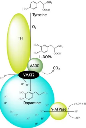
The monoamine DA is normally synthetized in a sequential reaction that starts with hydroxylation of the amino acid L-tyrosine into 3,4-dihydroxy-L-phenylalanine (L-DOPA) catalysed by cytosolic enzyme tyrosine hydroxylase (TH) (Nagatsu et al., 1964), the rate-limiting step on DA synthesis, followed the decarboxylation of L-DOPA into DA by the cytosolic enzyme aromatic L-amino acid decarboxylase (AADC) [(Christenson et al., 1972), reviewed by (Holtz, 1959)]. After synthesis, DA is transported from the cytosol into synaptic vesicles via vesicular monoaminergic transporter-2 (VMAT2). The uptake of a single molecule of DA from cytosol by VMAT2 is coupled to the release of two protons, and the gradient is maintained through the hydrolysis of adenosine triphosphate into adenosine diphosphate and phosphate by the action of vacuolar-type H+-ATPase present in membrane of synaptic vesicles
(SVs). This reaction of vacuolar-type H+-ATPase promotes the translocation of one proton into
the SV (Guillot and Miller, 2009, Chaudhry et al., 2008, Edwards, 2007). Recently, Cartier and colleagues (2010) demonstrated that TH and AADC are associated with VMAT2-containg SVs, supporting a coupling between DA synthesis and transport into monoaminergic SVs, figure 1. After the uptake of DA by VMAT2 to SVs, DA remains inside the vesicles along the axon terminals until it is released to extracellular space (Beckstead et al., 2004). In the extracellular space, DA can act on receptors of target neurons and/or be removed from the synapses, mainly, by the DA transporter (DAT), a crucial regulator of presynaptic DA homeostasis. This reuptake of the DA by the DAT back into the nerve terminals provides a mechanism to refill the SVs in a synthesis-independent manner (Pereira and Sulzer, 2012, Gainetdinov and Caron, 2003).
Over the past 40 years, the interest in research of the DAergic system has greatly increased among the scientific community. Much of this increased interest is due to the dysregulation of DA transmission on midbrain DAergic system reported in some neuropathologies, such as Parkinson’s disease or schizophrenia, as well as, the consequence of drugs addiction. Parkinson’s disease, the second most common neurodegenerative disorder, is a progressive bradykinetic disorder known by two main hallmarks, DAergic pars-compacta nigra-cell loss or degeneration and development of Lewy Bodies in these neurons, an abnormal aggregation of presynaptic protein α-synaclein (Dickson et al., 2009). In turn, schizophrenia, a severe and chronic mental disorder, often described as a neuronal connectivity disorder, has been associated with dysregulation of DAergic system of the ventral tegmental area (VTA) (Simpson et al., 2010, Meyer-Lindenberg et al., 2002). Together with the neurological disorders, impairment of DA transmission caused by drug addiction has increased the interest in studying DAergic system in the last 20 years (Saha et al., 2014, Yetnikoff et al., 2014, Bocklisch et al., 2013, Van den Oever et al., 2012, Robinson and Berridge, 1993).
Figure 1: Scheme with the process of dopamine synthesis and of uptake to the synaptic vesicles.
Synthesis of dopamine catalysed by tyrosine hydroxylase (TH) and aromatic amino acid decarboxylase (AADC), which are both associated with the vesicular monoaminergic transporter-2 (VAMT2). Uptake of dopamine into synaptic vesicles performed by VMAT2. Adenosine diphosphate (ADP); adenosine triphosphate (ATP); phosphate (Pi); vacuolar-type H+-ATPase (V-ATPase). Adapted from (Munoz et al., 2012).
1.1. Midbrain Dopaminergic System
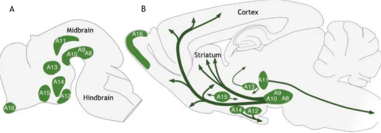
The midbrain DAergic neurons are part of a total of twenty catecholaminergic cell groups: three adrenergic (C1-C3), seven noradrenergic (A1-A7) and ten DAergic (A8-17) (Björklund and Hökfelt, 1984), figure 2. More precisely, the midbrain DAergic system is confined to tree major groups, retrorubral field, substantia nigra (SN), divided into pars compacta (SNc) and
pars reticulata (SNr), and VTA, i.e., A8, A9 and A10 cell group, respectively (Fallon and
Moore, 1978, Dahlstroem and Fuxe, 1964).
Among different species, the size and distribution of midbrain DAergic system differs significantly. In mice, the total number of TH-positive cells in three midbrain DAergic cell groups bilaterally is approximately 20,000-30,000, while in rats is about 40,000-50,000, with around 50% of cells located in A9 group (Blum, 1998, Nelson et al., 1996, German and Manaye, 1993, German et al., 1983). Otherwise, the human midbrain DAergic neurons are concentrated in SN, between 400,000–600,000 TH-positive cells that account to approximately 70% of all midbrain DAergic population (Bentivoglio and Morelli, 2005, Chu et al., 2002, Pakkenberg et al., 1991, German et al., 1983). However, with ageing, the DAergic population in midbrain tends to decrease around 4.7% neurons per year (Fearnley and Lees, 1991), a reduction that results in approximately less 40% of SN DAergic neurons by the age of 60 years, a number that significantly increases in neuropathologies such as Parkinson’s disease (McGeer et al., 1977).
Furthermore, the midbrain DAergic neurons are still classified in two different populations, dorsal and ventral tier, this nomenclature is based on the projections and spatial arrangement of the cell bodies. The dorsal tier includes the populations of cells with cell bodies located in dorsal part of SNc and VTA, as well as, the cells of the A8 cell group (Bjorklund and Dunnett, 2007, Bentivoglio and Morelli, 2005, Fallon and Moore, 1978). These neurons are loosely spaced and the dendrites are oriented mediolaterally in the plane of the SNc, while the major part of the axons are thin (0.1-0.4 µm) and relatively smooth, with few varicosities (0.3-0.6 µm) (Bentivoglio and Morelli, 2005, Smith and Kieval, 2000, Gerfen et al., 1987, Fallon and Moore, 1978). On the other hand, the ventral tier includes the cells with their cell bodies located in ventral part of SNc and VTA, and SNr (Bjorklund and Dunnett, 2007, Bentivoglio and Morelli, 2005, Fallon and Moore, 1978). Axons arising from ventral tier neurons are slightly thicker (0.2-0.6 µm) with more varicosities (0.4-1.0 µm) and an aspect more wrinkled or crinkled (Bentivoglio and Morelli, 2005, Gerfen et al., 1987).
The common need to classify midbrain DAergic neurons exceeds its simple spatial location or projections, going up to the transcription level (Roeper, 2013). Furthermore, although these nomenclatures are currently used we must be careful with the its use because of the significant differences observed in distribution, size and subdivisions among mammalian species (Smeets and Gonzalez, 2000).
Figure 2: Distribution of dopaminergic cell groups in the developing A) and adult B) rodent brain.
Dopaminergic neurons are localized in nine distinctive cell groups, as illustrated schematically, in a sagittal view. The principal dopaminergic projections are illustrated in b) by arrows. Adapted from (Bjorklund and Dunnett, 2007).
Midbrain Dopaminergic Pathways
1.1.1.
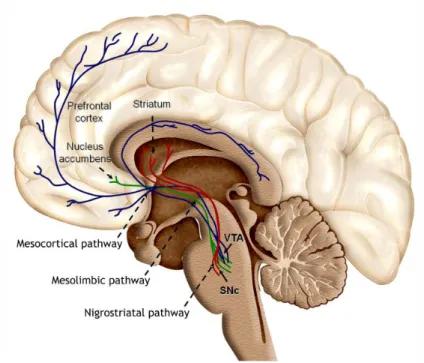
Midbrain neurons project widely throughout the brain and the DAergic neurons of the VTA and SNc represent, respectively, 60% and 80% of the neurons that project from these two regions (Yetnikoff et al., 2014). These projections are part of a group of midbrain DAergic projections, commonly grouped into three distinct pathways based on their brain targets, figure 2B and 3.
The nigrostriatal pathway roughly described as being derived from neurons located in both tiers of the SNc, in fact correspond to projections arising from SNc, lateral VTA and A8 cell group that project to the striatum (Bjorklund and Dunnett, 2007, Smith and Kieval, 2000). These projections correspond to the main source of innervation in sensorimotor striatum (Bjorklund and Dunnett, 2007, Joel and Weiner, 2000, Smith and Kieval, 2000), a pathway strongly involved in the control of voluntary movement (Arias-Carrion et al., 2010), whose loss of SNc DAergic neurons is the hallmark of Parkinson’s disease (Dickson et al., 2009, Arias-Carrion and Poppel, 2007).
Regarding to mesolimbic and mesocortical pathways whose main targets are, respectively, nucleus accumbens and prefrontal cortex, the projections arise mainly from VTA, however also projections from A8 cell group and dorsal tier of SN are present (Arias-Carrion et al., 2010, Arias-Carrion and Poppel, 2007, Bjorklund and Dunnett, 2007). These pathways have been suggested to be involved in the regulation of emotion and reward (Arias-Carrion et al., 2010, Arias-Carrion and Poppel, 2007, Bjorklund and Dunnett, 2007) and its dysregulation/alteration on neurotransmission has been associated in pathologies like schizophrenia and depression, as well as, in drug addiction (Meyer-Lindenberg et al., 2002, Robinson and Berridge, 1993).
Figure 3: Dopaminergic projections to the forebrain. Dopaminergic neurons located in the substantia
nigra pars compacta (SNc) and the ventral tegmental area (VTA) project their axons to the striatum,
nucleus accumbens and prefrontal cortex, representing, respectively, the nigrostriatal, mesolimbic and mesocortical pathway. These pathways are strongly involved in the control of voluntary movement, emotion and reward. Adapted from (Arias-Carrion et al., 2010).
2. From Synapse to Neurotransmitter Release
2.1. The Classic Chemical Synapse
Chemical synaptic transmission is the most common communication method between neurons, a one-way transmission with a typical delay of the information transmission of 1-5 ms or even more. In this type of transmission the pre- and postsynaptic membranes are physically separated in the order of 20-40 nm, by the synaptic cleft (Hormuzdi et al., 2004, Kandel et al., 2000).
As the name suggests, chemical transmission is characterized by their chemical agents of transmission, the neurotransmitters, which are clustered into SVs. Generally, after the action potential, the neurotransmitter-containing vesicles fuse with presynaptic membrane releasing the neurotransmitters into the synaptic cleft (Waites and Garner, 2011, Beckstead et al., 2004). While in the synaptic cleft, neurotransmitters interact with postsynaptic receptors present in the dendritic ‘spines’, a very tiny specialized protrusions emanating from dendritic branches, as is the case of excitatory glutamatergic synapses (Bito, 2010, Lin and Koleske, 2010), and trigger various postsynaptic events, figure 4.
Although the synapse comprises both the pre- and postsynaptic components, this review will focus only in the presynaptic terminals.
Figure 4: Synaptic vesicle cycle. Synaptic vesicles are filled with neurotransmitters and stored in
cytoplasm. Consequently, these vesicles can be translocated to active zone where they dock and prime. Subsequently, after a Ca2+-influx these vesicles fuse and neurotransmitters are released. After fusion-pore opening, synaptic vesicles undergo endocytosis and recycle via different pathways. Adapted from (Jahn and Fasshauer, 2012).
Synaptic Vesicle Pools
2.1.1.
Over the years, researchers have proposed the existence at the presynaptic terminals of the chemical synapses of distinct vesicle pools, with different properties. Rizzoli and Betz (2005) proposed a model in which every SV can be linked to one of three major vesicle pools: the readily releasable pool (RRP), the recycling pool and the reserve pool, figure 5A. In this simplistic model, the RRP, located at the presynaptic active zone, the site of neurotransmitter release, consist of vesicles with high fusion probability. The vesicles of this pool are the firsts to be released and depleted upon stimulation, being their vacancies repopulated by some of recycling vesicles docked at the active zone, showing an intense intermixing between these two pools. Due to their highest fusion probability, the RRP vesicles are crucial for synaptic strength however, despites its name, not all RRP vesicles are ready to be released. After a fast initial release, a slower release is maintained by the recycling pool, with 10-20% of all vesicles, which under physiological frequencies of stimulation continues to be released and refilled by new recycled vesicles. Finally, the reserve pool which are vesicles reluctant to release, comprise about 80-90% of all vesicles in presynaptic terminals. In their model, Rizzoli and Betz described a possibility of an intermixing between recycling and reserve pool at spatial but not functional level, figure 5B (Alabi and Tsien, 2012, Denker and Rizzoli, 2010, Schweizer and Ryan, 2006, Rizzoli and Betz, 2005).
More recently, new studies have introduced a refinement to the initial model proposed by Rizzoli and Betz (2005). In this ‘improved’ model, figure 5C, the recycling pool does not exist
new vision of the reserve pool, renamed as ‘resting pool’, consisting in 50-85% off all vesicles that remain unreleased even upon strong stimulation. Also, the initial thought about the spatial but not functional intermixing of recycling and reserve pool was knocked down, recent studies proved a differentiation among recycling and reserve pool, suggesting that recycling vesicles eventually will ‘mature’ into reserve vesicles. Docked reserve vesicles will occasionally exocytose replacing the recycling vesicles which have switched to the reserve pool. A new perspective arise with the introduction of the ‘superpool’ concept, in which the SVs/pools are no longer limited to single synaptic boutons, but instead can be exchanged across multiple synapses (Rizzoli, 2014, Alabi and Tsien, 2012, Denker and Rizzoli, 2010).
Figure 5: Organization of synaptic vesicle pool. A) The classical model of three distinctly localized
synaptic vesicle pools. The readily releasable pool (RRP; red) consists of vesicles docked at the active zone and primed for release. Behind the RRP are located the recycling pool vesicles (green), which are recruited for the active zone (left arrow) and released under moderate stimulation. Away from the active zone, under very high stimulation reserve pool vesicles (blue) are recruited for the active zone (right arrow) after depletion of recycling pool. B) A pool model taking into account the spatial, but not functional, intermixing between recycling and reserve pool (Rizzoli and Betz, 2005). C) The updated vesicle pool model. Recycling and reserve pool vesicles intermixing are not limited spatially, but also functionally with maturation of recycling pool vesicles into reserve pool vesicles (green-blue intermediate forms). Due to their permanent mobility, recycling pool vesicles reach the active zone that can be subsequently docked, forming the RRP vesicles. The frequent exchange of both recycling and reserve vesicles between synapses forms the ‘superpool’. Adapted from (Denker and Rizzoli, 2010).
Neurotransmitter Release
2.1.2.
Presynaptic Active Zone 2.1.2.1.
The active zone or synaptic active zone is a region located at the presynaptic plasma membrane precisely opposite to the postsynaptic neurotransmitter reception apparatus. Under electron microscopy it is characterized by a more or less regular array of electron-dense cone-shaped particles, together with a dense collection of proteins that form the cytoplasmic matrix at the active zone (CAZ) (Fejtova and Gundelfinger, 2006, Schoch and Gundelfinger, 2006, Zhai and Bellen, 2004, Dresbach et al., 2001). Therefore, the active zone can be divided into presynaptic membrane and CAZ, both playing an important role in neurotransmitters release.
While the presynaptic membrane is the region of Ca2+ entrance, mediated by the
voltage-gated Ca2+-channels (VGCCs), and SV fusion (Zhai and Bellen, 2004), the CAZ is
thought to define and organize neurotransmitter release sites, regulating the vesicle docking,
fusion and their proximity with Ca2+ channels (Gundelfinger and Fejtova, 2012, Zhai and
Bellen, 2004, Dresbach et al., 2001), and confer long-term stability to individual presynaptic sites (Tsuriel et al., 2009). Thus, to achieve these functions the protein meshwork of CAZ is
organized by a small set of multi-domain scaffolding proteins that included the Rab3-interacting molecules (RIMs), the RIM binding proteins (RIM-BPs), Bassoon and
Piccolo/Aczonin, the UNC-13/Munc-13 proteins, the CAST/ELKS/Bruchpilot proteins and the liprin-α (Sudhof, 2012, Sigrist and Schmitz, 2011, Fejtova and Gundelfinger, 2006, Schoch and Gundelfinger, 2006). RIM is a small protein identified as an interactor of Rab3, a small GTPase present on SVs. RIM proteins were identified as involved in recruiting of Ca2+ channels and
SVs to active zone (tethering), SV docking and priming, as well as, the Ca2+ channel-SV
colocalization at the presynaptic active zone (Gundelfinger and Fejtova, 2012, Sudhof, 2012, Han et al., 2011, Kaeser, 2011, Pernia-Andrade and Jonas, 2011, Sigrist and Schmitz, 2011). RIM-BPs were suggested to create a functional link between the synaptic-vesicle tethering apparatus and the Ca2+ channels, as well as, involved in the Ca2+ channels tethering (Kaeser et
al., 2011, Liu et al., 2011, Hibino et al., 2002). Bassoon and Piccolo/Aczonin, two very large proteins, were suggested to be involved in membrane trafficking and scaffolding (Kononenko et al., 2013, Waites et al., 2013, Tsuriel et al., 2009, Schoch and Gundelfinger, 2006, Dresbach et al., 2001). Furthermore, Bassoon has been suggested to be involved in long-term stability to individual presynaptic sites (Tsuriel et al., 2009), while Piccolo was shown to be a regulator of SV exocytosis and to be involved in the SV mobilization from the reserve pool to the RRP and endocytosis (Waites et al., 2011, Leal-Ortiz et al., 2008, Fenster et al., 2003). Munc13 proteins, were reported to play an important role in SV release and in the potentiation of neurotransmitter release in presynaptic short-term plasticity (Zhou et al., 2013, Sudhof, 2012, Fejtova and Gundelfinger, 2006, Schoch and Gundelfinger, 2006). Another important family of proteins in CAZ are the CAST/ELKS/Bruchpilot that contribute to the molecular organization of active zone (Ohtsuka, 2013, Fejtova and Gundelfinger, 2006, Schoch and Gundelfinger, 2006). Finally, liprin-α proteins are a group of proteins involved in synaptic cargo transport and anchoring, as well as, in the regulation of the size of active zone (Kittelmann et al., 2013, Sudhof, 2012, Sigrist and Schmitz, 2011).
Neurotransmitter Release by Exocytosis 2.1.2.2.
Tethered SVs are docked to the active zone, staying closely apposed to the presynaptic plasma membrane (Becherer and Rettig, 2006). In the presynaptic plasma membrane, docked SVs can stay more or less close to the VGCCs, a step extremely important for neurotransmitter release by exocytosis. The greater the distance between the SVs and VGCCs, the less likely is the release (Catterall and Few, 2008, Sudhof, 2004, Zhai and Bellen, 2004), resulting in reduced synaptic strength. With some delay, the docked SVs become fusion competent. A process that may involve molecular rearrangement and lipid modifications, collectively defined as priming (Murthy and De Camilli, 2003).
When an action potential arrive at the nerve terminal it depolarizes the presynaptic plasma membrane, VGCCs open, and Ca2+ influx occurs, stimulating SV exocytosis (Sudhof, 2004).
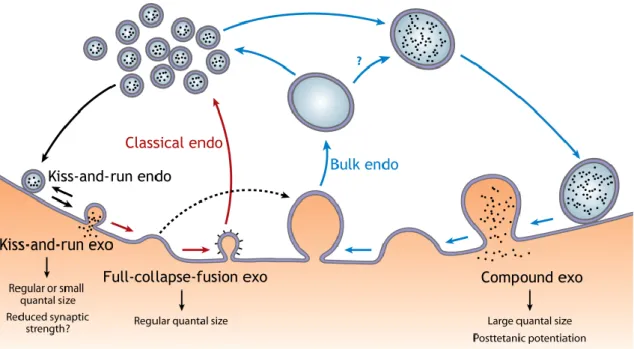
Ca2+-dependent SV exocytosis has been suggested to occur in three different modes, full
collapse fusion, kiss-and-run and compound exocytosis, figure 6. In a full collapse fusion, when fusion between SV and plasma membrane occur, a fusion pore opens and dilates until full collapse of the SV, a fast and complete transmitter release (Wu et al., 2014). On the other hand, in a kiss-and-run mode, a fusion pore opens and closes without vesicle collapse (Wu et al., 2014, Murthy and De Camilli, 2003). This is commonly associated with a narrow fusion pore, with slow and incomplete transmitter release, that results in a small quantal size, and consequently reduced synaptic strength. However, kiss-and-run may also open fusion pores large enough for a fast and complete exocytosis as in the full collapse mode. Finally, in compound exocytosis a giant vesicle, resultant from vesicle-vesicle fusion, fuses with the plasma membrane. Unlike the proposed to kiss-and-run mode, compound exocytosis leads to an increase of the quantal size, and thus of the synaptic strength (Wu et al., 2014).
Synaptic Vesicle Recycling 2.1.2.3.
Following exocytosis, SVs recycling is necessary for the maintenance of SV pools during sustained neurotransmission to prevent vesicle exhaustion. Thus, like exocytosis, also three different endocytosis modes have been suggested: the classic endocytosis, the bulk endocytosis and the kiss-and-run, mentioned above (Wu et al., 2014), figure 6.
The classic endocytosis consist in the generation of a vesicle via membrane invagination from the shallow and deep membrane invaginations, a profile like an Ω, and the fission of this Ω profile. This process is mediated by the molecular clathrin triskelion, which plays an important step in this type of endocytosis, the clatherin-coated and uncoated pits (Wu et al., 2014, Waites and Garner, 2011). Classic endocytosis consists in a slow endocytosis method and generally considered the most important endocytosis mode. This mode is usually thought to be preceded by the full-collapse exocytosis. On the other hand, bulk endocytosis consists in the retrieval of a large endosome-like structure. Bulk endocytosis contributes to rapid and slow endocytosis suggested to be associated with compound exocytosis. However, it also is possible that the bulk endocytosis follows full-collapse exocytosis. The kiss-and-run mode, previously mentioned, can also contributes to a slow endocytosis, however is the prime mediator of rapid endocytosis (Wu et al., 2014).
Figure 6: Schematic synaptic vesicle exo-endocytosis coupling. Three modes of exocytosis
(exo) — full-collapse fusion, kiss-and-run, and compound exocytosis — are coupled to classical endocytosis (endo), kiss-and-run, and bulk endocytosis, respectively. Full-collapse fusion may also be coupled to bulk endocytosis (dotted arrow), although this hypothesis remains to be tested. The functions of each exocytosis mode in regulating quantal size and generating synaptic plasticity are also listed. Question marks indicate an unclear function or transition. From (Wu et al., 2014).
2.2. Strategies to Study Axonal Terminals
Over the years, axons have been extensively studied, from RNA translation to axonal regeneration and also presynaptic studies (Larsen et al., 2014, Taylor et al., 2013, Denker et al., 2011, Kim et al., 2009, Olink-Coux and Hollenbeck, 1996). To achieve these multiplicities of studies, different culture techniques have been used, from more rudimental cultures, such as a conventional culture or micro-island culture, to more complex techniques, such as Campenot chamber or even, more recently, microfluidic devices.
One of most simply and practical culture methods that permits synaptic studies is the plate/dish cultures with or without coverslips (Hua et al., 2013, Bartolome-Martin et al., 2012, Levinson et al., 2005, Richmond et al., 1996), which I define here as conventional cultures. However, this methodology has some limitations when some type of controlled axonal isolation is required, problem partially resolved with the rise of micro-island cultures in the 60s (Millet and Gillette, 2012).
Micro-island cultures, figure 7A, have been widely used as model system for studying numerous molecular and cellular mechanisms of neuron development, synaptic transmission and plasticity including the influence of synaptic proteins and trophic factors on synapse formation and neurotransmitter release (Daniel et al., 2009, Schluter et al., 2006, Tarsa and
production and release (Sulzer et al., 1998, Johnson, 1994); short-term synaptic depression (Brody and Yue, 2000); long-term potentiation (Tong et al., 1996); long-term depression (Goda and Stevens, 1998). In micro-island culture, the axonal growth is confined to a restricted region of their own dendritic tree. Unlike conventional cultures, in a micro-island culture it is possible to isolate a single neuron or several neurons in a small restricted area. This aspect of micro-island culture improves the innervation density of the individual cells, which increases synapses formation and also the possibility of monosynaptic interactions (Allen, 2006). However, even with this technique axonal isolation remains impossible.
Some years later, a new method using the Campenot chambers, emerged and brought new insights into the axonal biology, but in this case, mainly focused on peripheral neurons (Mok et al., 2009, Hayashi et al., 2004, Campenot, 1977, Kimpinski et al., 1997), figure 7B. One of the main advantages of the Campenot chamber is the compartmentalization of neurons (Millet and Gillette, 2012) and the local fluid control (Campenot, 1982b, Campenot, 1982a, Campenot, 1977), a new characteristic in the culture techniques. Furthermore, this technique also permits an easy visualization of neurons using conventional microscopic methods and has the exceptional characteristic of permitting the analysis of many samples in parallel within the same chamber device. However, Campenot chamber had the big disadvantage of being incompatible with many central nervous system (CNS) neurons and of requiring neurotrophic factors to guide neurite outgrowth (Millet and Gillette, 2012).
More recently, the incompatibility of Campenot chambers with the major part of CNS was overcome with the emergence of the microfluidic chambers, figure 7C. Microfluidic chambers have more variable designs which are adapted to specific applications (Lu et al., 2012, Majumdar et al., 2011, Taylor et al., 2010, Vahidi et al., 2008, Ravula et al., 2007, Hung et al., 2005, Rhee et al., 2005, Taylor et al., 2005, Tourovskaia et al., 2005) and have served as support for different types of studies, such as local stimulation (Hosie et al., 2012, Taylor et al., 2010), development of sensory neurons-osteoblast (Neto et al., 2014) and cortico-striatal synaptic connections (Peyrin et al., 2011) and axonal regeneration studies (Vogelaar et al., 2009, Taylor et al., 2005). Together with microfluidic chambers, the microfluidic isolation arises (Millet and Gillette, 2012, Taylor et al., 2005, Taylor et al., 2003), enabling local stimulation. Additionally, microfluidic chambers are compatible with high-resolution microscopy, long-term culture of both CNS and peripheral nervous system, have high reproducibility and are highly versatile in materials and design (Millet and Gillette, 2012, Park et al., 2006). In contrast, this method has the big disadvantage of medium evaporation due to the working micro- and nano-volumes and the limitation of gas fluxes when compared with conventional cultures (Millet and Gillette, 2012).
In short, there are many culture techniques that can be used in axonal/synaptic studies and the combination of methodologies allows the reinforcement of results, making it impossible to select a single method as the best one.
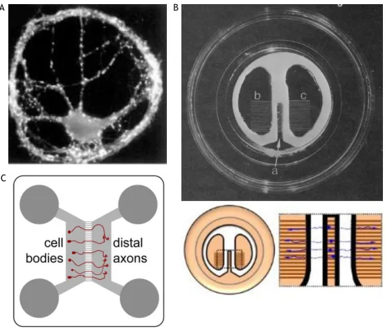
Figure 7: Cellular culture techniques for the study of synapses. A) Pyramidal neurons labelled against
synaptophysin showing a large number of puncta in micro-island cultures. B) Campenot chamber (upper) and schematic representation of neuronal growth within this (lower). C) Schematic representation of microfluidic device. A and B adapted from (Millet and Gillette, 2012) and C from (Hengst et al., 2009).
3. Dopaminergic Synapsis
3.1. The Dopamine Cycle
As already mentioned, DA is synthesised by a sequential reaction catalysed by TH and AADC and is transported from cytosol to SVs by VMAT2. However, if DA synthesis exceeds the SVs capacity, it is metabolized to 3,4-dihydroxyphenylacetic acid (DOPAC) by monoamine oxidase (MAO). DOPAC then rapidly diffused out of neurons, and is taken up by the glial cells in the neuropil and converted to homovanillic acid (HVA) by catechol-O-methyltransferase (Lookingland and Moore, 2005).
When DA is released to the synaptic cleft, DA is free to interact with stimulatory D1-like family receptors or inhibitory D2-like family receptors on postsynaptic target cells and inhibitory D2-like family receptors on presynaptic terminals (Lookingland and Moore, 2005). The D1-like family receptors are coupled to G proteins, which activate the adenylate cyclase,
A B
family receptors are also coupled to G proteins, which in this case inhibit adenylate cyclase, the consequent production of cAMP and the subsequent downstream signalling systems. Further, the D2-like family receptors are also involved in the modulation of Ca2+ levels (Yao et
al., 2008).
Some of extracellular DA is then tacked by glia and metabolized to 3-methoxytyramine and then to HVA, while a major portion of these DA is recaptured into DAergic neurons by the high affinity DAT located on presynaptic terminals. Once inside the cells, DA can be transported to SVs via VMAT2 or metabolized to DOPAC by MAO (Lookingland and Moore, 2005), figure 8.
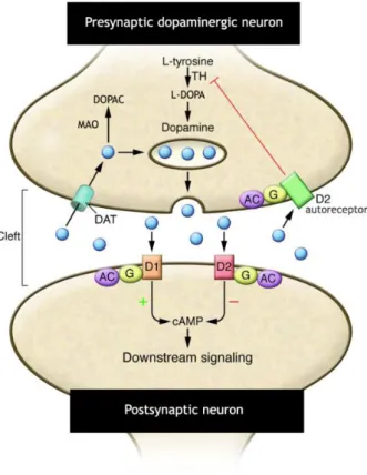
Figure 8: Schematic representation of dopamine cycle. D1- and D2-like family receptors are positively
or negatively coupled to adenylate cyclase (AC) via G proteins (G). Released dopamine (DA; blue circles) is then available to bind to the postsynaptic D1- and D2 receptors or presynaptically to D2 autoreceptors. Captured by DA transporters (DATs), DA can be subsequently stored in synaptic vesicles or metabolized into 3,4-dihydroxyphenylacetic acid (DOPAC) by monoamine oxidase (MAO). A small amount of DA is captured by catechol O-methyltransferase-containing glial cells where it is metabolized to homovanillic acid and 3-methoxytyramine. Adapted from (Blackstone, 2009).
3.2. Ultrastructure of Dopaminergic Presynaptic Structures
Most of synaptic studies and theories, like the described previously for the chemical synapse, were performed in excitatory hippocampal glutamatergic synapses; nonetheless they are not necessarily applicable to the modulatory DAergic system.
DA storage is believed to occur in two types of organelles, the SVs and large dense core vesicles. In DAergic terminals most of the DA, about 99%, is stored in SVs (Pothos et al., 1998, Nirenberg et al., 1997) and are commonly released by exocytosis from axonal varicosities, an
axonal swelling that is not the typical specialized presynaptic structures (Martin and Spuhler, 2013, Sudhof, 2008, Nirenberg et al., 1997). This lack of specialized postsynaptic structures apposite to varicosities together with previous results suggests that the mode of action of DA is non-synaptic (Martin and Spuhler, 2013, Descarries and Mechawar, 2000).
In a recent study, Martin and Spuhler (2013) showed that, although most of varicosities do not form synapses in TH-positive projections of the prefrontal cortex, they are in continuous contact with pre- and postsynaptic structures, what they called ‘synapse forming pair’, reinforcing the hypothesis for non-synaptic DA signalling. A similar configuration was described by Moss and Bolam (2008) for the striatum, where TH-positive axons often apposed to the glutamatergic synapses and/or their postsynaptic targets.
In fact, less frequently, DAergic synapses are also found in both symmetric terminal synapses and symmetric synapses en passant, i.e. non-terminal synapses (Moss and Bolam, 2008, Groves et al., 1994, Freund et al., 1984). Freund et al. (1984), using TH-labelling, reported the dendritic spine and dendritic shaft as the major postsynaptic target, while Groves et al. (1994), using 5-OHDA-labelling reported the spine necks or heads as the main targets for DAergic presynaptic structures. Similar to described by Martin and Spuhler (2013) for ‘synapse forming pair’, DAergic synapses often are part of a synaptic complex, where a spine forms a symmetric synapse with a DAergic bouton and an asymmetric synapses with other bouton, like excitatory glutamatergic synapses (Moss and Bolam, 2008, Groves et al., 1994, Goldman-Rakic et al., 1989, Freund et al., 1984), a structure defined by Goldman-Rakic et al. (1989) as ‘triadic complex’.
3.3. Axonal Dopamine Release
As already mentioned, axonal DAergic release was suggested to occur at two different sites, the synaptic boutons and non-synaptic varicosities, therefore it becomes practically impossible to talk about DAergic synapse without mentioning the ‘non-synaptic presynaptic’ structures.
Bryce Vissel and collaborators (2009) brought new insights into DAergic synapse. In their study, using VMAT2-labelling to identify the DAergic presynaptic structures and styryl dye FM1-43-labelling to study SV recycling, reported the existence of recycling SVs at both synaptic and non-synaptic sites in DAergic neurons. Moreover, almost all VMAT2- and FM1-43-positive synaptic sites and all VMAT2- and FM1-43-positive non-synaptic sites co-labelled with CAZ protein bassoon, suggesting that, in DAergic neurons, the DA release primarily occurs at sites that possess active sites. They also observed that the DAergic neurons exhibited a heterogeneous probability of release, which they suggested to be due to heterogeneity found at recycling pool size, and that the DAergic synapses are generally more
Classically, DAergic synaptic transmission is seen as a direct transmission at chemical synapses. In this type of transmission, the vesicular DA is released from the presynaptic terminal, diffuses across the synaptic cleft, and acts on DA receptors, clustered on the postsynaptic membrane, figure 9A. However, this theory may not correspond fully to the truth in DAergic synapses. Electron microscopy studies demonstrated that DAT are widely distributed from synaptic and non-synaptic sites at plasma membrane of axons, but at synaptic sites, DATs were found near, but not within the synaptic active zones (Hersch et al., 1997, Nirenberg et al., 1996), suggesting that DA diffuses from the synaptic cleft to the extrasynaptic space. Gonon (1997) proposed that DA diffuses in striatum at a distance of 12 µm and, more recently, Borland et al. (2005) estimated a diffusion and uptake of DA of at least 220 µm. Moreover, studies have demonstrated that DA receptors are predominantly extrasynaptic (Hersch et al., 1995, Yung et al., 1995, Sesack et al., 1994).
Taking this data, the theory proposed by Rice and Cragg (2008) may be a good model for transmission of DA in striatal DAergic synapse. In this model, DA synaptic signals do not exclusively depend of transmission across the synaptic cleft; instead, they act by diffusing from the release site and outside of synaptic cleft, figure 9B.
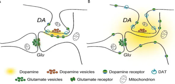
Figure 9: Two hypotheses for the dopaminergic synaptic transmission. A) Schematic representation of a
conventional dopamine (DA)ergic synapse on the neck of a dendritic spine on a striatal medium spiny neuron. Glutamatergic input forms synapses on the heads of these spines. DA released at synaptic cleft is constrained within to this region by DA transporters (DATs), where it is available to interact with subsynaptic DA receptors. B) Schematic representation of an updated DAergic synapse that shows the ability of DA to diffuse out of synaptic space in three dimensions. The resultant DA cloud interacts predominantly with extrasynaptic DA receptors, while the uptake of DA only occurs when it encounters DATs on DAergic axons. Adapted from (Rice and Cragg, 2008).
In this work, we pretend establish a new method that permits us study the DAergic nervous terminals and thus, in a future tried clarify some of these hypothesis/theories and doubts regarding the DAergic synapses. Moreover, we also pretend optimize a method that permits us monitoring the viability of our embrionary ventral midbrain DAergic cultures.
Chapter 2: Aims
The functions played by the ventral midbrain DAergic system makes this a field of high interest to the neurosciences. However, due to its nature, the presynaptic study of this system may represent a great challenge. Therefore new techniques or the development of existing ones will be of great impact in this field. This work had two main objectives:
- The first goal was to establish a new method to study DAergic terminals. In particular we aimed to develop DAergic cultures in microfluidic chambers, which allow the specific manipulation of pure axonal populations.
- The second goal consisted in the optimization of a method for measuring monoamine
neurotransmitters and their metabolites in ventral midbrain DAergic culture using high-performance liquid chromatography (HPLC) coupled to electrochemical detection (ECD).
Chapter 3: Methods
1. Animals
All experiments requiring the use of animals were conducted in compliance with protocols approved by the national ethical requirements for animal research, and with the European Convention for the Protection of Vertebrate Animals Used for Experimental and Other Scientific Purposes (Directive number 2010-63-EU).
All animals were kept under controlled conditions of temperature and light and with food and water available ad libitum.
2. Primary Midbrain Cultures
2.1. Collection of the Embryos
Embryonic day 15 to 16 Wistar rat embryos were collected from pregnant Wistar rats anesthetized with isoflurane or with a solution containing ketamine (87.5 mg/kg) and xylazine (12.5 mg/kg) followed by euthanasia induced by cervical dislocation. In a laminar flow hood, the collected uterine horns were placed in ice-cold phosphate buffer saline (PBS) and dissected to obtain the uterine sacs. Under same conditions, the uterine sacs were then dissected and the amniotic membranes removed to collect the embryos.
2.2. Dissection of Ventral Midbrain
Under a dissection microscope, the ventral midbrain region was dissected from the each embryo and putted in ice-cold PBS. First, using a scalpel blade the embryo brain was cut over the eye and the scalp tissue removed. Then, a forceps was used to stabilize the brain and fore- and hindbrain regions were carefully removed with scalpel blade. Finally, the dorsal part of the midbrain was removed using a scalpel blade and ventral part was stored in PBS until the end of dissection.
2.3. Preparation of Single Cell Suspension
The tissue chunk resultant from the dissection was incubated in an enzymatic solution containing 0.05% trypsin (cat. T7409; Sigma-Aldrich) and 0.2% deoxyribonuclease I (cat. DN25; Sigma-Aldrich) for 4 min at 37 ºC. The enzymatic digestion was then ended by resuspending the tissue in PBS containing 10% heat-inactivated (HI) fetal bovine serum (FBS) (cat. 10270; Life Technologies). After the enzymatic digestion, the tissue was washed in sterile PBS followed by mechanical digestion in neurobasal medium (cat. 21103-049; Invitrogen) through repetitive pipetting until a homogeneous suspension was observed. The resulting cell
suspension was then centrifuged at 405 G for 3 min (3K18C Bioblock Scientific; Sigma Laboratory Centrifuges). After centrifugation, the pellet was resuspended in 10 mL of neurobasal medium containing 2% of B27 supplement (cat. 17504-044; Invitrogen), 0.5 mM of L-glutamine (cat. G3126; Sigma-Aldrich), 25 µM of L-glutamic acid (cat. G8415; Sigma-Aldrich) and 1.2% of gentamicin (cat. G1272; Sigma-Aldrich) (complete neurobasal medium), and cells were counted in the haemocytometer using the trypan blue exclusion test (Strober, 2001a, Strober, 2001b).
3. Culture of Midbrain Cells (Conventional
Culture)
Dissociated ventral midbrain cells were plated at a density of 0.4x10⁶ cells per 10 or 13 mm
coverslip in 24-multiwell culture plates for analysis of DAergic survival and DAergic markers specificity. For in vitro DAergic viability tests were used cells at a density of 1x10⁶ cells per
12-multiwell culture plates and/or 0.5x10⁶ cells per13 mm coverslip in 24-multiwell culture
plates. To determine the better conditions to improve DAergic survival, cells were grown in coverslips pre-coated overnight with 0.1 mg/mL of poly-D-lysine (PDL; cat. P1024; Sigma-Aldrich) at 37 ºC or PDL-coating followed 2 µg/mL laminin (cat. L2020; Sigma-Sigma-Aldrich)-coating coverslips for 2 h at 37 ºC or PDL- and laminin-coating coverslips with addition of 10 ng/mL of the glial cell-derived neurotrophic factor (GDNF; cat. sc-4865; Santa Cruz Biotechnology, Inc) to the culture or PDL-coating coverslips. Cells were maintained for 5 or 6 days in vitro (DIV) under humidified 5% CO2 atmosphere at 37 ºC and then fixed until immunocytochemical
analysis.
4. Mesencephalic Microfluidic Cultures
4.1. Microfluidic Chamber Preparation
The microfluidic chamber used here was a model developed by Noo Li Jeon (Park et al., 2006, Taylor et al., 2003), figure 10, with some modifications in the assembly of the device. Briefly, glass coverslips were washed for 24 h in 65% nitric acid under constant agitation. To completely remove the nitric acid, the coverslips were rinsed five times with mQ H2O and five
times more, for 30 min, under constant agitation followed 20 min of sterilization under ultra
violet light in a laminar flow hood. Sterile coverslips were then incubated with 0.1 mg/mL PDL (cat. P7886, Sigma-Aldrich) overnight at 37 ºC, and then washed three times
for 5 min with mQ H2O. When laminin coating was performed, after PDL-coating and mQ H2O
washes, 2 µg/mL laminin in mQ H2O was incubated for 2 h at 37 ºC. Excess laminin was then
for 30 sec in 75% ethanol and dried under a laminar flow hood, and were finally assembled and softly pressed against the coverslips. To fluidicaly equilibrate the device, neurobasal medium, pre-heated at 37 ºC, was added to one reservoir of the device and allowed to fill the main channel, which was followed by addition of medium to the opposite reservoir. The medium progressively filled the microgrooves. After this, neurobasal medium was added to one of two reservoirs of the opposite main channel allowing the channel to fill. Finally the medium was added to the remainder reservoir.
Assembly of the devices was always performed in 90-mm Petri dishes.
Figure 10: The microfluidic chamber directs axonal growth. A) Schematic representation of PDMS
containing a relief pattern of somal and axonal compartments connected by microgrooves of 450 or 900 µm of length. B) Representation of axonal grown in microfluidic chambers with microgrooves of 450 µm or 900 µm of length at 14 DIV. In both models of microfluidic chambers the dendritic arborisation (green; MAP2) failed in the crossing of microgrooves to the axonal side, while axons (red; Tau) in both chambers crossed the microgrooves successfully. The bottom image represents the merge of both tau and MAP2 labelling. Adapted from (Taylor et al., 2005).
4.2. Culture of Midbrain Neurons in Microfluidic Chambers
Neurobasal medium was removed and fresh neurobasal medium was added to the upper reservoirs to completely clean the grooves. Then, medium was removed and 30 µL of neurobasal medium was added to one of axonal reservoirs to increase the pressure inside the axonal compartments and prevent cells from entering the microgrooves during cell seeding. Dissociated ventral midbrain cells were seeded at a density described in table 1. Cells were added to the somal side by a slow and gradual addition of 12 µL of cells at the entrance of the main channel. After the cell seeding, the flux inside the somal was allowed to stabilize for 45 min under a humidified 5% CO2 atmosphere, at 37 ºC. Then, 150 µL of complete
neurobasal medium was added to all reservoirs with or without 10 ng/mL of GDNF. Everyday 50 µL of neurobasal medium was added to one of the reservoirs in the somal side. To reduce medium evaporation, some drops of mQ H2O were added to the Petri dishes. Cells were
maintained in culture for at least 4.5 DIV (table 1) under humidified 5% CO2 atmosphere at
37 ºC and then fixed for immunocytochemical analysis.
Table1: Culture conditions tested in microfluidic chambers in an attempt to implement the method in
embrionary ventral midbrain dopaminergic cultures. Culture conditions tested: cell density, time in culture, length of the microfluidic chamber microgrooves. * Culture condition selected to perform the time and cell density tests.
Cell density Time in culture Microgroove Length (µm)
PDL-coating
432,000 cells 8 DIV 450
PDL- and Laminin-coating
432,000 cells 8 DIV 450
PDL- and Laminin-coating and GDNF stimulus
432,000 cells 8 DIV 450
PDL-coating and GDNF stimulus*
216,000 cells 6 DIV 450 8 DIV 450 270,000 cells 6 DIV 450 8 DIV 450 324,000 cells 4.5 DIV 450 5 DIV 450/900 6 DIV 450 8 DIV 450 378,000 cells 4.5 DIV 450 5 DIV 450/900 6 DIV 450 8 DIV 450 432,000 cells 4.5 DIV 450 5 DIV 450/900 6 DIV 450 8 DIV 450 10 DIV 450 12 DIV 450 14 DIV 450 486,000 cells 5 DIV 900
5. In Vitro Dopaminergic Viability
5.1. 1-Methyl-4-phenylpyridinium Stimulus
On the previous day before the stimulus, the culture medium was replaced with fresh medium.
To induce selective DAergic lesion, different concentrations (1 µM, 3 µM and 5 µM) of 1-methyl-4-phenylpyridinium (MPP+; cat. D048; Sigma-Aldrich) were applied at 5 DIV. Cells
were then incubated under humidified 5% CO2 atmosphere at 37 ºC for further 24 h and then
used for high-performance liquid chromatography or immunocytochemical analysis.
5.2. Preparation of Cell Extracts for High-Performance Liquid
Chromatography Analysis
For HPLC analysis, the culture medium was removed and cells were washed with pre-heated PBS. Then, PBS was removed and 60 µL of ice-cold 0.1M perchloric acid was added to the cells and the plates were immediately placed in ice. Cell lysates were collected and centrifuged three times at 21470 G (Mikro 200R; Hettich Zentrifugen), for 15 min, at 4 ºC, and the supernatant was stored at -20 ºC until HPLC analysis.
5.3. Dopamine Measurement
To determine the DA cell content, we used a quaternary HPLC system (1260 Serie Infinity; Agilent Technologies). Briefly, the analysis was performed using a HPLC pump (1260 Infinity Quaternaty Pump; Agilent Technologies) coupled to an electrochemical detector (Coulochem III; ESA). The working electrodes of the electrochemical detector (5011A; ESA) were set at -150 mV (reduction potential) in the first channel and +300 mV (oxidation potential) in the second channel. A sensibility of 10 nA and a filter time constant of 5 sec were used. Reverse phase ion pairing chromatography was used to assay DA and its metabolites DOPAC and HVA and the serotonin metabolite 5-hydoxyindoleatic (5-HIAA). The mobile phase used was based on mobile phase MD-TM 70-1332 (ESA) and consisted of a phosphate buffer in mQ H2O at
pH 3.0 and containing 75 mM sodium dihydrogen phosphate monohydrate (cat. S9638; Sigma-Aldrich), 1.7 mM sodium octyl sulfate (cat. O4003; Sigma-Aldrich) as the ion pairing reagent, 25 µM ethylenediaminetetraacetic acid disodium salt hydrate (cat. 20301; VWR) and 100 µL/L triethylamine (cat. T0424; TCI), kindly provided by Vítor Gaspar, MSc from Health Sciences Research Centre, University of Beira Interior, Portugal. Acetonitrile (cat. EM-AX0145; VWR) was used at 10% concentration as eluent. The column Zorbax 300S-C18 (4.6 × 150 mm, 5μm particle size − cat. 883995-902; Agilent Technologies) was used at 21 ºC. The analysis was performed at 0.6 mL/min flow rate and the injection volume was 30 µL. The reference standards, kindly provided by Filipa Campos, MSc (Health Sciences Research Centre,