Mammography in asymptomatic
women aged 40-49 years
Mamografia em mulheres
assintomáticas na faixa etária de 40
a 49 anos
I Centro de Atenção à Mulher. Instituto de Medicina Integral Prof. Fernando Figueira. Recife, PE, Brasil
II Departamento Materno Infantil. Universidade Federal de Pernambuco. Recife, PE, Brasil
III Departamento de Ginecologia e Obstetrícia. Universidade Federal de Campina Grande. Campina Grande, PB, Brasil
Correspondence:
Melania Maria Ramos Amorim
Rua Neuza Borborema de Souza, 300 Santo Antônio
58406-120 Campina Grande, PB, Brasil E-mail: [email protected] Received: 1/28/2014
Approved: 6/26/2014
Article available from: www.scielo.br/rsp
ABSTRACT
OBJECTIVE: To assess indings of mammography of and interventions resulting from breast cancer screening in women aged 40-49 years with no increased risk (typical risk) of breast cancer.
METHODS: This cross-sectional study evaluated women aged 40-49 years who underwent mammography screening in a mastology reference center in Recife, PE, Northeastern Brazil, between January 2010 and October 2011. Women with breast-related complaints, positive indings in the physical examination, or high risk of breast cancer were excluded.
RESULTS: The 1,000 mammograms performed were classiied into the following Breast Imaging-Reporting and Data System (BI-RADS) categories BI-RADS 0, 232; BI-RADS 1, 294; BI-RADS 2, 294; BI-RADS 3, 16; BI-RADS 4A, 2; BI-RADS 5, 1. There was one case of grade II invasive ductal carcinoma and various interventions, including 469 ultrasound scans, 53 referrals to mastologists, 11 cytological examinations, and 8 biopsies. CONCLUSIONS: Mammography screening in women aged 40-49 years with typical risk of breast cancer led to the performance of other interventions. However, it also resulted in increased costs without demonstrable eficacy in decreasing mortality.
DESCRIPTORS: Women. Mammography. Mass Screening. Breast Neoplasms, diagnosis. Cross-Sectional Studies.
Flávio Xavier SilvaI
Leila KatzI
Alex Sandro Rolland SouzaI,II
The annual incidence of breast cancer varies widely worldwide, from 19.3 per 100,000 women in East Africa to 89.9 per 100,000 in Western Europe.4 This is related to the urbanization process. Accordingly, although the rates are higher in developed countries, in recent years, breast cancer incidence has increased in developing countries.4
In Brazil, breast cancer is the second most frequent type of cancer in the female population, preceded only by nonmelanoma skin cancer.a It is estimated that
57,120 new cases will be diagnosed in 2014, with a risk of 56 cases per 100,000 women. Of these, 64.3% is predicted to occur in the northeast of Brazil.a Breast
cancer is the ifth most common cause of death due to cancer in the general population and the most frequent cause of death due to cancer in women.b
Because the early detection of breast cancer (before there is a palpable nodule) increases the chances of survival,1 routine mammography screening and phys-ical examination are recommended.15
Mammography is the best method for the early diagnosis of breast cancer, demonstrating a 15.0%-25.0% reduction
RESUMO
OBJETIVO: Avaliar os achados mamográicos e as intervenções decorrentes do rastreamento em mulheres de 40 a 49 anos de idade com risco habitual para o câncer de mama.
MÉTODOS: Estudo transversal com mulheres de 40 a 49 anos, submetidas ao rastreamento mamográico em centro de referência em mastologia, em Recife, PE, de janeiro de 2010 a outubro de 2011. Foram excluídas mulheres com queixas mamárias, alterações no exame físico e com alto risco para câncer de mama. RESULTADOS: Das 1.000 mamograias realizadas, 232 foram BI-RADS 0, 454 BI-RADS 1, 294 BI-RADS 2, 16 BI-RADS 3, duas BI-RADS 4A, uma BI-RADS 4C e uma BI-RADS 5. Observou-se um único caso de carcinoma ductal invasivo grau II e várias intervenções: 469 ultrassonograias, 53 encaminhamentos para a mastologia, 11 citologias e oito biópsias. CONCLUSÕES: O rastreamento mamográico em mulheres de 40 a 49 anos com risco habitual para o câncer de mama leva a outras intervenções e, assim, ao aumento dos custos com eicácia não mostrada para redução da mortalidade.
DESCRITORES: Mulheres. Mamograia. Programas de Rastreamento.
Neoplasias da Mama, diagnóstico. Estudos Transversais.
INTRODUCTION
in mortality among women undergoing breast cancer screening.6 Monthly breast self-examination could be an alternative to mammography screening owing to its simplicity and low cost.8 However, there is no evidence that it leads to decreased mortality. Furthermore, this practice is being abandoned because it causes more harm than good, such as unnecessary anxiety among women.10 Magnetic resonance imaging is recommended for screening only in women at high risk of breast cancer.9 Until date, there is no consensus about the perfor-mance of mammography screening among women aged 40-49 years.7 In this age group, breast cancer incidence is lower than that of patients aged 50-69 years,c but the occurrence of dense breasts and fast-growing tumors is higher.12 Breast cancer in young women remains poorly understood. It is believed that breast cancer is biologi-cally more aggressive in young women, with more frequent adverse histopathological characteristics and worse prognoses than in older women.12
Studies of women aged 40-49 years not at high risk are necessary and should consider the peculiarities of each population to determine the ideal age for starting a mammography-based breast cancer screening program.
a Ministério da Saúde. Instituto Nacional de Câncer. Estimativa 2014: incidência do câncer no Brasil. Rio de Janeiro; 2013.
The objective of this study was to assess the mammog-raphy indings and interventions resulting from breast cancer screening in women aged 40-49 years with no increased risk (typical risk) of breast cancer.
METHODS
This cross-sectional study was conducted between January 2010 and October 2011 in Recife, PE, Northeastern Brazil, in the Department of Radiology at the Instituto de Medicina Integral Prof. Fernando Figueira (IMIP), a specialty center in mastology.
The sample size was calculated using the public domain software OpenEpi (Atlanta, GA), version 7. An incidence of 4.6% for positive mammography was obtained from the irst mammography screening in this age group.14 Considering a 95% conidence level and power of 80.0%, a sample of 885 women was deemed required. To offset any potential losses, this number was increased to 1,000 women.
Women aged 40-49 years who underwent mammog-raphy screening between January 2010 and October 2011 were included in the study. Women with breast-related complaints (pain, nodule, nipple discharge, and increased breast volume) or posi-tive indings in the physical examination (shrinkage, bulging, nodules, hardening, and nipple discharge) at the time of the physical examination were excluded, along with those at high risk of breast cancer and those with absent mammography reports.
Women with the following characteristics were consid-ered to be at high risk of breast cancer: irst-degree relative with breast cancer before age 50 years, male relative with breast cancer, irst-degree relative with bilateral breast cancer or ovarian cancer at any age, histopathological diagnosis of a proliferative breast lesion with atypia or lobular neoplasia in situ, and personal history of breast or ovarian cancer.d Patients not presenting a high risk of the disease were consid-ered to have typical risk.
The variables studied were as follows: exposure-related variables [age (years), ethnicity, education (years), age at menarche (years), use of hormone therapy, use of oral contraceptives, breastfeeding in any previous pregnancy, and age at irst pregnancy (years)]; vari-ables associated with sample characterization (origin and state of menopause); outcome-associated vari-ables [Breast Imaging-Reporting and Data System (BI-RADS)];2 and descriptive variables [mammo-graphic characteristics (breast density, nodules, calci-fications, asymmetry, and structural distortions),
interventions performed (ultrasound scans, referral to mastologists, cytological examination, and biopsy), and the result of histopathological examination of the biopsy, in the order of lesions with worsening prognosis (nonproliferative lesions, proliferative lesions without atypia, proliferative lesions with atypia, in situ carci-noma, and invasive carcinoma)].
Cytological examination by ine-needle aspiration was indicated for patients with BI-RADS 4 and 5. For the other BI-RADS, cytological examination was indi-cated according to the results of other complementary examinations.e Biopsy was performed for the same
indi-cations, according to the lesion characteristics, in cases that were inconclusive, equivocal, or different from the clinical and radiological diagnoses.e The samples
were obtained through core biopsy or surgical biopsy.e
For patient selection, a list of all the women who had undergone mammography screening within the study period was obtained. Subsequently, patient records were obtained immediately afterward; the records were stamped to avoid the risk of selecting the same patients. The records of the patients aged 40-49 years were veri-ied according to the inclusion and exclusion criteria. The mammography reports were obtained through the institution’s computer system.
During the study period, 3,574 mammograms were performed among the population of interest. Of these, 2,076 were evaluated; the other 1,498 were excluded because they belonged to the same patient or because the patient records were not found. After veriication, 515 women were excluded from the total number of patients evaluated because they presented with complaints and/or physical changes and/or were at high risk, and 561 were excluded because the obtaining of mammography reports was not possible, leaving 1,000 women for analysis (Figure).
The data were analyzed using the EpiInfo software (Atlanta, GA), version 7. For the descriptive analysis, the mean and its standard deviation (SD) were calcu-lated for numeric variables, and frequency distribution was calculated for categorical variables.
To deine the association between biological, sociodemo-graphic, reproductive, and gynecological variables and BI-RADS categories 3, 4, and 5, we used the Chi-square test of association, or Fisher’s exact test where appro-priate, with a signiicance level of 5%. To determine the strength of the association, the prevalence ratio (PR) was calculated along with its 95% conidence interval. This study was approved by the Human Research Ethics Committee of the IMIP, under Presentation
Certificate for Ethical Consideration (CAAE − 03191212.0.0000.5201 from 6/26/2012). Approval was obtained without requiring informed consent because of the retrospective nature of the study and because the collection of informed consent forms from all the women subjected to mammographic examination was not feasible.
RESULTS
The mean age was 45.2 (SD = 3.5) years. With regard to patients’ location, 827 (82.7%) patients were from the Recife Metropolitan Area, 171 (17.1%) were from other cities in Pernambuco, and two (0.2%) came from other states. Most of the women were of mixed race (n = 368; 62.4%) and only one (0.2%) was of indig-enous origin. With regard to education, most women had completed 4-11 years of schooling (377; 64.3%) and 13 (2.2%) were illiterate (Table 1).
The mean age at menarche was 12.9 years (SD = 1.6), with a range of 9-19 years. Oral contraceptives were used by 58 (6.4%) women. Most patients were premenopausal (n = 681; 74.4%) and hormone-replacement therapy was used by only 19 women (2.1%) (Table 1).
The mean age at first pregnancy was 22.3 years (SD = 5.1), with a range of 12-43 years; 57 patients gave birth at age ≥ 30 years and 95 women were nulliparous. In addition, 74.4% reported that they breast-fed their infants from at least one previous pregnancy (Table 1). With regard to the mammography screening, 724 women (72.4%) had dense or moderately dense breasts, and
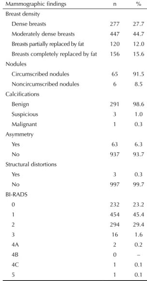
276 (27.6%) had breasts totally or partially replaced by fat. Nodules were observed in 71 women; 65 (91.5%) were circumscribed and six (8.5%) were not circum-scribed. Calciications were observed in 295 mammo-grams; 291 (98.6%) were benign, three (1.0%) were suspicious, and one (0.3%) was malignant. In addition, 63 mammograms (6.3%) were asymmetrical, and three (0.3%) showed structural distortions (Table 2). With regard to BI-RADS categories, 232 (23.2%) were inconclusive (BI-RADS 0), 454 (45.4%) were nega-tive for malignancy (BI-RADS 1), 294 (29.4%) were benign (BI-RADS 2), 16 (1.6%) were probably benign (BI-RADS 3), two (0.2%) had low suspicion of malig-nancy (BI-RADS 4A), one (0.1%) had moderate suspi-cion of malignancy (BI-RADS 4C), and one (0.1%) had high suspicion of malignancy (BI-RADS 5) (Table 2). Of the 1,000 mammograms evaluated, 160 (16.1%) were requested by mastologists, 833 (83.9%) by gyne-cologists, and the remaining seven by other special-ists. Among 833 examinations requested by gynceolo-gists, 53 (5.3%) cases were referred to mastologists. Ultrasound examination was requested for 469 women, with 182 of these being requested concurrently with mammography (Table 3). In the cases that underwent concurrent mammography and ultrasound examina-tions, mastologists requested for both mammography and ultrasound scans in 46.0% of the cases, whereas obstetricians requested these examinations in 13.0% of the cases (p = 0.0001).
Eleven cases underwent cytological examination. Two cytological examinations were considered unsatisfactory in patients with breast nodules and BI-RADS 0. Five examinations revealed nonproliferative benign lesions Figure. Flowchart of participant selection.
Ineligible (n = 515) Women with breast complaints
when the mammography was requested and/or changes in the physical examination and/or high risk Files without reports
(n = 561)
Eligible (n = 1,000) Women aged 40-49 years, asymptomatic and without high risk factors for breast cancer
IMIP Archive Collect files
(n = 2,076)
Files not found or duplicates
(n = 1,498) IMIP Radiology Department
Listing of mammographies of women aged 40-49 years
in patients with breast nodules, with three of these with BI-RADS 0 and two with BI-RADS 1. Four examinations revealed ibroadenoma in patients with breast nodules whose mammographies were BI-RADS 0 (Table 3).
Biopsies were performed in eight cases. Two cases showed nonproliferative lesions in patients with breast nodules
(one with noncircumscribed nodules and BI-RADS 2 and the other with BI-RADS 1 who had already under-gone cytological examinations). Five showed prolifera-tive lesions without atypia in patients with breast nodules, three of which had BI-RADS 0 (one with noncircum-scribed nodule and two with BI-RADS 2). The remaining case showed invasive ductal carcinoma, grade II, with the mammogram showing microcalciications and BI-RADS 5 (Table 3). In this case, the immunohistochemical exami-nation was positive for estrogen and progesterone recep-tors and negative for HER-2 (1+). The patient was in clinical stage IIIb and had undergone neoadjuvant chemo-therapy followed by mastectomy. After surgical treatment, the patient underwent radiotherapy followed by hormone therapy with tamoxifen. At present, the patient is being monitored for breast cancer, without clinical signs of recur-rence or distant disease 10 months after surgery. Table 1. Profile of women aged 40-49 years subjected to
mammographic screening. Recife, PE, Northeastern Brazil, 2010-2011. (N = 1,000)
Variable n %
Age (years)
40 to 44 465 46.5
45 to 49 535 53.5
Ethnicity
Caucasian 137 23.2
Mixed 368 62.4
Black 75 12.7
East Asian 9 1.5
Indigenous 1 0.2
Origin
Recife Metropolitan Area 827 82.7
Other cities in the state 171 17.1
Other states 2 0.2
Education
Illiterate 13 2.2
1 to 3 years completed 104 17.7
4 to 7 years completed 186 31.7
8 to 11 years completed 191 32.6
≥ 12 years completed 92 15.7
Menarche
< 12 years 157 17.6
≥ 12 years 735 82.4
Menopausal status
Premenopausal 681 74.4
Postmenopausal 234 25.6
Use of hormone therapy
Yes 19 2.1
No 886 97.9
Use of oral contraceptives
Yes 58 6.4
No 847 93.6
Breastfeeding
Yes 547 74.4
No 188 25.6
Age at first pregnancy
Nulliparous 95 13.4
< 30 years 556 78.5
≥ 30 years 57 8.0
Table 2. Mammographic findings in women aged 40-49 years who underwent mammography screening. Recife, PE, Northeastern Brazil, 2010-2011. (N = 1,000)
Mammographic findings n %
Breast density
Dense breasts 277 27.7
Moderately dense breasts 447 44.7
Breasts partially replaced by fat 120 12.0
Breasts completely replaced by fat 156 15.6
Nodules
Circumscribed nodules 65 91.5
Noncircumscribed nodules 6 8.5
Calcifications
Benign 291 98.6
Suspicious 3 1.0
Malignant 1 0.3
Asymmetry
Yes 63 6.3
No 937 93.7
Structural distortions
Yes 3 0.3
No 997 99.7
BI-RADS
0 232 23.2
1 454 45.4
2 294 29.4
3 16 1.6
4A 2 0.2
4B 0 –
4C 1 0.1
5 1 0.1
One of the patients with BI-RADS 4A showed dense right axillary lymph nodes and remains under inves-tigation. The other patient with BI-RADS 4A could not be located and the mammogram showed grouped pinpoint calciications. One patient with BI-RADS 4C and pleomorphic microcalciications did not follow up for medical care.
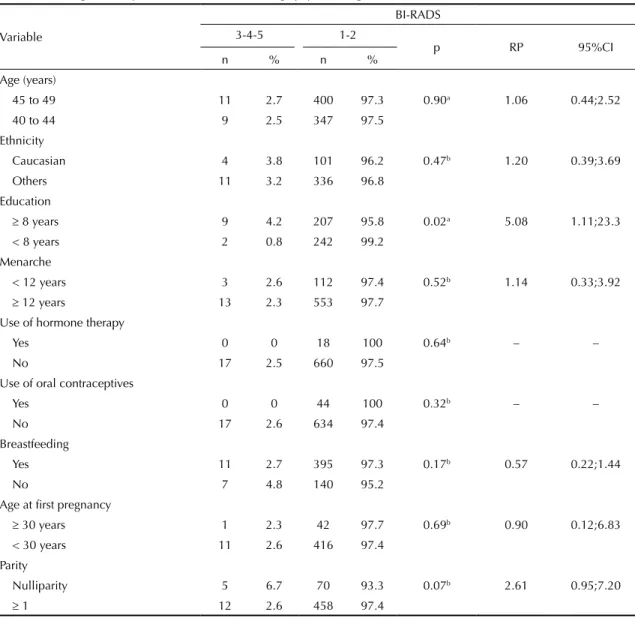
For the purposes of bivariate analysis, women were divided into two groups according to BI-RADS cate-gories (3, 4, and 5 versus 1 and 2). Because it is incon-clusive, BI-RADS 0 cases were excluded. Higher frequency of women with ≥ 8 years of education had BI-RADS categories 3, 4, or 5 than women with BI-RADS 1 and 2 (4.2% versus 0.8%; PR 5.08; 95%CI 1.11;23.3; p = 0.02). Nulliparous women had a higher frequency of BI-RADS categories 3, 4, and 5 (6.7% versus 2.6%) than others; however, this difference was not statistically signiicant (p = 0.07). For the variables age 45-49 years, Caucasian ethnicity, menarche at age < 12 years, use of hormone-replacement therapy, use of oral contraceptives, breastfeeding in at least one previous pregnancy, and age at irst pregnancy ≥ 30 years, no signiicant differences were detected between the two BI-RADS groups (Table 4).
DISCUSSION
Only one case of breast cancer was found among the 1,000 women aged 40-49 years who underwent routine mammography screening. In addition, several addi-tional procedures were performed, but the tests were inconclusive for many of these women.
Age continues to be one of the most important risk factors for breast cancer.17 Our results showed a prevalence of one case of breast cancer among 1,000 women aged 40-49 years. However, the study was limited by its retro-spective nature, and the infeasibility of obtaining histo-pathological results for two patients with BI-RADS 4A and for another with BI-RADS 4C. Nevertheless, even considering these three cases as positive, the number of cases of breast cancer in the age group of 40-49 years would have been four per 1,000. This prevalence is less than that estimated by theNational Cancer Institute in the United States (1 in 69)17 but higher than that of the general population. Results like these lead to controver-sies about the need for screening in this age group by national and international associations.17
The American Cancer Society and the American College of Obstetricians and Gynecologists recommend universal screening for women aged 40-49 years.17 However, according to the consensus reached between the US Preventive Services Task Force and the Canadian Task Force, routine mammography screening in women aged 40-49 years who are not at high risk is not recommended.13,17 In Brazil, the Ministry of Health and the National Cancer Institute (INCA) do not recommend routine mammography screening in this age group,d,e but other institutions have different screening protocols.
Considering the high frequency of breast cancer in Brazil and in the Northeast region as well as dificulties related to access to mammography screening, the current IMIP recommendation is routine examination after age 40 years even for patients with typical risk. In addition, mammography should be performed annually, but this suggestion differs from the INCA recommendations.d As a result of the early screening, 23.0% of the mammo-grams yielded inconclusive results (BI-RADS 0). This high rate of BI-RADS 0 was probably because most patients had dense breast tissue, which impaired the quality of the examination.6,12 Only 20 women had BI-RADS 3-5, i.e., of the 1,000 women who under-went screening, only 20 (2.0%) results required further investigation; on the other hand, 23.0% of the results were inconclusive (BI-RADS 0), and in the end, only one case was conirmed to have breast cancer. Because these mammograms were conducted on asymptomatic women, no women were classiied as BI-RADS 6. Other interventions were also performed, including 469 ultrasound scans, 53 referrals to mastologists, 11 cyto-logical examinations, and eight biopsies, totaling 541 interventions. Consequently, of the 1,000 women who underwent mammography, > 50.0% underwent complementary diagnostic methods, and these exami-nations contributed to a conclusive diagnosis in only a few cases. However, this study was not designed to address this issue. For this reason, future studies are Table 3. Frequency of complementary methods, procedures,
and histopathological findings in women aged 40-49 years who underwent mammography screening. Recife, PE, Northeastern Brazil, 2010-2011. (N = 1,000)
Methods and procedures n %
Ultrasound 469 46.9
Referral to mastologists 53 5.3
Cytological examination 11 1.1
Unsatisfactory 2 0.2
Nonproliferative lesions 5 0.5
Proliferative lesions without atypia 4 0.4
Proliferative lesions with atypia 0 –
Carcinoma 0 –
Biopsy 8 0.8
Histopathological findings
Nonproliferative lesions 2 0.2
Proliferative lesions without atypia 5 0.5
Proliferative lesions with atypia 0 –
In situ carcinoma 0 –
Table 4. Association of biological, sociodemographic, gynecological, and reproductive characteristics with BI-RADS categories 3, 4, and 5 in women aged 40-49 years who underwent mammography screening. Recife, PE, Northeastern Brazil, 2010-2011. (N = 1,000)
Variable
BI-RADS
3-4-5 1-2
p RP 95%CI
n % n %
Age (years)
45 to 49 11 2.7 400 97.3 0.90a 1.06 0.44;2.52
40 to 44 9 2.5 347 97.5
Ethnicity
Caucasian 4 3.8 101 96.2 0.47b 1.20 0.39;3.69
Others 11 3.2 336 96.8
Education
≥ 8 years 9 4.2 207 95.8 0.02a 5.08 1.11;23.3
< 8 years 2 0.8 242 99.2
Menarche
< 12 years 3 2.6 112 97.4 0.52b 1.14 0.33;3.92
≥ 12 years 13 2.3 553 97.7
Use of hormone therapy
Yes 0 0 18 100 0.64b – –
No 17 2.5 660 97.5
Use of oral contraceptives
Yes 0 0 44 100 0.32b – –
No 17 2.6 634 97.4
Breastfeeding
Yes 11 2.7 395 97.3 0.17b 0.57 0.22;1.44
No 7 4.8 140 95.2
Age at first pregnancy
≥ 30 years 1 2.3 42 97.7 0.69b 0.90 0.12;6.83
< 30 years 11 2.6 416 97.4
Parity
Nulliparity 5 6.7 70 93.3 0.07b 2.61 0.95;7.20
≥ 1 12 2.6 458 97.4
BI-RADS: Breast Imaging-Reporting and Data System
a Chi-squared test.
b Fisher’s exact t test.
needed to compare the number of interventions in the 40-49 age group with those of women > 50 years, and cost-beneit studies are needed to evaluate the cost of detection of one case of breast cancer and the cost of all examinations and interventions resulting from this screening. The risk group (BI-RADS categories 3, 4, and 5) included BI-RADS 3 because, despite having a low rate of malignancy (approximately 2.0%), it is considered a risk of developing breast cancer and requires complementary examinations, which may sometimes be unnecessary.
We do not have the natural history of the single case of breast cancer. It has been suggested that some cases of cancer diagnosed by mammography alone would never
have been diagnosed without impacting the women’s survival,5 similar to cases of prostate cancer diagnosed by screening with PSA and/or digital rectal examina-tion.5 The view that early detection of tumors allows curative treatment creates the so-called “time bias”. This scenario favors early detection but has not been supported by solid scientiic evidence.3 In this respect, it is possible that excessive diagnostic examinations are being conducted and that tumors that do not require treatment are being treated.3
the deinitive test results but even after they were declared cancer free.5 In addition, when a meta-anal-ysis was conducted for the subgroup of women aged < 50 years, stratiied by study quality, a signiicant difference in mortality from breast cancer was observed in the studies with a randomized sample.5
However, another meta-analysis evaluated the effec-tiveness of mammographic screening in decreasing mortality from breast cancer in women 39-49 years and yielded different results.10 Seven randomized trials were included and their joint analysis showed a signiicant reduction in mortality from breast cancer due to screening this age group.10 However, the studies included were of variable quality, and after exclusion of three randomized clinical trials conducted before 1980, the overall relative risk of mortality did not decrease signiicantly (RR = 0.87; 95%CI 0.56;1.13). The authors discuss the importance of false-positive results and the adverse effects of screening on the possible reduction of mortality.10 Therefore, women should be informed about the risks and beneits of screening before deciding whether or not to participate in a regular screening program before age 50.3 Furthermore, the number of mammograms performed in an annual screening program starting at age 40 years is almost twice those performed in a program starting at age 50 years and is done biannually. Consequently, radia-tion exposure is doubled.6 Although it has been argued that the amount of radiation from mammography is very low, repeated doses of radiation in more comprehen-sive screening programs pose potential risks that should not be disregarded. A cohort study of 100,000 women showed that annual screening between 40 and 55 years of age and biennial screening ≤ 74 years at a dose of 3.7 mGy for both breasts resulted in 86 radiation-induced cancers and 11 deaths from this type of cancer.19 It was also observed that 72.4% of mammogram results showed dense or moderately dense breasts. Dense breasts are expected for this age group,e although some
authors suggest that this density is a risk factor for breast cancer.16 Consequently, the false-positive rate and the rate of recall for imaging studies are higher and the predictive value of biopsies is lower.e
In addition to age, other studies showed risk factors that favor mammographic screening in the age group of 40-49 years, including breast density, family history, and previous biopsies.18 In the present study, nulliparous women with ≥ 8 years of education had a higher risk of BI-RADS categories 3, 4, and 5. Therefore, future studies should evaluate the beneits of individualized screening in this age group, according to the pres-ence of risk factors for breast cancer, in addition to the existing criteria for moderate risk, including nulliparity and family history of breast cancer after age 50 years. The study design has some limitations. Because the study was conducted in a hospital that solely serves the Uniied Health System (SUS), the proile of women treated at IMIP may be different from that of women assisted in private institutions. Therefore, it is not advis-able to extrapolate the results to the entire population of women aged 40-49 years. On the basis of the results of the present study, it is not possible to draw conclu-sions about mortality reduction. However, these ind-ings are relevant to SUS and should be considered when assessing the cost-effectiveness of the breast cancer screening program. The data from the Breast Cancer Information Systemc should be used for a large-scale evaluation of the results of breast cancer screening in Brazil, including the tests performed in the age group of 40-49 years. It is probable that excessive tests are being recommended and are not yielding consistent beneits for women.
1. American College of Obstetricians and Gynecologists Committee on Gynecologic Practice (US).
ACOG Practice Bulletin No. 122: Breast cancer
screening. Obstet Gynecol. 2011;118:372-82.
DOI:10.1097/AOG.0b013e31822c98e5
2. American College of Radiology. Breast imaging reporting
and data system (BI-RADS®). 4th ed. Reston (US); 2003.
3. Berry DA, Baines CJ, Baum M, Dickersin K, Fletcher SW, Gøtzsche PC, et. al. Flawed Inferences about screening mammography’s benefit based on
observational data. J Clin Oncol. 2009;27(4):639-52.
DOI:10.1200/JCO.2008.17.9341
4. El Saghir NS, Adebamowo CA, Anderson BO, Carlson RW, Bird PA, Corbex M, et. al. Breast cancer management in low resource countries (LRCs): consensus statement from the Breast Health
Global Initiative. Breast. 2011;20(Supll 2):3-11.
DOI:10.1016/j.breast.2011.02.006
5. Gøtzsche PC, Margrethe N. Screening for
breast cancer with mammography. Cochrane
Database Syst Rev. 2011;19;(1):CD001877.
DOI: 10.1002/14651858. CD001877. pub4
6. Heywang-Köbrunner SH, Hacker A, Sedlacek S. Advantages and disadvantages of mammography
screening. Breast Care (Basel). 2011;6(3):199-207.
DOI10.1159/000329005
7. Kettritz U. Screening of Breast Cancer - an
Eternal Discussion Revisited? Breast Care (Basel).
2010;5(2):119-120.
8. Kösters JP, Gøtzsche PC. Regular self-examination or clinical self-examination for
early detection of breast cancer. Cochrane
Database Syst Rev. 2003;(2):CD003373.
DOI: 10.1002/14651858. CD003373. pub2
9. Le-Petross HT, Shetty MK. Magnetic resonance imaging and breast ultrasonography as an adjunct to mammographic screening in high-risk patients.
Semin Ultrasound CT MR. 2011;32(4):266-72.
DOI:10.1053/j.sult.2011.03.005
10. Magnus MC, Ping M, Shen MM, Bourgeois J, Magnus JH. Effectiveness of mammography screening in reducing breast cancer mortality in women aged 39-49
years: a meta-analysis. J Womens Health (Larchmt).
2011;20(6):845-52. DOI:10.1089/jwh.2010.2098
11. Mark K, Temkin SM, Terplan M. Breast self-awareness. the evidence behind the euphemism.
Obstet Gynecol. 2014;123(4):734-6.
DOI:10.1097/AOG.0000000000000139
12. Martins CA, Guimarães RM, Silva RL, Ferreira AP, Gomes FL, Sampaio JR, et al. Evolução da mortalidade por câncer de mama em mulheres jovens: desafios
para uma Política de Atenção Oncológica. Rev Bras
Cancerol. 2013;59(3):341-9.
13. Miller AB, Wall C, Cornelia J Baines CJ, Sun P, To T, et al. Twenty five year follow-up for breast cancer incidence and mortality of the Canadian National Breast Screening Study: randomised screening trial.
BMJ. 2014;348:g366. DOI:10.1136/bmj.g366
14. Moss S, Thomas I, Evans A, Thomas B, Johns L; Trial Management Group. Randomised controlled trial of mammographic screening in women from age 40:
results of screening in the first 10 years. Br J Cancer.
2005;92(5):949-54. DOI:10.1038/sj.bjc.6602396
15. The Canadian Task Force on Preventive Health Care. Recommendations on screening for breast cancer
in average-risk women aged 40-74 years. CMAJ.
2011;183(17):1991-2001. DOI:10.1503/cmaj.110334
16. Tice JA, Cummings SR, Smith-Bindman R, Ichikawa L, Barlow WE, Kerlikowske K. Using clinical factors and mammographic breast density to estimate breast cancer risk: development and validation of a new
predictive model. Ann Intern Med. 2008;148(5):337-47.
DOI:10.7326/0003-4819-148-5-200803040-00004
17. US Preventive Services Task Force. Screening for breast cancer: U.S. Preventive Services Task Force recommendation statement.
Ann Intern Med. 2009;151(10):716-26.
DOI:10.7326/0003-4819-151-10-200911170-00008
18. Van Ravesteyn NT, Miglioretti DL, Stout NK, Lee SJ, Schechter CB, Buist DS, et al. Tipping the balance of benefits and harms to favor screening mammography starting at age 40 years: a comparative modeling
study of risk. Ann Intern Med. 2012;156(9):609-17.
DOI:10.7326/0003-4819-156-9-201205010-00002
19. Yaffe MJ, Mainprize JG. Risk of radiation-induced breast
cancer from mammographic screening. Radiology.
2011;258(1):98-105. DOI:10.1148/radiol.10100655
REFERENCES
Based on the master's dissertation of Silva FX, titled: “Mamografia em mulheres de 40 a 49 anos com risco habitual para
o câncer de mama”, which was submitted to the Postgraduate Stricto Sensu Program in Mother and Child Health at the
Instituto de Medicina Integral Prof. Fernando Figueira, in 2013.