UNIVERSIDADE FEDERAL DO CEARÁ
FACULDADE DE FARMÁCIA, ODONTOLOGIA E ENFERMAGEM PROGRAMA DE PÓS-GRADUAÇÃO EM ODONTOLOGIA
MESTRADO EM ODONTOLOGIA
JULIANNE COELHO DA SILVA
ESTUDO IN VIVO E IN VITRO DA CITOTOXICIDADE E DAS PROPRIEDADES FÍSICO-QUÍMICAS DE RESINAS COMPOSTAS EXPERIMENTAIS PARA
CAPEAMENTO PULPAR DIRETO
JULIANNE COELHO DA SILVA
ESTUDO IN VIVO E IN VITRO DA CITOTOXICIDADE E DAS PROPRIEDADES FÍSICO-QUÍMICAS DE RESINAS COMPOSTAS EXPERIMENTAIS PARA
CAPEAMENTO PULPAR DIRETO
Dissertação de Mestrado apresentada ao
Programa de Pós-Graduação em
Odontologia da Faculdade de Farmácia, Odontologia e Enfermagem da Universidade Federal do Ceará, como requisito parcial para a obtenção do Título de Mestre em Odontologia.
Área de Concentração: Clínica Odontológica. Orientador: Prof. Dr. Victor Pinheiro Feitosa. Co-orientadora: Prof. Dra. Ana Paula Negreiros Nunes Alves
JULIANNE COELHO DA SILVA
ESTUDO IN VIVO E IN VITRO DA CITOTOXICIDADE E DAS PROPRIEDADES FÍSICO-QUÍMICAS DE RESINAS COMPOSTAS EXPERIMENTAIS PARA
CAPEAMENTO PULPAR DIRETO
Dissertação de Mestrado apresentada ao
Programa de Pós-graduação em
Odontologia da Faculdade de Farmácia,
Odontologia e Enfermagem da
Universidade Federal do Ceará como requisito parcial para a obtenção do Título de Mestre em Odontologia.
Aprovada em: ____/____/____
BANCA EXAMINADORA
__________________________________________________ Prof. Dr. Victor Pinheiro Feitosa (Orientador)
Universidade Federal do Ceará (UFC)
__________________________________________________ Prof. Dr. Sérgio Lima Santiago
Universidade Federal do Ceará (UFC)
__________________________________________________ Prof. Dr. Paulo Goberlânio de Barros Silva
AGRADECIMENTOS
A Deus, por ter me concedido saúde e força para superar todos os desafios ao longo desses dois anos de Mestrado.
Aos meus pais, Julio e Graça, pelo amor, incentivo e apoio incondicional; por sempre acreditarem em mim e viver comigo todos os meus sonhos. Amo vocês, e serei eternamente grata.
As minhas irmãs, Isabelle e Nathállia, pelo amor, carinho, compreensão, apoio e colo nas horas difíceis.
Ao meu noivo, Edson Cetira, por sempre acreditar em mim e ser meu grande incentivador nos desafios que surgiram nesses dois anos.
As minhas tias-mães, Gláucia, Núbia e Socorro, pelo amor, colo e afago. Obrigada por estarem sempre presentes ao longo dessa caminhada e a toda a minha família, pelo apoio, carinho e torcida.
Aos grandes amigos que tive a oportunidade de conviver durante o Mestrado, Talita Arrais, David Queiroz, Ana Laura, Adeílson Alves, Diana Cunha, Nara Rodrigues, Luciana Arcanjo, Héllen Suzane, Breno Souza, Marcelo Sidou, Sthefani, Bernardo Aguiar e a todos os demais, agradeço pelo companheirismo, apoio, amizade e por tornarem mais leves os desafios ao longo desses dois anos.
A minhas amigas Talita Albuquerque, Amilene Lemos e Ianara Paz, aos meus amigos do PSOI, Caroline Salema, Pedro Fernandes, Bianca Palhano, Marcella Lima, Sabrina Saldanha e Débora Torquato, e aos amigos da Turma XVIII pela amizade, apoio e torcida.
Ao Team Feitosa, o meu muito obrigada pelo apoio e amizade, Madiana Magalhães, Elisa Martins, Diego Martins, Nayara Sousa e Nara Sena.
Ao meu orientador, Dr. Victor Feitosa, obrigada por todo aprendizado e oportunidades que a mim foram concedidas ao longo do Mestrado.
os integrantes da estomatopatologia pelo apoio e ajuda, em especial ao Doutorando Ernando e ao Dr. Paulo Goberlânio.
Á banca, Dr. Sergio Santiago e Dr. Paulo Goberlânio, por prontamente aceitarem o convite e pelas contribuições que certamente vislumbrarão ao engrandecimento deste trabalho.
RESUMO
O objetivo foi analisar as propriedades físico-químicas de resinas compostas experimentais com partículas bioativas e avaliar a citotoxicidade e a resposta in vivo da polpa ao capeamento pulpar direto. As resinas experimentais foram preparadas e, posteriormente, as nanopartículas de hidroxiapatita adsorvidas com sinvastatina e glutationa foram adicionadas em 0% (Resina Controle), 1% (Res 1%) e 10% (Res 10%) em peso. O hidróxido de cálcio [Ca(OH)2] fotopolimerizável (Ultra-blend Plus, Ultradent) foi utilizado como grupo controle. As propriedades físico-químicas estudadas foram a resistência à flexão e o módulo de elasticidade através de ensaio de flexão de três pontos (n=10), avaliação da liberação de cálcio (n=6) e grau de conversão (n=3) avaliado por espectroscopia Raman. Para a avaliação da citotoxicidade in vitro foram realizados os ensaios de proliferação e contagem celular e as células foram observadas em microscopia confocal com os corantes live/dead. Para a avaliação da resposta pulpar foi realizado o capeamento pulpar direto nos molares inferiores de ratos Wistar (n=6). Os animais foram eutanasiados após 7 dias de realizado o procedimento, e as hemimandíbulas removidas para o preparo de lâminas e análise histológica. A análise estatística foi feita com Kruskall-Wallis e ANOVA com pós-teste de Tukey. O nível de significância adotado foi de 5%. Na resistência à flexão não houve diferença estatística entre os grupos (p>0,05). No módulo de elasticidade, Res 10% apresentou os melhores resultados, mas sem diferença estatística com Res 1%, que não diferiu estatisticamente dos demais grupos. No grau de conversão os resultados da Res 10% foram estatisticamente maiores que as resinas 0% e 1%. Ca(OH)2 demonstrou maior liberação de cálcio em 28 e 45 dias de armazenamento, sem diferença estatística no segundo período com o grupo Res 10%. No ensaio de proliferação celular não foi observada diferença estatística significante. Na análise em microscopia confocal as resinas em menores concentrações mostraram maior viabilidade celular. Na avaliação da resposta pulpar foi possível observar dano pulpar no grupo tratado com o hidróxido de cálcio. Assim, pode-se concluir que a resina experimental pode ser uma alternativa viável ao hidróxido de cálcio Ultra-blend Plus, entretanto são necessários mais estudos que comprovem o reparo do tecido pulpar e a neoformação dentinária.
ABSTRACT
The aim was to investigate the physicochemical properties of experimental composite resins with bioactive particles and to evaluate cytotoxicity and in vivo pulp response to direct pulp capping. The experimental resins were prepared and, subsequently, the hydroxyapatite nanoparticles adsorbed with simvastatin and glutathione were added at 0% (Control Resin), 1% (Res 1%) and 10% (Res 10%) by weight. Photopolymerizable calcium hydroxide [Ca(OH)2] (Ultra-blend Plus, Ultradent) was used as the control group. The physicochemical properties studied were flexural strength and elastic modulus through three-point bending test (n = 10), calcium release (n=6) and degree of conversion (n=3) assessed by spectroscopy Raman. Proliferation and cell counting assays were performed to evaluate in vitro cytotoxicity and the cells were observed in confocal microscopy with live/dead dyes. To evaluate the pulp response, pulp capping was performed directly on the lower molars of Wistar rats (n=6). The animals were euthanized after 7 days of the procedure, and the hemimandibules removed for the preparation of slides and histological analysis. Statistical analysis was performed with Kruskall-Wallis and ANOVA with Tukey post-test. The level of significance was 5%. In flexural strength there was no statistical difference between groups (p> 0.05). In the elastic modulus, Res 10% presented the best results, but revealed no statistical difference with Res 1%, which did not differ statistically from the further groups. In the degree of conversion, the results of the Res 10% were statistically higher than the resins 0% and 1%. Ca(OH)2 showed higher calcium release after 28 and 45 days of storage, with no statistical difference in the second period with the Res 10% group. No significant statistical difference was observed in the cell proliferation assay. By the confocal microscopy analysis, the resins at lower concentrations showed higher cell viability. In the evaluation of the pulp response, it was possible to observe pulp damage in the group treated with calcium hydroxide. Therefore, it can be concluded that the experimental resin may be a viable alternative to Ultra-blend Plus calcium hydroxide; however, further studies are necessary to prove the repair of pulp tissue and dentin neoformation.
SUMÁRIO
1 INTRODUÇÃO GERAL ... 10
2 PROPOSIÇÃO ... 15
2.1 Objetivo Geral... 16
2.2 Objetivos Específicos... 16
3 CAPÍTULO ... 17
4 CONCLUSÃO GERAL... 39
REFERÊNCIAS... 41
ANEXO A – APROVAÇÃO DA COMISSÃO DE ÉTICA NO USO DE ANIMAIS... 48
ANEXO B – APROVAÇÃO DO COMITÊ DE ÉTICA... 49
1 INTRODUÇÃO GERAL
Apesar dos avanços nos materiais dentários e na odontologia preventiva nas últimas décadas, a cárie dentária prevalece até os dias de hoje como uma doença que afeta grande parte da população mundial, estando a sua ocorrência relacionada com o status socioeconômico e o nível de escolaridade (GAÇE; KELMENDI; FUSHA, 2014). Em estudos conduzidos no Brasil, observou-se uma associação entre a maior prevalência da doença com condições socioeconômicas vulneráveis (BOING et al., 2014).
Geralmente, a doença cárie tem seu início no esmalte dentário e a sua progressão pode resultar em lesões profundas, ocasião em que a remoção do tecido cariado pode gerar exposição pulpar, sendo necessário a execução de procedimentos mais invasivos, como tratamento de canal ou extração do dente. O capeamento pulpar direto é um tratamento alternativo com a finalidade de preservar a vitalidade da polpa e pode ser usado em situações de exposição pulpar. A técnica consiste na aplicação de um material protetor diretamente sobre o tecido exposto, entretanto, esse procedimento mostra-se até hoje bastante desafiador para a odontologia restauradora com taxas de sucesso questionáveis (CHEN et al., 2015; ESMERALDO et al., 2013; MODENA et al., 2009; PASHLEY, 1996).
O material dentário a ser utilizado para capeamento pulpar direto deve preencher alguns requisitos, como capacidade de controlar a inflamação e a infecção, apresentar adesão à dentina, promover a formação de dentina terciária e ser biocompatível e bioativo, ou seja, capaz de modular a resposta do tecido pulpar à agressão (FERRACANE; COOPER; SMITH, 2010; MODENA et al., 2009).
Os materiais à base de hidróxido de cálcio são os mais utilizados no capeamento pulpar direto, pois apresentam biocompatibilidade, propriedades antibacterianas, capacidade de estimular a formação de dentina esclerótica e reparadora e custo acessível (KATO et al., 2016; MODENA et al., 2009; SUZUKI et al., 2016). Eles atuam liberando íons de cálcio e hidroxila, os primeiros são responsáveis pelo recrutamento e pela proliferação de células indiferenciadas da polpa dentária, e os últimos exercem ação antibacteriana (GANDOLFI et al., 2015a).
adequadas propriedades mecânicas, mas a grande vantagem do seu uso é a sua capacidade seladora, o que dificulta a infiltração marginal (PARIROKH; TORABINEJAD, 2010; NATALE et al., 2014; SUZUKI et al., 2016). Por outro lado, o MTA apresenta algumas desvantagens, como o longo tempo de presa, o alto custo e a difícil manipulação (PARIROKH; TORABINEJAD, 2010; VARALAKSHMI et al., 2013). Outro material que vem sendo utilizado para capeamento pulpar é o Biodentine®, um novo cimento bioativo à base de silicato de tricálcio, que tem efeitos positivos quando em contato com as células pulpares, estimulando a formação da dentina terciária e promovendo um bom selamento marginal, entretanto apresenta um alto custo (KOUBI et al., 2012; NATALE et al., 2014).
Estudos vêm mostrando que a sinvastatina, uma estatina reconhecida como um medicamento seguro e barato para o tratamento de hiperlipidemia apresenta outras funções, que são os chamados efeitos pleiotrópicos, como efeito anti-inflamatório, indução de angiogênese e melhora na formação óssea (BLANCO-COLIO et al., 2003; MORASCHINI et al., 2018). Em relação aos efeitos anti-inflamatórios, as estatinas têm mostrado regular as moléculas de adesão celular. Em 2002, Seljeflot et al demonstraram que a sinvastatina reduz os níveis plasmáticos de molécula de adesão intercelular-1 (ICAM-1), molécula de adesão vascular (VCAM-1), E-selectina e P-selectina, as quais são essenciais para o primeiro passo de recrutamento dos monócitos. Além disso, estudos apontam que elas interferem no processo de entrada dos monócitos na parede arterial, passo seguinte ao seu recrutamento, através da redução da expressão da proteína quimiotática de monócitos (MCP-1) e de interleucina-8 (IL-8). Alguns marcadores inflamatórios também mostraram ser afetados pelas estatinas, como a proteína-C reativa. A sinvastatina também mostrou reduzir os níveis das citocinas pró-inflamatórias fator de necrose tumoral-alfa (TNF-α) e interleucina-1-beta (IL-1β). Esses mecanismos podem estar relacionados tanto com a diminuição da produção de lipídeos, como ser independentes desse processo (BLANCO-COLIO et al., 2003; SELJEFLOT et al., 2002).
pirofosfato (FPP) e geranilgeranilpirofosfato (GGPP) que são usados para a modulação de importantes proteínas celulares, como a proteína G (BLANCO-COLIO et al., 2003).
Outro efeito é a indução de angiogênese, mecanismo relacionado com a habilidade das estatinas em ativar as proteínas quinase Akt / PKB em células endoteliais, que possuem a capacidade de fosforilar a enzima óxido nítrico sintetase (NOS) endotelial, responsável pela inibição da NOS induzida que ocasiona inflamação. Esta enzima leva a um aumento da produção de óxido nítrico derivado do endotélio, que por sua vez é um importante mediador dos mecanismos pró-angiogênicos com funções protetoras ao endotélio, como inibição da adesão plaquetária e inflamação, ocasionando a inibição da apoptose das células endoteliais e formação óssea (KUREISHI et al., 2000).
A sinvastatina melhora a função dos osteoblastos através da expressão de proteínas morfogenéticas ósseas e suprime a função dos osteoclastos, resultando em formação óssea aumentada (MAEDA et al., 2001; MORASCHINI et al., 2018). Também já foi demonstrado uma melhor função dos odontoblastos, levando à diferenciação de células pulpares em secretoras de matriz dentinária (OKAMOTO et al., 2009). Varalakshmi et al. (2013) demonstraram que a associação entre sinvastatina e fosfato de tricálcio induziu a diferenciação odontogênica, o que foi evidenciado pela identificação de marcadores osteogênicos/odontogênicos, como proteína óssea morfogenética (BMP-2), fosfatase alcalina (ALP), osteonectina (OCD), dentre outros, além disso observaram a formação de nódulos de mineralização. A sinvastatina induz significativamente a expressão do gene sialofosfoproteína dentinária (DSPP) e mostra uma alta taxa de formação de tecido duro após sete dias nas células tronco da polpa dental (OKAMOTO et al., 2009; ASL AMINABADI et al, 2013).
As pesquisas relatam que a citotoxicidade provocada pelas resinas pode ser ocasionada pela depleção dos níveis de glutationa, que é um essencial agente antioxidante intracelular, e também pela produção das espécies reativas de oxigênio no tecido pulpar (NASSAR et al., 2013). Samuelsen et al em 2011 sugeriram que os monômeros interagem com a glutationa intracelular, aumentando o estresse oxidativo no interior das células. Dessa forma, a utilização de sistemas adesivos e de resinas compostas sobre a polpa exposta está contraindicada, devendo ser assegurada uma espessura de um milímetro de dentina remanescente para a sua utilização (HEBLING et al., 1999; NASSAR et al., 2013; NASSAR et al., 2014). Entretanto, estudos vêm mostrando que algumas drogas podem ser utilizadas para reduzir a citotoxicidade causada pelas resinas, como a glutationa que tem um significativo efeito antioxidante, e assim, podem atuar favorecendo a viabilidade celular. Contudo, o principal mecanismo para a eliminação da citotoxicidade pode ser explicado através da interação direta da cisteína com o 2-hidroxietil metacrilato (HEMA) e com outros monômeros resinosos, que dificulta a entrada destes agentes no interior das células pulpares (NASSAR et al., 2013; NASSAR et al., 2014).
2 PROPOSIÇÃO
2.1 Objetivo Geral
Formular e avaliar as propriedades físico-químicas de resinas experimentais contendo partículas bioativas adsorvidas com sinvastatina e glutationa, sua citotoxicidade in vitro e sua influência como material de capeamento na polpa dental de ratos.
2.2 Objetivos Específicos
- Formular uma resina experimental contendo partículas bioativas adsorvidas com sinvastatina e glutationa.
- Avaliar o grau de conversão da resina experimental.
- Analisar a resistência à flexão e módulo de elasticidade da resina experimental.
- Quantificar a liberação de cálcio da resina experimental.
3 CAPÍTULO
Esta dissertação está baseada no Artigo 46 do Regimento Interno do Programa de Pós-Graduação em Odontologia da Universidade Federal do Ceará que regulamenta o formato alternativo para dissertações de Mestrado e teses de Doutorado, e permite a inserção de artigos científicos de autoria ou coautoria do candidato. Por se tratar de estudos envolvendo animais e seres humanos, ou parte deles, o projeto de pesquisa foi submetido à apreciação da Comissão de Ética no Uso de Animais –CEUA e do Comitê de Ética da Universidade Federal do Ceará, tendo sido aprovado. Assim sendo, esta dissertação é composta de um artigo científico que será submetido ao periódico Journal of Dental Research (JDR), conforme descrito abaixo:
CYTOTOXICITY AND PHYSICAL PROPERTIES OF NEW COMPOSITES FOR DIRECT PULP CAPPING
SILVA JC, FERREIRA-JÚNIOR AEC, SILVA PGB, MOTA MRL, NUNES AP, MIRON
ABSTRACT
The aim was to investigate the physicochemical properties of experimental composite resins with bioactive particles and to evaluate cytotoxicity and in vivo pulp response to direct pulp capping. The experimental resins were prepared and, subsequently, the hydroxyapatite nanoparticles adsorbed with simvastatin and glutathione were added at 0% (Control Resin), 1% (Res 1%) and 10% (Res 10%) by weight. Photopolymerizable calcium hydroxide [Ca(OH)2] (Ultra-blend Plus, Ultradent) was used as the control group. The physicochemical properties studied were flexural strength and elastic modulus through three-point bending test (n = 10), calcium release (n=6) and degree of conversion (n=3) assessed by spectroscopy Raman. Proliferation and cell counting assays were performed to evaluate cytotoxicity in vitro and the cells were observed in confocal microscopy with live/dead dyes. To evaluate the pulp response, pulp capping was performed directly on the lower molars of Wistar rats (n=6). The animals were euthanized after 7 days of the procedure, and the hemimandibles removed for the preparation of slides and histological analysis. Statistical analysis was performed with Kruskall-Wallis and ANOVA with Tukey post-test. The level of significance was 5%. In flexural strength there was no statistical difference between groups (p> 0.05). In the elastic modulus, Res 10% presented the best results, but revealed no statistical difference with Res 1%, which did not differ statistically from the further groups. In the degree of conversion, the results of the Res 10% were statistically higher than the resins 0% and 1%. Ca(OH)2 showed higher calcium release after 28 and 45 days of storage, with no statistical difference in the second period with the Res 10% group. No significant statistical difference was observed in the cell proliferation assay. By the confocal microscopy analysis, the resins at lower concentrations showed higher cell viability. In the evaluation of the pulp response, it was possible to observe pulp damage in the group treated with calcium hydroxide. Therefore, it can be concluded that the experimental resin may be a viable alternative to Ultra-blend Plus calcium hydroxide; however, further studies are necessary to prove the repair of pulp tissue and dentin neoformation.
INTRODUCTION
Dental caries remains today as a disease that affects much of the world
population, mainly in the countries with low socioeconomic conditions (Boing et al.
2014; Gaçe et al. 2014). Deep caries, as well as others damages, such as traumatic
injury can lead to pulpal exposure (Gaçe et al. 2014). In this regard, pulp therapy is
performed to maintain pulp vitality of a tooth (Gandolfi et al. 2015). The treatment
consists on the application of a material directly onto the exposed tissue (Modena et
al. 2009). These protective materials should attain bioactive properties to promote pulp
cell activity and pulp healing, and the formation of new reparative dentin (Ferracane et
al. 2010; Gandolfi et al. 2015).
Calcium hydroxide-based products have been recommended for the treatment
of exposed pulp due to their property of stimulating dentin formation and antibacterial
action (Kato et al. 2016; Modena et al. 2009). However, high solubility and fast
dissolution are well-known shortcomings of conventional calcium hydroxide materials
(Gandolfi et al. 2015). Although there are several materials available, neither of them
possesses all desirable characteristics. Therefore, direct pulp capping remains
challenging for operative dentistry.
Hydroxymethylglutaryl-coenzyme A reductase inhibitors are used as
cholesterol-lowering drugs (Blanco-colio et al. 2003). Previous papershave observed
that statins may increase the mRNA expression of bone morphogenetic protein 2
(BMP-2) in osteoblasts, promoting bone formation as result (Moraschini et al. 2018;
Mundy et al., 1999). In last decade, the introduction of simvastatin in Dentistry has
increased (Aminabadi et al. 2013; Moraschini et al. 2018; Varalakshmi et al. 2013).
formation of mineralized nodules, thereby showing to be a promising pulp capping
agent (Okamoto et al. 2009; Varalakshmi et al. 2013).
Resin composites are materials with good physical properties. However, they
cannot be applied directly on the pulp tissue due to harsh pulp irritation, necrosis and
stimulation of glutathione depletion, (Chang et al. 2005; Hanks et al. 1991; Hebling et
al. 1999; Modena et al. 2009; Samuelsen et al. 2011; Stanley et al. 1975). Recent
studies have shown that some drugs may be used to reduce this cytotoxicity, such as
glutathione, which interacts with resin monomers thereby impairing the entrance in
pulp cells and increasing cell viability (Nassar et al. 2013; Nassar et al. 2014).
The aim of this study was to evaluate selected physicochemical properties, cell
proliferation and cell viability of human dental pulp (HDP) cells an experimental
composite containing bioactive particles adsorbed with simvastatin and glutathione.
Furthermore, other objective was to assay the in vivo response of exposed pulps in
rats to these materials. Study hypothesis is that the incorporation of bioactive particles
on experimental composite resin interferes directly in the physicochemical properties,
cytotoxicity in pulp cells and in vivo response.
MATERIALS AND METHODS
Experimental composites
The monomeric blend of experimental resin composite was composed of 20
wt% Bisphenol-A-glycerolate-dimethacrylate (BisGMA), 68.5 wt%
urethane-dimethacrylate (UDMA) and also by 10 wt% glycerol-urethane-dimethacrylate-phosphate
(GDMA-P), the acidic functional monomer. The photoinitiator system was composed
benzoate and 0.5 wt% camphorquinone. Then, the experimental composites were
produced by mixing filler particles: i) Res 0% - without bioactive fillers + 50 wt%
silanated barium glass fillers (0.7 µm mean size, Esstech Inc. Essignton, USA); ii) Res
0.1% - 0.1 wt% hydroxyapatite nanoparticles adsorbed with glutathione + 0.1 wt%
hydroxyapatite nanoparticles adsorbed with simvastatin + 0.1 wt% beta tricalcium
phosphate fillers (Sigma Aldrich, St. Louis, USA) + 49.7 wt% silanated barium glass
(Esstech) (exclusively for the cell proliferation assay); iii) Res 1% - 1 wt%
hydroxyapatite with glutathione + 1 wt% hydroxyapatite with simvastatin + 1 wt% beta
tricalcium phosphate + 47 wt% silanated barium glass; iv) Res 10% - 10 wt%
hydroxyapatite with glutathione + 10 wt% hydroxyapatite with simvastatin + 10 wt%
beta tricalcium phosphate + 20 wt% silanated barium glass. In total, all composites had
50wt% resin blend and 50wt% fillers. Photopolymerizable calcium hydroxide [Ca(OH)2]
(Ultra-blend Plus, Ultradent) was used as the control group.
Degree of conversion
The degree of conversion (DC) of the experimental composites was undertaken
following a modified protocol previously described by Feitosa et al. (2014). Briefly, each
material (n=3) was placed in a glass plate and positioned in a Raman
micro-spectrophotometer (Xplora, Horiba, Paris, France) with 532 nm wavelength argon laser
and 3.2 mW power. The spectra were assessed before and subsequent to light
activation (40 s; 1100 mW/cm2, DB685, Dabi Atlante, Ribeirão Preto, Brazil). All
spectra were obtained in a range of 1500–1700 cm-1. The peak height was determined subsequent to baseline subtraction and normalization process. The residual unreacted
carbon–carbon double bond content (% C=C) was determined from the ratio of absorbance intensities of aliphatic C=C bonds (peak at 1637 cm-1) against an internal
after starting photo-curing. Degree of conversion was determined by subtracting the
C=C% from 100%. The analyses were performed in triplicate.
Flexural strength and elastic modulus
Flexural strength and elastic modulus were tested in bar-shaped specimens (7
x 1 x 1 mm, n = 10). After 24 h at 37 °C and 100% humidity, the specimens were
fractured under three-point bending in a universal testing machine (Instron, Canton,
USA) at 1 mm/min crosshead speed. Flexural strength (FS) and elastic modulus (E)
were calculated according to the equations below:
𝜎𝐹𝑆 =2 𝑥 𝑤 𝑥 ℎ3 𝑥 𝐿 𝑥 𝑑2 𝐸 = 4 𝑥 𝑤 𝑥 ℎ𝐿 𝑥 𝐷33 𝑥 𝑑
where L is the failure load (for flexural strength) or the load recorded in the elastic
region of the load x displacement curve (for elastic modulus), in Newton’s, d is the distance between the supports (5 mm), w is the width and h in the thickness of the
specimen (both in mm), and D is the displacement (in mm) of the cross-head
corresponding to L. The machine software (Bluehill, Instron) automatically calculated
and reported the maximum flexural strength and elastic modulus in megapascals.
Calcium release
Six disk-shaped samples from each material (n=3) were prepared by using
silicone models, according to the manufacturers' instructions. The samples were stored
in distilled water and storage solutions were changed after 28 and 45 days. The
calcium release was determined by mixing the storage solutions with arsenazo III in
20mM HEPES at pH 7.4 (Sigma Aldrich, St. Louis, USA). The analysis of calcium
release through this solution was performed using a UV-Visible spectrophotometer
Arsenazo III colorimetric method (Grienberger and Konnerth 2012). Aliquots of 5 μL of the samples (diluted 1:10 and partially neutralized) were added to 50 μL of deionized water before UV-Vis analysis. For calibration, standards containing 40 to 200 μg Ca/mL solutions (Sigma Aldrich) were used.
Cell culture and Proliferation assay
Human dental pulp (HDP) fibroblasts and MC3T3 osteoblastic cells were
seeded onto tiny disks of each material in 96-well plates at a density of 10,000 cells
per well either group. During cell seeding, α-MEM medium (Invitrogen, Basel, Switzerland) was supplemented with 50μg/mL ascorbic acid (Invitrogen) and 2 mM β -glycerophosphate (Invitrogen) to promote proliferation/differentiation (Miron et al.
2016). Cells were quantified using fluorescent MTT assay (Invitrogen) at 1, 3, and 5
days for cell proliferation as previously described (Miron et al., 2013). At desired time
points, cells were washed with phosphate-buffered solution (PBS) and quantified using
a fluorescence plate reader (Infinite 200, Tecan Group Ltd. Männedorf, Switzerland).
Experiments were performed in triplicate with three independent experiments for each
condition.
Pulpal Response
According to the sample calculation based on the study of LIU et al. (2015) that
after one week of exposure of the pulp chamber of rats submitted to direct pulp capping
treatment, no animals of the control group (n = 0, 0.0%) presented mineralized tissue
deposition versus 75.0% of the animals treated with MTA (n = 3), a sample of 6 mice
per study group was estimated in order to obtain a sample that represents 80% of the
power and 95% confidence to modify the biological behavior after capping pulping with
(Rattus novergicus) were used (n=6). Each animal had two of the pulp capping
materials randomly applied to each of the two lower molars according to the material
that was used: i) without restorative procedure; ii) photopolymerizable calcium
hydroxide (Ultra-blend Plus, Ultradent Products Inc., South Jordan, USA); iii) Res 0%
- without bioactive fillers + 50 wt% silanated barium glass fillers (0.7 µm mean size,
Esstech Inc. Essignton, USA); iv) Res 10% - 10 wt% hydroxyapatite with glutathione +
10 wt% hydroxyapatite with simvastatin + 10 wt% beta tricalcium phosphate + 20 wt%
silanated barium glass. The animals were euthanized by barbiturates (sodium
pentobarbital, 150 mg/kg, intraperitoneal) after 7 days, and the hemimandibules were
removed surgically for the preparation of histological slides (hematoxylin-eosin) as
previously described by Timm (2005). Then, qualitative analysis was performed by
conventional light microscopy.
Statistical Analysis
All data were compiled and tested by Shapiro-Wilk normality test and
homoscedasticity test (α=0.05). After passing these first tests, they were analyzed with one-way ANOVA and Tukey’s post-hoc test (p<0.05). In the case variance was not equal and data were not normal, Kruskall-Wallis test was performed (p<0.05) to
compare groups.
RESULTS
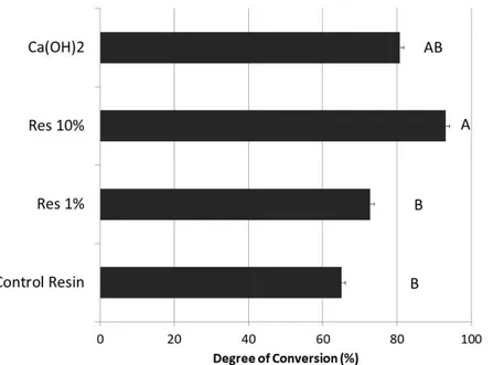
In the results of degree of conversion, depicted in Figure 1, there was no
statistical differences between groups Res 0%, Res 1% and Ca(OH)2. Nevertheless,
(p=0.004) and Res 1% (p=0.022), but was similar to Ca(OH)2. The flexural strength
(Fig. 2) showed no significant differences among groups (p>0.05). Contrariwise,
outcomes of elastic modulus (Fig. 3) demonstrated significant differences among
groups. Res 10% exhibited higher modulus than Res 0% and Ca(OH)2 (p<0.001 and
p=0.005 respectively), but without statistically significant difference with the Res 1%
(p=0.14), which depicted intermediate results without significant difference to all other
groups. The findings of calcium release assay (Fig. 4) demonstrated the highest
release for Ca(OH)2 group in 28 and 45 days of storage, but without significant
difference with Res 10% in 45 days.
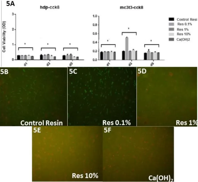
The results of cell proliferation assay showed no statistical significant difference
in the cell counting among all groups in the culture of hdp-cck8 cells, although in the
culture of mc3t3-cck8 the cell viability was higher with the Res 0.1% in 3 or 5 days.
The images of live/dead confocal investigation (Fig. 6) obtained by confocal laser
microscopy demonstrated that Res 0.1% (Fig. 6-B) improved the cell viability when
compared with Res 0% (Fig. 6-A). However, with the increase in concentration of
bioactive fillers, the rate of cell death was higher (Fig. 6-D).
In the results of pulpal response, it was possible to observe cell ectasia in the
group without procedure in the deep pulp. The teeth restored with Res 10%
demonstrated moderate cell ectasia in the deep pulp. The findings of the group
restored with Res 0% demonstrated a large cell ectasia. In some teeth restored with
Ca(OH)2 it was possible to find cellular damage in a different pattern from the previous
DISCUSSION
The results herein demonstrated that tested composite resin with high (10%)
concentration of nanoparticles is cytotoxic and causes cellular damage. However, the
incorporation of bioactive fillers in an experimental resin in low concentrations reduced
its cytotoxicity without significantly impairing their physicochemical properties.
Therefore, the hypothesis that the incorporation of bioactive particles in an
experimental composite resin interferes directly in the physicochemical properties,
cytotoxicity in pulp cells and in vivo response should be accepted.
According to findings of degree of conversion (DC), Res 10% demonstrated
higher polymerization when compared with resin without nanoparticles (Res 0%). This
composite formulation had large amount of drugs (simvastatin and glutathione)
adsorbed onto nanoparticles. Both compounds are antioxidant and might have created
blocking of free radicals during photoinitiation, thereby hampering polymerization.
However, during preparation of these experimental composites, drugs were adsorbed
onto hydroxyapatite nanocarriers, rather than dissolved in the co-monomer blend.
Such procedure was intentionally undertaken with the aim to avoid higher
polymerization inhibition.
Furthermore, the calcium phosphates (hydroxyapatite and beta-tricalcium
phosphate) added to the composite increased polymerization, what is in accordance
with Andrade Neto et al. (2016) which concluded that crystalline hydroxyapatite
nanorods included in resin infiltrants improves the DC. This may occur due to
crystallinity of hydroxyapatite, which increase light scattering and consequently the
calcium phosphates, when dissolved in the resin, might lead to higher polymerization
thanks to the proton-releasing ability of phosphates, thereby acting as catalysts what
may even induce spontaneous polymerization (Liu et al. 2016; Zhang et al. 2013,
Zhang and Wang 2012).
The flexural strength outcomes (Fig. 2) depicted no significant difference among
groups. However, contrasting elastic moduli were attained (Fig. 3), as Res 10%
showed highest stiffness. Indeed, a suitable explanation for this finding relies on the
higher polymerization of composite resin containing 10% bioactive fillers (Fig. 1).
Ultra-blend Plus [Ca(OH)2 material]showed lower elastic modulus, what may be attributed
to its formulation composed basically by calcium hydroxide with minor concentrations
of hydroxyapatite and urethane-dimethacrylate, whilst the composites presented a
great quantity of cross-linking monomers and reinforcing filler particles which improved
the rigidity (Aljabo et al. 2015; Kangwankai et al. 2017).
By analyzing calcium release (Fig. 4), light-curable calcium hydroxide
(Ultra-blend Plus, Ultradent Products Inc., South Jordan, United States) showed the higher
results. In fact, such outcome was expected thanks to the composition of this material.
The high release indeed has occurred because soluble calcium is the base
composition of this material. Furthermore, in accordance with a preliminary study,
Ultra-blend provide the greatest release of calcium ionswith a significant difference
observed between others cements based on calcium hydroxide, which might also be
attributed to the presence of hydroxyapatite (Duarte et al. 2007).
The results of cell viability demonstrated no statistical difference among groups
(Fig. 5), which may indicate that the new pulp capping might be a potential replacement
live/dead obtained by confocal microscopy suggested that composite resin with 0.1%
bioactive fillers (Fig. 6B) may improve the cell viability when compared with composites
containing 10% bioactive fillers and the that without bioactive fillers (0%), which is the
conventional composite resin used in the clinics. These findings may indicate that
glutathione in low concentrations is able to reduce the oxidative stress and the cell
death caused by resin monomers.
These results corroborate with the findings of Nassar et al. (2013) that evaluated
the effect of glutathione on cell viability. They concluded that glutathione in controlled
concentrations had a protective effect against HEMA cytotoxicity. The high
concentration of simvastatin may have decreased cell viability in groups with 1% and
10% bioactive nanoparticles (Fig. 6C and 6D), hypothesis that is supported by the
investigation of Aminabadi et al., (2013) whose findings revealed a higher rate of pulp
inflammation and necrosis by increasing the concentration of simvastatin. Okamoto et
al. (2009) examined the effects of statin on the cell proliferation of human dental pulp
stem cell and their results indicated suppression of proliferation, which is mediated
through the inhibition of the mevalonate and Rho pathways caused by statins. Besides,
they inhibited actin fiber formation and cell cycle progression that is regulated by Rho
(Baek et al. 2005; Tanaka et al. 2000).
Although the qualitative analysis of the slides produced with hemi jaws of the
rats (Fig. 7), it is possible to conclude that calcium hydroxide caused more cellular
damage in pulp. These is in agreement with previous studies, such as that of Scarano
et al. (2003), which found a small area of necrosis in pulps capped with Ultra-blend
Plus. When compared with Res 10%, the standard of damage is very different, likely
due to presence of bioactive fillers releasing glutathione and simvastatin, both showing
(Varalakshmi et al. 2013). The group without particles demonstrated vastly
disorganized pulpal tissue that may indicate necrosis, which was reported in previous
studies due to cytotoxicity caused by resin monomers (Hebling et al. 2009).
The findings of this study showed that the incorporation of adsorbed simvastatin
and glutathione on calcium phosphate nanoparticles in an experimental resin
composite did not impair the physicochemical properties, even increasing the
polymerization. However, low concentrations of bioactive fillers improve the cell
viability when compared with the traditional resin and light-curable calcium hydroxide.
There are preliminary studies, which report that simvastatin has been tested and
proven both in the laboratory and in vivo the excellent ability to manage pulpal tissue
biomodulation. Moreover, glutathione has the ability to reduce the cytotoxicity caused
by the resin monomers and there are no reports so far regarding a composite resin for
direct pulp capping that incorporates such bioactive substances.
Indeed, the major advantage of this composite is to be adhesive and
self-etching, eliminating acid conditioning and adhesive application, which when performed
directly on the pulp tissue causes cellular damage and intense inflammation. The
clinical relevance of the topic may be the possibility of turning pulp capping procedures
faster and more efficient without the use of temporary cements and restorations.
Indeed, pulp capping is still a challenging procedure for dental practice, where
materials still show a relevant annual failure rate (Hilton et al. 2013). However, further
studies are needed to prove the regeneration of pulp tissue and its mineralization over
REFERENCES
Aljabo A, Xia W, Liaqat S, Khan MA, Knowles JC, Ashley P, Young AM. 2015. Conversion, shrinkage, water sorption, flexural strength and modulus of re-mineralizing dental composites. Dental Materials. 31(11):1279-89.
Andrade Neto DM, Carvalho EV, Rodrgues EA, Feitosa VP, Sauro S, Melec G, Carboned L, Mazzeto SE, Rodrigues LF, Fechine PBA. 2016. Novel hydroxyapatite nanorods improveanti-caries efficacy of enamel infiltrants. Dental Materials. 32(6): 784–793.
Asl Aminabadi N, Maljaei E, Erfanparast L, Aghbali AA, Hamishehkar H, Najafpour E. 2013. Simvastatin versus Calcium Hydroxide Direct Pulp Capping of Human Primary Molars: A Randomized Clinical Trial. J Dent Res Dent Clin Dent Prospects. 7(1):8-14.
Baek KH, Lee WY, Oh KW, Tae HJ, Lee JM, Lee EJ, Han JH, Kang MI, Cha BY, Lee KW, Son HY, Kang SK. 2005. The effect of simvastatin on the proliferation and differentiation of human bone marrow stromal cells. J Korean Med Sci. 20(3):438-44
Blanco-colio LM, Tuñon J, Martín-ventura JL, Egido J. 2003. Anti-inflamatory and immunomodulatory effects of statins. Kidney International. 63:12-23.
Boing AF, Bastos JL, Peres KG, Antunes JLF, Perese MA. 2014. Social determinants of health and dental caries in Brazil: a systematic review of the literature between 1999 and 2010. Rev Bras Epidemiol Suppl D.S.S. 17(2):102-115
Chang HH, Guoa MK, Kastenb FH, Chang MC, Huanga GF, Wanga YL, Wanga RS, Jenga JH. 2005. Stimulation of glutathione depletion, ROS production and cell cycle arrest of dental pulp cells and gingival epithelial cells by HEMA. Biomaterials 26:745– 753.
Duarte MA, Martins CS, de Oliveira Cardoso Demarchi AC, de Godoy LF, Kuga MC, Yamashita JC. 2007. Calcium and hydroxide release from different pulp-capping materials. Oral Surg Oral Med Oral Pathol Oral Radiol Endod.104(1):66-69.
on the physicochemical properties of self-etch dental adhesives. J Dent. 42(5):565-574.
Ferracane JL, Cooper PR, Smith AJ. 2010. Can interaction of materials with the dentin-pulp complex contribute to dentin regeneration? Odontology. 98(1):2-14.
Gaçe E, Kelmendi M, Fusha E. 2014. Oral Health Status of Children with disability living in Albania. ORIGINAL PAPER. Mater Sociomed. 26(26):392-394.
Gandolfi MG, Spagnuolo G, Siboni F, Procino A, Rivieccio V, Pelliccioni GA, Prati C, Rengo S. 2015. Calcium silicate/calcium phosphate biphasic cements for vital pulp therapy: chemical-physical properties and human pulp cells response. Clin Oral Invest. 19(8):2075-2089.
Grienberger C, Konnerth A. 2012. Imaging Calcium in Neurons. Neuron. 73(5):862-885.
Hanks CT, Strawn SE, Watacha JC, Craig RG. 1991. Cytotoxic effects of resin components on cultured mammalian fibroblasts. J Dent Res. 70(11):1450-1455.
Hilton TJ, Ferracane JL, Mancl L; 2013. Comparison of CaOH with MTA for direct pulp capping: a PBRN randomized clinical trial. J Dent Res. 92(7 Suppl):16-22.
Hebling J, Giro EMA, Costa CA. 1999. Biocompatibility of an adhesive system applied to exposed human dental pulp. J Endod. 25(10):676-682.
Hebling J, Lessa FC, Nogueira I, Carvalho RM, Costa CA. 2009. Cytotoxicity of resin-based light-cured liners. Am J Dent. 22(3):137-42.
Kangwankai K, Sani S, Panpisut P, Xia W, Ashley P, Petridis H, Young AM. 2017. Monomer conversion, dimensional stability, strength, modulus, surface apatite precipitation and wear of novel, reactive calcium phosphate and polylysine-containing dental composites. PLoS One. 12(11):e0187757.
Liu S, Wang S, Dong Y. 2015. Evaluation of a Bioceramic as a Pulp Capping Agent In Vitro and In Vivo. JOE. 41(5):652-657.
Liu Y, Bai X, Liu YW, Wang Y. 2016. Light-Cured Self-Etch Adhesives Undergo Hydroxyapatite-Triggered Self-Cure. J Dent Res. 95(3):334-41.
Miron RJ, Saulacic N, Buser D, Iizuka T, Sculean A. 2013. Osteoblast proliferation and differentiation on a barrier membrane in combination with BMP2 and TGFbeta1. Clinical oral investigations. 17:981-988.
Miron RJ, Chandad F, Busert D, Sculean A, Cochran D, Zhang Y. 2016. Effect of Enamel Matrix Derivative (EMD)-Liquid on Osteoblast and Periodontal Ligament Cell Proliferation and Differentiation. Journal of Periodontology. 87(1): 91-99.
Modena KC, Casas-apayco LC, Atta MT, Costa CA, Hebling J, Sipert CR, Navarro MFL, Santos CF. 2009. Cytotoxicity and biocompatibility of direct and indirect pulp capping materials. J Appl Oral Sci. 17(6):544-554.
Moraschini V, Almeida DCF, JM. Diuana, Calasans-Maia JA, Diuana Calasans-Maia M, 2018. The ability of topical and systemic statins to increase osteogenesis around dental implants: a systematic review of histomorphometric outcomes in animal studies. Int J Oral Maxillofac Surg. 47(2):1-9
Mundy G, Garret R, Harris S, Chan J, Chen D, Rossini G, Boyce B, Zhao M, Gutierrez G. 1999. Stimulation of Bone Formation in Vitro and in Rodents by Statins. Science. 286:1946-1949.
Nassar M, Hiraishi N, Islam MS, Tamura Y, Otsuki M, Kasugai S, Ohya K, Tagami J, Tay FR. 2013a. The effect of glutathione on 2-hydroxyethylmethacrylate cytotoxicity and on resin–dentine bond strength. International Endodontic Journal. 47:652–658.
Nassar M, Hiraishi N, Shimokawac H, Tamura Y, Otsuki M, Kasugai S, Ohya K, Tagami J. 2013b. The inhibition effect of non-protein thiols on dentinal matrixmetalloproteinase activity and HEMA cytotoxicity. Journal of dentistry. 42:312–318.
Differentiation of Human Dental Pulp Stem Cells In Vitro and In Vivo. Journal of Endodontics. 35(3):367-372. .
Samuelsen JT, Kopperud HM, Holme JA, Dragland IS, Christensen T, Dahl JE. 2011. Role of thiol-complex formation in 2-hydroxyethyl-methacrylate-induced toxicity in vitro. Journal of Biomedical Materials Research. 96:395-401.
Scarano A, Manzon L, Di Giorgio R, Orsini G, Tripodi D, Piattelli A. 2003. Direct capping with four different materials in humans: histological analysis of odontoblast activity. J Endod. 29(11):729-34.
Stanley HR, Going RE, Chauncey HH. 1975. Human pulp response to acid pretreatment of dentin and to composite restoration. J Am Dent Assoc. 91:817-825.
Tanaka T, Tatsuno I, Uchida D, Moroo I, Morio H, Nakamura S, Noguchi Y, Yasuda T, Kitagawa M, Saito Y, et al. 2000. Geranylgeranyl-pyrophosphate, an isoprenoid of mevalonate cascade, is a critical compound for rat primary cultured cortical neurons to protect the cell death induced by 3-hydroxy-3-methylglutaryl-CoA reductase inhibition. J Neurosci. 20(8):2852-9.
Timm LL. 2005. Routine techniques of preparation and analysis of histological slides. Caderno La Salle XI. 2(1):231 – 239.
Varalakshmi PR, Kavitha M, Govindan R, Narasimhan S. 2013. Effect of Statins with a-Tricalcium Phosphate on Proliferation, Differentiation, and Mineralization of Human Dental Pulp Cells. Journal of Endodontics. 39(6):806-812.
Zhang Y, Wang Y. (2012). The effect of hydroxyapatite presence on the degree of conversion and polymerization rate in a model self-etching adhesive. Dental Materials. 28(3): 237–244
FIGURES
Figure 1. Graph depicting the degree of conversion results. Different capital letters indicate statistically significant differences (p<0.05) among different materials.
Figure 4. Graph depicting the calcium release results. Different capital letters indicate statistically significant differences (p<0.05) among different materials in the period of 28 days. Different lowercase letters indicate significant differences (p<0.05) between the materials in the period of 45 days.
Figure 5A. Graph depicting the cell viability results. Asterisk indicate no statistically
significant differences (p<0.05) among groups. Figure 5B, 5C, 5D, 5E and 5F. Images
obtained by confocal laser scanning microscopy using live/dead dyes for cells
submitted to different resins. (B), (C), (D), (E) and (F) represent cells cultured in Control
Resin, Res 0.1%, Res 1%, Res 10% and Ca(OH)2, respectively. Fig. A shows relatively
intermediary amount of cell death. Fig. C demonstrates a smaller amount of cell death
when compared to (B). Fig. D indicates higher quantity of cell death as the
concentration increased. Fig. E shows the increase in the number of dead cells. Fig. F
exhibits high amount dead cells similar with the Resin with 10% bioactive
treatment, in which is possible to observe slight deep PE. Fig. B depicts a tooth treated
with Res 10%, which is possible to observe moderate PE. Fig. C show teeth restored
with Ca(OH)2, on the former superficial PE was found whereas on the latter it is
possible to observe necrosis represented by asterisks. Fig. E revealed a tooth restored
with Control Resin (0% bioactive fillers) showing superficial PE.
Figure 6. Hematoxylin-eosin
staining of slices obtained
from in vivo study of the
materials on direct pulp
capping. (A2), (B2), (C2)
and (D2) represent the high
magnification (200x) of (A1),
(B1), (C1) and (D1),
respectively. Black arrows
indicate dentinal tissue,
white arrows show
pre-dentin, open white arrows
depict odontoblast zone,
black arrows indicate
connective tissue and of
white arrowheads indicate
pulp ectasia (PE). Fig. A
4 CONCLUSÃO GERAL
ALJABO, A.; XIA, W.; LIAQAT, S.; KHAN, M. A.; KNOWLES, J. C.; ASHLEY, P.; YOUNG, A. M. Conversion, shrinkage, water sorption, flexural strength and modulus of re-mineralizing dental composites. Dental Materials Journal, Tokyo v. 31, n.11, p.1279-89, 2015.
ANDRADE NETO, D. M.; CARVALHO, E. V.; RODRIGUES, E. A.; FEITOSA, V. P.; SAURO, S.; MELEC, G.; CARBONED, L.; MAZZETO, S. E.; RODRIGUES, L. F. FECHINE, PBA. Novel hydroxyapatite nanorods improve anti-caries efficacy of enamel infiltrants. Dental Materials Journal, Tokyo, v.32, n. 6, p. 784–793, 2016.
ASL AMINABADI, N.; MALJAEI, E.; ERFANPARAST, L.; ALA AGHBALI, A.; HAMISHEHKAR, H.; NAJAFPOUR, E. Simvastatin versus Calcium Hydroxide Direct Pulp Capping of Human Primary Molars: A Randomized Clinical Trial. J Dent Res Dent Clin Dent Prospects, Tabriz, v. 7, n. 1, p. 8-14, 2013.
BAEK, K. H.; LEE, W. Y.; OH, K. W.; TAE, H. J.; LEE, J. M.; LEE, E. J.; HAN, J. H.; KANG, M. I.; CHA, B. Y.; LEE, K. W.; SON, H. Y.; KANG, S. K. The effect of simvastatin on the proliferation and differentiation of human bone marrow stromal cells. J Korean Med Sci, Seoul, v. 20, n. 3, p. 438-44, 2005.
BLANCO-COLIO, L. M.; TUÑON, J.; MARTÍN-VENTURA, J. L.; EGIDO, J. Anti-inflamatory and immunomodulatory effects of statins. Kidney International, New York, v.63, p. 12-23, 2003.
BOING, A. F.; BASTOS, J. L.; PERES, K. G.; ANTUNES, J. L. F.; PERESE, M. A. Social determinants of health and dental caries in Brazil: a systematic review of the literature between 1999 and 2010. Rev Bras Epidemiol Suppl D.S.S., São Paulo, v. 17, n. 2; p.102-115, 2014.
CHANG, H. H.; GUOA, M. K.; KASTENB, F. H.; CHANG, M. C.; HUANGA, G. F.; WANGA, Y. L.; WANGA, R. S.; JENGA, J. H. Stimulation of glutathione depletion, ROS production and cell cycle arrest of dental pulp cells and gingival epithelial cells by HEMA. Biomaterials, Amsterdam, v.26, n.1; p.745–753, 2005.
Dentine Using Scaffold of CMC/ACP Nanocomplexes in an In Vitro Tooth Model of Deep Caries. PLoS One, San Francisco, v. 14, n. 1, e0116553, 2015.
DUARTE, M. A.; MARTINS, C. S.; DEMARCHI, A. C. O. C.; DE GODOY, L. F.; KUGA, M. C.; YAMASHITA, J. C. Calcium and hydroxide release from different pulp-capping materials. Oral Surg Oral Med Oral Pathol Oral Radiol Endod, St. Louis, v. 104, n. 1, p. 66-69, 2007.
ESMERALDO, M. R. A.; CARVALHO, M. G. F.; CARVALHO, R. A.; LIMA, R. F.; COSTA, E. M. B. Inflammatory effect of green propolis on dental pulp in rats. Braz Oral Res., São Paulo, v. 27, n. 5, p. 417-422, set-out, 2013.
FEITOSA, V. P.; SAURO, S.; OGLIARI, F. A.; STANSBURY, J. W.; CARPENTER, G. H.; WATSON, T. F.; SINHORETI, M. A.; CORRER, A. B. The role of spacer carbon chain in acidic functional monomers on the physicochemical properties of self-etch dental adhesives. J Dent., Shīrāz, v.42, n.5, p. 565-574, fev. 2014.
FERRACANE, J. L.; COOPER, P. R.; SMITH, A. J. Can interaction of materials with the dentin-pulp complex contribute to dentin regeneration? Odontology, Tokyo, v. 98, n. 1, p.2-14, fev. 2010.
GAÇE, E.; KELMENDI, M.; FUSHA, E. Oral Health Status of Children with disability living in Albania. ORIGINAL PAPER. Mater Sociomed., Sarajevo, v. 26, n. 26, p. 392-394, 2014.
GANDOLFI, M. G.; SIBONI, F.; BOTERO, T.; BOSSÙ, M.; RICCITIELLO, F.; PRATI, C. Calcium silicate and calcium hydroxide materials for pulp capping: biointeractivity, porosity, solubility and bioactivity of current formulations. J Appl Biomater Funct Mater., Milano, v. 13, n. 1, p. 43-60, 2015a.
GANDOLFI, M. G.; SPAGNUOLO, G.; SIBONI, F.; PROCINO, A.; RIVIECCIO, V.; PELLICCIONI, G. A.; PRATI, C.; RENGO, S. Calcium silicate/calcium phosphate biphasic cements for vital pulp therapy: chemical-physical properties and human pulp cells response. Clin Oral Invest., Berlin, v. 19, n.8, p. 2075-2089, 2015b.
GRIENBERGER, C.; KONNERTH, A. Imaging Calcium in Neurons. Neuron.,
HANKS, C. T.; STRAWN, S. E.; WATACHA, J. C.; CRAIG, R. G. Cytotoxic effects of resin components on cultured mammalian fibroblasts. J Dent Res, Chicago, v. 70, n. 11, p. 1450-1455, 1991.
HEBLING, J.; GIRO, E. M. A.; COSTA, C. A. Biocompatibility of an adhesive system applied to exposed human dental pulp. J Endod., New York, v. 25, n. 10, p. 676-682, 1999.
HEBLING, J.; LESSA, F. C.; NOGUEIRA, I.; CARVALHO, R. M.; COSTA, C. A. Cytotoxicity of resin-based light-cured liners. Am J Dent., San Antonio, v. 22, n.3, p. 137-42, 2009.
HILTON, T. J.; FERRACANE, J. L.; MANCL, L. Comparison of CaOH with MTA for direct pulp capping: a PBRN randomized clinical trial. J Dent Res., Chicago, v. 92, n. 7, p. 16-22, 2013.
KANGWANKAI, K.; SANI, S.; PANPISUT, P.; XIA, W.; ASHLEY, P.; PETRIDIS, H.; YOUNG, A. M. Monomer conversion, dimensional stability, strength, modulus, surface apatite precipitation and wear of novel, reactive calcium phosphate and polylysine-containing dental composites. PLoS One, San Francisco, v. 12, n. 11, p. e0187757, 2017.
KATO, C.; SUZUKI, M.; SHINKAI, K.; KATOH, Y. Histopathological and immunohistochemical study on the effects of a direct pulp capping experimentally developed adhesive resin system containing reparative dentin-promoting agents. Dental Materials Journal, Tokyo, v.30, n.5, p. 583-597, 2011.
KOUBI, G.; COLON, P.; FRANQUIN, J. C.; HARTAMANN, A.; RICHARD, G.; FAURE, M. O.; LAMBERT, G. Clinical evaluation of the performance and safety of a new dentine substitute, Biodentine, in the restoration of posterior teeth — a prospective study. Clin Oral Invest, Berlin, v. 17, p. 243–249, 2013.
LIU, S.; WANG, S.; DONG, Y. Evaluation of a Bioceramic as a Pulp Capping Agent In Vitro and In Vivo. JOE, Chicago, v. 41, n. 5, p.652-657, mai. 2015.
LIU, Y.; BAI, X.; LIU, Y. W.; WANG, Y. Light-Cured Self-Etch Adhesives Undergo Hydroxyapatite-Triggered Self-Cure. J Dent Res., Chicago, v. 95, n. 3, p. 334-41, 2016.
MAEDA, T.; MATSUNUMA, A.; KAWANE, T.; HORIUCHI, N. Simvastatin promotes osteoblast differentiation and mineralization in MC3T3-E1 cells. Biochem Biophys Res Commun, New York, v. 26, n.3, p. 874-877, jan. 2001.
MIRON, R. J.; SAULACIC, N.; BUSER, D.; IIZUKA, T.; SCULEAN, A. Osteoblast proliferation and differentiation on a barrier membrane in combination with BMP2 and TGFbeta1. Clinical oral investigations, Berlin, v. 17, n. 1, p. 981-988, 2013.
MIRON, R. J.; CHANDAD, F.; BUSERT, D.; SCULEAN, A.; COCHRAN, D.; ZHANG, Y. Effect of Enamel Matrix Derivative (EMD)-Liquid on Osteoblast and Periodontal Ligament Cell Proliferation and Differentiation. Journal of Periodontology, Chicago, v. 87, n.1, p. 91-99, 2016.
MODENA, K. C.; CASAS-APAYCO, L. C.; ATTA, M. T.; COSTA, C. A.; HEBLING, J.; SIPERT, C. R.; NAVARRO, M. F. L.; SANTOS, C. F. Cytotoxicity and biocompatibility of direct and indirect pulp capping materials. J Appl Oral Sci, Bauru, v. 17, n. 6, p. 544-554, 2009.
MORASCHINI, V.; ALMEIDA, D. C. F.; MAIA, J. A.; DIUANA CALASANS-MAIA, M. The ability of topical and systemic statins to increase osteogenesis around dental implants: a systematic review of histomorphometric outcomes in animal studies. Int J Oral Maxillofac Surg.,Copenhagen, v. 47, n. 2, p. 1-9, jan. 2018.
MUNDY, G.; GARRET, R.; HARRIS, S.; CHAN, J.; CHEN, D.; ROSSINI, G.; BOYCE, B.; ZHAO, M.; GUTIERREZ, G. Stimulation of Bone Formation in Vitro and in Rodents by Statins. Science., Nova York, v. 286, n. 5446, p.1946-1949, dez. 1999.
NASSAR, M.; HIRAISHI, N.; SHIMOKAWAC, H.; TAMURA, Y.; OTSUKI, M.; KASUGAI, S.; OHYA, K.; TAGAMI, J. The inhibition effect of non-protein thiols on dentinal matrix metalloproteinase activity and HEMA cytotoxicity. Journal of dentistry., Bristol, v. 42; n. 3, p.312–318, mar. 2014.
NATALE L. C.; RODRIGUES M. C.; XAVIER T. A.; SIMÕES A.; SOUZA D. N.; BRAGA R. R. Ion release and mechanical properties of calcium silicate and calcium hydroxide materials used for pulp capping. International Endodontic Journal, Oxford, v. 48, n. 1, p. 89-94, apr. 2014.
OKAMOTO, Y.; SONOYAMA, W.; ONO, M.; AKIYAMA, K.; FUJISAWA, T.; OSHIMA, M.; TSUCHIMOTO, Y.; MATSUKA, Y.; YASUDA, T.; SHI, S.; KUBOKI, T. Simvastatin Induces the Odontogenic Differentiation of Human Dental Pulp Stem Cells In Vitro and In Vivo. Journal of Endodontics., Nova York, v. 35, n. 3, p.367-372, mar. 2009. PARIROKH M.; TORABINEJAD M. Mineral trioxide aggregate: A comprehensive literature review—Part I: Chemical, physical, and antibacterial properties. J Endod, Chicago, v. 36, n. 1, p. 16-27, 2010.
PASHLEY D.H. Dynamics of the pulpo-dentin complex. Crit Rev Oral Biol Med, Boca Raton, v. 7, n. 2, p. 104-133, 1996.
SAMUELSEN, J. T.; KOPPERUD, H. M.; HOLME, J. A.; DRAGLAND, I. S.; CHRISTENSEN, T.; DAHL, J. E. Role of thiol-complex formation in 2-hydroxyethyl-methacrylate-induced toxicity in vitro. Journal of Biomedical Materials Research., Hoboken, v. 96, n. 2, p. 395-401, fev. 2011.
SCARANO A.; MANZON, L.; DI GIORGIO, R.; ORSINI, G.; TRIPODI, D.; PIATTELLI, A. Direct capping with four different materials in humans: histological analysis of odontoblast activity. J Endod., Nova York, v. 29, n. 11, p.729-34, nov. 2003.
STANLEY, H. R.; GOING, R. E.; CHAUNCEY, H. H. Human pulp response to acid pretreatment of dentin and to composite restoration. J Am Dent Assoc., Chicago, v. 91, p. 817-825, 1975.
SUZUKI M.; TAIRA Y.; KATO C.; SHINKAI K.; HATOH Y. Histological evaluation of direct pulp capping of rat pulp with experimentally developed low-viscosity adhesives containing reparative dentin-promoting agents. J Dent, Shīrāz, v. 44, p. 27-36, jan, 2016.
TANAKA, T.; TATSUNO, I.; UCHIDA, D.; MOROO, I.; MORIO, H.; NAKAMURA, S.; NOGUCHI, Y.; YASUDA, T.; KITAGAWA, M.; SAITO, Y.; HIRAI, A. Geranylgeranyl-pyrophosphate, an isoprenoid of mevalonate cascade, is a critical compound for rat primary cultured cortical neurons to protect the cell death induced by 3-hydroxy-3-methylglutaryl-CoA reductase inhibition. J Neurosci., Washington, v. 20, n.8, p.2852-2859, abr. 2000.
TIMM, L. L. Routine techniques of preparation and analysis of histological slides. Caderno La Salle XI., Canoas, v. 2, n. 1, p. 231-239, 2005.
VARALAKSHMI, P. R.; KAVITHA, M.; GOVINDAN, R.; NARASIMHAN, S. Effect of Statins with a-Tricalcium Phosphate on Proliferation, Differentiation, and Mineralization of Human Dental Pulp Cells. Journal of Endodontics, Nova York, v. 39, n. 6, p. 806-812, fev. 2013.
ZHANG, Y.; WANG, Y. The effect of hydroxyapatite presence on the degree of conversion and polymerization rate in a model self-etching adhesive. Dental Materials, Oxford, v. 28, n.3, p. 237–244, mar. 2012.