ORIGINAL
ARTICLE
Coinfection by HTLV-I/II is associated with an
increased risk of strongyloidiasis and delay in starting
antiretroviral therapy for AIDS patients
Authors
Brites C1 Goyanna F1 França LG1 Pedroso C1 Netto EM1 Adriano S1 Sampaio J1 Harrington Jr W2
1Universidade Federal da Bahia, Salvador, Bahia, Brazil
2University of Miami School of Medicine, FL, USA
Submitted on: 03/08/2010 Approved on: 06/10/2010
Correspondence to:
Carlos Brites
Laboratório de Pesquisa em Infectologia-Virologia Rua João das Botas , SN, Canela - Salvador, Bahia, Brazil
CEP-40110-160 Phone: 55-71-32354901 Fax: 55-71-32472756 crbrites@ufba.br
We declare no confl ict of interest
ABSTRACT
Objective: To compare the clinical characteristics and outcomes of HIV-1-HTLV-1 coinfected pa-tients, in Bahia, Brazil. Methods: Retrospective, comparative study. Results: Among a total of 123 consecutive HIV infected patients, 20 men (20.6%) and 6 women (23.1%) had detectable an-tibodies against HTLV-I/II. The major risk factor associated with coinfection by HTLV was in-travenous drug use (57.7% of coinfected patient versus 9.2% of HTLV seronegative patients, p < 0.0001). Coinfected patients had higher absolute lymphocyte counts (1,921 + 762 versus
1,587 + 951, p = 0.03). Both groups of patients had similar means of CD4+ and CD8+ cell counts. However, among patients with AIDS CD4+ cell counts were signifi cantly higher among those coin-fected with HTLV-I/II (292 ± 92 cells/mm3, versus 140 ± 177cells/mm3, p = 0.36). The frequency and
type of opportunistic infections were similar for both groups, but strongyloidiasis and encephalopa-thy were more frequently diagnosed in coinfected patients (p < 0.05). On the other hand, patients coinfected with HTLV-I/II received signifi cantly less antiretroviral therapy than singly infected by HIV-1. Conclusion: Coinfection by HTLV-I/II is associated with an increased risk of strongyloidiasis for HIV patients. Higher CD4 count may lead to underestimation of immunodefi ciency, and delay to initiate antiretroviral therapy.
Keywords: HIV; HTLV-I/II; coinfection Strongyloidiasis; CD4/CD8.
[Braz J Infect Dis 2011;15(1):6-11]©Elsevier Editora Ltda.
INTRODUCTION
The human immunodefi ciency virus type-I (HIV-1) is the causative agent of the acquired immunodeficiency syndrome (AIDS). Hu-man T-cell leukemia/lymphoma virus type I (HTLV-I) is conclusively associated with adult T-cell leukemia/lymphoma (ATLL), and tropical spastic paraparesis/HTVL-associated myelopa-thy (TSP/HAM). The role of HTLV-II infection as a cause of disease is still controversial.1-5 These infectious agents all share a similar pattern of transmission. The established routes of infection are sexual, parenteral (blood transfusion, needle sharing or percutaneous exposure), and vertical (congenital or via breastfeeding).6-11 Coinfection with these two distinct retroviruses has been fre-quently reported in the last few years.12-16
There is some evidence suggesting that double infection by HIV-HTLV may alter the clinical and laboratory course of AIDS, but the real impact of coinfection on clinical presentation and outcome of AIDS patients remains to be determined.17-19
Bahia, a northeastern state of Brazil, is an endemic area for HTLV infection. A re-cent report showed a prevalence rate of 1.8% of HTLV-I/II antibodies among the general population.20 The reported incidence of AIDS cases in Salvador, the capital city of Bahia state, is 16.3/100,000 inhabitants.21 In a previ-ous, cross-sectional study of 895 HIV-infected patients we detected a rate of coinfection by HTLV-I/II of 16.7%.16
MATERIAL AND METHODS
Patient population
A total of 123 consecutive patients seen at the AIDS clinics of the
Hospital Universitário Prof. Edgard Santos, in Salvador, Brazil, dur-ing the years of 1994 and 1995, were selected. To be eligible for the study, patients had to have a Western blot (WB)-confi rmed infec-tion by HIV-1, a previous test for HTLV-I/II antibodies (or stored serum sample), and a follow-up period of more than 6 months. The period of time was chosen in order to evaluate the impact of coinfection among patients before triple antiretroviral therapy was introduced as the standard of care for Brazilian patients with AIDS. Information on age, gender, route of transmission of retroviral infection, date of diagnosis, and clinical stage, both at the time of diagnosis and at the fi nal evaluation were obtained from medical records or directly from the physicians responsi-ble for the patients care. Clinical course, including the frequency and type of opportunistic infections (OI) or malignancies, use of antiretroviral drugs, the duration of therapy, and duration of follow-up was also recorded. The 1993’s CDC revised classifi ca-tion was used for disease staging. All data were retrospectively collected from patients’ medical charts.
Laboratory data used for comparison between the two groups were CD4 and CD8 cell counts, hemoglobin levels, and absolute number of lymphocytes. Two independent reviewers, who were blinded to the results of HTLV-I/II serology, per-formed data collection.
Serology tests
Screening for HIV-1 antibodies was performed by EIA (Genetic Systems, Seattle, WA) and confi rmed by WB (Cambridge-Bio-tech, Rockville-MD). Antibodies against HTLV-I/II were detect-ed using an EIA kit (Coulter Corporation, Hialeah, FL), and con-fi rmed with WB (Fujirebio, Tokyo, Japan). The reactive samples (presence of any band on WB) were then retested with a synthet-ic peptide-based ELISA (Coulter Select - Coulter Corporation) and by a WB (Diagnostics Biotechnology, Singapore) containing envelope recombinant proteins (rgp46-I and rgp46-II), which al-lows for the discrimination between HTLV-I and II infection.22-24
Statistical analysis
Descriptive statistics were calculated and proportions were com-pared by calculating odds ratio (OR) and estimating 95% con-fi dence intervals (CI) by the Corncon-fi eld method. Yates-corrected chi-square analysis was used to measure associations and cal-culate 2-tailed P values. Means were compared using Student’s
t test, and the Kruskal-Wallis test, when applicable. All calcula-tions were performed by using EPIINFO 6.01b.
RESULTS
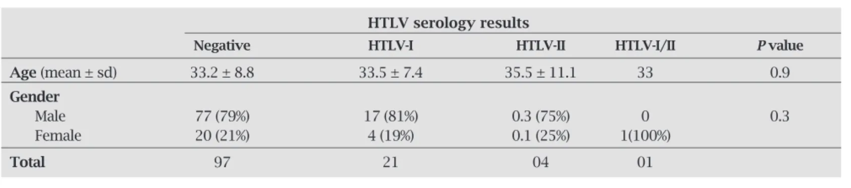
Medical records were reviewed for 123 patients (97 male), who fulfi lled the inclusion criteria for the study. Twenty men (20.6%) and 6 women (23.1%) had detectable antibodies against HTLV-I/II (Table 1). Median follow-up time was 16
Table 1. Frequency of episodes of diagnosed infections according to the serological status for HTLV-I/II infection
Infection HTLV serology
Negative HTLV-I HTLV-II Pvalue*
P. jiroveci pneumonia 20 1 1 0.3
CNS toxoplasmosis 14 5 - 0.8
Oral candidiasis 146 15 1 0.2
Esophageal candidiasis 17 1 1 0.4
Tuberculosis 37 10 1 0.8
Strongyloidiasis 2 3 1 0.04
CNS cryptococosis 2 0 1 0.8
Recurrent herpes 31 2 1 0.4
CMV 10 2 0 0.9
Kaposi’s sarcoma 9 1 0 0.8
Bacterial infections 119 26 1 0.3
Cryptosporidiasis 6 0 0 0.6
Isosporiasis 4 1 0 0.9
Diarrhea 178 25 5 0.3
Peripheral neuropathy 5 2 0 0.8
Herpes zoster 12 4 1 0.8
Encephalopathy 1 2 0 0.04
Table 3. Risk factors for acquisition of retroviral infection among singly and dually infected patients
Risk factors Negative HTLV-I HTLV-II HTLV-I/II P
value*
Blood 0.5 0.1 0.1 0.1 0.4
transfusion
IVDU 0.9 12 0.3 - < 0.0001
Homosexual 45 10 - - 0.2
activity
Previous STD 29 08 0.2 - 0.7
Sex with 0.2 0.1 - - 0.9
prostitutes
None of the 10 - - NS
above
IVDU, intravenous drugs user; STD, sexually transmitted diseases; NS, nonsignificant.
*P value comparing negative and positive patients for HTLV-I/
II antibodies. The total may exceed the sample size, due to positive history for more than one risks in the list.
months (range 656 months). The mean age was signifi -cantly higher for men as compared to women (34.5 ± 8.1 and 29 ± 8.8 respectively, P = 0.002) but there was no dif-ference between the mean age for HTLV-positive and nega-tive patients (Table 2).
Risk factors for coinfection: the major risk factor asso-ciated with coinfection by HTLV was intravenous drug use (IVDU). Among HTLV seropositive patients, 57.7% had a history of IVDU as the risk behavior for acquiring retrovi-ral infections, compared to 9.2% among patients without HTLV coinfection (P < 0.0001). Infection by HTLV-I/II was not associated with blood transfusion, sexually trans-mitted diseases (STD), or sexual behavior (Table 3).
Clinical characteristics: at the time of the first visit to the clinic, 73 (59.3%) patients were asymptomatic, while the remaining 50 patients had an established diagnosis of AIDS (category C by CDC criteria). The frequency of epi-sodes of OI diagnosed during follow-up were compared according to the serological results for HTLV-I/II infec-tion: there was no difference in the overall frequency of
Table 2. Mean age and gender distribution for HIV-1 infected patients according to their serology results for HTLV-I and HTLV-II
HTLV serology results
Negative HTLV-I HTLV-II HTLV-I/II Pvalue
Age (mean ± sd) 33.2 ± 8.8 33.5 ± 7.4 35.5 ± 11.1 33 0.9
Gender
Male 77 (79%) 17 (81%) 0.3 (75%) 0 0.3
Female 20 (21%) 4 (19%) 0.1 (25%) 1(100%)
Total 97 21 04 01
OI for both groups, but strongyloidiasis was diagnosed in 4/26 coinfected, compared to 2/97 in singly infected patients (OR = 8.55; 95% CI: 1.21-73.62, P = 0.02, Fisher exact test). In addition, two episodes of encephalopathy were diagnosed during the study period in HTLV-posi-tive, but no case was detected among singly infected pa-tients (P = 0.04, Fisher exact test).
Laboratories results
Hemoglobin levels were similar for both singly (11.9 ± 2.2 g/dL) and doubly infected (12 ± 2.3 g/dL) patients (P > 0.05).
Patients coinfected by HTLV-I/II had absolute lym-phocytes counts significantly higher than those in-fected by HIV-1 alone (1,921 ± 762 versus 1,587 ± 951,
P = 0.03). However, when they were stratified by clini-cal status, asymptomatic patients presented a similar number of lymphocytes, regardless of their HTLV-I/II serology status, while coinfected, symptomatic patients, had significantly higher lymphocyte counts (887 ± 515 cells/mm3 versus 1,687 ± 731 cells/mm3, P = 0.02, Kruskal-Wallis test).
Table 4. CD4+, CD8+ and lymphocyte counts (mean ± SD) of patients according to their HTLV serology
Laboratory HTLV serology
Values/clinical Negative HTLV-I HTLV-II HTLV-I/II Pvalue*
Status n 102 25 0.4 0.1
Symptomatic 50
CD4 cell counts 25 140 ± 177 292 ± 92 - - 0.036
CD8 cell counts 25 695 ± 403 839 ± 537 - - 0.6
Lymphocytes 50 887 ± 515 1,687 ± 731 - - 0.02
Asymptomatic* 73
CD4 cell counts 50 434 ± 281 537 ± 269 504 832 0.5
CD8 cell counts 50 1,300 ± 883 797 ± 308 465 1,965 0.1
Absolute lymphocyte count 72 2,013 ± 912 2,257 ± 835 1,845 ± 630 2,255 0.8
All patients 123
CD4 cell counts 75 334 ± 284 432 ± 237 504 832 0.2
CD8 cell counts 75 1,112 ± 813 810 ± 339 465 1,965 0.2
Absolute lymphocyte count 123 1,563 ± 952 958 ± 816 1,638 ± 490 2,255 0.1
P values were calculated by Kruskal-Wallis test, and compare HTLV I/II negative and positive patients.
*Only 50 patients had available CD4 cell counts.
Progression to clinical disease during the follow-up period was similar for singly and coinfected pa-tients (24/60 and 3/13, respectively, RR = 0.58; CI 0.20 - 1.63 P = 0.3). Use of antiretroviral drugs was more fre-quent among patients infected by HIV-1 alone (64.3%) compared to those coinfected by HTLV-I/II (42.3%) (P = 0.04, Yates corrected). This difference was more evident when we analyzed patients without a clini-cal picture of AIDS (non-category C disease): only 16.4% of coinfected patients were on antiretroviral therapy, in contrast to 51% of singly infected patients (RR = 0.23; 95% CI: 0.06-0.97 P = 0.04, Yates corrected).
DISCUSSION
Chronic infection by HIV is associated with immunodefi -ciency, caused by progressive CD4 lymphocyte depletion. Infection by HTLV-I may also be related to some degree of perturbation of the immune response among patients that harbor the virus, but usually do not exhibit clinical symptoms.25
While there is evidence suggesting that coinfection by HIV-HTLV viruses may modify the clinical and laboratory fi ndings during the course of HIV infection, the lack of conclusive studies leaves the clinical impact of HTLV in-fection on HIV infected patients open to controversy. The present report provides additional support for the conten-tion that patients harboring HIV and HTLV may present a modifi ed clinical course of the HIV infection, character-ized by a higher frequency of some clinical events.
The demographic characteristics for singly and dou-bly infected patients were comparable in this report. In contrast, a previous, larger study, detected a higher preva-lence of HTLV-I coinfection among women, in Bahia.26 This discrepancy may be due to the smaller sample size of this work, which probably lacked sufficient power to detect differences in coinfection rates by gender (only 6 women were coinfected).
Surrogate markers for HIV infection showed a distinct pattern for singly and doubly-infected patients. The ab-solute lymphocyte counts were quite different in the two groups, with higher counts among HTLV coinfected pa-tients. Similar number of lymphocytes in asymptomatic patients in both groups suggests that lymphocyte depletion occurs more slowly in coinfected patients during the course of HIV infection.
The analysis of CD4 cell count provided a similar pic-ture. Asymptomatic patients had comparable counts of CD4 cells, but in contrats it was higher among coinfected patients presenting with clinical disease. CD8 cell counts were not related to the status HTLV infection, regardless of the patient’s clinical status. Similarly, hemoglobin levels were comparable in both groups.
HIV-positive patients, compared to seronegatives.31 The pre-sent report provides evidence that this association in HIV- HTLV coinfected patients is even higher than in HIV-1 singly-infected individuals.
The association between coinfection and encephalopa-thy is less clear: HTLV-I infection is associated with TSP/ HAM, a clinical entity characterized by signs and symp-toms of myelopathy, but displaying some evidence of brain involvement, such as perivascular infi ltrates, demyeliniza-tion, and white matter destruction. In addidemyeliniza-tion, other se-vere involvement of central nervous system has been re-ported.32-35 Unfortunately, the retrospective design of the present study did not allow to clarify the exact cause of death of these two patients.
The rate of clinical evolution from asymptomatic infec-tion to AIDS during the follow-up period was similar for both groups. However, the use of antiretroviral drugs was more frequent among singly-infected patients suggesting that the abnormalities in the number of lymphocytes and the CD4+ count induced by HTLV coinfection may de-lay the initiation of antiretroviral therapy in coinfected pa-tients, as predicted by Schechter et al. in a previous report.15 Since CD4 cell count is still the main marker to defi ne the best moment to start antiretroviral therapy and/or prophy-laxis for OI, this fact may add a clinically relevant problem to physicians, when attending patients in endemic areas for these two agents.
The fi nding that cell subsets count is similar among asymptomatic patients, but signifi cantly different among those with clinical symptoms suggests that the decrease in CD4 cell counts occurs later in the evolution of coinfected patients. Nonetheless, the higher CD4 cell counts do not pro-vide any immunological benefi t. It is likely that immunolog-ical changes secondary to the progression of HIV infection may also be affected, or may modify the lymphoproliferative response that usually follows HTLV infection. However, the mechanisms and intensity of such immune modifi cations are still unclear.
Finally, the extent to which HIV/HTLV coinfection modifies clinical outcomes of HIV infected patients re-mains unclear. There are some evidence supporting the hypothesis that coinfection by these agents may modulate the clinical presentation of these individuals, with an in-crease in the frequency of some parasitic diseases, and of severe forms of scabies.31,36 In addition, there are evidence supporting a potential impact of coinfection on survival, which was shorter for coinfected patients than for those infected by HIV-1 alone.37,38
Our study also presents some limitations, that could have influenced the results: first, the retrospective de-sign may prevent definitive conclusions, although, since HAART is currently the standard of care for all patients in need of treatment we could not evaluate the impact of
in-creased CD4 cell counts on physicians’ decision on when to start therapy. On the other hand, the higher proportion of IVDU among coinfected patients could also be con-founding factor of lower rate of antiretroviral treatment among coinfected patients. It could be true for asymp-tomatic patients, but our data showed that even patients with a clinical picture of AIDS had decreased chance of receiving therapy, which could not be explained on the basis of patients’ risk behavior. It makes more sense to conclude that the discrepancy between CD4 cell count and clinical status was probably the factor that misled physicians in deciding the optimal time to start treat-ment. In addition, we did not have information on how many patients had stools examination performed for
S. stercoralis parasitism, but in AIDS patients such para-site usually causes severe diarrhea and the stools exami-nation routinely performed in these situations, making unlikely that a symptomatic patient not having had a proper diagnostic investigation.
Our results confi rm previous studies reporting higher CD4 cell counts in coinfected patients, and suggest that such phenomenon may mislead physicians in the choice of the optimal time to start antiretroviral therapy.15,39 It can not be ruled out the delay in starting antiretroviral therapy as one of the potential explanation for the higher mortality detect-ed among coinfectdetect-ed individuals causdetect-ed by increasdetect-ed CD4 cell counts, rather than a direct effect of HTLV-I on AIDS progression. The increased frequency of S. stercoralis infec-tion and encephalopathy also indicates that coinfecinfec-tion may modify the course of HIV disease. However, the available data provide only limited evidence and defi nitive answers will require larger, prospective studies.
REFERENCES
1. Kalyanaraman VS, Sarngadharan MG, Nakao T et al. Natural
antibodies to the structural core protein (p24) of the human t-cell leukemia (lymphoma) retrovirus found in sera of leuke-mia patients. Proc Natl Acad Sci. USA 1982; 79:1653-57. 2. Robert-Guroff M, Gallo RC. Establishment of an etiologic
relationship between the human T-cell leukemia/lymphoma virus (HTLV) and adult T-cell leukemia. Blut 1983; 47:1-12.
3. Gessain A, Barin F, Vernanr JC, et al. Antibodies to human
T-lymphotropic virus type I in patients with tropical spastic paraparesis. Lancet 1986; 2:99-100.
4. Bartholomew C, Cleghorn F, Charles W, et al. HTLV-I and
tropical spastic paraparesis. Lancet 1986; 2:99-100.
5. Osame M, Usuku K, Izumo S, et al. HTLV-I associated
my-elopathy, a new clinical entity. Lancet 1986; 1:1031-2. 6. Ocochi K, Sato H, Hinuma Y. A retrospective study on
trans-mission of adult T cell leukemia virus by blood transfusion. Seroconversion in recipients. Vox Sang 1984; 46:245-53.
7. Ando Y, Nakano S, Saito K, et al. Transmission of adult T cell
8. Bartholomew C, Saxiger WC, Clark JW, et al. Transmission of HTVL-I and HIV among homosexual men in Trinidad. JAMA 1987; 257:2605-8.
9. Kajiyama W, Kashiwagi S, Ikematsu H, et al. Intrafamilial
transmission of adult T cell leukemia virus. J Infect Dis. 1986; 154:851-7.
10. Delaporte E, Peters M, Bardy JL, et al. Blood transfusion as
a major risk factor for HTLV-I infection among hospitalized children in Gabon (Equatorial Africa). J Acq Immune Def Syndr. 1993; 6:424-8.
11. Blattner WA. Epidemiology of HYLV-I and associated diseases. In: Blattner, WA ed Human Retrovirollogy: HTLV. New York: Raven Press 1990; 251-65.
12. Bartholomew C, Blattner W, Cleghorn F. Progression to AIDS in homosexual men coinfection with HIV and HTLV-I Trini-dad. Lancet 1987; 2:1469-1987.
13. Hattori T, Koito A, Takatsuli K, Ikematsu S. et al. Frequent
in-fection with human T-cell lymphotropic virus type I in patients with AIDS, but not in carriers of human immunodefi -ciency virus type 1. J Acq Immune Def Synd. 1989; 2:272-276. 14. Gotuzzo E, Escamilla J, Phillips IA, Sanchez J, Wignall FS, Anti-goni J. The impact of human T-lymphotropic virus type I/II in-fection on the prognosis of sexually acquired immunodefi ciency syndrome. Archives of Internal Medicine 1992; 152:1429-52. 15. Schechter M, Harrison LH, Halsey NA, Trade G, Santino M,
Moulton CH, Quinn TC. Coinfection with human T-cell lym-photropic virus type I and HIV in Brazil: impact on markers of HIV disease progression. JAMA 1994; 271:353-357.
16. Brites C, Harrington Jr W, Pedroso C, Netto EM, Badaró R. Epide-miological characteristics of HTLV-I and II coinfection in Brazil-ian subjects infected by HIV-1. Braz J Infect Dis. 1997; 43-48. 17. Zack JA, Cann AJ, Lugo JP, Chen ISY. HIV-1 production from
infected peripheral blood T cell after HTLV-I induced mi-togenic stimulation. Science 1988; 240:1026-9.
18. De Rossi A, Saggioro D, Calabró ML, Cenzato R, Chieco-Bi-anchi. Reciprocal activation of human T-lymphotropic viruses in HTLV-I transformed cells superinfected with HIV-1. J Acq Immune Def Synd 191; 4:380-385.
19. Lusso P, Lori F, Gallo RC. CD4-independent infection by human immunodefi ciency virus type 1 after phenotypic mixing with human T-cell leukemia viruses. J Virol. 1990; 64(12):6341-4. 20. Dourado I, Alcantara LC, Barreto ML, da Gloria Teixeira
M, Galvão-Castro B. HTLV-I in the general population of Salvador, Brazil: a city with African ethnic and sociodemo-graphic characteristics. J Acquir Immune Defi c Syndr. 2003 15;34(5):527-31.
21. Ministério da Saúde. AIDS - Boletim epidemiológico 2002, Ano XVI, N0 01. Semana epidemiológica 14 a 52.
22. Lal RB, Brodine S, Kazura J, Mbidde Katonga E, Yanagihara R, Roberts C. Sensitivity and specifi city of a recombinant trans-membrane glycoprotein (rgp 21) - spiked Western immunoblot for serological confi rmation of human T-cell lymphotropic virus type I and II infections. J Clin Microbiol. 1992; 30(2):296-299. 23. Lipka JJ, Santiago P, Chan L, Reyes GR, Samuel KP, Blattner
WA, Shaw GM, Hanson CV, Sninsky JJ, Foung SKH. Modifi ed Western blot assay for confi rmation and differentiation of hu-man T-cell lymphotropic virus type I and II. J Infect Dis. 1991; 164:400-403.
24. Lipka JJ, Miyoshi I, Hadlol KG, Reyes GR, Chow TP, Blattner WA, Shaw GM, Hanson CV, Gallo D, Chan L, Foung SKH. Segregation of human T-cell lymphotropic virus type I and II infections by antibody reactivity to unique viral epitopes. J Infect Dis. 1992; 165:268-272.
25. Welles SL, Tachibana N, Okayama A, et al. Decreased reactivity
to PPD among HTLV-I carriers in relation to virus and hema-tologic status. Intl J Cancer 1994; 56:337-40.
26. Brites C, Harrington Jr W, Pedroso C, Badaró R. HTLV-I/II in-fection among HIV-1 infected individuals in Brazil. The First National Conference on Human Retroviruses and Related In-fections, Washington, DC, 1993.
27. Nakada K, Kokagura M, Komoda H, Hinuma Y. High incidence of HTLV antibody in carriers of Strongyloides stercoralis [let-ter]. Lancet 1984; 1:633.
28. Dixon AC, Yanagihara ET, Kwock DW, Nakamura JM. Stron-gyloidiasis associated with human T-cell lymphotropic virus type I infection in a nonendemic area. West Indian J Med. 1989; 151:410-3.
29. Robinson RD, Lindo JF, Neva FA, et al. Immunoepidemiologic
studies of Strongyloides stercoralis and human T lymphotrop-ic virus type I infections in Jamalymphotrop-ica. West Indian Med J. 1990; 169:692-6.
30. Sato Y, Toma H, Takara M, Kiyuna S, Shiroma Y. Seroespidemi-ological studies on the concomitance of strongyloidiasis with T-cell leukemia viral infection in Okinawa, Japan. Japanese Journal of Parasitology 1990; 39(4):376-83.
31. Feitosa G, Bandeira AC, Sampaio DP, Badaró R, Brites C. High prevalence of giardiasis and strongyloidiasis among HIV-infected patients in Bahia, Brazil. Braz J Infect Dis. 2001; (6):339-44.
32. Newton MR, Rudge P, Cruickshank K. Magnetic resonance imaging in HTLV-I antibody positive patients. Lancet 1987; ii:514.
33. Tournier-Lasserve E, Gout D, Gessain A, et al. HTLV-I, brain
abnormalities on magnetic resonance imaging, and relation with multiple sclerosis. Lancet 1987; ii:49-50.
34. Shibasaki H, Endo C, Kuroda Y, Kakigi R, Oda K, Komine S. Clinical picture of HTLV-I associated myelopathy. J Neurol Sci. 1988; 87:15-24.
35. Puccione-Söhler M, Chimelli L, Merçon M, et al.
Pathologi-cal and virologiPathologi-cal assessment of acute HTLV-I-associated myelopathy complicated with encephalopathy and systemic infl amation. J Neurol Sci. 2003; 207:87-93.
36. Brites C, Weyll M, Pedroso C, Badaró R. Severe and Norwegian scabies are strongly associated with retroviral (HIV-1/HTLV-I) infection in Bahia, Brazil. AIDS 2002; 16(9):1292-3.
37. Sobesky M, Couppie P, Pradinaud R, et al. Coinfection with
HIV and HTLV-I infection and survival in AIDS stage. French Guiana Study. GECVIG (Clinical HIV Study Group in Guiana). Presse Med. 2000 Mar 4; 29(8):413-6.
38. Brites C, Alencar R, Gusmão R, et al. Coinfection by HTLV-I
is associated with a shorter survival among HIV-infected pa-tients, in Bahia, Brazil. AIDS 2001 Oct 19; 15(15):2053-5. 39. Schechter M, Moulton L, Harrison L. HIV viral load and