Surgical Infections: A Microbiological Study
Santosh Saini1, Naveen Gupta1, Aparna1, Department of Microbiology1, Department of Lokveer1 and M.S. Griwan2 Surgery2, Pt. B.D. Sharma PGIMS, Rohtak, India
Surgical infections are mostly polymicrobial, involving both aerobes and anaerobes. One hundred seventeen cases comprised of abscesses (n=51), secondary peritonitis (n=25), necrotizing fascitis (n=22) and wounds with devitalized tissues (n=19) were studied. The number of microorganisms isolated per lesion was highest in secondary peritonitis (2.32). The aerobe/ anaerobe ratio was 0.81 in secondary peritonitis and 1.8 in necrotizing fascitis. Most secondary peritonitis (80%), necrotizing fascitis (75%) and wounds with devitalized tissues (66.7%) were polymicrobial. Common microorganisms isolated in our study were E. coli, Staphylococcus aureus, Klebsiella spp., Pseudomonas aeruginosa, Bacteroides fragilis and Peptostreptococcus spp. The most effective antibiotics for S. aureus were clindamycin (79.1%) and cefuroxime (70.8%). For Gram-negatives (Klebsiella spp., E. coli and Proteus spp.), the most effective antibiotics were cefotaxime, ceftizoxime, amikacin and ciprofloxacin. Pseudomonas aeruginosa was maximally sensitive to amikacin (35.2%) and ciprofloxacin (35.2%). The greatest degree of multidrug resistance to all the drugs was found in P. aeruginosa (52.9%), followed by Klebsiella spp. (33.3%), Proteus spp. (33.3%), E. coli (22.2%), and S. aureus (12.5%). All the anaerobes that we isolated were 100% sensitive to metronidazole and chloramphenicol, followed by clindamycin (95% to 100%). Apart from antibiotic therapy, non-antimicrobial methods, such as hyperbaric oxygen therapy and debridement also play an important role in the treatment of surgical infections. Key Words: Abscess, secondary peritonitis, necrotizing fascitis and wounds with devitalized tissue.
Received on 15 October 2003; revised 24 March 2004. Address for correspondence: Dr. Naveen Gupta. Department of Microbiology, Pt. B.D. Sharma Post Graduate, Institute of Medical Sciences, Rohtak (Haryana), India. E-mail: [email protected]
The Brazilian Journal of Infectious Diseases 2004;8(2):118-125 © 2004 by The Brazilian Journal of Infectious Diseases and Contexto Publishing. All rights reserved.
The microbiology laboratory plays a key role in providing information about surgical infections that slowly worsen or otherwise fail to heal. The main pathogens or groups of microorganisms that a microbiology laboratory should routinely detect and report (with antibiograms being provided when appropriate) are as follows: Staphylococcus aureus, Pseudomonas aeruginosa, beta hemolytic streptococci, coliform bacteria, pigmented Gram-negative anaerobes (Prevotella and Porphyromonas
spp.) non - pigmented Gram-negative anaerobes
(primarily Bacteroides, Prevotella and
Fusobacterium spp.), Peptostreptococcus spp., and
Clostridium spp. [1].
The introduction of microorganisms into a previously sterile site, such as a wound, is termed contamination. Many contaminating bacteria find the tissues of the wound a hostile environment and succumb. Other species that are able to survive begin to actively multiply and the wound may be said to have been colonized. Colonization does not always lead to infection. Which of the colonizing bacterial species eventually emerge as actual etiological agents of infection depends upon their virulence, relative numbers, and on selective factors, such as wound environment and antibiotics. The bacterial flora of the open wound is seldom static; it is usually changing, new organisms appear in wounds and old ones disappear [2].
are polymicrobial, involving both aerobes and anaerobes. These pathogens cause delayed healing and infection [1]. We investigated the role of anaerobes and aerobes in abscesses, secondary peritonitis, necrotising fascitis, and in wounds with devitalized tissue, to determine the optimal antimicrobial therapy.
Material and Methods
The investigation was conducted at PGIMS, Rohtak from July 2001 and June 2002. The study group included:
(a) Abscess group (Group A) - having closed abscesses (single/multiple) with redness and brownish induration at the periphery.
(b) Secondary peritonitis (Group B) - patients from the emergency operation room who had signs and symptoms of peritonitis.
(c) Necrotising fascitis (Group C) - patients having unexplained fever with pain, brownish edema, tenderness and brownish grey interfascial planes.
(d) Wounds with devitalized tissue (Group D) -gangrenous tissue with no sensation, no blood supply, blackening of the affected organ or portion, and foul odor coming from the affected part.
Specimen collection
Skin or mucus membranes were decontaminated using alcohol or povidone iodine. The specimens were purulent exudate aspirated from abscesses, peritoneal fluid or exudate from peritonitis, swab, exudate or aspirate from deep necrotising fascitis and devitalized gangrenous tissue. For anaerobic culture, the specimens were collected in cooked meat broth (CMB, Hi-Media) and incubated at 37°C for 48 hours. The media used for aerobic incubation were 5% sheep blood agar, MacConkey agar, 7% salt agar and chocolate agar. Chocolate agar was incubated aerobically with 5% to 10% CO2. The media used for anaerobic incubation were Brain Heart Infusion agar (BHI), neomycin BHI agar, Bacteroides Bile esculin agar. Anaerobic incubation was done with P. aeruginosa as a biological
indicator and alkaline methylene blue glucose as a chemical indicator.
Aerobes were identified using standard microbiological methods [3] and anaerobes were processed for identification up to level III as per the Wadsworth Anaerobic Bacteriology Manual [4]. Various commonly used antimicrobial agents that are recommended by NCCLS were used to ascertain the susceptibility pattern of aerobes and anaerobes by the disc diffusion method [5]. Reference strain S. aureus
NCTC 6571 was used as a control for Gram-positive cocci, E. coli NCTC 10418 for Gram-negative bacilli, and P. aeruginosa NCTC 10662 for Pseudomonas.
Results
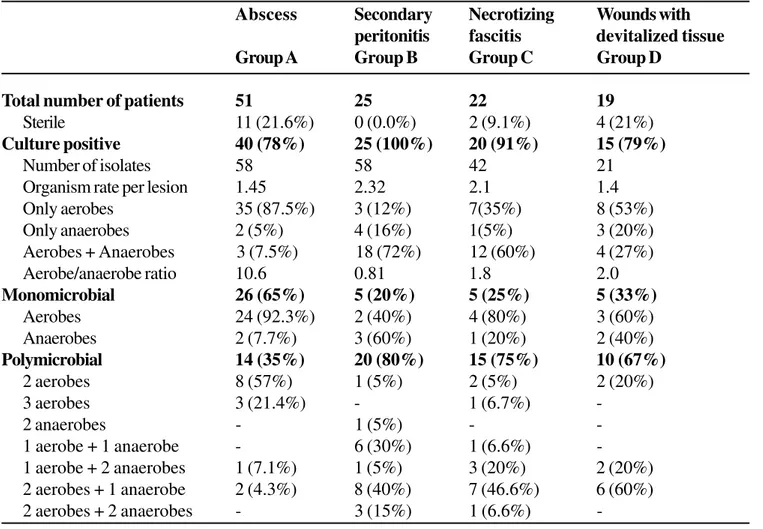
Table 1. Analysis of isolates from surgical infections
Abscess Secondary Necrotizing Wounds with peritonitis fascitis devitalized tissue Group A Group B Group C Group D
Total number of patients 51 25 22 19
Sterile 11 (21.6%) 0 (0.0%) 2 (9.1%) 4 (21%)
Culture positive 40 (78%) 25 (100%) 20 (91%) 15 (79%)
Number of isolates 58 58 42 21
Organism rate per lesion 1.45 2.32 2.1 1.4
Only aerobes 35 (87.5%) 3 (12%) 7(35%) 8 (53%)
Only anaerobes 2 (5%) 4 (16%) 1(5%) 3 (20%)
Aerobes + Anaerobes 3 (7.5%) 18 (72%) 12 (60%) 4 (27%)
Aerobe/anaerobe ratio 10.6 0.81 1.8 2.0
Monomicrobial 26 (65%) 5 (20%) 5 (25%) 5 (33%)
Aerobes 24 (92.3%) 2 (40%) 4 (80%) 3 (60%)
Anaerobes 2 (7.7%) 3 (60%) 1 (20%) 2 (40%)
Polymicrobial 14 (35%) 20 (80%) 15 (75%) 10 (67%)
2 aerobes 8 (57%) 1 (5%) 2 (5%) 2 (20%)
3 aerobes 3 (21.4%) - 1 (6.7%)
-2 anaerobes - 1 (5%) -
-1 aerobe + -1 anaerobe - 6 (30%) 1 (6.6%)
-1 aerobe + 2 anaerobes 1 (7.1%) 1 (5%) 3 (20%) 2 (20%) 2 aerobes + 1 anaerobe 2 (4.3%) 8 (40%) 7 (46.6%) 6 (60%)
2 aerobes + 2 anaerobes - 3 (15%) 1 (6.6%)
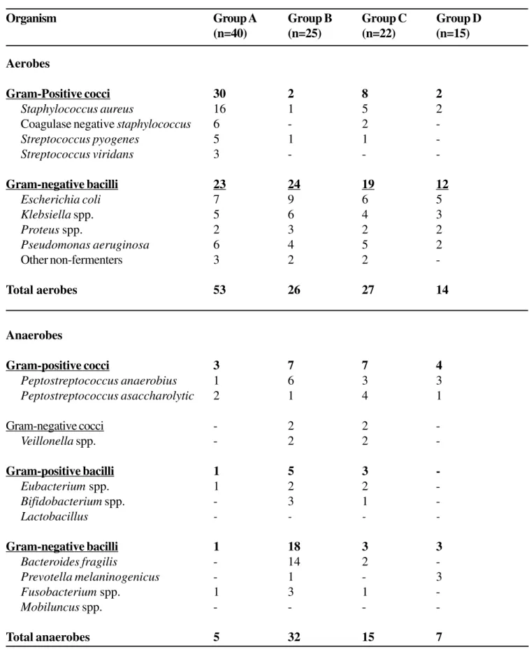
-and Peptostreptococcus anaerobius were predominant among the anaerobes, whereas among the aerobes, E. coli and Klebsiella spp. were predominant in group B infections. In group C (n=22), 42 microorganisms were isolated, 27 (64.2%) of which were aerobes. The predominant aerobic and anaerobic isolates were E. coli, S. aureus, Peptostreptococcus
spp. and B. fragilis. In group D (n=15) infections, 21 microorganisms were isolated, 14 (66.6%) of which were aerobes, and the predominant microorganisms were E. coli, Klebsiella spp., Prevotella melaninogenicus and Peptostreptococcus spp.
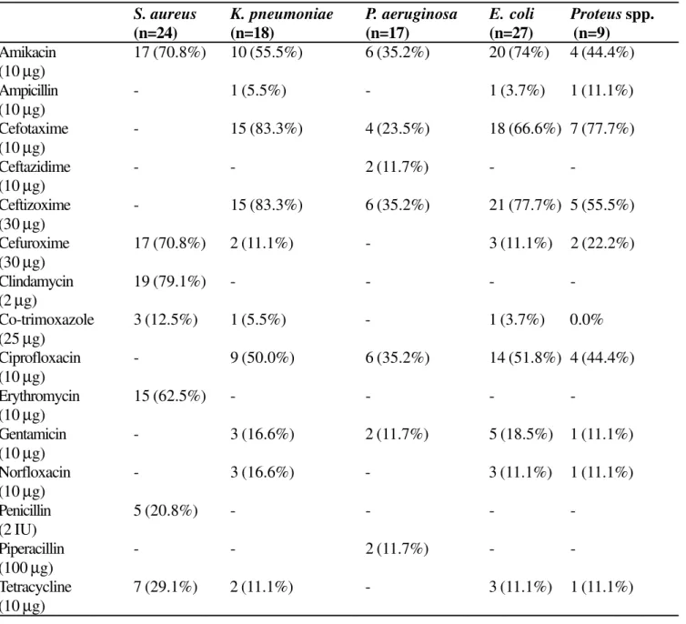
The antibiotic susceptibility pattern of the aerobes and anaerobes was determined (Tables 3 and 4, respectively). The most effective antibiotics for S. aureus
were clindamycin (79.1%), amikacin (70.8%), and cefuroxime (70.8%). While, the most effective antibiotics for the Gram-negatives (K. pneumoniae, E. coli and
Proteus spp.) were cefotaxime, ceftizoxime, amikacin and ciprofloxacin (Table 3). Pseudomonas aeruginosa
was most sensitive to amikacin (35.2%) and ciprofloxacin (35.2%). The highest degree of multidrug resistance to all the drugs was found in P. aeruginosa (52.9%), followed by Klebsiella spp. (33.3%), Proteus spp. (33.3%), E. coli (22.2%), and S. aureus (12.5%).
Table 2. Aerobes and anaerobes isolated in the study group
Organism Group A Group B Group C Group D
(n=40) (n=25) (n=22) (n=15)
Aerobes
Gram-Positive cocci 30 2 8 2
Staphylococcus aureus 16 1 5 2
Coagulase negative staphylococcus 6 - 2
-Streptococcus pyogenes 5 1 1
-Streptococcus viridans 3 - -
-Gram-negative bacilli 23 24 19 12
Escherichia coli 7 9 6 5
Klebsiella spp. 5 6 4 3
Proteus spp. 2 3 2 2
Pseudomonas aeruginosa 6 4 5 2
Other non-fermenters 3 2 2
-Total aerobes 53 26 27 14
Anaerobes
Gram-positive cocci 3 7 7 4
Peptostreptococcus anaerobius 1 6 3 3
Peptostreptococcus asaccharolytic 2 1 4 1
Gram-negative cocci - 2 2
-Veillonella spp. - 2 2
-Gram-positive bacilli 1 5 3
-Eubacterium spp. 1 2 2
-Bifidobacterium spp. - 3 1
-Lactobacillus - - -
-Gram-negative bacilli 1 18 3 3
Bacteroides fragilis - 14 2
-Prevotella melaninogenicus - 1 - 3
Fusobacterium spp. 1 3 1
-Mobiluncus spp. - - -
Table 3. Sensitivity pattern of common aerobic isolates from surgical infections
S. aureus K. pneumoniae P. aeruginosa E. coli Proteus spp.
(n=24) (n=18) (n=17) (n=27) (n=9)
Amikacin 17 (70.8%) 10 (55.5%) 6 (35.2%) 20 (74%) 4 (44.4%) (10 µg)
Ampicillin - 1 (5.5%) - 1 (3.7%) 1 (11.1%)
(10 µg)
Cefotaxime - 15 (83.3%) 4 (23.5%) 18 (66.6%) 7 (77.7%)
(10 µg)
Ceftazidime - - 2 (11.7%) -
-(10 µg)
Ceftizoxime - 15 (83.3%) 6 (35.2%) 21 (77.7%) 5 (55.5%)
(30 µg)
Cefuroxime 17 (70.8%) 2 (11.1%) - 3 (11.1%) 2 (22.2%)
(30 µg)
Clindamycin 19 (79.1%) - - -
-(2 µg)
Co-trimoxazole 3 (12.5%) 1 (5.5%) - 1 (3.7%) 0.0%
(25 µg)
Ciprofloxacin - 9 (50.0%) 6 (35.2%) 14 (51.8%) 4 (44.4%)
(10 µg)
Erythromycin 15 (62.5%) - - -
-(10 µg)
Gentamicin - 3 (16.6%) 2 (11.7%) 5 (18.5%) 1 (11.1%)
(10 µg)
Norfloxacin - 3 (16.6%) - 3 (11.1%) 1 (11.1%)
(10 µg)
Penicillin 5 (20.8%) - - -
-(2 IU)
Piperacillin - - 2 (11.7%) -
-(100 µg)
Tetracycline 7 (29.1%) 2 (11.1%) - 3 (11.1%) 1 (11.1%)
(10 µg)
Table 4. Susceptibility pattern of anaerobic isolates from surgical infectionsism
Organism GPC (n=21) GNC (n=4) GPB (n=9) GNB (n=25)
Metronidazole (5 µg) 21 4 9 25
Penicillin (2IU) 15 3 6 7
Clindamycin (2 µg) 20 4 9 24
Cefuroxime (30 µg) 16 4 8 14
Chloramphenicol (10 µg) 21 4 9 25
Erythromicin (10 µg) 10 2 6 9
Cefotaxime (30 µg) 17 4 9 22
Discussion
Surgical infections, such as abscesses, secondary peritonitis, necrotising fascitis and wounds with devitalized tissues are largely polymicrobial, and the role of both aerobic and anaerobic bacteria in the pathogenesis of these infections is well recogniszed. Microbial synergy may increase the net pathogenic effect and hence the severity of infection in several ways: (i) oxygen consumption by aerobic bacteria induces tissue hypoxia and a lowering of the redox potential, which favors the growth of anaerobic bacteria; (ii) specific nutrients produced by one bacterium may encourage the growth of fastidious and potentially pathogenic cohabiting microorganisms; and (iii) some anaerobes are able to impair host immune cell function and thus provide a competitive advantage for themselves as well as for other, cohabiting, microorganisms [1].
An abscess is a localized collection of purulent inflammatory tissue caused by suppuration deep within a tissue, an organ or a confined space. It is produced by deep seeding of pyogenic bacteria into a tissue. It may involve skin, dermis, fasciae, muscles, and even bones. Abscess inside the cavities poses a great problem for treatment, e.g. brain abscess, pleural abscess, and intra-abdominal abscess [6]. Among our group A (n=51) infections, 11 (21.6%) were sterile. The mean number of species of organisms per lesion was 1.45 and the aerobe/anaerobe ratio was 10.6; 65% of the infections were monomicrobial. Staphylococcus aureus and E. coli were the predominant aerobes, while Peptostreptococci were common among the anaerobes. Wren reported Bacteroides spp. (40.4%),
Fusobacterium (10.1%), Clostridium spp. (2.2%), gram-positive non-sporulating bacilli (13.4%) and
Veillonella spp. (5.6%), from pus samples aspirated from closed abscesses or pus-filled cavities [7]. Brook reported in 1995 that Bacteroides spp. (32%) is the most common bacteria in abscesses, followed by E. coli and Peptostreptococcus spp. [8]. Sunmonen et al. studied 86 abscesses in intravenous drug users (IVDU); these yielded 173 aerobes and 131 anaerobes, among which S. aureus was most common (50%), while among the anaerobes, Prevotella spp.,
F. nucleatum, P. micros, A. odontolyticus and
Veillonella were isolated. In non IVDU, S. aureus was the most common (53%), followed by coagulase-negative staphylococcus (CONS) (19%),
Streptococcus millieri and Streptococcus pyogenes. The main anaerobes isolated were Peptostreptococcus
spp., Bacteroides spp. and Gram-positive bacilli [9]. Peritonitis is a localized or generalized inflammatory process of the peritoneum, which may appear in both acute and chronic forms. In the acute form, the motor activity of the intestine is decreased and the intestinal lumen becomes distended with gas and fluid. Secondary peritonitis may be due to the entry of bacilli into the peritoneal cavity through perforations of the gut or from an external penetrating wound. The most common causes of secondary peritonitis are appendicitis, perforations associated with diverticulitis, peptic ulcer, gangrenous gall bladder and gangrenous obstructions of the small bowel from adhesive bands, incarcerated hernia or volvulus [10]. In study group B, a mean of 2.32 organisms were isolated per lesion, with both aerobes and anaerobes in 72% of the cases, and an aerobe/anaerobe ratio of 0.81. Eighty percent of the group B infections were polymicrobial, among which
E. coli, Klebsiella spp., and P. aeruginosa were the most common aerobes, while B. fragilis, Peptostreptococcus spp. were common among the anaerobes. Other researchers have reported E. coli, Klebsiella spp., B. fragilis, Peptostreptococcus spp. in intra-abdominal infections, in accordance with our findings [11,12].
aeruginosa and Klebsiella spp., while
Peptostreptoccocus spp. and Bacteroides spp. were common anaerobes. Giuliano et al. reported 124 isolates detected from 15 patients with similar sources of isolates [14]. Perra and Howard reported an average of 3.5 organisms isolated per patient, among which
Staphylococcus spp., group D streptococci,
Pseudomonas, S. viridans, and Proteus mirabilis
were common aerobes, while among anaerobes
Bacteroides spp., Peptostreptococcus spp. and C. perfringens were predominant [15]. Other researchers have reported similar results [16,17].
Wounds and devitalized tissues are characterized by rapidly spreading edema, myositis, tissue necrosis , gas production, and profound toxaemia, occurring as a complication of wound infection. Infection usually results from contamination of the wound with soil, particularly that from cultivated land. It may be indirectly derived from dirty clothing, street dust and even the air of a poorly ventilated theatre [18]. In our study, 66.7% of the group D infections were polymicrobial and 1.4 organisms were isolated per lesion, with an aerobe/anaerobe ratio of 2.0. The most common aerobes isolated in our study were E. coli, Klebsiella, S. aureus and Proteus spp., while the most common anaerobes were Peptostreptococcus spp. and P. melaninogenicus. Baradkar et al., who made a study of 63 clinically suspected cases of gangrene, found that 82.5% of the samples yielded both aerobes and anaerobes. In their study E. coli (38.4%) emerged as the most common pathogen, followed by Proteus
spp. (12.8%), Klebsiella spp. (35.8%) and S. aureus
(12.8%), while among the anaerobes, Clostridium spp. and Peptostreptococci were predominant [19].
Today it is clear that there is significant problem with increasing resistance to antimicrobial agents among anaerobic bacteria. Multiple mechanisms of resistance have been encountered in anaerobes, as well as in aerobes [20], e.g. β-lactam is the enzyme responsible for inactivating the β-lactam ring of β-lactam antibiotics in both Gram-positives and Gram-negatives, whereas loss of the outer membrane protein and altered target sites are mechanisms of resistance in Bacteroides spp. [21]. We found that culturing and determining the
sensitivity of aerobes and anaerobes is of the utmost importance in cases of abscesses, secondary peritonitis, necrotising fascitis and wounds with devitalized tissues. We recommend the use of metronidazole, chloramphenicol or clindamycin for the treatment of anaerobic infections and third generation cephalosporins, amikacin and ciprofloxacin for Gram-negative aerobes and clindamycin or cefuroxime for S. aureus. The choice of prophylactic antibiotics should cover both facultatively anaerobic and anaerobic bacteria, with high concentrations during surgery and throughout the duration of the surgical procedures. Newer classes of antibiotics, such as ureidopenicillin, carbapeneous and the β-lactam /β-lactamase inhibitor combinations, has expanded the choice for both prophylactic and therapeutic treatment. Combination therapy with an aminoglycoside (e.g. amikacin) or a cephalosporin (cefotaxime or ceftizoxime), plus clindamycin or metronidazole, is very effective. Apart from antibiotic therapy, nonantimicrobial methods, such as hyperbaric oxygen therapy, and debridement also play an important role in the treatment of surgical infections.
References
1. Bowler P.G., Duerden B.I., Armstrong D.G.. Wound microbiology and associated approaches to wound management. Clin Microbiol Rev 2001;244-69. 2. Ellner P.D. Microbiology of wounds. In: Lorian V, editor.
Significance of Medical Microbiology. 2nd ed. Baltimore: Williams & Wilkins; 1982:158-68.
3. Koneman E.W., Allen S.D., Janda W.M., et al. Colour Atlas and Textbook of Diagnostic Microbiology. 5th ed. USA: Lippincott, 1997.
4. Sutter V.L., Citron D.M., Edelstein M.A.C., Finegold S.M. Processing of clinical specimens and isolation and identification procedures. In: Wadsworth Anaerobic Bacteriology Manual. 4th ed. Belmont: Star Publishing;
1985:23-70.
5. National Committee for Clinical Laboratory Standards (NCCLS). Performance Standards for Antimicrobial Susceptibility Testing. Sixth ed., Approved Standards M2-A6, 1997: Wayne, Pennsylvania.
7. Wren M.W.D. The culture of clinical specimen for anaerobic bacteria: a comparison of three regimen. J Med Microbiol 1977;10:195-201.
8. Brook I.. Bacteroides infections in children. J Med Microbiol 1995;43:92-8.
9. Summanen P.H., Talan D.A., Strong C., et al. Bacteriology of skin and soft tissue infections: Comparison of infections in intravenous drug users and individuals with no history of intravenous drug use. Clin Infect Dis
1995;20(2):S279-82.
10. Isselbacher K.J., Epstein A. Diverticular, vascular and other disorders of intestine and peritoneum. In: Fauci A.S., Braunwald E., Isselbacher K.J., et al. [editors]. Harrison’s Principles of Internal Medicine. 14th ed. New York: McGraw Hill; 1998:1648-56.
11. Gorbach S.L., Norsen J. Anaerobic microorganisms in intraabdominal infections. In: Balows A, Dehaan RH, Dowell VR Jr. editors. Anaerobic bacteria: Role in Disease. Springfield: Charles C. Thomas; 1974:212-38. 12. Swenson R.M., Lorber B., Michaelson T.C. The bacteriology of intraabdominal infections. Arch Surg
1974;109:398.
13. Stevens D.L. Infections of the skin, muscle and soft tissues. In: Fauci AS, Braun WE, Isselbacher KJ, Wilson JD, Martin JB, Kasper DL et al editors. Harrison’s Principles of Internal Medicine. 14th ed. New York: McGraw Hill; 1998:827-30.
14. Guiliano A., Lewis F., Hadley K., Blaisdell F.W. Bacteriology of necrotizing fascitis. Am J Surg
1977;134:52-7.
15. Perra M.E., Howard R.J. Necrotizing fascitis. Surg Gynecol Obstet 1985;161:357-61.
16. Brook I., Frazier E.H. Clinical and microbiological features of necrotizing fascitis. J Clin Microbiol
1995;33(9):2382-7.
17. Anuradha D., Biswas J., Saraswathi K., Gogate A.Microbiological features of necrotising fascitis. Ind J Med Microbiol 1999;17(1):18-21.
18. Colle J.G. Clostridium: gas gangrene, tetanus, food poisoning, pseudomembranous colitis. In: Greenwood D, Slack R, Pentherer J, editors. Medical Microbiology. 15th ed. London: Churchill Livingston; 1998:232-42. 19. Banadkar V.P., Patwardhan N.S., Deshmukh A.B., et al.
Bacteriological study of clinically suspected cases of gas gangrene. Ind J Med Microbiol 1999;17(3):133-4. 20. Bawdon R.E., Crune L.R., Palchaudhary S. Antibiotic
resistance in anaerobic bacteria: molecular biology and clinical aspects. Rev Infect Dis 1982;4:1075-95. 21. Chaudhary R., Mazumdar J., Dhawan B. Genus