Ide ntificatio n o f a pro te in kinase
activity that pho spho rylate s co nne xin43
in a pH-de pe nde nt manne r
1Department of Microbiology and Immunology and
2Departmentof Pharmacology, SUNY Health Science Center at Syracuse,
Syracuse, NY, USA P. Yahuaca2,
J.F. Ek-Vitorin2,
P. Rush2, M. Delmar2
and S.M. Taffet1
Abstract
The carboxyl-terminal (CT) domain of connexin43 (Cx43) has been implicated in both hormonal and pH-dependent gating of the gap junction channel. An in vitro assay was utilized to determine whether the acidification of cell extracts results in the activation of a protein kinase that can phosphorylate the CT domain. A glutathione S-transferase (GST)-fusion protein was bound to Sephadex beads and used as a target for protein kinase phosphorylation. A protein extract produced from sheep heart was allowed to bind to the fusion protein-coated beads. The bound proteins were washed and then incubated with 32
P-ATP. Phosphorylation was assessed after the proteins were resolved by SDS-PAGE. Incubation at pH 7.5 resulted in a minimal amount of phosphorylation while incubation at pH 6.5 resulted in significant phosphorylation reaction. Maximal activity was achieved when both the binding and kinase reactions were performed at pH 6.5. The protein kinase activity was stronger when the incubations were performed with manganese rather than magnesium. Mutants of Cx43 which lack the serines between amino acids 364-374 could not be phosphorylated in the in vitro kinase reaction, indicating that this is a likely target of this reaction. These results indicate that there is a protein kinase activity in cells that becomes more active at lower pH and can phosphorylate Cx43.
Co rre spo nde nce
S.M. Taffet
Department of Microbiology and Immunology
SUNY Health Science Center at Syracuse
750 E. Adams Street Syracuse, NY 13210 USA
Fax: + 1-315-464-4417 E-mail: taffets@ hscsyr.edu
Presented at the Meeting “Gap Junctions in the Nervous and Cardiovascular Systems: Clinical Implications”, Rio de Janeiro, RJ, Brazil, June 6-11, 1998.
Research supported by grant PO 1-HL39707 from the National Heart, Lung and Blood Institute and by a grant-in-aid from the American Heart Association, New York Affiliate. The work was performed during Mario Delmar’s tenure as an Established Investigator of the American Heart Association.
The current address of P. Yahuaca is Department of Pharmacology, School of Medicine, Universidad Autónoma de Zacatecas, Carretera a la Bufa No. 5, Zacatecas, Zac., 98000, México.
Received July 30, 1999 Accepted September 15, 1999
Ke y words
·Connexin ·Phosphorylation ·Phosphotransferases ·Protein kinase
Intro ductio n
Connexin43 (Cx43) is one of a group of integral membrane proteins that form inter-cellular channels called gap junctions. Six connexin proteins form a hexameric struc-ture called a connexon, which constitutes a hemichannel. Two connexons, each provid-ed by respective neighboring cells, can be aligned and assembled to form an intercellu-lar channel.
that the CT is not an essential component of the channel pore. It has been demonstrated that the CT domain of Cx43 plays an impor-tant role in regulation of intercellular com-munication. Changes in Cx43-mediated gap junctional communication by such factors as acidification, growth factors, oncogenes and activators of protein kinases have been shown to require an intact CT domain.
Cx43 is commonly found as a phospho-protein. A number of potential consensus sites for both serine/threonine and tyrosine kinases can be identified.Detailed biochemi-cal studies have shown that Cx43 acts as a substrate for phosphorylation by v-Src (7,8), protein kinase C (PKC) and mitogen-acti-vated protein kinase (MAPK) (9-11). Recent evidence also suggests that Cx43 was phos-phorylated by the mitosis-associated cdc-2 kinase (12) and cGMP-dependent phospho-rylation of rat Cx43 has also been demon-strated (13). The functional consequence of Cx43 phosphorylation is rather complex. For example, activation of PKC leads to a shift in the unitary conductance of the channel to-ward lower conductance states (14). How-ever, there is a seemingly paradoxical in-crease in macroscopic conductance, prob-ably due to an increase in open probability (15,16). These results show that electrical and metabolic coupling are not necessarily directly related, and can be regulated differ-ently. Phosphorylation of Cx43 by MAPK also leads to a decrease in coupling (17,18). With respect to tyrosine kinases, co-expres-sion of v-Src leads to phosphorylation of Cx43 and prevents the formation of junc-tional conductance in Cx43-expressing oo-cytes and this regulation has been shown to be dependent on the CT domain (19). Fi-nally, it is interesting to note that the Src-homology 3 (SH3) domain of v-Src binds to the carboxyl terminal domain of Cx43 (7). The latter opens the possibility that the CT of Cx43 may associate with SH3-containing proteins thus modifying the degree of com-munication between cells.
Our long-term goal is to understand the mechanisms responsible for chemical regu-lation of gap junctions. We have focused on the molecular steps involved in the closure of Cx43 by low intracellular pH (pHi). Sev-eral protein kinases are activated under con-ditions of lower intracellular pH including MAPK (20), pp60 Src (21) and stress-acti-vated protein kinases (SAPK) (22). Interest-ingly, both Src and MAPK are known to phosphorylate Cx43 in the CT domain. Due to the requirement for the CT domain for regulation by both lower pHi and protein kinases, we sought to determine whether we could detect a protein kinase activity that would result in the phosphorylation of Cx43 under low pH-dependent conditions. To do this we have generated a fusion protein con-sisting of glutathione-S-transferase (GST) with the CT domain of Cx43. We refer to this 40-kDa fusion protein as GST-Cx43-CT. We used an in vitro assay to determine whether lower pH could increase the binding to or phosphorylation of Cx43-CT by a protein kinase present in a cell or tissue extract. We were able to demonstrate that a protein ki-nase activity did exist that phosphorylated Cx43-CT on a serine residue and that this phosphorylation was greatly enhanced if the reaction occurred at low pH (6.5).
Expe rime ntal proce dure s
Production of Cx43 fusion prote ins
amino acids. We studied the phosphoryla-tion of the following Cx43 mutants; 261-280, 281-300 and 364-373.
To clone the wild type or mutant CT domains the following primers were used: forward GAA GGA TCC ATG AGC GAT CCT TAC CAC GCC and reverse - GCT TGA ATT CCA AGC CGG TTT AAA TCT CC. PCR was performed with pfu polymerase (Stratagene, La Jolla, CA, USA). After liga-tion into pGEX-2T, the plasmids were grown in E. coli DH5a. The fusion protein was expressed by incubating the bacteria in LB broth with 0.5 mM IPTG for 3-5 h. The bacteria were washed in Tris-buffered saline (TBS) and resuspended in TBS (1/10 of the volume of the LB broth culture) with 1 mM DTT. The cells were then sonicated on ice for 1 min. After sonication, NP-40 and PMSF were added to final concentrations of 1% and 1 mM, respectively. The extract was then cleared by centrifugation for 20 min at 17,000 g and the extracts were stored at -70o
C. The presence of the fusion protein was confirmed by polyacrylamide gel elec-trophoresis (SDS-PAGE). To prepare fusion protein-coated beads, 1 ml of bacterial lysate was bound to 50 µl of glutathione-Sepharose 4B beads (Pharmacia) at 4o
C for 2.5 h while gently rocking. After loading, the beads were washed with TBS. The beads were then in-cubated with 2 mM ATP and 10 mM MgCl2
for 20 min at 37o
C in order to remove a high molecular weight contaminant from the beads. The beads were then washed three times with TBS and stored for up to one week in l ml of TBS with 1% NP-40 and 1 mM DTT.
He art e xtract pre paration and in vitro kinase
assay
Sheep heart extracts were prepared by homogenizing 1 g of tissue (ventricle) in 10 ml of lysis buffer (50 mM MOPS (3-[N-morpholino] propanesulfonic acid, pH 7.5), 50 mM NaCl, 1 mM EGTA, 2 mM MgCl2,
0.1% ß-mercaptoethanol, 1% Triton X-100, and complete protease inhibitor cocktail (Boehringer Mannheim, Mannheim, Ger-many). The extracts were cleared by cen-trifugation (14,500 rpm, 20 min), assessed for protein content by the method of Bradford, and stored at -70o
C until use.
The in vitro kinase assay was derived from the assay system described by Turner et al. (24,25). For analysis of in vitro protein kinase activity, the fusion protein-coated beads were washed once and resuspended in 500 µl of lysis buffer. For each individual tube of the kinase assay, 30 µl of these beads were placed in 1 ml of lysis buffer (adjusted to the appropriate pH) with 1 mg (protein content) of sheep heart extract. Binding of the extract to the beads was at 4o
C for 90 min with gentle rocking. Following binding, the beads were then washed three times with lysis buffer and then washed two times in kinase buffer (50 mM MOPS, 3 mM MnCl2,
again at the appropriate pH).
The pellet was resuspended in 20 µl of kinase buffer and 5 µCi of [g-32
P] ATP (4500 Ci/mmol, ICN Biomedicals, Inc., Costa Mesa, CA, USA). Unless otherwise noted, the ki-nase reaction was for 20 min at 20o
C. The reaction was terminated by the addition of 6X SDS sample buffer and the sample was placed in boiling water for 5 min. Samples were then resolved by SDS-PAGE, using an 11% resolving gel. Following SDS-PAGE, the proteins were visualized by staining with Coomassie blue (GelCode®
, Pierce Chemi-cal Co., Millford, IL, USA). Gels were dried, and analyzed by PhosphoImager (Molecular Dynamics, Sunnyvale, CA, USA).
Re sults
allowed to interact with the GST-CT fusion protein bound to a Sepharose bead. After binding, the beads were washed to remove unbound proteins and incubated with [g-32
P] ATP. The proteins were resolved by SDS-PAGE and phosphorylation was assessed by PhosphoImager analysis. Figure 1 represents an example of one such experiment. When both the binding and kinase steps of this assay were performed at pH 7.5 there was minimal phosphorylation of the GST-CT fu-sion protein. Under these conditions, there was also minimal phosphorylation of a sec-ond protein of approximately 60 kDa. In previous studies, we have determined that an intracellular pH of 6.5 was sufficient to cause the closure of Cx43 channels expressed in
Xenopus oocytes. When both the binding and kinase steps were performed at pH 6.5 there was a dramatic increase in the phos-phorylation of GST-Cx43-CT and a signifi-cant increase in phosphorylation of the 60-kDa protein. No detectable phosphorylation of a control GST protein was observed (data not shown). When only the kinase or binding step was performed at pH 6.5 the result was an intermediate level of phosphorylation. This indicates that the kinase was both better
retained in the bead-GST-CX43-CT com-plex at lower pH and was more active at lower pH.
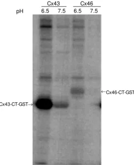
In order to determine whether this might be a general activation of protein kinases, we produced a second GST fusion protein con-struct with Cx46-CT. This protein was pro-duced and used in a manner identical to Cx43. Both Cx43-CT and Cx46-CT, GST protein constructs, were used as targets for binding and phosphorylation by sheep heart extracts. Figure 2 shows the phosphoryla-tion of Cx43-CT-GST fusion proteins at pH 6.5 and pH 7.5 (for both the binding and kinase steps). As expected, there was an increase in phosphorylation of Cx43-CT. When Cx46-CT was used in place of Cx43 there was no detectable phosphorylation at pH 7.5 and only a small, although detect-able, phosphorylation at pH 6.5. The amount of connexin-GST fusion protein was shown to be similar based on the intensity of Coo-massie staining. This phosphorylation reac-tion was, therefore, not a general increase in protein kinase activity but had some speci-ficity. Testing of CT constructs derived from other connexin proteins would be required to determine the range of possible substrates of this protein kinase.
In order to better define the role of pH in the binding and kinase assays, the pH of one of these steps was held constant while the pH of the other was varied. In panel A of Figure 3 the kinase assay was held at either pH 6.5 (open bars) or pH 7.5 (closed bars), while the pH of the binding reaction was changed. The results were quantified and then normal-ized so that the highest value was 100%. As expected, the curve obtained when the ki-nase assay was performed at lower pH was significantly higher. This curve indicates that the lower the pH, the more kinase can bind to the GST-Cx43-CT. Due to the very low level of activity in this experiment, it is not pos-sible to make a definitive comment about the kinase activity obtained when the kinase reaction was performed at pH 7.5. In panel B Figure 1 - Phosphorylat ion of
Cx43. An in vitro kinase reaction w as performed using a Cx43-CT-GST fusion protein. The fusion protein w as immobilized on gluta-thione Sephadex resin and incu-bated w ith 1 mg of sheep heart extract for 90 min. Binding reac-tions w ere performed in M OPS buffer at either pH 6.5 or 7.5. Af-ter w ashing, the beads w ere incu-bated w ith 5 µCi of [g-32P] ATP in
50 mM M OPS and 3 mM M nCl2
at the pH indicated. The reaction w as stopped by the addition of SDS sample buffer and the pro-t eins w ere resolved by SDS-PAGE. Visualizat ion w as per-formed using a PhosphoImager. The image show n w as represen-tative of at least three experi-ments. The migration of the Cx43-CT-GST fusion protein is marked. Phosphorylat ion is enhanced w hen the binding and/or kinase incubations are at pH 6.5. CT-GST, Carboxy terminal-glutathione-S-transferase fusion protein.
Cx43-CT-GST Binding
Kinase
6.5 6.5 7.5 7.5
6.5 7.5 6.5 7.5
123®
85®
50®
33®
of Figure 3, the pH of the binding step was held constant at either pH 6.5 (open bars) or pH 7.5 (closed bars). The pH of the kinase assay was then altered as indicated in the chart. The activity was higher when the bind-ing was performed at the lower pH as is expected from the results shown in panel A. At both pHs of binding, the activity was higher when the kinase reaction was per-formed at a lower pH, reaching a maximum at pH 6.5. However, unlike the binding step, this activity did not seem to continue to increase as the pH was reduced.
In our initial studies, the kinase assays were performed in a buffer that utilized man-ganese as the source of divalent cations. In order to further characterize the kinase activ-ity the experiments were performed with either 3 mM MgCl2 or3 mM MnCl2. Figure 4
demonstrates that the activation of the pro-tein kinase for phosphorylation of both the GST-Cx43-CT protein and the other phos-phorylation targets in the extract was greatly enhanced by the presence of manganese rather than magnesium.
To further analyze the phosphorylation reaction, mutants of Cx43-CT were cloned into the GST expression vector. These pro-teins were expressed and analyzed for reac-tion with the acidificareac-tion-activated protein kinase activity as before. The results of one such experiment are presented in Figure 5. When wild type Cx43-CT was used there was a significant increase in phosphoryla-tion seen when the pH was dropped from 7.5 to 6.5. Conversely, no phosphorylation was observed when GST alone was used as a potential phosphate acceptor. Mutants of Cx43-CT were chosen based on the pres-ence of potential phosphorylation sites. A deletion of the protein from amino acid 261 to 280 was phosphorylated in an acidifica-tion dependent manner although the overall level of phosphorylation was slightly less intense than that observed for the wild type protein. Similarly, there was phosphoryla-tion of a mutant peptide that lacked amino
R
e
la
ti
v
e
k
in
a
s
e
a
c
ti
v
it
y
125
100
75
50
0 25
Figure 2 - Comparison of in vitro phosphorylation of Cx43 and Cx46. GST (glutathione-S-trans-ferase) fusion proteins derived from Cx43 or Cx46 w ere pre-pared and analyzed for phospho-rylation by sheep heart extracts. The pH of both the binding and kinase reactions w as the same and is indicated in the figure. The results indicate that the ki-nase reaction w as enhanced against Cx43 at low er pH. Cx46-CT-GST
® Cx43
pH 6.5 7.5 6.5 7.5
Cx46
R
e
la
ti
v
e
k
in
a
s
e
a
c
ti
v
it
y
125
100
75
50
0 25
6.0 6.5 7.0 8.0
pH of binding reaction
6.0 6.5 7.0 7.5 8.0
pH of kinase reaction
Figure 3 - Titration of pH in the in vitro kinase assay. In this as-say, the pH of either the binding or kinase reaction w as held con-stant w hile the pH of the other reaction w as varied. The phos-phorylation of the Cx43-CT-GST fusion protein w as quantified by PhosphoImager analysis. Tw o lanes for each pH w ere quanti-fied and the results w ere aver-aged. The figure is representa-tive of three experiments. Panel A, The pH of the kinase reaction w as held at either pH 6.5 (open bars) or pH 7.5 (closed bars) w hile the pH of the binding re-action w as varied from 6 to 8 at 0.5-pH unit intervals. Panel B, The pH of the binding reaction w as held at either pH 6.5 (open bars) or pH 7.5 (closed bars) w hile the pH of the kinase reac-tion w as varied from 6 to 8 at 0.5-pH unit intervals.
acids 281 to 300. There was, however, a dramatic loss of demonstrable kinase activ-ity when a deletion of amino acids 364-373 was tested. This deletion removes six serines that make up consensus sites for several protein kinases. From these results, the site
A
B
Cx43-CT-GST®
stand multiple washing steps. Therefore, we can conclude that a relatively strong binding interaction must take place between the ki-nase and Cx43. The phosphorylation was somewhat specific. There was no detectable phosphorylation of the GST carrier protein and only weak phosphorylation of the Cx46-CT domain used as a control.
Many studies have shown that phospho-rylation is a common modification of the connexin molecule (26, see also 27). Others have demonstrated that phosphorylation of Cx43 results in an alteration of the unitary conductance of connexin channels (14,28-30). The activation of kinases coincides with a shift of the unitary conductance of gap junction channels from neonatal cardiac myocytes (31). While under dephosphory-lating conditions, human Cx43 channels show a predominance of larger unitary conduc-tance (28). Furthermore, it has been sug-gested that permeability and single channel conductance of Cx43 gap junction channels are independently regulated, and that electri-cal and metabolic coupling are differentially modulated by various phosphorylating con-ditions (15). Taken together, these data sug-gest a relationship between phosphorylation of connexins and modification of intercellu-lar communication.
We have previously demonstrated that the CT domain of Cx43 is required for acidi-fication-induced gating (32,33). This domain is also required for other gating reactions (19,34). A number of protein kinases are known to phosphorylate Cx43 and modify the gating of the channels. In many cases, the phosphorylation reactions have been shown to occur in the CT domain. Truncation or mutation of the CT domain of Cx43 results in a loss of the ability of Cx43 to be modu-lated by a number of protein kinases or fac-tors known to activate protein kinases. In one example, Src kinase was unable to in-duce the gating of a truncated form of Cx43 (19) unless the CT domain was also ex-pressed in the cell. In another example, mu-Figure 5 - Analysis of
phospho-rylation of Cx43 mutants. GST (glutathione-S-transferase) fu-sion proteins w ere prepared that had deletions in the CT (carboxyl terminal) domain of Cx43. The mutants tested had delet ions f rom am ino acids 261-280, 281-300 and 364-373. In addit ion, bot h w ild t ype Cx43-CT and GST alone w ere t est ed. The m ut ant s w ere tested w ith the reactions per-formed at both pH 6.5 and pH
7.5. The presence of equal amounts of fusion protein in the reaction w as confirmed by the Coomassie blue staining of the gel. The migration of the Cx43-CT-GST fusion protein is marked. The mutated isoforms migrate slightly faster. The phosphorylation of the fusion protein w as greatly reduced by the removal of the region from amino acids 364 and 373.
60 kDa
Cx43-CT-GST® M g
pH 6.5 7.5 6.5 7.5 M n
40 kDa Figure 4 - The pH-dependent
ki-nase is more active in the pres-ence of M n than M g. The in vitro kinase assay w as performed at pH 6.5 and 7.5 (the pH of the binding and kinase steps w as constant). The divalent cation present in the kinase reaction w as either 3 mM M gCl2 or 3
mM M nCl2 as indicated. Kinase
act ivit y w as signif icant ly en-hanced by the presence of man-ganese. CT-GST, Carboxyl ter-minal-glutathione-S-transferase fusion protein.
of phosphorylation appears to be primarily in the serines at the C-terminal end of the molecule.
D iscussio n
In this report, we demonstrate that tissue extracts contain a protein kinase or kinases that can specifically interact with the car-boxyl terminal domain of Cx43 and phos-phorylate the protein. The unique property of these kinases is that the interaction with, and the phosphorylation of, connexin are enhanced by reducing the pH to 6.5. This coincides with the intracellular pH required to gate Cx43. The assay system chosen re-quires that the interaction of the protein ki-nase with the target protein be able to
with-Cx43-CT-GST
tation of three serines that represent poten-tial MAPK phosphorylation sites reduces the ability of growth factors to gate connex-ins (17). It is possible that the acidification-induced phosphorylation of connexin de-scribed in this manuscript may act as a me-diator in acidification-induced gating.
It has been suggested that many regula-tors of cell-to-cell coupling act in concert. For example, pHi and Ca have been reported to act together to regulate channel activity (35-37). More recent studies have proposed that acidification-induced gating does not directly affect cell coupling, but may act through the ubiquitous calcium-receptor pro-tein, calmodulin (38). It has also been sug-gested that calcium-induced gating of gap junction channels could result from the acti-vation of specific kinases, with consequent connexin phosphorylation (39). It is interest-ing to note that there are multiple calcium/ calmodulin-dependent kinase II (CAMK II)
phosphorylation sites in the carboxyl-termi-nal domain of Cx43.
Our studies localized the region of phos-phorylation to a serine-rich region at the C-terminal end of the Cx43 molecule. This region contains consensus phosphorylation sites for PKC, cAMP-dependent protein ki-nase and CAMK. To date, our studies have not been able to specifically detect any of these kinases in the proteins bound to Cx43-CT at lower pH. We have also not been able to block the acidification-induced phospho-rylation reactions using inhibitors of these kinases. Future studies will be performed to identify the kinase or kinases involved.
Ackno wle dgm e nts
We thank Wanda Coombs and Marta Mastroianni for their excellent technical sup-port.
Re fe re nce s
1. Werner R, Levine E, Rabadan-Diehl C & Dahl G (1991). Gating properties of con-nexin32 cell-cell channels and their mu-tants expressed in Xenopus oocytes. Pro-ceedings of the Royal Society of London, 243: 5-11.
2. Stergiopoulos K, Alvarado JL, Ek-Vitorin JF, Taffet SM & Delmar M (1999). Hetero-domain interactions as a mechanism for the regulation of connexin channels. Cir-culation Research, 84: 1144-1155. 3. Dunham B, Liu S, Taffet SM , Trabka-Janik
E, Delmar M , Petryshyn R, Zheng S, Perzova R & Vallano M L (1992). Immu-nolocalization and expression of functional and nonfunctional cell-to-cell channels from w ild-type and mutant rat heart con-nexin43 cDNA. Circulation Research, 70: 1233-1243.
4. Koval M , Geist ST, W est phale EM , Kemendy AE, Civitelli R, Beyer EC & Steinberg TH (1995). Transfected con-nexin45 alters gap junction permeability in cells expressing endogenous con-nexin43. Journal of Cell Biology, 130: 987-995.
5. Lin JS, Eckert R, Kistler J & Donaldson P (1998). Spatial differences in gap junction
gating in the lens are a consequence of connexin cleavage. European Journal of Cell Biology, 76: 246-250.
6. Lin JS, Fitzgerald S, Dong Y, Knight C, Donaldson P & Kistler J (1997). Process-ing of the gap junction protein connexin50 in the ocular lens is accomplished by calpain. European Journal of Cell Biology, 73: 141-149.
7. Kanemitsu M Y, Loo LW, Simon S, Lau AF & Eckhart W (1997). Tyrosine phosphory-lation of connexin 43 by v-Src is mediated by SH2 and SH3 domain interactions. Journal of Biological Chem istry, 272: 22824-22831.
8. Loo LW, Berestecky JM , Kanemitsu M Y & Lau AF (1995). pp60src-mediated phos-phorylation of connexin 43, a gap junction protein. Journal of Biological Chemistry, 270: 12751-12761.
9. Warn-Cramer BJ, Lampe PD, Kurata WE, Kanemitsu M Y, Loo LW, Eckhart W & Lau AF (1996). Characterization of the mito-gen-activated protein kinase phosphoryla-tion sites on the connexin-43 gap juncphosphoryla-tion protein. Journal of Biological Chemistry, 271: 3779-3786.
10. Kim DY, Kam Y, Koo SK & Joe CO (1999).
Gating connexin 43 channels reconsti-tuted in lipid vesicles by mitogen-acti-vated protein kinase phosphorylation. Journal of Biological Chem istry, 274: 5581-5587.
11. Hossain M Z, Jagdale AB, Ao P, Kazlauskas A & Boynton AL (1999). Disruption of gap junctional communication by the platelet-derived grow th factor is mediated via mul-tiple signaling pathw ays. Journal of Bio-logical Chemistry, 274: 10489-10496. 12. Kanemitsu M Y, Jiang W & Eckhart W
(1998). Cdc2-mediated phosphorylation of the gap junction protein, connexin43, dur-ing mitosis. Cell Grow th and Differentia-tion, 9: 13-21.
13. Kw ak BR, Saez JC, Wilders R, Chanson M , Fishman GI, Hertzberg EL, Spray DC & Jongsma HJ (1995). Effects of cGM P-de-pendent phosphorylation on rat and hu-man connexin43 gap junction channels. Pflügers Archiv, European Journal of Physiology, 430: 770-778.
62: 51-53.
15. Kw ak BR & Jongsma HJ (1996). Regula-tion of cardiac gap juncRegula-tion channel per-meability and conductance by several phosphorylating conditions. M olecular and Cellular Biochemistry, 157: 93-99. 16. Kw ak BR, van Veen TA, Analbers LJ &
Jongsma HJ (1995). TPA increases con-ductance but decreases permeability in neonatal rat cardiomyocyte gap junction channels. Experimental Cell Research, 220: 456-463.
17. Warn-Cramer BJ, Cottrell GT, Burt JM & Lau AF (1998). Regulation of connexin-43 gap junctional intercellular communication by mitogen-activated protein kinase. Jour-nal of Biological Chemistry, 273: 9188-9196.
18. Hossain M Z, Ao P & Boynton AL (1998). Platelet-derived grow th factor-induced disruption of gap junctional communica-tion and phosphorylacommunica-tion of connexin43 involves protein kinase C and mitogen-activated protein kinase. Journal of Cellu-lar Physiology, 176: 332-341.
19. Zhou L, Kasperek EM & Nicholson BJ (1999). Dissection of the molecular basis of pp60(v-src) induced gating of connexin 43 gap junction channels. Journal of Cell Biology, 144: 1033-1045.
20. Thomas D, Ritz M F, M alviya AN & Gaillard S (1996). Intracellular acidification medi-ates the proliferative response of PC12 cells induced by potassium ferricyanide and involves M AP kinase activation. Inter-national Journal of Cancer, 68: 547-552. 21. Yamaji Y, Tsuganezaw a H, M oe OW &
Alpern RJ (1997). Intracellular acidosis ac-tivates c-Src. American Journal of Physiol-ogy, 272: C886-C893.
22. Zanke BW, Lee C, Arab S & Tannock IF (1998). Death of tumor cells after intracel-lular acidification is dependent on stress-activated protein kinases (SAPK/JNK) pathw ay activation and cannot be inhib-ited by Bcl-2 expression or interleukin
1beta-converting enzyme inhibition. Can-cer Research, 58: 2801-2808.
23. Ek JF, Delmar M , Perzova R & Taffet SM (1994). Role of histidine 95 on pH gating of the cardiac gap junction protein con-nexin43. Circulation Research, 74: 1058-1064.
24. Turner CE & M iller JT (1994). Primary se-quence of paxillin contains putative SH2 and SH3 domain binding motifs and mul-tiple LIM domains: identification of a vinculin and pp125Fak-binding region. Journal of Cell Science, 107: 1583-1591. 25. Brow n M C, Perrotta JA & Turner CE
(1998). Serine and threonine phosphoryla-tion of the paxillin LIM domains regulates paxillin focal adhesion localization and cell adhesion to fibronectin. M olecular Biol-ogy of the Cell, 9: 1803-1816.
26. Lau AF, Kurata WE, Kanemitsu M Y, Loo LW , W arn-Cram er BJ, Eckhart W & Lampe PD (1996). Regulation of con-nexin43 function by activated tyrosine protein kinases. Journal of Bioenergetics and Biomembranes, 28: 359-368. 27. Bruzzone R, White TW & Goodenough
DA (1996). The cellular internet: on-line w ith connexins. Bioessays, 18: 709-718. 28. M oreno AP, Saez JC, Fishman GI & Spray
DC (1994). Human connexin43 gap junc-tion channels. Regulajunc-tion of unitary con-ductances by phosphorylation. Circulation Research, 74: 1050-1057.
29. Kw ak BR & Jongsma HJ (1996). Regula-tion of cardiac gap juncRegula-tion channel per-meability and conductance by several phosphorylating conditions. M olecular and Cellular Biochemistry, 157: 93-99. 30. Kw ak BR, van Veen TA, Analbers LJ &
Jongsma HJ (1995). TPA increases con-ductance but decreases permeability in neonatal rat cardiomyocyte gap junction channels. Experimental Cell Research, 220: 456-463.
31. Takens-Kw ak BR & Jongsma HJ (1992). Cardiac gap junctions: three distinct single
channel conductances and their modula-t ion by phosphorylamodula-t ing modula-t reamodula-t m enmodula-t s. Pflügers Archiv, European Journal of Physiology, 422: 198-200.
32. Liu S, Taffet SM , Stoner L, Delmar M , Vallano M L & Jalife J (1993). A structural basis for the unequal sensitivity of the major cardiac and liver gap junctions to intracellular acidification: the carboxyl tail length. Biophysical Journal, 64: 1422-1433.
33. M orley GE, Ek-Vitorin JF, Taffet SM & Delmar M (1997). Structure of connexin43 and its regulation by pHi. Journal of Car-diovascular Electrophysiology, 8: 939-951. 34. Hom m a N, Alvarado JL, Coom bs W , Stergiopoulos K, Taffet SM , Lau AF & Delmar M (1998). A particle-receptor mo-del for the insulin-induced closure of con-nexin43 channels. Circulation Research, 83: 27-32.
35. Arellano RO, Rivera A & Ramon F (1990). Protein phosphorylation and hydrogen ions modulate calcium-induced closure of gap junction channels. Biophysical Jour-nal, 57: 363-367.
36. De M ello WC (1984). M odulation of junc-tional permeability. Federation Proceed-ings, 43: 2692-2696.
37. Spray DC, White RL, de Carvalho AC, Har-ris AL & Bennett M V (1984). Gating of gap junction channels. Biophysical Journal, 45: 219-230.
38. Peracchia C, Wang X, Li L & Peracchia LL (1996). Inhibition of calmodulin expres-sion prevents low -pH-induced gap junc-t ion uncoupling in Xenopus oocyjunc-t es. Pflügers Archiv, European Journal of Physiology, 431: 379-387.